Article

Optimizing Green Globular Body Induction for Micropropagation of Microsorum pteropus ‘Windeløv’

1
Department of Tropical Agriculture and International Cooperation, National Pingtung University of Science and Technology, Pingtung 91201, Taiwan
2
Department of Aquaculture, National Pingtung University of Science and Technology, Pingtung 91201, Taiwan
3
Department of Aquaculture, Faculty of Fisheries, Kasetsart University, Bangkok 10900, Thailand
4
Department of Plant Medicine, National Pingtung University of Science and Technology, Pingtung 91201, Taiwan
*
Authors to whom correspondence should be addressed.
Horticulturae 2024, 10(7), 673; https://doi.org/10.3390/horticulturae10070673
Submission received: 26 May 2024 / Revised: 14 June 2024 / Accepted: 21 June 2024 / Published: 25 June 2024
(This article belongs to the Section Floriculture, Nursery and Landscape, and Turf)

Abstract

:
Microsorum pteropus ‘Windeløv’ is a water fern from Asia commonly used in aquarium landscapes. This study aimed to develop a mass production method through the induction of green globular bodies (GGBs) from leaf explants of M. pteropus. Surface sterilization was performed on adventitious buds on the fronds of M. pteropus ‘Windeløv’ as explants. The results showed that the optimal explant disinfection condition was 1% sodium hypochlorite solution for 20 min, achieving a successful rate of 87.5 ± 9.6%. The most effective GGB induction was achieved with MS medium supplemented with 5.0 mg/L of NAA (1-Naphthaleneaceticacid) and 0.5 mg/L of 6-BA (6-benzylaminopurine), producing the highest number and size of GGBs, with a mean value of 4.0 ± 0.8 GGBs per explant and a diameter of 2.45 ± 0.61 mm after 8 weeks of culture. The regeneration of multiple sporophytes from the GGBs was only observed under hormone-free MS medium, with a mean value of 34.2 ± 7.3 sporophytes per GGB after 6 weeks of culture. Subsequently, rootless and separated sporophytes could be directly transplanted into the aquarium with a 100% survival rate.

1. Introduction

Aquatic plants play a crucial role in both the aesthetic and ecological balance of aquascapes, utilized in hobbyist setups and commercial ventures. Beyond their visual appeal, these plants significantly influence the hydrological, geomorphological, and physicochemical aspects of their environments. They provide a habitat and sustenance for a diverse array of organisms, including microbes and vertebrates, therefore enriching the overall visual and ecological harmony of aquariums and water gardens [1,2,3]. The cultivation and transplantation of these plants are essential not only for enhancing visual aesthetics, but also for meeting the growing demands of the global aquarium and water garden markets [4,5,6]. Moreover, the art of aquascaping, which involves skillfully arranging these plants into captivating underwater landscapes, highlights the need for a wide variety of species adapted to different aquatic conditions [7].
Among these species, Microsorum pteropus, especially the ‘Windeløv’ cultivar, is renowned for its distinct leaf structure and adaptability to aquatic conditions [8]. The ‘Windeløv’ cultivar, with its intricate lace-like fronds, is a favored choice among aquascapers seeking to add texture and depth to their designs [9]. However, the propagation of this and similar species faces challenges, notably their limited natural reproductive capabilities. In its natural habitat, this species reproduces through spores, a process that can be slow and unreliable due to environmental factors and the need for specific conditions for spore germination and growth. Additionally, traditional propagation methods, such as division or cuttings, are limited by a slow growth rate and susceptibility to pests and diseases [10]. These constraints impede large-scale commercial cultivation and threaten the preservation of rare and delicate varieties.
Additionally, aquatic plants have been acknowledged for their significant role in bioremediation. Aquatic phytoremediation utilizes macrophytes, including freshwater-adapted angiosperms, pteridophytes, and ferns, to remove and degrade pollutants in aquatic environments [11]. Macrophytes possess a remarkable ability to absorb nutrients and other substances from their growth medium, thereby reducing the pollution levels in targeted water bodies [12]. For example, Ceratophyllum demersum, also known as hornwort or coontail, is a submerged, free-floating aquatic plant with a broad distribution and is noted for its resilience to various abiotic stresses [13,14,15]. These features highlight the essential role of aquatic plants not only in preserving ecosystem health, but also in potential environmental remediation efforts.
Addressing these cultivation challenges, tissue culture techniques offer an effective alternative to the rapid, true-to-type propagation of various plant species, thus facilitating the large-scale micropropagation of high-quality clones in a relatively short timeframe [5,6,16,17]. Recent advancements in tissue culture technology, particularly the development of green globular bodies (GGBs), present promising opportunities for efficient and sustainable propagation with a high multiplication rate [18]. GGBs are clusters of undifferentiated plant cells that can develop into new plant structures under suitable conditions. They have become essential for the in vitro vegetative propagation of ferns and show potential in cryopreservation [18,19] and somatic embryogenesis research [20]. These techniques significantly advance aquatic plant propagation, ensuring rapid, sterile, and controlled growth conditions.
This research focuses on the micropropagation of M. pteropus ‘Windeløv’, particularly on the induction of GGBs. The main objectives include obtaining sterile explants, initiating and multiplying GGB tissue, regenerating sporophytes, and acclimatizing plants after removal from culture conditions. These efforts are directed towards establishing more efficient micropropagation techniques. By leveraging advanced tissue culture technologies, this study aims to address the limitation of traditional propagation methods, thereby improving ecological sustainability and commercial viability in the aquascaping industry.

2. Materials and Methods

2.1. Plant Material and Sterilization

Microsorum pteropus ‘Windeløv’ plants were collected from the aquaculture farm of the National Pingtung University of Science and Technology’s Aquaculture Department. The plants were grown in emersed leaf form, using driftwood as the planting substrate, under greenhouse conditions with flowing groundwater. Natural light was used for illumination, and the plants were cultured at room temperature. Fronds with adventitious buds on the back were cut into 0.5 cm2 as explants for surface sterilization. The explants were surface-sterilized for 10, 20, and 30 min with 1.0%, 2.0%, 3.0%, and 4.0% sodium hypochlorite supplemented with Tween 20 (Sigma Aldrich, St. Louis, MO, USA) (2 drops, approximately 80 μL, per 100 mL). Subsequently, the explants were washed five times with sterile distilled water and then dried on sterile tissue paper for 1–3 min until visible moisture was removed. The explants were then placed on full-strength Murashige and Skoog [21] basal medium supplemented with 3% (w/v) sucrose, with the pH adjusted to 5.7, and 0.5% (w/v) Agar A (Bio Basic, Markham, Canada). The medium was aliquoted into a 20 mL volume, autoclaved, and poured into Petri dishes (90 mm × 20 m, Alpha Plus Scientific Corp., Taoyuan, Taiwan). Each batch included four Petri dishes, with each dish containing ten explants. The explants were incubated in a controlled-environment growth chamber (F-740, HiPoint, Kaohsiung, Taiwan) at 25 ± 1 °C. The explants were incubated under a light intensity of 30 μmol m–2 s–1 provided by white fluorescent lights (FL40D, China Electric, TOA Lighting, Taipei, Taiwan) with a 12 h photoperiod. After four weeks in vitro, the prevalence of explant contamination was noted, calculating the percentage of contamination by dividing the number of affected explants by their total number. The axenic explants were subcultured in the same solid MS medium to obtain enough numbers of explants for the following studies.

2.2. Green Globular Bodies (GGBs) Induction

To determine the optimal concentration of cytokinins, 6-Benzylaminopurine (6-BA; Phyto Technology Laboratories, St. Lenexa, KS, USA), Thidiazuron (TDZ; Phyto Technology Laboratories, Kansas, USA), and auxin 1-Naphthaleneacetic acid (NAA; Phyto Technology Laboratories) were used for inducing green globular bodies (GGBs). Aseptic sporophytes approximately 1 cm in height with 5 fronds were placed in Petri dishes containing Murashige and Skoog [21] basal medium supplemented with varying concentrations of 6-BA or TDZ (0, 0.5, 1, or 5 mg/L) and NAA (0, 1, 5, or 10 mg/L) for a period of 4 weeks. Each treatment included four Petri dishes, each with five explants. The culture conditions in the growth chambers were as described above. The percentage of GGBs formation was calculated by dividing the number of explants showing GGB formation by the total number of explants. The primary parameter measured was the average number of GGBs.

2.3. Green Globular Bodies’ Proliferation

Based on the GGB induction evaluation, we further assessed the influence of 11 PGR combinations on GGB proliferation [8,9,18]. GGBs, approximately 0.8 mm in diameter, were isolated from the explants and subcultured back onto their original induction medium. Each treatment consisted of four Petri dishes, with ten GGBs per dish. The culture conditions in the growth chambers were as previously described. After four weeks, the diameter of the GGBs was measured using Imageview software Version 2.4.0 (Bestscope, Beijing, China). Additionally, sporophyte regeneration was monitored as an indicator of GGB multiplication at this stage. The sporophyte regeneration rate (%) was calculated using the formula: (number of GGBs with sporophyte development/total number of GGBs incubated) × 100%.

2.4. Regeneration of Sporophyte

To determine the optimal concentration of cytokinins (6-BA or TDZ) and NAA for sporophyte regeneration, GGBs (~1.8 mm in diameter), produced from the most suitable GGB proliferation medium, were placed in Petri dishes containing Murashige and Skoog [21] basal medium with varying concentrations of 6-BA or TDZ (0, 0.5, or 1 mg/L) and NAA (0, 0.5, or 1 mg/L) for a period of 6 weeks. Each treatment was replicated in four Petri dishes, with ten explants per replicate. The growth chamber conditions were maintained as previously described. The percentage of sporophyte regeneration was calculated by dividing the number of explants with frond formation by the total number of explants. The study parameter was the average number of plantlets.

2.5. Acclimatization

For the acclimatization process of the plantlets, seedlings derived from GGB progenitors, exhibiting leafy extensions around 2.5 cm in height, were utilized. These seedlings were affixed onto stainless-steel woven wire mesh (5 × 5 cm) using super glue gel (MXBON, Chia-Yi, Taiwan). There were seven pieces of stainless-steel woven wire mesh, each containing five seedlings. Each woven mesh was considered as a replicate. Subsequently, they were cultured within a crystal shrimp aquarium for two months, where the plants were fully submerged. The survival rate was calculated as the number of surviving seedlings divided by the total number of seedlings. The parameter used in the study was based on the average of the total plantlets’ weight per woven mesh.

2.6. Statistical Analysis

The data obtained from the experiment were analyzed using the SAS 9.3 (Statistical Analysis System) statistical suite software. After one-way analysis of variance, if there was a significant difference between treatments, Duncan’s multiple range test was used to compare whether there were significant differences between each treatment group (p < 0.05).

3. Results

3.1. Sterilization Stage

In this experiment, various concentrations of sodium hypochlorite solutions in combination with different soaking times were tested to identify the optimal disinfection conditions for the M. pteropus ‘Windeløv’. According to the results in Table 1, groups treated with 1% and 2% sodium hypochlorite solutions for various durations and the group treated with a 3% solution for 10 min were all successfully disinfected and showed leafy ex-plant growth (Figure 1A). Furthermore, the disinfection condition of soaking in a 1% sodium hypochlorite solution for 20 min achieved a disinfection success rate of 47.5 ± 12.6%, which was identified as the optimal disinfection condition. Groups treated with a 3% sodium hypochlorite solution for 20 and 30 min and a 4% solution for 10, 20, and 30 min had a 0% success rate in disinfection. In these groups, the explants were mostly browned and dead (Figure 1B,C). Figure 1D shows an explant contaminated with fungi; it is evident that many mycelia surrounded the explant, which were mostly found in groups treated with a 1% sodium hypochlorite solution for short periods (10 min).

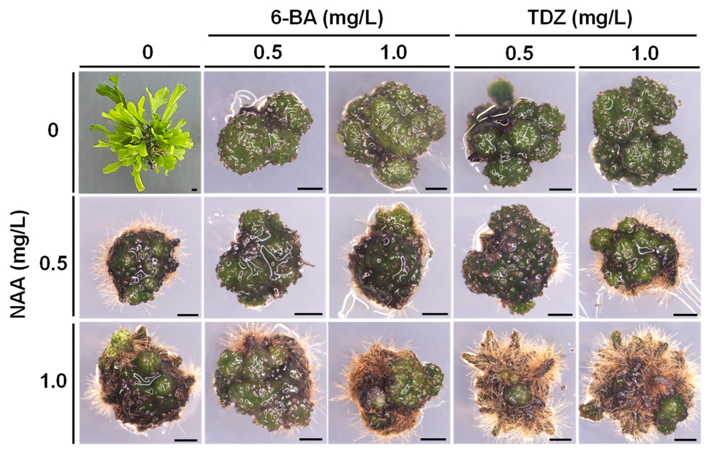
3.2. Green Globular Bodies’ Induction

The results in Table 2 show that the induction of green globular bodies (GGBs) required the presence of plant growth regulators (PGRs). Groups without PGRs did not form GGBs, while those with PGRs showed varying rates of induction. The highest induction rates, over 90%, were observed in the groups with 6-BA at 0.5 mg/L combined with NAA at different concentrations. Specifically, the combination of 6-BA at 0.5 mg/L and NAA at 5.0 mg/L produced the highest number of GGBs, averaging 4.0 ± 0.8 per explant.
Groups with 6-BA at 1.0 mg/L combined with NAA at 5.0 mg/L or 10.0 mg/L showed some browning, whereas other combinations exhibited greener and more granular GGBs. Notably, rhizoids were present in groups with higher concentrations of NAA (5.0 and 10.0 mg/L), indicating robust GGB formation. In contrast, GGBs from the group with TDZ at 5.0 mg/L combined with NAA at 10.0 mg/L appeared yellow-green and had a looser structure (Figure 2).

3.3. Green Globular Bodies’ Proliferation

As shown in Table 3, four weeks after initiating the experiment, the diameters of the GGBs varied among the groups, ranging from 1.28 ± 0.14 mm to 2.45 ± 0.61 mm. None of the groups exhibited the regeneration of sporophytes. The treatment with 6-BA at 0.5 mg/L and NAA at 5.0 mg/L yielded the largest average GGB diameter of 2.45 ± 0.61 mm.

3.4. The Regeneration of Sporophytes

To determine the optimal conditions for the regeneration of sporophytes in M. pteropus ‘Windeløv’, various concentrations of the auxin NAA were combined with different types and concentrations of the cytokinins 6-BA and TDZ. We calculated the number of explants with frond formation of each sporophyte to reflect the regeneration rate. According to the results shown in Table 4, only the group without any plant growth regulators successfully regenerated sporophytes, beginning at four weeks of cultivation. After six weeks of cultivation, each sporophyte regenerated an average of 34.2 ± 7.3 seedlings. The groups supplemented with cytokinins (6-BA and TDZ) displayed the differentiation of GGBs. When combined with NAA, an increase in concentration made the rhizoid structures on the GGBs more pronounced (Figure 3).

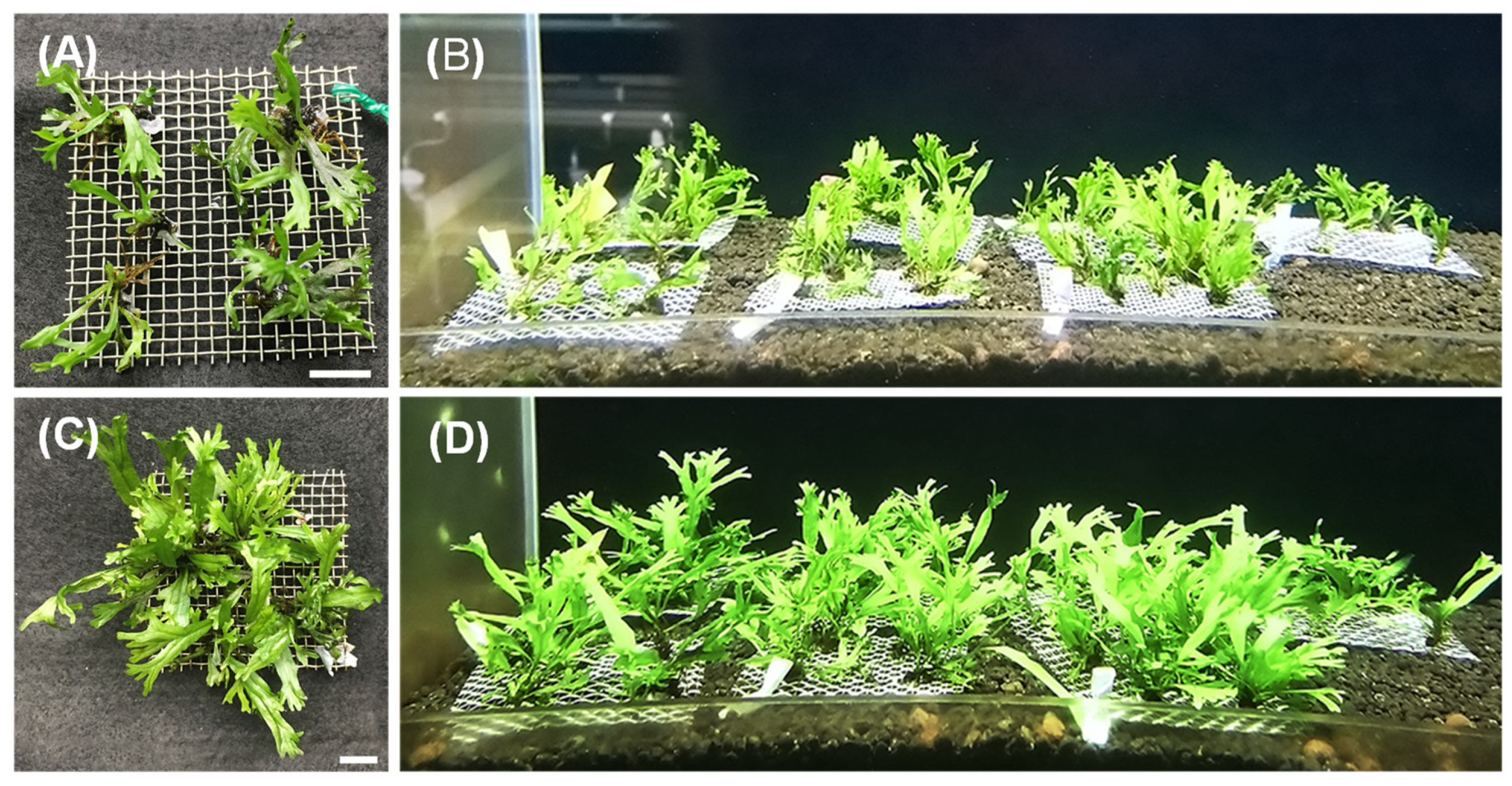
3.5. Acclimatization

In this study, tissue-cultured seedlings of M. pteropus ‘Windeløv’ were extracted directly from culture bottles and affixed onto stainless-steel woven wire mesh using adhesive (Figure 4A). Subsequently, they were introduced into an aquarium setting (Figure 4B) for a cultivation period of two months. An analysis of the data presented in Table 5 revealed that following the two-month cultivation period, the average weight increase per woven wire mesh was measured at 0.86 ± 0.21 g. Additionally, examination of the tissue-cultured seedlings indicated that the tissue-cultured seedlings exhibited a high vitality (Figure 4C,D), characterized by the absence of sporophyte yellowing or other deleterious manifestations, alongside achieving a survival rate of 100%.

4. Discussion

This study aimed to establish effective protocols for the sterilization, induction, proliferation, and acclimatization of M. Pteropus ‘Windeløv’. By optimizing various parameters, including the concentrations of sterilizing agents and plant growth regulators, we sought to enhance the tissue culture techniques for this aquatic fern. The findings provide valuable insights into the challenges and solutions associated with the micropropagation of aquatic plants, which are often hindered by biological contamination and difficulties in tissue regeneration.
Aquatic plants present unique challenges in tissue culture due to their susceptibility to contamination. For the explants in this study, we selected aerial leaves rather than submerged leaves, because submerged leaves are more prone to biological contamination, making surface sterilization more challenging, as previous research has demonstrated that aquatic plants, due to their growth in high-humidity environments, are susceptible to biological contamination [8]. Additionally, the use of surface sterilization solutions, which are mostly strong oxidants or highly toxic chemicals, can damage plant tissues. Aquatic plant leaves lack a waxy epidermis layer and have thinner leaf blades [22], making them more susceptible to direct contact with surface sterilization solutions, leading to the whitening and death of the explants.
Successful sterilization was achieved in all low-concentration groups; however, fungal contamination was common in the group sterilized with 1% sodium hypochlorite. In higher-concentration groups, except for the group sterilized with 3% sodium hypochlorite for 10 min, the browning of explants was observed (Figure 1). Previous research supported our findings, in which the death rate of explants increased proportionally with the concentration of sterilizing solution [23]. We found that lower concentrations exhibited higher numbers of vital explants, while higher concentrations resulted in the browning of the explants. Moreover, it was found that the use of 5% sodium hypochlorite solution for 5 min on the ex vitro propagation of mini dwarf pearls resulted in the complete whitening of the explants. This experiment also confirmed that even a short immersion of explants in high-concentration sterilizing solutions can cause explant death.
Conditions for the induction and proliferation of GGBs in M. pteropus were established. Plant growth regulators played a significant role in the induction and proliferation of GGBs, as all experiment groups, except the control group without PGRs, saw induced production. In seed plants, PGRs are commonly utilized to induce adventitious buds and callus tissue. Early reports often used a single cytokinin for GGBs’ induction, such as in fishbone fern (Nephrolepis cordifolia) [24], bird’s nest fern (Asplenium nidus) [25], and Polypodium cambricum [26]. More recently, the study on Pteris aspericaulis var. tricolor reported that GGB formation is induced by combining appropriate concentrations of plant growth regulators [8]. This controls tissue proliferation or differentiation into buds by altering the concentration combination of PGRs. Moreover, studies are increasingly supporting that the combination of cytokinins and auxins results in higher GGB induction rates compared to using a single cytokinin, as seen in Platycerium bifurcatum [27], Cibotium barometz [9], and Adiantum reniforme [28]. In this study, combinations of the cytokinins 6-BA and TDZ with the auxin NAA generally caused higher induction rates of GGBs than using a single cytokinin or auxin, once again demonstrating that the use of cytokinins combined with auxins is superior to using a single plant growth regulator for GGBs’ induction. While Yu et al. [9] stated in their report that the induction and proliferation of GGBs of C. barometz require PGRs, particularly the cytokinin TDZ, our study found that a higher concentration of TDZ combined with NAA led to the browning or a loose morphology of the GGBs by the fourth week. This indicates that high concentrations of TDZ are not conducive to GGB induction in M. pteropus. In the proliferation experiment on GGBs, except for the group without plant growth regulators, none of the groups containing plant growth regulators produced sporophytes during the experiment. A previous study by Higuchi and Amaki [25] on Nephrolepis indicated that 6-BA inhibits plant regeneration while promoting continuous GGBs’ proliferation. Amaki and Higuchi [29] also observed that 6-BA inhibits the formation of sporophytes in several fern GGBs and stably promotes GGBs’ proliferation. Yu et al. [9] found, in their study on the proliferation of GGBs of C. barometz, that TDZ has a similar effect to 6-BA in inhibiting sporophyte regeneration on GGBs. Cárdenas-Aquino et al. [30] stated in their report that adding 6-BA or 2-isopentenyl adenine (2-ip)to culture media can promote GGBs proliferation and inhibit sporophyte formation. These findings collectively suggest that cytokinins promote GGBs’ proliferation and inhibit sporophyte regeneration.
In the sporophyte regeneration experiments, only the GGBs in the control group without PGRs regenerated sporophytes, whereas those in the PGR-containing groups continued to proliferate GGBs without sporophytes. Shelikhan [31] cultured GGBs of N. biserrata in culture media without plant growth regulators and observed natural differentiation into buds after two weeks. Similarly, fishbone fern (N. cordifolia) [24], bird’s nest fern (A. nidus) [25], sword brake fern (P. ensiformis), delta maidenhair fern (Adiantum raddianum), leather fern (Rumohra adiantiformis) [29], and scaly male fern (Dryopteris affinis) [32] all differentiated into sporophytes when cultured in culture media without plant growth regulators. Hsu [33] also observed that GGBs of Platycerium hillii and P. alcicorne regenerated new buds in MS culture media, suggesting that endogenous plant hormones may influence spontaneous bud formation. This indicates that M. pteropus ‘Windeløv’ does not require additional PGRs for sporophyte regeneration and root induction, potentially reducing labor costs in its commercial production.

5. Conclusions

This study developed an effective micropropagation protocol for M. pteropus ‘Windeløv’. Using 1% sodium hypochlorite for 20 min to disinfect adventitious buds on the plant’s dorsal fronds achieved the optimal disinfection. The cultivation of M. pteropus ‘Windeløv’ explants in MS medium with 0.5 mg/L of 6-BA and 5.0 mg/L of NAA resulted in the successful induction of GGBs after four weeks. These GGBs, after an additional four weeks in the same medium, reached an average size of 2.45 ± 0.61 mm. Subsequent culturing in growth-regulator-free MS medium led to each GGB producing about 34.20 ± 7.33 seedlings within six weeks.
These results indicate that the optimized conditions for GGB induction and proliferation can be reliably used for the large-scale micropropagation of this aquatic fern. The high survival rate of 100% for the tissue-cultured seedlings further underscores the viability of this approach. By achieving consistent and high-quality GGB formation and subsequent sporophyte regeneration, this study addresses the limitations of traditional propagation methods, such as slow growth rates and susceptibility to pests and diseases. Consequently, the findings support the commercial scalability of the micropropagation protocol, offering a practical solution for the efficient production of Microsorum pteropus ‘Windeløv’ and potentially other aquatic plants.

Author Contributions

Conceptualization, W.S., C.-C.S., Y.T. and T.-M.W.; methodology, W.S., Y.T., C.-C.S., K.-T.W. and T.-M.W.; validation, W.S., C.-C.S., K.-T.W. and P.K.; formal analysis W.S., C.-C.S., K.-T.W. and P.K.; investigation, W.S., C.-C.S. and K.-T.W.; resources, Y.T. and T.-M.W.; data curation, W.S., Y.T. and T.-M.W.; writing—original draft preparation, W.S. and K.-T.W.; writing—review and editing, Y.T. and T.-M.W.; visualization, W.S., Y.T. and T.-M.W.; supervision, Y.T. and T.-M.W.; project administration, Y.T. and T.-M.W. All authors have read and agreed to the published version of the manuscript.

Funding

This research was partially funded by the Agricultural Technology Park Administration Center, Ministry of Agriculture, Taiwan (109AS-1.2.1-PT-F1(16)).

Data Availability Statement

Data are contained within the article.

Acknowledgments

We thank Skyfish Aqua Co. Ltd. (Pingtung, Taiwan) for providing cultivated environment for seedlings acclimation.

Conflicts of Interest

The authors declare no conflict of interest.

References

  1. Brix, H. Do macrophytes play a role in constructed treatment wetlands? Water Sci. Technol. 1997, 35, 11–17. [Google Scholar] [CrossRef]
  2. Engelhardt, K.A.M.; Ritchie, M.E. Effects of macrophyte species richness on wetland ecosystem functioning and services. Nature 2001, 411, 687–689. [Google Scholar] [CrossRef] [PubMed]
  3. Wood, K.A.; O’Hare, M.T.; McDonald, C.; Searle, K.R.; Daunt, F.; Stillman, R.A. Herbivore regulation of plant abundance in aquatic ecosystems. Biol. Rev. 2017, 92, 1128–1141. [Google Scholar] [CrossRef] [PubMed]
  4. Alavijeh, M.; Ebadi, A.; Zarei, A.; Omidi, M. Somatic embryogenesis from anther, whole flower, and leaf explants of some grapevine cultivars. Plant Tissue Cult. Biotechnol. 2016, 26, 219. [Google Scholar] [CrossRef]
  5. Karimi Alavijeh, M.; Safi, S.; Zarei, A. An efficient method for economic micropropagation of three aquatic plant species (Lobelia cardinalis, Staurogyne repens, and Alternanthera reineckii). Aquac. Int. 2023, 31, 1623–1636. [Google Scholar] [CrossRef]
  6. Mansour, A.T.; Ashour, M.; Alprol, A.E.; Alsaqufi, A.S. Aquatic plants and aquatic animals in the context of sustainability: Cultivation techniques, integration, and blue revolution. Sustainability 2022, 14, 3257. [Google Scholar] [CrossRef]
  7. Hiscock, P. Encyclopedia of Aquarium Plants; Interpet: London, UK, 2003. [Google Scholar]
  8. Yu, R.; Li, F.; Wang, G.; Ruan, J.; Wu, L.; Wu, M.; Yang, C.; Shan, Q. In vitro regeneration of the colorful fern Pteris aspericaulis var. tricolor via green globular bodies system. Vitr. Cell. Dev. Biol.-Plant 2021, 57, 225–234. [Google Scholar] [CrossRef]
  9. Yu, R.; Zhang, G.; Li, H.; Cao, H.; Mo, X.; Gui, M.; Zhou, X.; Jiang, Y.; Li, S.; Wang, J. In vitro propagation of the endangered tree fern Cibotium barometz through formation of green globular bodies. Plant Cell Tissue Organ Cult. (PCTOC) 2017, 128, 369–379. [Google Scholar] [CrossRef]
  10. Nofal, E.M.S.; Sayed, S.S.; Hassan, H.H.M. Micropropagation protocol of rabbit foot fern Davallia fejeensis Hook. Appl. Ecol. Environ. 2022, 20, 699–709. [Google Scholar] [CrossRef]
  11. Rai, P.K. Heavy metal phytoremediation from aquatic ecosystems with special reference to macrophytes. Crit. Rev. Environ. Sci. Technol. 2009, 39, 697–753. [Google Scholar] [CrossRef]
  12. Dhote, S.; Dixit, S. Water quality improvement through macrophytes—A review. Environ. Monit. Assess. 2009, 152, 149–153. [Google Scholar] [CrossRef] [PubMed]
  13. Devi, S.R.; Prasad, M. Copper toxicity in Ceratophyllum demersum L. (Coontail), a free floating macrophyte: Response of antioxidant enzymes and antioxidants. Plant Sci. 1998, 138, 157–165. [Google Scholar] [CrossRef]
  14. Sun, Y.; Guo, H.; Yu, H.; Wang, X.; Wu, J.; Xue, Y. Bioaccumulation and physiological effects of tetrabromobisphenol A in coontail Ceratophyllum demersum L. Chemosphere 2008, 70, 1787–1795. [Google Scholar] [CrossRef] [PubMed]
  15. Duman, F.; Koca, F.D.; Sahan, S. Antagonist effects of sodium chloride on the biological responses of an aquatic plant (Ceratophyllum demersum L.) exposed to hexavalent chromium. Water Air Soil Pollut. 2014, 225, 1865. [Google Scholar] [CrossRef]
  16. Khamushi, M.; Dehestani-Ardakani, M.; Zarei, A.; Kamali Aliabad, K. An efficient protocol for micropropagation of old cypress of Abarkuh (Cupressus sempervirens var. horizontalis [Mill.]) under in vitro condition. Plant Cell Tissue Organ Cult. 2019, 138, 597–601. [Google Scholar] [CrossRef]
  17. Ozcan, E.; Onlu, S.; Sezgin, M.; Barpete, S. The effect of improvised media and sugar concentration on in vitro shoot multiplication of Riccia fluitans L.: An amphibious liverwort. Fresenius Environ. Bull. 2021, 30, 1696–1702. [Google Scholar]
  18. Liao, Y.; Wu, Y. In vitro propagation of Platycerium bifurcatum (Cav.) C. Chr. via green globular body initiation. Bot. Stud. 2011, 52, 455–463. [Google Scholar]
  19. Pence, V. Propagation and cryopreservation of Asplenium scolopendrium var. americanum, the American Hart’s-Tongue fern. Am. Fern J. 2015, 105, 211–225. [Google Scholar] [CrossRef]
  20. Li, X.; Fang, Y.-H.; Han, J.-D.; Bai, S.-N.; Rao, G.-Y. Isolation and characterization of a novel somatic embryogenesis receptor kinase gene expressed in the fern Adiantum capillus-veneris during shoot regeneration in vitro. Plant Mol. Biol. Report. 2015, 33, 638–647. [Google Scholar] [CrossRef]
  21. Murashige, T.; Skoog, F. A revised medium for rapid growth and bio assays with Tobacco tissue cultures. Physiol. Plant. 1962, 15, 473–497. [Google Scholar] [CrossRef]
  22. Haque, S.M.; Ghosh, B. A submerged culture system for rapid micropropagation of the commercially important aquarium plant, ‘Amazon sword’ (Echinodorus ‘Indian Red’). Vitr. Cell. Dev. Biol.-Plant 2019, 55, 81–87. [Google Scholar] [CrossRef]
  23. Yildiz, M. The prerequisite of the success in plant tissue culture: High frequency shoot regeneration. In Recent Advances in Plant In Vitro Culture; Leva, A., Rinaldi, L., Eds.; IntechOpen: Rijeka, Croatia, 2012. [Google Scholar]
  24. Higuchi, H.; Amaki, W.; Suzuki, S. In vitro propagation of Nephrolepis cordifolia Prsel. Sci. Hortic. 1987, 32, 105–113. [Google Scholar] [CrossRef]
  25. Higuchi, H.; Amaki, W. Effects of 6-benzylaminopurine on the organogenesis of Asplenium nidus L. through in vitro propagation. Sci. Hortic. 1989, 37, 351–359. [Google Scholar] [CrossRef]
  26. Bertrand, A.M.; Albuerne, M.A.; Fernández, H.; González, A.; Sánchez-Tamés, R. In vitro organogenesis of Polypodium cambricum. Plant Cell Tissue Organ Cult. 1999, 57, 65–69. [Google Scholar] [CrossRef]
  27. Lu, D.; Huang, Q.; Deng, C.; Zheng, Y. Phytoremediation of copper pollution by eight aquatic plants. Pol. J. Environ. Stud. 2018, 27, 175–181. [Google Scholar] [CrossRef] [PubMed]
  28. Lin, C.-W.; Shih, C.-H.; Chang, H.-C.; Kao, C.-Y. Green Globular Body Induction and Plantlet Regeneration of Endangered Fern Adantum Reniforme var. sinense. Indian J. Agric. Res. 2024, 58, 56–62. [Google Scholar]
  29. Amaki, W.; Higuchi, H. A possible propagation system of Nephrolepis, Asplenium, Pteris, Adiantum and Rumohra (Arachniodes) through tissue culture. Acta Hortic. 1992, 300, 237–244. [Google Scholar] [CrossRef]
  30. Cárdenas-Aquino, M.d.R.; Camas-Reyes, A.; Valencia-Lozano, E.; López-Sánchez, L.; Martínez-Antonio, A.; Cabrera-Ponce, J.L. The cytokinins BAP and 2-iP modulate different molecular mechanisms on shoot proliferation and root development in lemongrass (Cymbopogon citratus). Plants 2023, 12, 3637. [Google Scholar] [CrossRef]
  31. Shelikhan, L.A. In vitro regeneration of fern via green globular bodies. Russ. J. Plant Physiol. 2023, 70, 16. [Google Scholar] [CrossRef]
  32. Fernandez, H.; Bertrand, A.M.; Sanchez-Tames, R. Influence of tissue culture conditions on apogamy in Dryopteris affinis sp. affinis. Plant Cell Tissue Organ Cult. 1996, 45, 93–97. [Google Scholar] [CrossRef]
  33. Hsu, W. In vitro plant regeneration from sporophytic root explants of Platycerium hillii and Platycerium alcicorne. Master’s Thesis, National Ilan University, Yilan, Taiwan, 2011. [Google Scholar]
Figure 1. M. pteropus ‘Windeløv’ explants after disinfection. (A) Explants that survived successfully after disinfection; (B,C) explants that turned brown and died; and (D) explants that failed to be disinfected and were contaminated by fungi. Scale bar = 1.0 mm.
Horticulturae 10 00673 g001
Figure 2. The differentiation of green globular bodies in M. pteropus ‘Windeløv’ responses to varying concentrations of NAA combined with different concentrations of 6-BA or TDZ. Scale bar = 1.0 mm.
Horticulturae 10 00673 g002
Figure 3. The effect of different concentrations of NAA combined with varying concentrations of cytokinins, 6-BA and TDZ, on the regeneration of sporophytes in M. pteropus ‘Windeløv’. Scale = 1.0 mm.
Horticulturae 10 00673 g003
Figure 4. Growth performance of M. pteropus ‘Windeløv’ two months after being acclimatized in an aquarium. (A,B) Initial state and (C,D) plant growth after two months of cultivation. Scale bar = 1.0 mm.
Horticulturae 10 00673 g004
Table 1. The effect of sodium hypochlorite concentration and time on the success rate of disinfection of M. pteropus ‘Windeløv’.
NaOCl (%)Duration Time (Min)% Vital Aseptic Explants *
1.010.00.0 ± 0.0 c
20.047.5 ± 12.6 a
30.015.0 ± 12.9 c
2.010.012.5 ± 9.6 c
20.010.0 ± 8.2 c
30.015.0 ± 5.8 c
3.010.02.5 ± 5.0 bc
20.00.0 ± 0.0 c
30.00.0 ± 0.0 c
4.010.00.0 ± 0.0 c
20.00.0 ± 0.0 c
30.00.0 ± 0.0 c
* Data are presented as mean ± standard deviation (n = 4). Different lowercase letters in each column indicate significant differences between treatments (Duncan’s multiple range test, p < 0.05).
Table 2. The effect of different concentrations of NAA combined with different concentrations of 6-BA or TDZ on the induction of green globular bodies in M. pteropus ‘Windeløv’.
Cytokinin (mg/L)NAA (mg/L)% GGB FormationGGB No. per Explant
0.00.00.0 ± 0.0 e0.0 ± 0.0 f
1.05.0 ± 8.7 e0.2 ± 0.2 f
5.065.0 ± 8.7 c1.0 ± 0.4 de
10.075.0 ± 8.7 bc1.3 ± 0.1 d
6-BA0.50.095.0 ± 8.7 ab2.8 ± 0.8 abc
1.090.0 ± 10 ab1.5 ± 0.1d
5.0100.0 ± 0.0 a4.0 ± 0.8 a
10.0100.0 ± 0.0 a2.5 ± 0.3 b
1.00.0100.0 ± 0.0 a4.0 ± 0.8 a
1.0100.0 ± 0.0 a3.1 ± 0.7 ab
5.095 ± 8.7 ab2.0 ± 0.2 c
10.065 ± 8.7 c1.2 ± 0.4 de
5.00.030.0 ± 10.0 d0.7 ± 0.1 e
1.015.0 ± 8.7 de0.1 ± 0.1 f
5.050.0 ± 10.0 cd1.2 ± 0.2 d
10.055.0 ± 8.7 c1.6 ± 0.6 cde
TDZ0.50.070 ± 10.0 bc1.5 ± 0.1 d
1.055 ± 8.7 c1.2 ± 0.4 de
5.075 ± 8.7 bc1.3 ± 0.3 d
10.085 ± 8.7 b1.2 ± 0.4 de
1.00.090 ± 10.0 ab1.7 ± 0.3 cd
1.095 ± 8.7 ab1.9 ± 0.1 c
5.080 ± 14.1 bc1.5 ± 0.5 cd
10.085 ± 8.7 b1.8 ± 0.4 cd
5.00.075 ± 16.6 bc1.5 ± 0.5 cd
1.090 ± 10.0 ab2.0 ± 0.1 c
5.080 ± 14.1 bc1.9 ± 0.3 c
10.090 ± 10.0 ab1.8 ± 0.2 cd
Different lowercase letters in each column indicate significant differences between treatments (Duncan’s multiple range test, p < 0.05).
Table 3. The growth conditions of green globular bodies of M. pteropus ‘Windeløv’ under the optimal proliferation culture medium conditions among the 11 experimental groups.
Combination No.PGR (mg/L)GGB Diameter * (mm)Sporophyte
Regeneration * (%)
6-BATDZNAA
10.5 0.01.63 ± 0.4 cd0.0 ± 0.0
2 1.01.74 ± 0.28 bcd
3 5.02.45 ± 0.61 a
4 10.01.91 ± 0.34 bc
51.0 0.01.28 ± 0.14 e
6 1.01.53 ± 0.33 de
7 5.01.69 ± 0.37 bcd
8 1.00.01.75 ± 0.27 bcd
9 1.01.65 ± 0.12 cd
10 5.01.02.07 ± 0.43 b
11 10.01.59 ± 0.28 cde
* Data are presented as mean ± standard deviation. Different lowercase letters in each column indicate significant differences between treatments (Duncan’s multiple range test, p < 0.05).
Table 4. The effect of different concentrations of NAA combined with varying concentrations of cytokinins, 6-BA and TDZ, on the regeneration of sporophytes in M. pteropus ‘Windeløv’.
PGRs (mg/L)Sporophyte Regeneration * (%)Plantlet * (ind.)
6-BATDZNAA
0100 ± 0.0 a34.2 ± 7.3 a
0 0.50.0 ± 0.0 b0.0 ± 0.0 b
1.00.0 ± 0.0 b0.0 ± 0.0 b
00.0 ± 0.0 b0.0 ± 0.0 b
0.5 0.50.0 ± 0.0 b0.0 ± 0.0 b
1.00.0 ± 0.0 b0.0 ± 0.0 b
00.0 ± 0.0 b0.0 ± 0.0 b
1 0.50.0 ± 0.0 b0.0 ± 0.0 b
1.00.0 ± 0.0 b0.0 ± 0.0 b
00.0 ± 0.0 b0.0 ± 0.0 b
0.50.50.0 ± 0.0 b0.0 ± 0.0 b
1.00.0 ± 0.0 b0.0 ± 0.0 b
00.0 ± 0.0 b0.0 ± 0.0 b
10.50.0 ± 0.0 b0.0 ± 0.0 b
1.00.0 ± 0.0 b0.0 ± 0.0 b
* Data are presented as mean ± standard deviation (n = 4). Different lowercase letters in each column indicate significant differences between treatments (Duncan’s multiple range test, p < 0.05).
Table 5. Survival rate and plant weight two months after acclimatization of M. pteropus ‘Windeløv’ tissue-cultured seedlings.
Survival Rate *
(%)
Initial Weight *
(g/Piece)
Final Weight *
(g/Piece)
Increased Weight *
(g/Piece)
100.0 ± 0.00.86 ± 0.161.72 ± 0.370.86 ± 0.21
* Data are presented as mean ± standard deviation (n = 7).
Disclaimer/Publisher’s Note: The statements, opinions and data contained in all publications are solely those of the individual author(s) and contributor(s) and not of MDPI and/or the editor(s). MDPI and/or the editor(s) disclaim responsibility for any injury to people or property resulting from any ideas, methods, instructions or products referred to in the content.

