
Development of Primary Monolayer Cell Model and Organotypic Model of Uterine Leiomyoma


Abstract
:1. Introduction
2. Experimental Design
2.1. Materials
- Serological pipettes (Pasteur, 3 mL, PE, PU, sterile, graduated) (Biologix, Shandong, China; Cat. no.: 30-0138A1)
- Culture bottle “T-25”, for work with adhesive cell cultures (TC treated), lid with filter, sterile (Corning; Wiesbaden, Germany; Cat. no.: Corning-3815)
- Plastic sterile containers, volume 120 mL(Medpolymer, Saint-Petersburg, Russia; Cat. no.: 2620304)
- Sterile plastic Petri dishes, 94 × 16 mm (Greiner, Frickenhausen, Germany; Cat. no.: Greiner-633181)
- Sterile plastic Petri dishes, 35 mm, for working with adherent cell cultures, ventilated (Corning, Wiesbaden, Germany; Cat. no.: Corning-353001)
- 15 mL volume centrifuge tubes (Corning, Wiesbaden, Germany; Cat. no.: 430053)
- 1.7 mL volume centrifuge tubes (Corning, Wiesbaden, Germany; Cat. no.: 3207)
- 2.0 mL volume centrifuge tubes (Corning, Wiesbaden, Germany; Cat. no.: 3213)
- PCR tubes 0.5 µL (SSI, Lodi, CA, USA, Cat. no.: SSI-3320-00)
- PCR tubes 0.2 µL (SSI, Lodi, CA, USA, Cat. no.: SSI-3225-00)
- Tips for automatic pipettes 200 µL, 1000 µL, 5000 µL (Axygen, Union, CA, USA; Cat. no.: TF-200-R-S, T-1000-B-R-S, T-5000-C)
- Surgical scissors, sterile (Surgicon, Punjab, Pakistan; Cat. no.: 1269184)
- Sharp-tipped metal J-16-140, sterile (Surgicon, Punjab, Pakistan; Cat. no.: 1269194)
- Cryotubes, sterile (Deltalab S.L., Barcelona, Spain; Cat. no.: 409106.1)
- PCR plates; 96-well; ABI-compatible (SSI, USA, Cat. no.: SSI-3425-00)
- Gibco®AmnioMAX™ C-100 Complete Medium (Thermo Fisher Scientific, Waltham, MA, USA; Cat. no.: 12558011)
- Gibco®AmnioMAX™ C-100 Basal Medium (Thermo Fisher Scientific, Waltham, MA, USA; Cat. no.: 17001074)
- Gibco®AmnioMAX™ C-100 Supplement (Thermo Fisher Scientific, Waltham, MA, USA; Cat. no.: 12556023)
- Eagle’s MEM nutrient medium (BioloT, SaintPetersburg, Russia; Cat. no.: 1.3.3.)
- Collagenase type IV (Clostridium histolyticum), 200 U/mL(Sigma Aldrich, Steinheim, Germany; Cat. no.: Si C5138-1G)
- 1 × 0.25% trypsin solution (BioloT, SaintPetersburg, Russia; Cat. no.: 1.2.2.5.)
- 0.3% Versen’s solution (BioloT, SaintPetersburg, Russia; Cat. no.: 1.2.3.2.)
- Sterile saline solution 0.9% NaCl (BioFarmGarant, Vladimir, Russia; Cat. no.: 322664)
- DPBS without Ca and Mg (SigmaAldrich, Steinheim, Germany; Cat. no.: 59331C-1000ML)
- 96° ethyl alcohol P.O.A. (Merck, Gernsheim, Germany; Cat. no.: 8.18760.1000)
- Penicillin-Streptomycin Solution 10.000 IU/mL (Gibco, ThermoFisher Scientific, Waltham, MA, USA; Cat. no.: 15140122)
- Kit for reverse transcription with MMLV-RH (DiAM, Moscow, Russia; Cat. no.: 1967.0050)
- RT-PCR Kit with EVA Green dye (Syntol, Moscow, Russia; Cat. no.: R-441)
- Fetal bovine serum (BioloT, SaintPetersburg, Russia; Cat. no.: 1.1.10.7.)
- Dimethylsulfoxide (DMSO) (VWR (Amresco), Mont-Royal, QC, Canada; Cat. no.: Am-0231-0.1)
- RNAlaterTM solution (Invitrogen, Thermo Fisher Scientific, Waltham, MA, USA; Cat. no.: Am-7020)
- Big Dye Terminator v.3.1 Kit (Applied Biosystems, Troy, NY, USA; Cat. no.:4337455)
- Big Dye XTerminator Purification Kit (Applied Biosystems, Troy, NY, USA; Cat. no.: 4376487)
- Kit for reverse transcription (Syntol, Moscow, Russia; Cat. no.: OT-1)
- Mixture of dNTP, 10 mM (10 µmol) each (Syntol, Moscow, Russia; Cat. no.: N1103)
- Taq polymerase, buffer without Mg2+ and 25 mM MgCl2 (Syntol, Moscow, Russia; Cat. no.: E0120)
2.2. Equipment
- Set of automatic pipettes volume 10–100 µL, 100–1000 µL, 1000–5000 µL (Sartorius BIOHIT, Göttingen, Germany; Cat. no.: 725050,725070, 725080)
- Biosafety Cabinet Class II (Laminar systems, Miass, Russia; Cat. no.: 1R-D.001-12ada)
- Ultraviolet germicidal irradiator (recirculator) Desar (Himmedservis, Tver, Russia; Cat. no.: av345)
- Centrifuge for 15 mL tubes up to 2300× g (ELMI Ltd., Riga, Latvia; Cat. no.: Elmi CM-6M)
- Centrifuge MiniSpin (Eppendorf, Hamburg, Germany; Cat. no.: 00000030762)
- CO2 incubator MCO-19AIC(UV)(SANYO Electr. Co., Osaka, Japan; Cat. no.: SA-MCO19)
- Incubator +37 °C (Memmert, Germany; Cat. no.: 9537930)
- Two-compartment refrigerator: +40 °C and −20 °C (POZIS, Zelenodolsk, Tatarstan, Russia; Cat. no.: 00000031036)
- Refrigerator −80 °C (SANYO Electr. Co., Osaka, Japan; Cat. no.: MDF-U32V)
- Inverted microscope MIBR with a digital camera (LOMO, Saint Petersburg, Russia; Cat. no.: 00000074356)
- Centrifuge mini-vortex microspin (BIOSAN, Riga, Latvia; Cat. no.: 00000026197)
- Thermocycler Rotor-Gene 3000 (Corbett Research, Mortlake, NSW, Australia)
- Laboratory roller mixer-rotator (BIOSAN, Riga, Latvia; Cat. no.: BS-010133-AAG)
- Capillary genetic analyzer (Sanger DNA sequencer) (Applied Biosystems, Carlsbad, CA, USA; Cat. no.: A28978)
- Thermocycler T100 (BIO-RAD, Hercules, CA, USA; Cat. no.: 00010014822)
3. Procedure
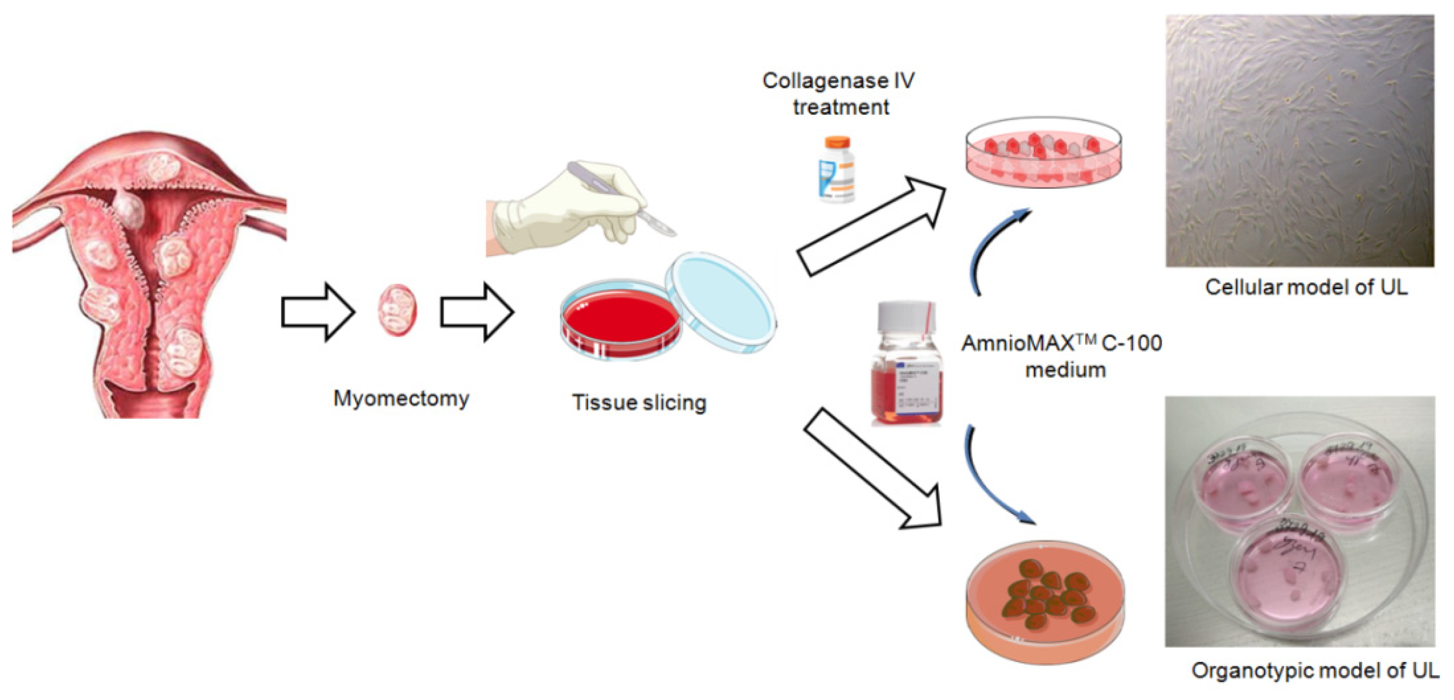
3.1. Obtaining Primary Cell Culture from Fragments of Collagenase-Treated Leiomyoma Nodules
 CRITICAL STEP One hour before handling the material, turn on the Desar and UV lamp in the Biosafety Cabinet Class II. Ensure that all the procedures are completed under sterile conditions.
- Wash tumor fragments, total volume not less than 3.0–3.5 cm3, in three changes of saline, then place in a Petri dish with 15 mL of saline and shred into fragments about 3 × 3 × 3 mm in diameter using a scalpel (scissors).
- Then, transfer the tissue fragments to 15 mL centrifuge tubes with 5 mL of Eagle’s MEM nutrient medium (Biolot, Saint-Petersburg, Russia) and 2.5 mg of type IV collagenase (Sigma Aldrich, Saint Louis, MO, USA), which corresponds to enzyme activity of 200 U/mL.
- Incubate tubes with tumor fragments in a tilted position for 90–120 min at 37 °C, shake or resuspend periodically.
- After incubation, add 5 mL of DPBS to the tubes and resuspend the tissue fragments using a 5-mL automatic pipette to obtain a cell suspension.
- Then, centrifuge the cells for 10 min at 200× g, remove the supernatant, and separate the precipitate.
- Then, add 5 mL of DPBS to the tubes again, well resuspend the precipitate, and centrifuge for 10 min at 200× g, remove the supernatant, and resuspend the precipitate again.
- Then, add 2 mL of pre-diluted AmnioMax Basal Medium with AmnioMax Supplement serum to the tubes. Transfer the resulting suspension to culture flasks T-25 (Thermo Fisher Scientific, Waltham, MA, USA) using a disposable sterile Pasteur pipette and incubate for 12–18 days at 37 °C and 5% CO2 to reach 80% confluence. Then the culture medium should be changed every 3–4 days.
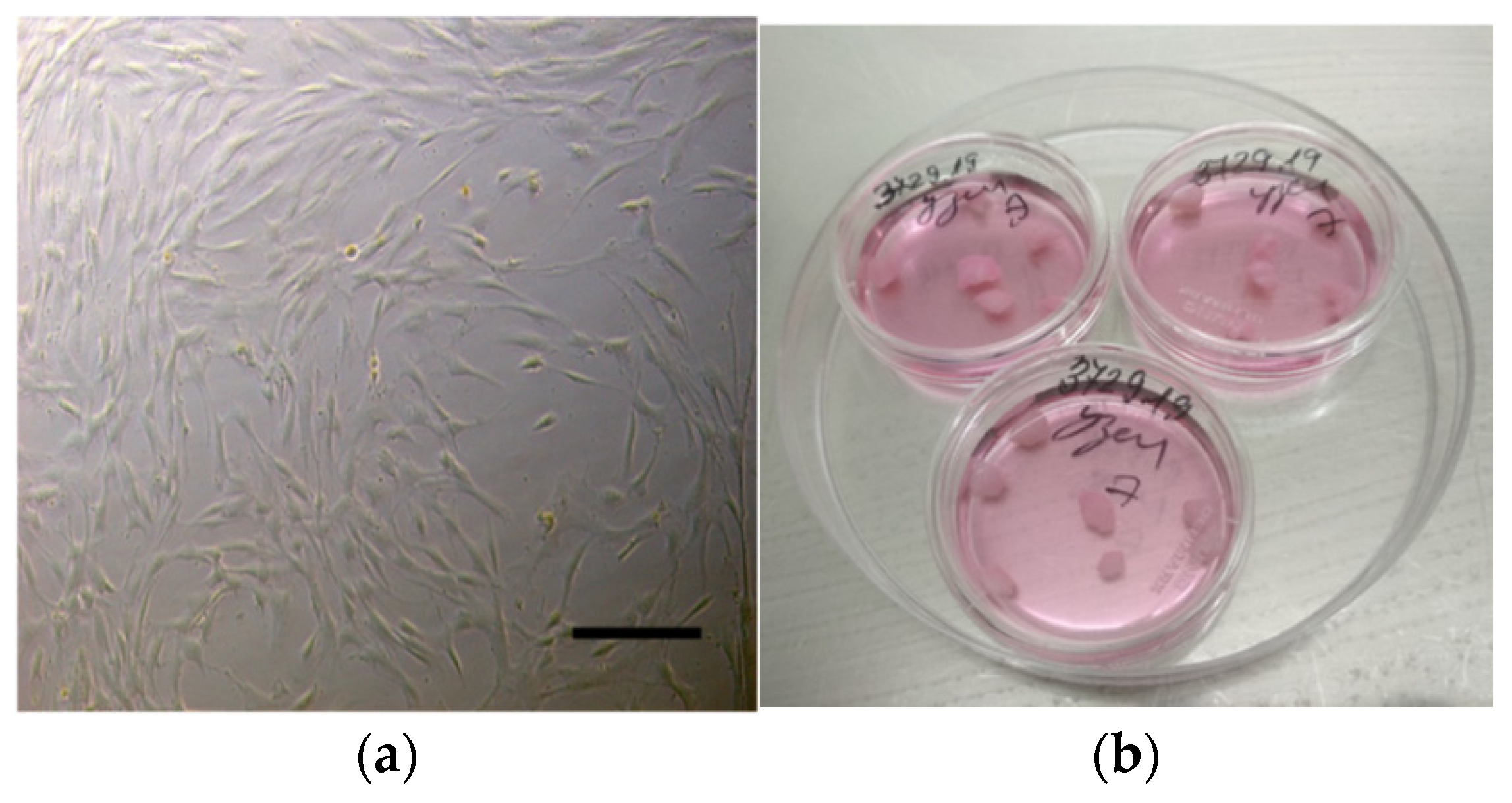
- When primary (p0) UL cultures reach 80% confluence (Figure 2a), they can be used for experiments or for the first passaging. We recommend taking at least 10–15% of p0 cells to achieve 80% confluence of p1 culture in 7–12 days. The viability of cells after passage can be monitored by the Trypan Blue exclusion test.
3.2. Freezing of Leiomyoma Cell Cultures and Nucleic Acid Isolation
CRITICAL STEP The following protocol steps should be performed in the Biosafety Cabinet Class II.- Wash the cells twice with Versen’s solution.
- Remove the cells by adding 700 µL of Trypsin-Versen solution to each well (1:3 ratio).
- Incubate for 4–5 min at +37 °C. 4.
- Add 5.0 mL of 0.9% NaCl and gently wash cells from the bottom of the vial.
- Transfer the cell suspension to 15.0 mL centrifuge tubes.
- Then, centrifuge the cells for 10 min at 200× g, remove the supernatant, and separate the precipitate.
- Then, add 5 mL of DPBS to the tubes again, well resuspend the precipitate, and centrifuge for 10 min at 200× g, then remove the supernatant.
- Add 300 μLof DPBS solution without Ca and Mg, mix gently.
- Transfer 100 µL of the cell suspension to a 2.0 mL tube containing 1.0 mL of RNA stabilizer. Leave at room temperature for 1 h. Then use for RNA isolation or store in a −80 °C freezer for up to 1 year if necessary.
- Transfer 100 µL of cell suspension to a 2.0 mL dry tube and use for DNA extraction. If necessary, store in a −80 °C freezer for up to 1 year.
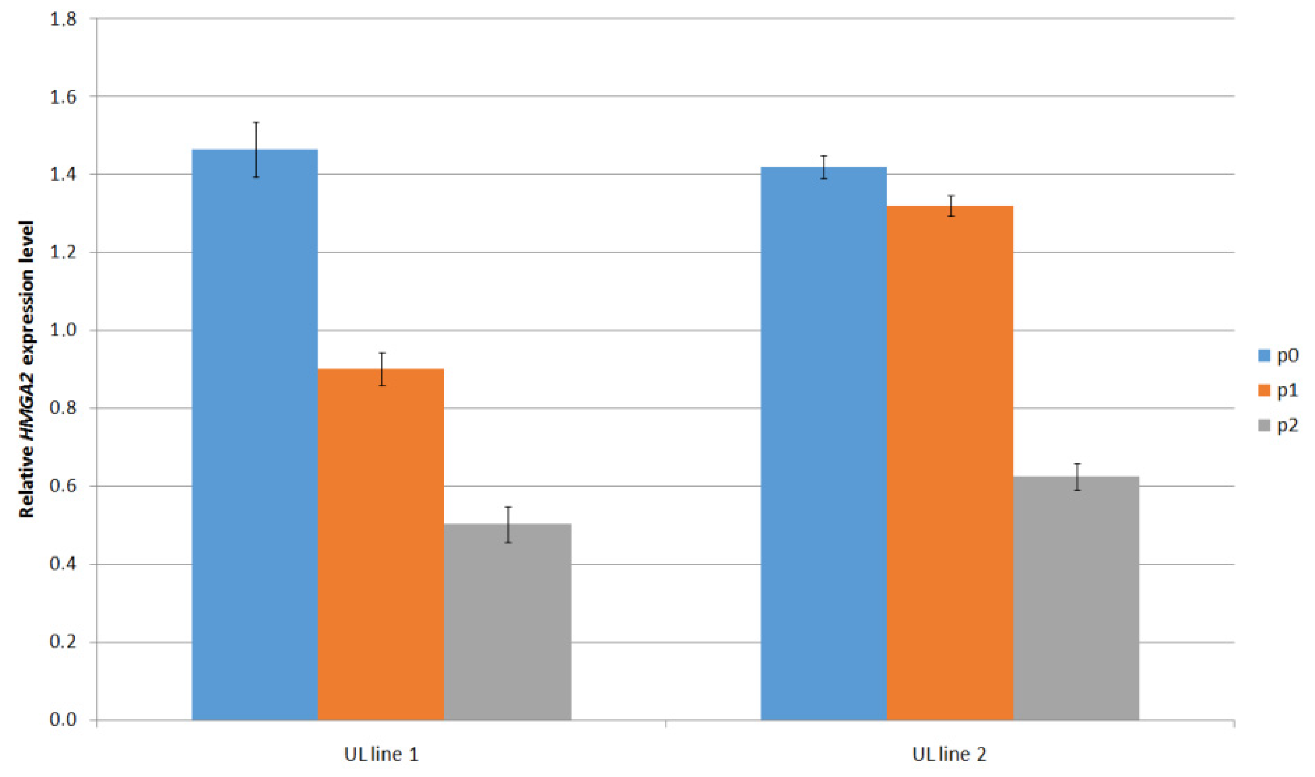
- Transfer 100 µL of cell suspension to a 2.0 mL cryotube containing a mixture of 100 µL dimethyl sulfoxide and 900 µL bovine serum. Mix gently by pipetting. Cryotubes should bescrewed thoroughly. Label cryovials with a water- and low-temperature resistant marker. Label cryoprobes with the date, culture name, and passage. If necessary, store in a freezer at −80 °C for no more than 1 year. According to our observations, when p0 leiomyoma cell lines are thawed, cells carrying the MED12 mutation and/or increased expression of the HMGA2 gene are preserved in p1 culture.
3.3. Organotypic Model from Native Fragments of Leiomyoma Nodules
- Using scissors or a scalpel, divide a large fragment of the node into smaller ones (approximately 3 × 3 × 3 or 4 × 4 × 4 mm) in the number necessary for subsequent experiments.
- Transfer 5–7 fragments to each 35 mm diameter Petri dish (Figure 2b).
- Add 1.0 mL of Gibco®AmnioMAX™ C-100 Complete Medium. Place in a CO2 incubator. According to literature data and personal observations, cells in tumor slices remain viable at least for 7–21 days [10]. It is recommended to keep tumor fragments ex vivo in culture for up to 7 days with the medium change every 3–4 days when using them in experimental work.
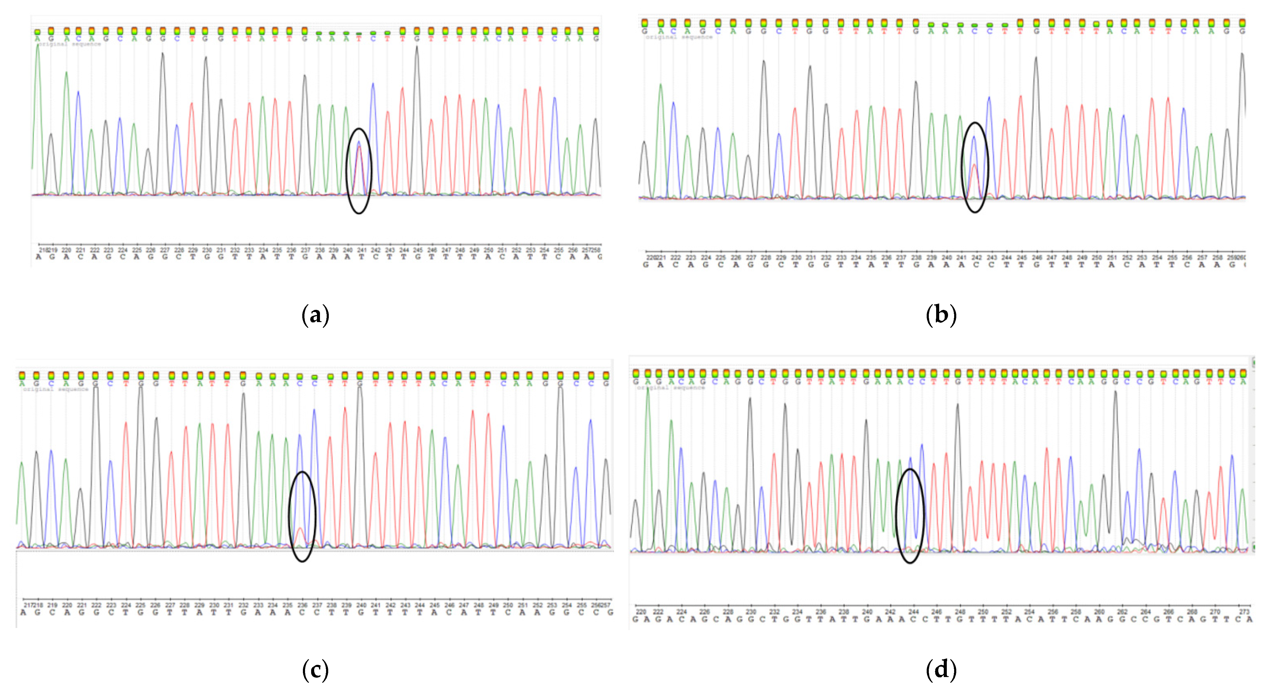
CRITICAL STEP The culture medium should not completely cover the fragments, providing contact with air (10–15% of the fragment surface) in the CO2 incubator and preventing it from floating up. According to our observations, on the 5–7th day of cultivation, the cells begin to migrate into the culture medium and ensure the fixation of the UL fragment on the substrate.3.4. MED12 Mutation Analysis
3.5. Reverse Transcription of RNA and HMGA2 Gene Expression Analysis
3.5.1. Reverse Transcription of RNA
- Perform eachreaction in a total volume of 20 μL containing 10 mmol d-NTP and 200 U/mL MMLUV in the presence of oligo-dT16 (2.5 mM) and random hexamers (3 mg/mL) with the addition of 100 to 500 ng total RNA.
- Place strips of random oligonucleotide hexamers with the reaction mixture into Thermocycler T100. The reverse transcription reaction was performed at: +25 °C–10 min; +37 °C–120 min; +85 °C–5 min; +4 °C–5 min. The cDNA was stored at +4 °C. The obtained cDNA was used for real-time PCR.
3.5.2. HMGA2 Gene Expression Analysis
- 12 µL cDNA mixture [x ng/mkl] + 12 µL H2O, stir by pipetting, incubate for 10 min.
- 12 µL of [x/2 ng/mkl] cDNA mixture + 12 µL H2O, stir by pipetting, incubate for 10 min.
- 12 µL of [x/4 ng/mkl] cDNA mixture + 12 µL H2O, stir by pipetting, incubate for 10 min.
- 12 µL of [x/8 ng/mkl] cDNA mixture + 12 µL H2O, stir by pipetting, incubate for 10 min.
- 12 µL of [x/16 ng/mkl] cDNA mixture + 12 µL H2O, stir by pipetting, incubate for 10 min.
- The calibration dilutions must be stored at −20 °C.
CRITICAL STEP The following steps of the protocol are performed in the PCR box.- 1.
- Take out the strips and the lids for the strips using tweezers.
- 2.
- In 1.5 mL tubes (or 2 mL-depending on the number of samples), make separate mixtures for each set of primers at a concentration of 5 pmol (HMGA2 gene: forward 5′-AGA GTC CCT CTA AAG CAG CTC A-3′; reverse 5′-CAA CTG CTGCTG AGG TAG AAA TCG-3′), including primers to the housekeeping gene (β-actingene:5′-TGC CGA CAG GAT GCA GAA G-3′; reverse 5′-GCC GAT CCACAC GGA GTA CT-3′). Tubes for the mixtures should be wrapped in foil.
- 3.
- Scheme of mixture preparation (per sample taking into account prepared standards and one blank sample; in order of adding reagents):
- µL H2O
- 4 µL MgCl2 (25 mM)
- µL dNTP’s (2.5 mM)
- 2.5 µL EvaGreen (10×)
- 1 µL F-primer
- 1 µL R-primer
- 0.3 µL Taq-pol.
- 4.
- Stir the mixture by pipetting; pipette into 22.5 µL strips. Try to dig close to the bottom of the strips; do not leave any droplets on the walls at the top.
- 5.
- Using separate spouts, dig 2.5 µL of cDNA into strips. Drop each sample in two replicates.
- 6.
- Close the test tubes with caps. Place in the amplifier Rotor-Gene 3000 (Corbett, Australia) and run the appropriate program (Single cycle 95°–5 min; then 38 cycle: 94°–15 s; 600–60 s. Then heating from 60° to 95° in 0.5 degree increments, 10 s each step).
- 7.
- The relative level of mRNA expression was calculated by the ΔΔCt method (Livak method) using ExpressionSuit V1.0.3 software.
4. Expected Results
Supplementary Materials
Author Contributions
Funding
Institutional Review Board Statement
Informed Consent Statement
Data Availability Statement
Conflicts of Interest
References
- Ohara, N. Action of progesterone receptor modulators on uterine leiomyomas. Clin. Exp. Obstet. Gynecol. 2008, 35, 165–166. [Google Scholar] [PubMed]
- Zhu, Y.; Zhang, T.; Xie, S.; Tu, R.; Cao, Y.; Guo, X.; Zhou, J.; Zhou, X.; Cao, L. Gestrinone inhibits growth of human uterine leiomyoma may relate to activity regulation of ERα, Src and P38 MAPK. Biomed. Pharmacother. 2012, 66, 569–577. [Google Scholar] [CrossRef] [PubMed]
- Segars, J.H.; Parrott, E.C.; Nagel, J.D.; Guo, X.C.; Gao, X.; Birnbaum, L.S.; Pinn, V.W.; Dixon, D. Proceedings from the third national institutes of health international congress on advances in uterine leiomyoma research: Comprehensive review, conference summary and future recommendations. Hum. Reprod. Update 2014, 20, 309–333. [Google Scholar] [CrossRef] [PubMed]
- Shtykalova, S.V.; Egorova, A.A.; Maretina, M.A.; Freund, S.A.; Baranov, V.S.; Kiselev, A.V. Molecular Genetic Basis and Prospects of Gene Therapy of Uterine Leiomyoma. Russ. J. Genet. 2021, 57, 1002–1016. [Google Scholar] [CrossRef]
- Nadine Markowski, D.; Tadayyon, M.; Bartnitzke, S.; Belge, G.; Maria Helmke, B.; Bullerdiek, J. Cell cultures in uterine leiomyomas: Rapid disappearance of cells carrying MED12 mutations. Genes Chromosomes Cancer 2014, 53, 317–323. [Google Scholar] [CrossRef] [PubMed]
- Holzmann, C.; Markowski, D.N.; Bartnitzke, S.; Koczan, D.; Helmke, B.M.; Bullerdiek, J. A rare coincidence of different types of driver mutations among uterine leiomyomas (UL). Mol. Cytogenet. 2015, 8, 76. [Google Scholar] [CrossRef] [PubMed] [Green Version]
- Bloch, J.; Holzmann, C.; Koczan, D.; Helmke, B.M.; Bullerdiek, J. Factors affecting the loss of MED12-mutated leiomyoma cells during in vitro growth. Oncotarget 2017, 8, 34762–34772. [Google Scholar] [CrossRef] [PubMed] [Green Version]
- Klemke, M.; Meyer, A.; Nezhad, M.H.; Bartnitzke, S.; Drieschner, N.; Frantzen, C.; Schmidt, E.H.; Belge, G.; Bullerdiek, J. Overexpression of HMGA2 in uterine leiomyomas points to its general role for the pathogenesis of the disease. Genes Chromosomes Cancer 2009, 48, 171–178. [Google Scholar] [CrossRef] [PubMed]
- Wang, J.; Ohara, N.; Takekida, S.; Xu, Q.; Maruo, T. Comparative effects of heparin-binding epidermal growth factor-like growth factor on the growth of cultured human uterine leiomyoma cells and myometrial cells. Hum. Reprod. 2005, 20, 1456–1465. [Google Scholar] [CrossRef] [PubMed] [Green Version]
- Salas, A.; López, J.; Reyes, R.; Évora, C.; de Oca, F.M.; Báez, D.; Delgado, A.; Almeida, T.A. Organotypic culture as a research and preclinical model to study uterine leiomyomas. Sci. Rep. 2020, 10, 5212. [Google Scholar] [CrossRef] [PubMed]
- Osinovskaya, N.S.; Malysheva, O.V.; Shved, N.Y.; Ivashchenko, T.E.; Sultanov, I.Y.; Efimova, O.A.; Yarmolinskaya, M.I.; Bezhenar, V.F.; Baranov, V.S. Frequency and Spectrum of MED12 Exon 2 Mutations in Multiple Versus Solitary Uterine Leiomyomas From Russian Patients. Int. J. Gynecol. Pathol. 2016, 35, 509–515. [Google Scholar] [CrossRef] [PubMed]
- Dzhemlikhanova, L.K.; Efimova, O.A.; Osinovskaya, N.S.; Parfenyev, S.E.; Niauri, D.A.; Sultanov, I.Y.; Malysheva, O.V.; Pendina, A.A.; Shved, N.Y.; Ivashchenko, T.E.; et al. Catechol-O-methyltransferase Val158Met polymorphism is associated with increased risk of multiple uterine leiomyomas either positive or negative for MED12 exon 2 mutations. J. Clin. Pathol. 2017, 70, 233–236. [Google Scholar] [CrossRef] [PubMed]
- Islam, M.S.; Protic, O.; Ciavattini, A.; Giannubilo, S.R.; Tranquilli, A.L.; Catherino, W.H.; Castellucci, M.; Ciarmela, P. Tranilast, an orally active antiallergic compound, inhibits extracellular matrix production in human uterine leiomyoma and myometrial cells. Fertil. Steril. 2014, 102, 597–606. [Google Scholar] [CrossRef] [PubMed]
- Moore, A.B.; Yu, L.; Swartz, C.D.; Zheng, X.; Wang, L.; Castro, L.; Kissling, G.E.; Walmer, D.K.; Robboy, S.J.; Dixon, D. Human uterine leiomyoma-derived fibroblasts stimulate uterine leiomyoma cell proliferation and collagen type I production, and activate RTKs and TGF beta receptor signaling in coculture. Cell Commun. Signal. 2010, 8, 10. [Google Scholar] [CrossRef] [PubMed] [Green Version]


Publisher’s Note: MDPI stays neutral with regard to jurisdictional claims in published maps and institutional affiliations. |
© 2022 by the authors. Licensee MDPI, Basel, Switzerland. This article is an open access article distributed under the terms and conditions of the Creative Commons Attribution (CC BY) license (https://creativecommons.org/licenses/by/4.0/).
Share and Cite
Shved, N.; Egorova, A.; Osinovskaya, N.; Kiselev, A. Development of Primary Monolayer Cell Model and Organotypic Model of Uterine Leiomyoma. Methods Protoc. 2022, 5, 16. https://doi.org/10.3390/mps5010016
Shved N, Egorova A, Osinovskaya N, Kiselev A. Development of Primary Monolayer Cell Model and Organotypic Model of Uterine Leiomyoma. Methods and Protocols. 2022; 5(1):16. https://doi.org/10.3390/mps5010016
Chicago/Turabian StyleShved, Natalia, Anna Egorova, Natalia Osinovskaya, and Anton Kiselev. 2022. "Development of Primary Monolayer Cell Model and Organotypic Model of Uterine Leiomyoma" Methods and Protocols 5, no. 1: 16. https://doi.org/10.3390/mps5010016