Novel Protocol for the Preparation of Porcine Bone Marrow Primary Cell Culture for African Swine Fever Virus Isolation
Abstract
:1. Introduction
2. Equipment
- Class II biological safety cabinet (model: Streamline—E Series, catalog number: SC2-E).
- Humidified 37 ± 2 °C, 5 ± 1% CO2 incubator (model: S-Bt Smart Bio Term, catalog number: BS-010425-A01).
- −70 °C freezer (Sanyo MDF-U53V, catalog number: 9782).
- −20 °C freezer (model: LG, catalog number: GA-B419SQQL).
- Refrigerator, 4 °C, with an acceptable range of 2 °C to 8 °C (model: LG, catalog number: GA-B419SQQL).
- Water bath, 37 °C (model: WB-4MS Stirred water bath, BioSan, catalog number: BS-010406-AAA).
- Portable Pipettor (Dragon Laboratory Instruments Limited, Beijing, China, Levo plus, catalog number: 710932).
- Micropipettors: Single channel, 1–10 µL, 2–20 µL, 50–200 µL, 100–1000 µL (Eppendorf catalog numbers: Q11390H, K12770H, Q24196H, L18745H).
- Micropipettors: Multi-channel, 0.5–10 µL, 5–50 µL, 50–300 µL (BTLab systems, Saint Louis, USA, Lab System, catalog numbers: N87539, D23745, E21512).
- Multi-channel for larger volumes (RaininTM Pipet-Life XLS, L-1200, 100–1200 µL, catalog number: 17014497).
- Inverted Microscope (Euromex Microscopen, Arnhem, Netherlands, model: Olympus Euromex Oxion, catalog number: SKU: EOX.2053-PLPH).
- Tabletop Centrifuge (HERMLE Labortechnik GmbH, Wehingen, Germany, model: HERMLE Z 306, catalog number: 310.00 v01).
- Shaker incubator (BioSan, Riga, Latvia, model: Biosan ES-20, catalog number: BS-010135-AAA).
- Electronic balance (model: OHAUS Navigator NV212, catalog number: 83033081).
- Countess™ automatic cell counter.
3. Materials and Reagents
- Pipette tips: 10 μL, 20 μL, 200 μL, 1000 μL (Thermo Fisher Scientific, Waltham, MA, USA, catalog numbers: 2140-05, 72830-440, 72830-044, 72830-042).
- Sterile serological pipettes: 1 mL, 5 mL, 10 mL, 25 mL, 50 mL (Thermo Fisher Scientific, catalog numbers: 14672-918, 14672-920, 4101, 14672-900, 53384-451).
- Conical tubes: 15 and 50 mL (Corning, AZ, USA, Falcon®, catalog numbers: 353110 and 352098)
- T-75 flask (Corning, catalog number: 430641U).
- T-25 flask (Corning, catalog number: 430825).
- T-225 flask (Corning, catalog number: 431082).
- Millex-HV Syringe Filter Unit, 0.45 µm, PVDF, 33 mm, gamma sterilized (Catalog number: SLHV033RS).
- Absorbent paper (Kimtech Pure CL4, Kimberly-Clark Professional, GA, USA, catalog number: 7646).
- Reservoirs (Costar Reagent Reservoir, Corning®, catalog number: 29442-474).
- TrypLE™ Express Enzyme (1X), no phenol red (Thermofisher, catalog number: 12604-013).
- EZFlow® cell strainers (catalog number 410-0002-OEM).
- Minimum Essential Medium (MEM) (Thermofisher, catalog number: 11095-080).
- ASFV field strain: For example an ASFV-Arm07 field strain (Arm07).
- Trypan blue solution, 0.4% (Thermofisher scientific, catalog number: 15250061).
- Gentamicin 40mg/ml Solution (Wockhardt UK Ltd. Wrexham, UK, catalog number: J01GB03).
- Penicillin G sodium salt (Sigma 1000000 Units, Sigma-Aldrich, Sofia, Bulgaria, Catalog Number P3032-1MU).
- Streptomycin sulfate (Sigma, Cat. Number 5711-100GM).
- Saline solution (Sigma-Aldrich, catalog number: S8776).
- Bacto™ TC Lactalbumin Hydrolysate (Thermofisher, catalog number 259962).
- 70% Ethanol.
- Fetal Bovine Serum (Sigma-Aldrich, Cat. Number F7524-500ML).
4. Animals
5. Procedure
5.1. Selection of Tubular Bones
5.2. Isolation of PBMP Cells
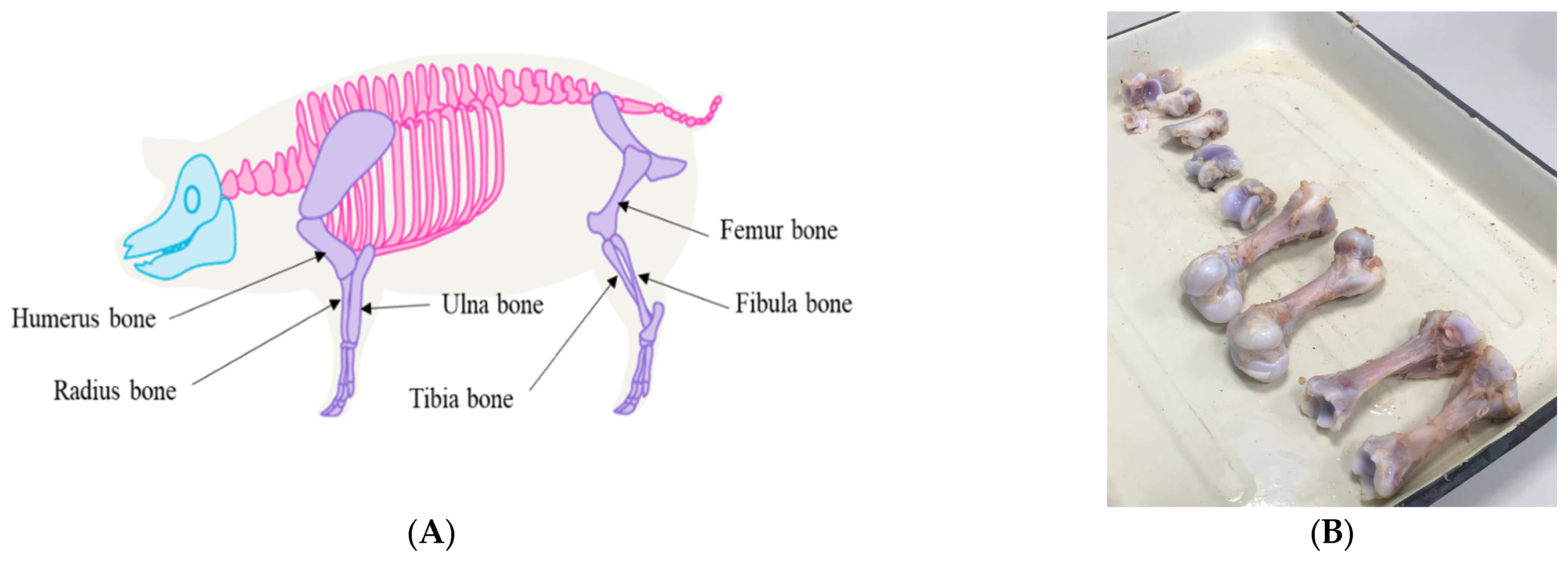
- Place isolated tubular bones (humerus, ulna, radius, femur, and tibia) in a vessel and wash with 70% ethanol by incubating in a shaker-incubator at room-temperature and 250 rpm for 10–15 min in order to disinfect the surfaces of the samples (Figure 1A).
- Remove the muscle and tissue mass from the bones with a surgical scalpel and rewash the bones, similar to step 1 (Figure 1B).
- 3.
- Cut the bones with a sterile hand pruner into small cubes approximately 2 cm2, thereby exposing the bone marrow as much as possible (Figure 2A).
- 4.
- Place the bone pieces uniformly in a T-225 plastic culture flask (Figure 2B).
- 5.
- Add washing buffer (Table 1) to the bone pieces in a ratio of 1:1 (m/v), after which the vials are carefully inverted 1–2 times. Incubate at room temperature (RT) for 2 min, and then discard the buffer.
- 6.
- Add saline solution in a ratio of 1:1.5 (m/v) into the flasks with bone pieces, and incubate in a shaker-incubator at 37 °C and 250 rpm for 20 min to remove the bone marrow cells from the bone cavities.
- 7.
- Filter the cell suspension through an 8-layer medical gauze filter into a sterile glass vial (about 120 mL each). Step 6 can be repeated in order to gather more cells from the bone marrow. The filtrate is evenly poured into sterile centrifuge beakers and centrifuged at +4 °C and 1400 rpm for 20 min.
- 8.
- After centrifugation, discard the supernatant and re-suspend the pellet in growth medium (Table 1) (about 25 mL for one glass vial). The cell suspension is successively passed through an 8-layer medical gauze filter and through nylon cell strainers with a mesh size of 100 and 70 µm.
- 9.
- Calculate the cell concentration using a CountessTM automatic cell counter or analogue (ex. cell suspension sample is diluted 10 times in saline solution, and 0.4% trypan blue solution is added to the diluted volume in a ratio of 1:1 and loaded into the chamber of a disposable counter slide.)
- 10.
- Dilute the cell suspension in growth medium, and adjust to an inoculation concentration of 20–30 million cells/cm3.
- 11.
- Add the diluted suspension of PBMP cells to cell culture flasks as indicated in Table 2.
- 12.
- Incubate the flasks in a humidifier at +37 °C with 5% CO2 for 48 h.
- 13.
6. Expected Results (Isolation of African Swine Fever Virus from Biological Samples)
6.1. Sample Preparation and Infection
6.2. ASF Virus Identification
7. Validation
8. Conclusions
Supplementary Materials
Author Contributions
Funding
Acknowledgments
Conflicts of Interest
References
- Sánchez-Vizcaíno, J.M.; Laddomada, A.; Arias, M.L. African Swine Fever Virus. In Diseases of Swine; Wiley: Hoboken, NJ, USA, 2019; pp. 443–452. [Google Scholar] [CrossRef]
- Krug, P.W.; Holinka, L.G.; O’Donnell, V.; Reese, B.; Sanford, B.; Fernandez-Sainz, I.; Gladue, D.P.; Arzt, J.; Rodriguez, L.; Risatti, G.R.; et al. The Progressive Adaptation of a Georgian Isolate of African Swine Fever Virus to Vero Cells Leads to a Gradual Attenuation of Virulence in Swine Corresponding to Major Modifications of the Viral Genome. J. Virol. 2015, 89, 2324–2332. [Google Scholar] [CrossRef] [PubMed]
- Mazloum, A.; van Schalkwyk, A.; Shotin, A.; Zinyakov, N.; Igolkin, A.; Chernishev, R.; Debeljak, Z.; Korennoy, F.; Sprygin, A.V. Whole-genome sequencing of African swine fever virus from wild boars in the Kaliningrad region reveals unique and distinguishing genomic mutations. Front. Vet. Sci. 2023, 9, 1019808. [Google Scholar] [CrossRef]
- De León, P.; Bustos, M.J.; Carrascosa, A.L. Laboratory methods to study African swine fever virus. Virus Res. 2013, 173, 168–179. [Google Scholar] [CrossRef]
- Hess, W.R.; DeTray, D.E. The use of leukocyte cultures for diagnosing African swine fever (ASF). Bull. Office Intern. Epizoot. 1960, 8, 317–320. [Google Scholar]
- Malmquist, W.A.; Hay, W. Hemadsorption and cytopathic effect produced by African swine fever virus in swine bone marrow and buffy coat cultures. Am. J. Vet. Res. 1960, 21, 104–108. [Google Scholar]
- Malmquist, W.A. Propagation, modification, and hemadsorption of African swine fever virus in cell cultures. Am. J. Vet. Res. 1962, 23, 241–247. [Google Scholar] [PubMed]
- Wardley, R.C.; Wilkinson, P.J.; Hamilton, F. African Swine Fever Virus Replication in Porcine Lymphocytes. J. Gen. Virol. 1977, 37, 425–427. [Google Scholar] [CrossRef]
- Greig, A.S.; Boulanger, P.; Bannister, G.L. African swine fever. V. Cultivation of the virus in primary pig kidney cells. Can. J. Comp. Med. Vet. Sci. 1967, 31, 24–31. [Google Scholar]
- Breese, S.S.; Stone, S.S.; Deboer, C.J.; Hess, W.R. Electron microscopy of the interaction of African swine fever virus with ferritin-conjugated antibody. Virology 1967, 31, 508–513. [Google Scholar] [CrossRef]
- De Castro, M.P. Compartamento do virus aftoso em cultura de celulas: Susceptibilidade da Rinhagem de celulas swinas IB-RS-2. Arq. Inst. Biol. Sao Paulo 1964, 31, 63–78. [Google Scholar]
- Breese, S.S.; DeBoer, C.J. Electron microscope observations of African swine fever virus in tissue culture cells. Virology 1966, 28, 420–428. [Google Scholar] [CrossRef] [PubMed]
- Almeida, J.D.; Waterson, A.P.; Plowright, W. The morphological characteristics of African swine fever virus and its resemblance to tipula iridescent virus. Arch. Virol. 1967, 20, 392–396. [Google Scholar] [CrossRef] [PubMed]
- Dardiri, A.H.; Bachrach, H.L.; Heller, E. Inhibition by Rifampin of African Swine Fever Virus Replication in Tissue Culture. Infect. Immun. 1971, 4, 34–36. [Google Scholar] [CrossRef]
- Cox, B.F.; Hess, W.R. Note on the African swine fever investigation in Nysaland. Bull. epizoot. Dis. Afr. 1962, 10, 439–440. [Google Scholar]
- Gil-Fernandez, C.; Vilas-Minondo, P.; García-Gancedo, A. Plaque formation by African swine fever virus in chick embryo fibroblasts in the absence of CO2 atmosphere. Arch. Virol. 1976, 52, 207–216. [Google Scholar] [CrossRef] [PubMed]
- Ribeiro, M.J.; Azevedo, J.A.R. La peste porcine Africaine au Portugal. Bull. Off. int. Epizoot. 1961, 55, 88–106. [Google Scholar]
- Enjuanes, L.; Carrascosa, A.L.; Moreno, M.A.; Vinuela, E. Titration of African Swine Fever (ASF) Virus. J. Gen. Virol. 1976, 32, 471–477. [Google Scholar] [CrossRef]
- Tabarés, E.; Martinez, J.; Gonzalvo, F.R.; Sánchez-Botija, C. Proteins specified by African swine fever virus: II. Analysis of proteins in infected cells and antigenic properties. Arch. Virol. 1980, 66, 119–132. [Google Scholar] [CrossRef]
- Vigario, J.D.; Castro Portugal, F.L.; Ferreira, C.A.; Festas, M.B. Purification and Study of the Structural Polypeptides of African Swine Fever Virus; Commision of the European Union: Brussels, Belgium, 1977; pp. 469–482. [Google Scholar]
- Tabarés, E.; Olivares, I.; Santurde, G.; Garcia, M.J.; Martin, E.; Carnero, M.E. African swine fever virus DNA: Deletions and additions during adaptation to growth in monkey kidney cells. Arch. Virol. 1987, 97, 333–346. [Google Scholar] [CrossRef]
- Carrascosa, A.L.; Bustos, M.J.; Galindo, I.; Viñuela, E. Virus-specific cell receptors are necessary, but not sufficient, to confer cell susceptibility to African swine fever virus. Arch. Virol. 1999, 144, 1309–1321. [Google Scholar] [CrossRef]
- Hurtado, C.; Bustos, M.J.; Carrascosa, A.L. The use of COS-1 cells for studies of field and laboratory African swine fever virus samples. J. Virol. Methods 2010, 164, 131–134. [Google Scholar] [CrossRef] [PubMed]
- Masujin, K.; Kitamura, T.; Kameyama, K.-I.; Okadera, K.; Nishi, T.; Takenouchi, T.; Kitani, H.; Kokuho, T. An immortalized porcine macrophage cell line competent for the isolation of African swine fever virus. Sci. Rep. 2021, 11, 4759. [Google Scholar] [CrossRef] [PubMed]
- Gómez-Villamandos, J.; Bautista, M.; Sánchez-Cordón, P.; Carrasco, L. Pathology of African swine fever: The role of monocyte-macrophage. Virus Res. 2013, 173, 140–149. [Google Scholar] [CrossRef] [PubMed]
- Rai, A.; Pruitt, S.; Ramirez-Medina, E.; Vuono, E.A.; Silva, E.; Velazquez-Salinas, L.; Carrillo, C.; Borca, M.V.; Gladue, D.P. Identification of a Continuously Stable and Commercially Available Cell Line for the Identification of Infectious African Swine Fever Virus in Clinical Samples. Viruses 2020, 12, 820. [Google Scholar] [CrossRef] [PubMed]
- Kwon, H.-I.; Do, D.T.; Van Vo, H.; Lee, S.-C.; Kim, M.H.; Nguyen, D.T.T.; Tran, T.M.; Le, Q.T.V.; Ngo, T.T.N.; Nguyen, N.M.; et al. Development of optimized protocol for culturing African swine fever virus field isolates in MA104 cells. Can. J. Vet. Res. 2022, 86, 261–268. [Google Scholar]
- Zhao, D.; Sun, E.; Huang, L.; Ding, L.; Zhu, Y.; Zhang, J.; Shen, D.; Zhang, X.; Zhang, Z.; Ren, T.; et al. Highly lethal genotype I and II recombinant African swine fever viruses detected in pigs. Nat. Commun. 2023, 14, 3096. [Google Scholar] [CrossRef]
- Mazloum, A.; Igolkin, A.S.; Zinyakov, N.G.; Van Schalkwyk, A.; Vlasova, N.N. Changes in the genome of African swine fever virus (Asfarviridae: Asfivirus: African swine fever virus) associated with adaptation to reproduction in continuous cell culture. Probl. Virol. 2021, 66, 211–216. [Google Scholar] [CrossRef]
- Mazloum, A.; van Schalkwyk, A.; Shotin, A.; Igolkin, A.; Shevchenko, I.; Gruzdev, K.N.; Vlasova, N. Comparative Analysis of Full Genome Sequences of African Swine Fever Virus Isolates Taken from Wild Boars in Russia in 2019. Pathogens 2021, 10, 521. [Google Scholar] [CrossRef]
- Bastos, A.D.; Penrith, M.L.; Cruciere, C.; Edrich, J.L.; Hutchings, G.; Roger, F.; Couacy-Hymann, E.G.; Thomson, G.R. Genotypingfield strains of African swine fever virus by partial p72 gene characterisation. Arch. Virol. 2003, 148, 693–706. [Google Scholar] [CrossRef]
- Quembo, C.J.; Jori, F.; Vosloo, W.H.; Heath, L. Genetic characterization of African swine fever virus isolates from soft ticks at thewildlife/domestic interface in Mozambique and identification of a novel genotype. Transbound. Emerg. Dis. 2018, 65, 420–431. [Google Scholar] [CrossRef]
- Mazloum, A.; van Schalkwyk, A.; Chernyshev, R.; Igolkin, A.; Heath, L.; Sprygin, A. A Guide to Molecular Characterization of Genotype II African Swine Fever Virus: Essential and Alternative Genome Markers. Microorganisms 2023, 11, 642. [Google Scholar] [CrossRef] [PubMed]


| Wash Buffer (1×) | ||
|---|---|---|
| Volume Added | Final Concentration | |
| Saline solution | Up to 500 mL | |
| Gentamicin 40 mg/mL Solution | 10 mL | 1.6 mg/mL (1.6%) |
| Streptomycin sulfate | 1g | 0.1% |
| Penicillin G sodium salt | 106 Units | 103 Units |
| Growth Medium | ||
| Volume Added | Final Concentration | |
| Minimum Essential Medium (MEM) | Up to 1000 mL | |
| Lactalbumin hydrolysate | 2.5 g | 0.25% |
| Fetal Bovine Serum (FBS) | 150 mL | 15% |
| Gentamicin 40 mg/mL Solution | 2 mL | 0.08 mg/mL (0.08%) |
| Maintenance Medium | ||
| Volume Added | Final Concentration | |
| Minimum Essential Medium (MEM) | Up to 1000 mL | |
| Lactalbumin hydrolysate | 2.5 g | 0.25% |
| Fetal Bovine Serum (FBS) | 50 mL | 5% |
| Gentamicin 40 mg/mL Solution | 2 mL | 0.08 mg/mL (0.08%) |
| Area (cm2) | Cell/cm3 | |
|---|---|---|
| Flask | ||
| T-25 | 25 | 10–15 × 107 |
| T-75 | 75 | 30–45 × 107 |
| T-225 | 225 | 15–20 × 108 |
| Culture plates | ||
| 48-well | 1.1 | 6–10 × 107 |
| 96-well | 0.32 | 2–3 × 10⁷ |
| Area (cm2) | Volume (mL) | |
|---|---|---|
| Flask | ||
| T-25 | 25 | 3–5 |
| T-75 | 75 | 8–15 |
| T-225 | 225 | 45–68 |
| Culture plates | ||
| 48-well | 1.1 | 0.2–0.4 |
| 96-well | 0.32 | 0.1–0.2 |
Disclaimer/Publisher’s Note: The statements, opinions and data contained in all publications are solely those of the individual author(s) and contributor(s) and not of MDPI and/or the editor(s). MDPI and/or the editor(s) disclaim responsibility for any injury to people or property resulting from any ideas, methods, instructions or products referred to in the content. |
© 2023 by the authors. Licensee MDPI, Basel, Switzerland. This article is an open access article distributed under the terms and conditions of the Creative Commons Attribution (CC BY) license (https://creativecommons.org/licenses/by/4.0/).
Share and Cite
Puzankova, O.; Gavrilova, V.; Chernyshev, R.; Kolbin, I.; Igolkin, A.; Sprygin, A.; Chvala, I.; Mazloum, A. Novel Protocol for the Preparation of Porcine Bone Marrow Primary Cell Culture for African Swine Fever Virus Isolation. Methods Protoc. 2023, 6, 73. https://doi.org/10.3390/mps6050073
Puzankova O, Gavrilova V, Chernyshev R, Kolbin I, Igolkin A, Sprygin A, Chvala I, Mazloum A. Novel Protocol for the Preparation of Porcine Bone Marrow Primary Cell Culture for African Swine Fever Virus Isolation. Methods and Protocols. 2023; 6(5):73. https://doi.org/10.3390/mps6050073
Chicago/Turabian StylePuzankova, Olga, Vera Gavrilova, Roman Chernyshev, Ivan Kolbin, Alexey Igolkin, Alexandr Sprygin, Ilya Chvala, and Ali Mazloum. 2023. "Novel Protocol for the Preparation of Porcine Bone Marrow Primary Cell Culture for African Swine Fever Virus Isolation" Methods and Protocols 6, no. 5: 73. https://doi.org/10.3390/mps6050073


