Article

The Combined Effects of Cadmium and Microplastic Mixtures on the Digestion, Energy Metabolism, Oxidative Stress Regulation, Immune Function, and Metabolomes in the Pearl Oyster (Pinctada fucata martensii)

1
Fisheries College, Guangdong Ocean University, Zhanjiang 524088, China
2
Pearl Breeding and Processing Engineering Technology Research Centre of Guangdong Province, Guangdong Ocean University, Zhanjiang 524088, China
3
Guangdong Provincial Key Laboratory of Aquatic Animal Disease Control and Healthy Culture, Guangdong Ocean University, Zhanjiang 524088, China
4
Guangdong Science and Innovation Center for Pearl Culture, Guangdong Ocean University, Zhanjiang 524088, China
5
Pearl Research Institute, Guangdong Ocean University, Zhanjiang 524088, China
*
Author to whom correspondence should be addressed.
Fishes 2025, 10(3), 133; https://doi.org/10.3390/fishes10030133
Submission received: 12 February 2025 / Revised: 13 March 2025 / Accepted: 14 March 2025 / Published: 18 March 2025
(This article belongs to the Special Issue Advances in Bivalve Aquaculture)

Abstract

:
The accumulation of cadmium (Cd) and microplastics (MPs) can have major deleterious effects on the health of marine ecosystems and organisms, including the pearl oyster Pinctada fucata martensii. Here, we characterized the effects of Cd and MPs on key biochemical parameters of P. f. martensii via an experiment with various treatments. Pearl oysters were exposed to either only Cd (5 or 50 μg/L), only MPs (5 mg/L), or both Cd and MPs for 2 d, and this was followed by a 5-day recovery period. Measurements of the activities of lipase, amylase, protease, T-ATPase, catalase, glutathione peroxidase, acid phosphatase, and alkaline phosphatase enzymes, as well as the malondialdehyde content in the hepatopancreas, were made at various time points during the experiment. Metabolomics analysis of the gills was also performed. Significant interactions between time and treatment on lipase, protease, and catalase activities were observed. However, no significant effect of time–treatment interactions on amylase and T-ATPase activities was observed. Enzyme activities varied among groups both during the exposure period (6 to 48 h) and the recovery period. The malondialdehyde content was also increased throughout the experiment. Pathway analysis indicated that the purine metabolism, glycerophospholipid metabolism, nucleotide metabolism, arachidonic acid metabolism, neuroactive ligand–receptor interaction, and linoleic acid metabolism pathways were the most commonly affected under different treatments. The findings of our study revealed the differential effects of exposure time and treatment on enzyme activities and metabolites and their respective pathways. Our findings enhance our understanding of the biochemical responses of the pearl oyster P. f. martensii to environmental stressors, particularly Cd and MPs.
Key Contribution: Our findings will aid future studies of the mechanisms underlying the toxicity of mixtures of heavy metals and microplastics on various shellfish species.

1. Introduction

Plastics are important materials in all aspects of our daily life because of their low price, durability, light weight, high performance, and machinability. Plastics are a concerning source of pollution in many ecosystems [1]. Plastics discarded in the environment have physical, chemical, and biological effects on organisms. Microplastics (MPs), defined as plastic particles smaller than 5 mm in size, can be formed from these discarded plastics [2] and eventually enter the marine environment. The total amount of MPs in the ocean ranges from approximately 1.5 × 10¹³ to 5.1 × 10¹³ particles [3]. MPs can enter the organs and tissues of organisms through various pathways because of their small size [4,5], which affects the organism’s behavior [6], growth [7], immunity [8], and reproduction [9]. MPs can also induce gastrointestinal damage, energy metabolism disorders, behavioral changes, endocrine disorders, oxidative stress, and DNA damage [10]. Exposure to MPs can significantly alter the activities of catalase (CAT), superoxide dismutase, and glutathione S-transferase, the total oxidation state, the total antioxidant capacity, lipase activity, and lipid peroxidation in shellfish [11]. The specific surface area/volume ratio and hydrophobicity of MPs facilitate their ability to associate with other toxic contaminants, such as persistent organic pollutants, heavy metals (HMs), and microorganisms [12].
The rapid development of the coastal economy and increases in various offshore operations, lake reclamation, and manufacturing industries have led to increases in the amount of HMs entering the ocean and, thus, the content of HMs in coastal waters. HMs continuously discharged into the ocean accumulate in algae and sediment and are adsorbed by the bodies of fish and shellfish; these HMs are then transmitted through the food chain and accumulate in the ecosystem [13]. HMs have toxic side effects on organisms and pose serious threats to human health [14]. In addition to industrial wastewater, the large-scale discharge of agricultural wastewater leads to HM pollution and poses a major threat to the aquaculture industry, the safety of aquatic products, and the stability of the ecological environment [15,16]. Cadmium (Cd) is widely distributed in aquatic systems and has received much research attention in the toxicology and regulatory fields; it has been listed as a key pollutant by many institutions [17]. In normal water bodies, the concentration of Cd generally ranges from 10 to 500 ng/L. However, in some polluted water bodies in China, the Cd concentration far exceeds this value; in some industrialized areas of China, the Cd concentration even exceeds 10 μg/L [18]. Cd does not have biological functions, and there are a few organisms that have evolved systems to regulate Cd ions. Cd can accumulate in individuals exposed to Cd for long periods [19,20,21]. Aquatic organisms are highly sensitive to HM pollution, and Cd can induce negative effects such as oxidative stress [22], apoptosis [23], and metabolic disorders. Cd can also interfere with the energy metabolism of aquatic organisms, alter behavior, disrupt the endocrine system, and lead to imbalances in osmotic pressure [24,25], which threaten the survival, growth, and development of aquatic organisms.
Pinctada fucata martensii is a pearl oyster species cultured in seawater in China and Japan. It is also an important part of the marine aquaculture of mollusks in southern China and is mainly used for the large-scale production of marine pearls. Liusha Bay is located in the southwestern part of the Leizhou Peninsula and is the largest marine pearl culture area in China [26]. Bivalve mollusks are sedentary animals that rely on filter feeding [27]. Here, we performed an experiment to determine the effects of Cd and MPs on the digestion, energy metabolism, oxidative stress regulation, and immune function of pearl oysters. Overall, our aim was to explore the effects of HM–MP mixtures on pearl oysters to shed light on the mechanisms underlying their toxic effects.

2. Materials and Methods

2.1. Experimental Animals and Design

Pearl oysters P. f. martensii, with a shell length of 58.46 ± 4.14 mm, were collected from a farm (20°27′ N, 109°55′ E) in Liusha Bay, China. Pearl oysters were cleaned with seawater, brought to the laboratory, and acclimated for 14 d at a temperature of 24 ± 0.5 °C. Throughout the acclimatization and experimental period, seawater was filtered by a sand filtration basin and continuously aerated with oxygen. The pearl oysters were fed cultured Chlorella spp. daily at a concentration of 30,000 cells/mL.
To analyze the individual and combined effects of MPs and Cd on the digestion, energy metabolism, oxidative stress regulation, immune function, and metabolomics of pearl oysters, an experiment in a 2 × 3 factorial design was performed. This consisted of two levels of MP concentrations (0 and 5 mg/L) and three levels of Cd concentrations (0, 5, and 50 μg/L).
Following a laboratory acclimatization period, pearl oysters were randomly divided into a control group (C, with 0 μg/L Cd + 0 mg/L MPs) and five experimental groups, which were designated as B (with 5 mg/L MPs + 0 μg/L Cd), D (with 5 mg/L MPs + 5 μg/L Cd), F (with 5 mg/L MPs + 50 μg/L Cd), G (with 0 mg/L MPs + 5 μg/L Cd), and H (with 0 mg/L MPs + 50 μg/L Cd). Each tank contained 45 pearl oysters. Commercial MPs (300 nm ~ 40 μm) were procured from Guangdong Fengtai Polymer Materials Co., Ltd. (Dongguan, China). The morphological characteristics of MPs were analyzed by TESCAN MIRA scanning electron microscope at 1.10 kx and 330 kx magnifications (Supplementary Figure S1). Mixtures of MPs and dissolved Cd were prepared following methods described in a previous study [8]. In the experiments, pearl oysters were subjected to treatments for 2 d, and this was followed by a 5-day recovery phase. Each tank was filled with aerated artificial seawater with a salinity of 30 ± 0.5 psu and a temperature of 24 ± 0.5 °C. The exposure media were completely replaced daily to maintain consistent concentrations of compounds.

2.2. Sample Collection

At 6 h, 12 h, 24 h, 48 h, and 7 d of the experiment, 6 randomly selected pearl oysters were collected from each group. I means that pearl oysters in experimental group B were subjected to treatments for 48 h. II means that pearl oysters in experimental group D were subjected to treatments for 48 h. III means that pearl oysters in experimental group G were subjected to treatments for 48 h. IV means that pearl oysters in experimental group B were followed by a 5-day recovery phase. V means that pearl oysters in experimental group D were followed by a 5-day recovery phase. VI means that pearl oysters in experimental group G were followed by a 5-day recovery phase. Hepatopancreas and gill tissues were taken from each pearl oyster and immediately stored in liquid nitrogen. The activities of immune function-related enzymes, digestive enzymes, and oxidative stress-related enzymes in hepatopancreas tissues were measured. Metabolites were extracted from gill tissues.

2.3. Biochemical Measurements

The hepatopancreas tissues that were collected were homogenized. The resulting homogenates were centrifuged at 12,000× g for 20 min at 4 °C. The supernatants were then carefully transferred to a 2 mL tube. The content of protein in the supernatants was analyzed using the Lowry method with bovine serum albumin as the standard.
The activities of digestive enzymes (protease, amylase, and lipase), immune function-related enzymes (acid phosphatase (ACP) and alkaline phosphatase (AKP)), oxidative stress-related enzymes (glutathione peroxidase (GPx) and CAT), energy metabolism enzymes, and total ATPase (T-ATP) were measured. Kits from Nanjing Jiancheng Bioengineering Institute (Nanjing, China) were used to measure the activities of these enzymes following the manufacturer’s instructions. Absorbance measurements were taken using an automatic microplate analyzer (EnSpire, PerkinElmer, Springfield, IL, USA). All assays were performed within 24 h after tissue extraction.

2.4. Metabolite Evaluation

2.4.1. Extraction of Metabolites

Tissue samples (25 mg ± 1 mg) were taken and mixed with beads and 500 μL of the extracted solution (MeOH:ACN:H2O, 2:2:1 v/v), which contained deuterated internal standards. The mixture was vortexed for 30 s. Next, the samples were homogenized (35 Hz, 4 min) and then sonicated for 5 min in a 4 °C water bath; this process was repeated three times. The samples were incubated at −40 °C for 1 h to precipitate proteins. The samples were centrifuged at 12,000 rpm (RCF = 13,800× g, R = 8.6 cm) for 15 min at 4 °C. The resulting supernatant was transferred to a new glass vial for analysis. A quality control sample was generated by blending an equal amount of the supernatants from the samples.

2.4.2. LC-MS/MS Analysis

LC-MS/MS analyses were conducted using a UHPLC system (Thermo Fisher Scientific, Waltham, MA, USA) coupled with an Orbitrap Exploris 120 mass spectrometer (Orbitrap MS, Thermo) with a Waters ACQUITY UPLC BEH Amide column (2.1 mm × 50 mm, 1.7 μm). The mobile phase comprised 25 mmol/L ammonium acetate and 25 mmol/L ammonia hydroxide in water (pH = 9.75) (A) and acetonitrile (B). The auto-sampler temperature was maintained at 4 °C, and the injection volume was 2 μL. The Orbitrap Exploris 120 mass spectrometer (Orbitrap MS; Thermo Scientific, Waltham, MA, USA) was utilized to perform MS/MS spectra analysis in the information-dependent acquisition (IDA) mode using Xcalibur software 4.4. In the IDA mode, the software continuously assesses the full-scan MS spectrum. The ESI source conditions were set as follows: sheath gas flow rate at 50 Arb, Aux gas flow rate at 15 Arb, capillary temperature at 320 °C, full MS resolution at 60,000, MS/MS resolution at 15,000, collision energy at SNCE 20/30/40, and spray voltage at 3.8 kV (positive) or −3.4 kV (negative).

2.5. Data Analysis

2.5.1. Metabolomic Analysis

The raw data were converted to mzXML format using ProteoWizard 3.0.22317 and analyzed using an in-house program developed in R based on XCMS. This program was used for peak detection, extraction, alignment, and integration [28]. Metabolite identification was performed using the R package along with BiotreeDB (V3.0).
After relative standard deviation de-noising, the missing values were replaced by half of the minimum value. The internal standard normalization method was used in this data analysis. The final dataset containing information on the peak number, sample name, and normalized peak area was imported into the SIMCA16.0.2 software package (Sartorius Stedim Data Analytics AB, Umea, Sweden) for multivariate analysis. The data were scaled and logarithmically transformed to minimize the effect of noise and the high variance of the variables. After these transformations, principal component analysis (PCA) was performed to characterize variation in the data and the grouping of the samples. Potential outliers in the dataset were identified based on the 95% confidence interval in the PCA score plot. Supervised orthogonal projections to latent structures-discriminant analysis (OPLS-DA) was performed to characterize separation among groups and identify significantly changed metabolites. Next, a 7-fold cross-validation was performed to calculate R2 and Q2 values. R2 indicates how well the variation in a variable is explained, and Q2 indicates how well a variable can be predicted. To evaluate the robustness and predictive ability of the OPLS-DA model, permutation tests were performed with 200 replications, and the R2 and Q2 intercept values were obtained. The intercept value of Q2 represents the robustness of the model, the risk of overfitting, and the reliability of the model; smaller Q2 values indicate a more reliable model. Furthermore, the value of variable importance in projection (VIP) of the first principal component in the OPLS-DA was determined, which summarizes the contribution of each variable to the model. Metabolites with VIP > 1 and p < 0.05 (Student’s t-test) were considered significantly changed metabolites. Pathway enrichment analysis was performed using the Kyoto Encyclopedia of Genes and Genomes (KEGG; http://www.genome.jp/kegg/ (accessed on 10 February 2025)) and MetaboAnalyst (http://www.metaboanalyst.ca/ (accessed on 10 February 2025)) databases.

2.5.2. Biochemical Analysis

All data analyses were conducted in SPSS 27.0 software. The Shapiro–Wilk test and Levene’s F-test were initially used to evaluate the normality and homoscedasticity of the data, respectively. The effects of treatment, time, and their interactions on enzyme activity were evaluated using a two-way analysis of variance (ANOVA), followed by Fisher’s significant difference test. The threshold for statistical significance was p < 0.05.

3. Results

3.1. Detection and Analysis of Enzyme Activity in Pearl Oysters

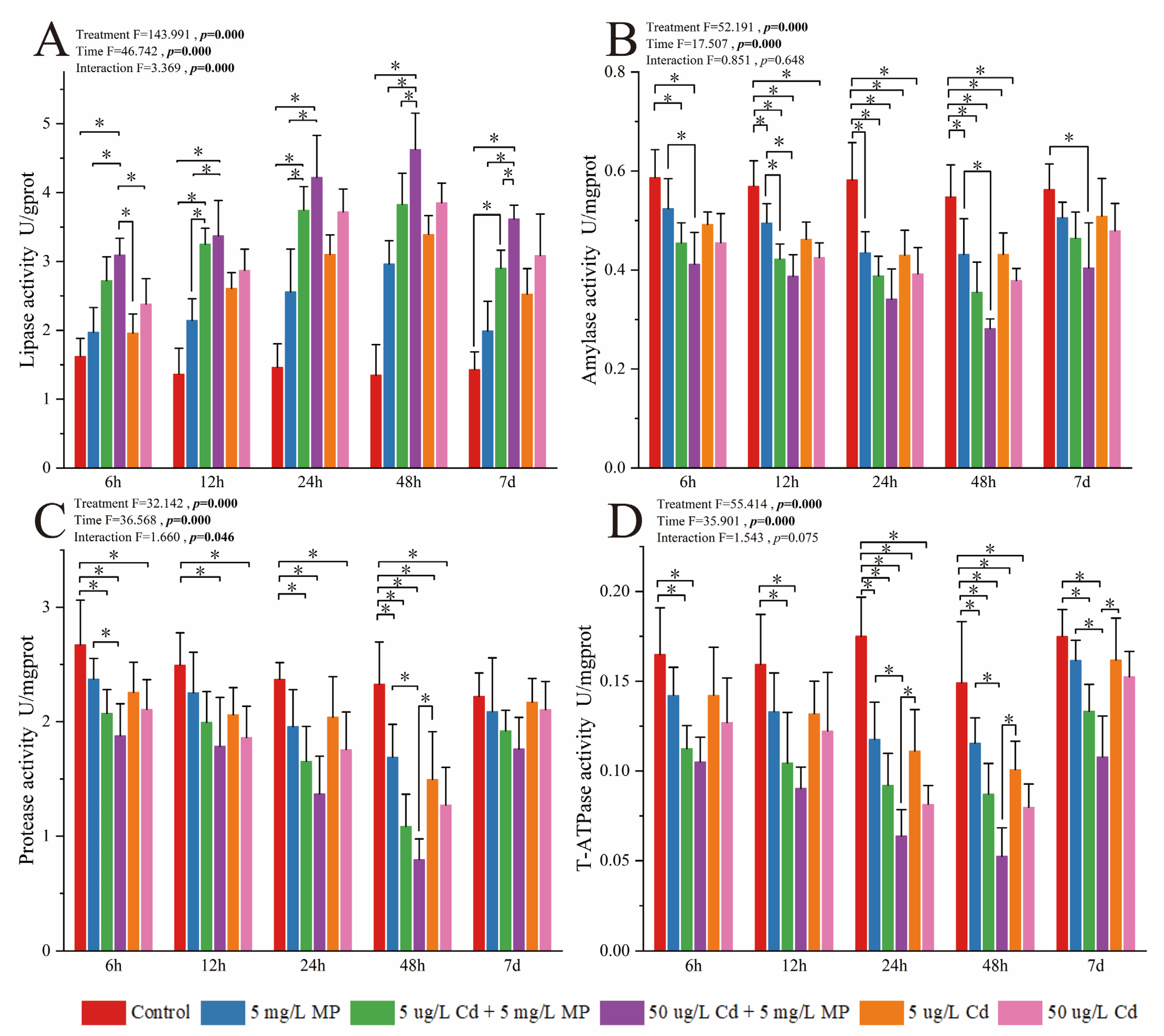
3.1.1. Activity of Enzymes Involved in Digestion and Energy Metabolism

The interaction between time and treatment had a significant effect on the activity of lipase and protease in the hepatopancreatic tissues of pearl oysters under exposure to MPs, Cd, and both MPs and Cd (Figure 1, p < 0.05). No significant differences in the activities of amylase and T-ATPase were observed among treatments (Figure 1, p > 0.05). The activities of lipase, amylase, and T-ATPase decreased significantly over the experimental period. With the exception of ATPase in group C and group D, the activities of the enzymes in most of the treatment groups returned to normal levels after the 5-day recovery period. The activities of lipase, amylase, and T-ATPase were significantly lower in group D than in the other experimental groups.

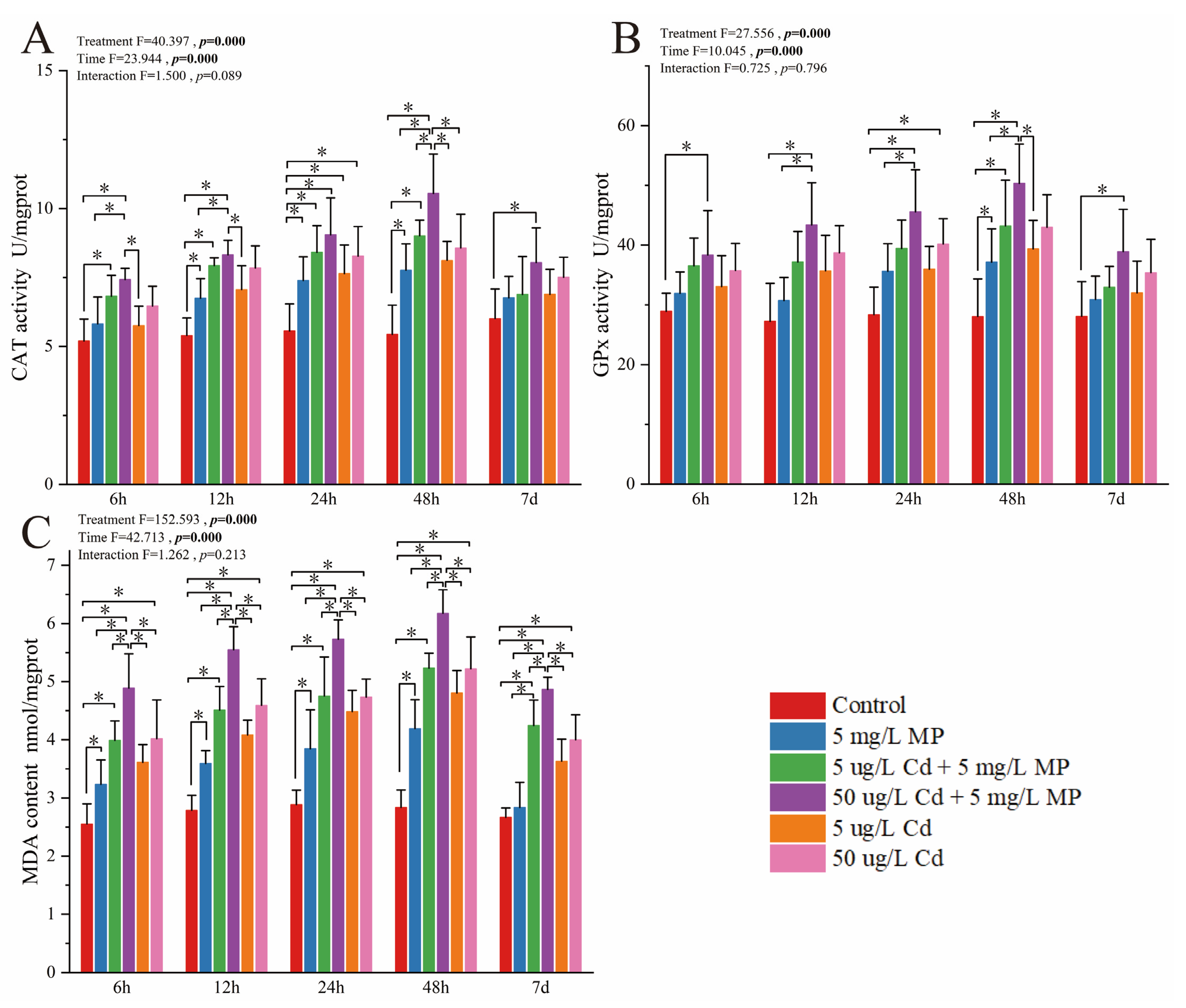
3.1.2. Activity of Enzymes and Oxidants Involved in Oxidative Stress Regulation

The interaction between time and concentration did not significantly affect the activities of CAT and GPx and the content of MDA (Figure 2, all p > 0.05). No significant changes were observed in the activities of CAT and GPx at 6 h, 12 h, and 24 h. The activities of CAT and GPx were highest in the D group at 48 h. The content of MDA increased significantly at each time point, and the MDA content was highest in group D.

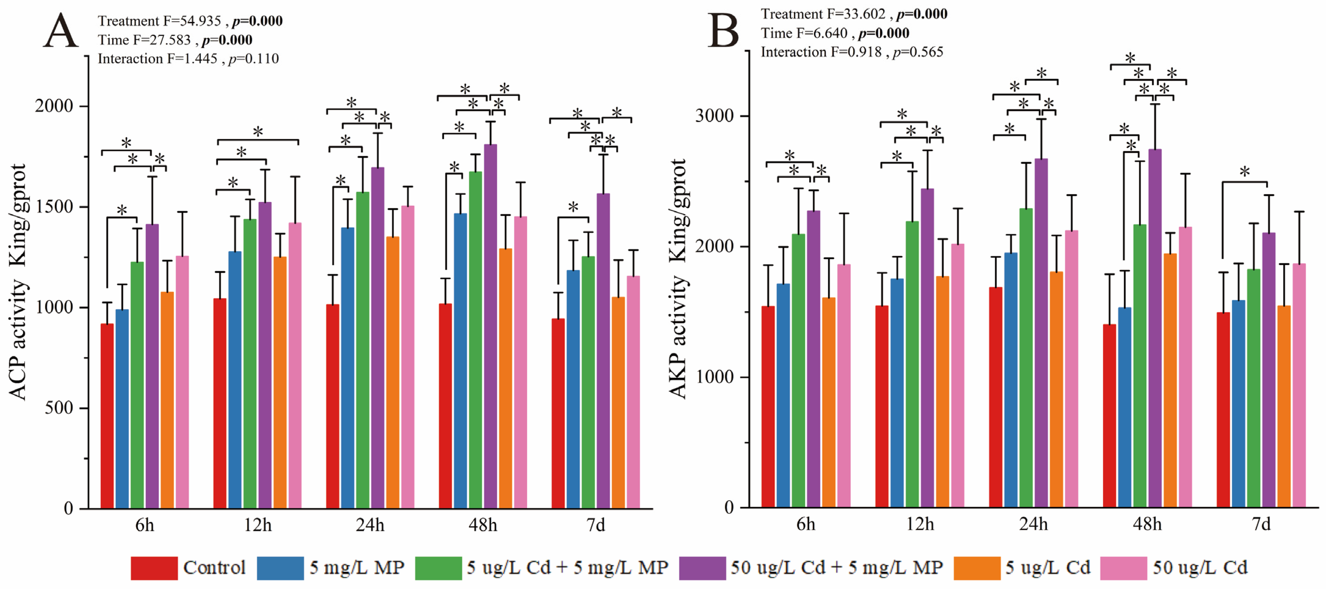
3.1.3. Activity of Enzymes Involved in Immune Function

The interaction between time and concentration had no significant effect on the activities of AKP and ACP (Figure 3, p > 0.05). The activities of these two enzymes continuously increased; however, the activity of AKP in group B did not change significantly among all time points during the experiment. The activities of AKP and ACP were highest in group D at each time point. The activities of ACP and AKP gradually decreased to their original level following a 5-day recovery period.

3.2. Analysis of the Pearl Oyster Metabolome

3.2.1. Multivariate Analysis of Metabolites

A total of 29,082 valid peaks were identified for the pearl oyster, which yielded a total of 1798 metabolites after data preprocessing using the in-house MS2 database. The R2X values for the PCA and OPLS-DA models ranged from 0.507 to 0.595 and from 0.231 to 0.375, respectively (Figure 4), for the following comparisons: C vs. I, C vs. II, C vs. III, C vs. IV, C vs. V, C vs. VI, I vs. II, I vs. III, II vs. III, IV vs. V, IV vs. VI, V vs. VI, I vs. IV, II vs. V, and III vs. V. Moreover, the R2Y and Q2 values of the OPLS–DA for the same comparisons ranged from 0.982 to 0.995 and from 0.0335 to 0.758, respectively (Figure 5), indicating good stability with no overfitting for the OPLS-DA model. These observations suggested the suitability and application of the model in subsequent analyses.

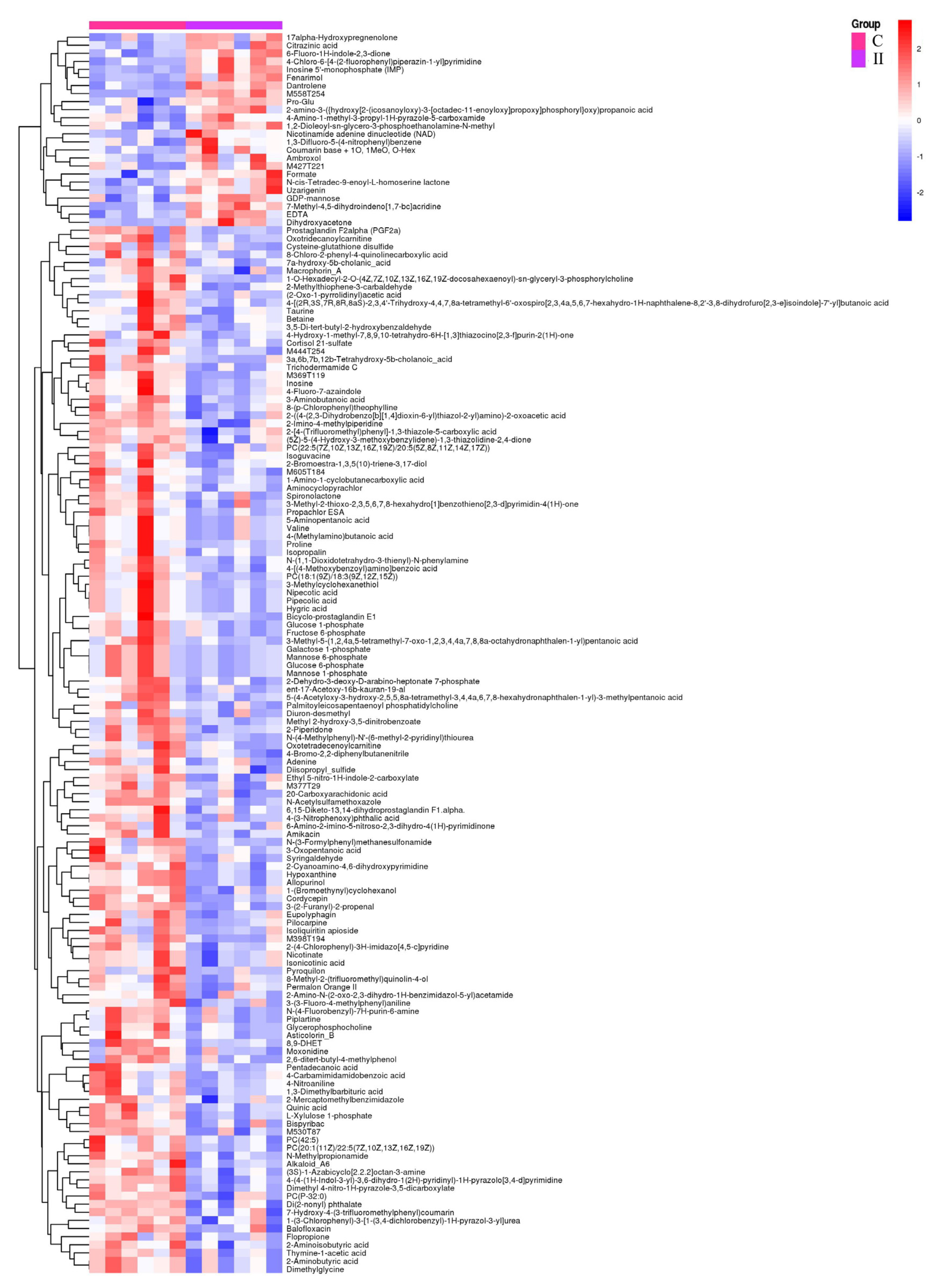
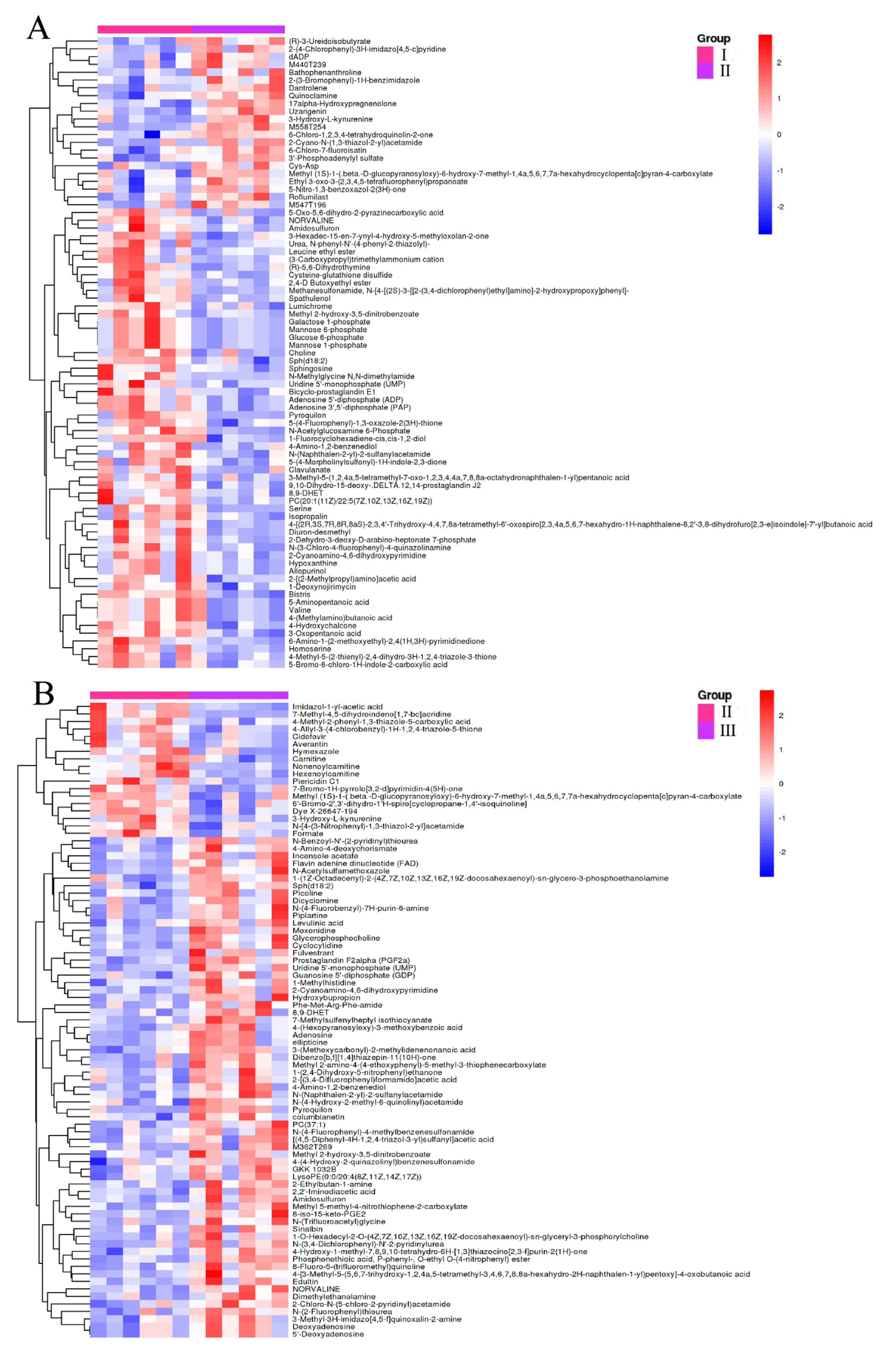
3.2.2. Identification of Significantly Different Metabolites (SDMs)

A total of 1301 (459 up-regulated and 842 down-regulated), 1603 (703 up-regulated and 900 down-regulated), 1487 (752 up-regulated and 735 down-regulated), 5325 (2070 up-regulated and 3255 down-regulated), 2882 (1645 up-regulated and 1237 down-regulated), 2148 (1021 up-regulated and 1127 down-regulated), 1123 (632 up-regulated and 491 down-regulated), 624 (466 up-regulated and 158 down-regulated), 1051 (566 up-regulated and 485 down-regulated), 3013 (2150 up-regulated and 863 down-regulated), 2792 (1884 up-regulated and 908 down-regulated), 1476 (583 up-regulated and 893 down-regulated), 4200 (2297 up-regulated and 1903 down-regulated), 1446 (845 up-regulated and 601 down-regulated), and 1636 (1139 up-regulated and 497 down-regulated) SDMs were identified in the C vs. I, C vs. II, C vs. III, C vs. IV, C vs. V, C vs. VI, I vs. II, I vs. III, II vs. III, IV vs. V, IV vs. VI, V vs. VI, I vs. IV, II vs. V, and III vs. V comparisons, respectively (Figure 6 and Figure 7).

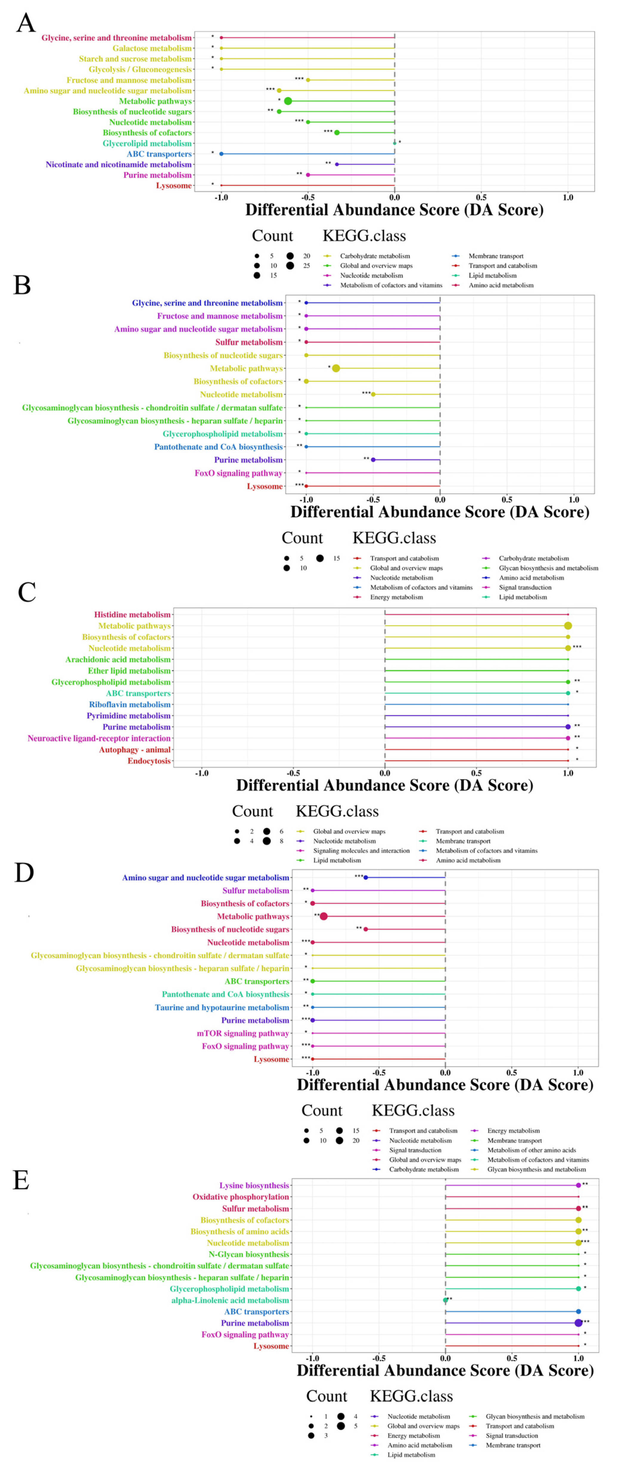
3.2.3. Characterization and Functional Analysis of Metabolic Pathways

The SDMs in the C vs. I, C vs. II, C vs. III, C vs. IV, C vs. V, C vs. VI, I vs. II, I vs. III, II vs. III, IV vs. V, IV vs. VI, V vs. VI, I vs. IV, II vs. V, and III vs. V comparisons were annotated to 44, 43, 32, 74, 29, 33, 38, 10, 14, 53, 26, 35, 45, 36, and 16 KEGG pathways, respectively (Figure 8). The most significantly enriched pathways across all comparisons were purine metabolism, glycerophospholipid metabolism, nucleotide metabolism, arachidonic acid metabolism, and neuroactive ligand–receptor interaction, except for the C vs. III and C vs. VI comparisons, and linoleic acid metabolism, except for the II vs. III comparison.

4. Discussion

4.1. Effects of MPs, Cd, and Combined MPs and Cd Exposure on Digestion and Energy Metabolism

Purine metabolism in organisms, including the pearl oyster P. f. martensii, involves the synthesis and degradation of purine nucleotides, which are essential for energy transfer and cellular signaling [29]. In our study, exposure to MPs and Cd significantly altered the abundance of several metabolites associated with the purine metabolism pathway, especially inosine 5′-monophosphate (IMP, C00130). The observed shift in IMP levels indicated an energy imbalance within the pearl oysters, which likely induced the utilization of AMP molecules to promote ATP synthesis [30]. Similar results have been obtained in other bivalves, such as the razor clam Sinonovacula constricta [31] and Mytilus galloprovincialis [32], under nanoplastic and MP exposure, respectively. However, the responses of the deep-sea mussel Bathymodiolus platifrons to Cd and copper exposure differed from the responses of pearl oysters observed in our study [33]. Nucleotide metabolism involves the biosynthesis and degradation of nucleotides, which are fundamental for DNA and RNA synthesis. This pathway also supports the proliferation of cells and energy metabolism via the regulation of nucleotide pools [34]. We found that metabolites related to nucleotide metabolism, such as IMP (C00130), inosine (C00294), and hypoxanthine (C00262), were altered in the pearl oyster P. f. martensii following exposure to MPs and Cd. The changes observed in inosine levels indicated that the utilization of ATP for energy production was reduced, which led to a decrease in the production of IMPs. The decreased production of IMP led to a decrease in inosine production, which subsequently resulted in lower hypoxanthine levels [29,35]. Decreased IMP production has also been observed in Corbicula fluminea under MP exposure but not in B. platifrons under Cd and copper exposure [29,33]. Therefore, the exposure of P. f. martensii to Cd and MPs possibly disrupted various biological components and processes, such as purine nucleotide pools, DNA and RNA synthesis and repair, energy metabolism, and other purine-dependent cellular functions. The energy produced through these metabolic processes is critically important for the ability of organisms to cope with environmental stressors and essential biological processes, including biomineralization [30,36].
The digestive enzymes of aquatic animals are important indicators of material digestion. Marine invertebrates use various digestive enzymes to decompose substrates and meet their diverse nutritional needs. The biological effects of MPs and HMs on the digestive system of aquatic organisms have received widespread attention. We found that exposure to MPs, Cd, and combined exposure to MPs and Cd significantly reduced the amylase and protease activities of pearl oysters. Amylase converts starch into sugars and plays a key role in carbohydrate metabolism and energy generation. The decrease in amylase activity observed in this study has also been observed in Crassostrea virginica, Pelteobagrus fulvidraco, S. henanense, and M. galloprovincialis under Cd and MP exposure [37,38,39]. This decrease in amylase activity likely stems from changes in the composition of digestive enzymes induced by Cd, as Cd can bind to the cysteine residue in sulfhydryl groups, thus reducing enzyme activity [40,41]. Additionally, Trestrail et al. [39] reported that MP leachates can be toxic to microbes; altering the enzyme-secreting microbiome may be an additional way that MPs can alter digestive enzyme production in M. galloprovincialis. Protease facilitates the digestion of proteins by breaking them down into peptides, which is essential for nutrient absorption. Decreases in protease activity have also been observed in C. virginica [37], Lymantria dispar [42], and Bufo gargarizans [40] under Cd exposure. However, decreases in protease activity were not observed in P. f. martensii [43] and M. galloprovincialis [39] under MP exposure. Similar to amylase, the inhibition of protease activity is linked to the ability of Cd to alter the composition of protease [42]. However, the lipase activity of pearl oysters significantly increased under Cd exposure. Lipase decomposes triglycerides into fatty acids and glycerol. Its activity is critically important for lipid metabolism and energy production. The increase in lipase activity is conducive to the digestion and absorption of fat, and metabolic activity is usually accompanied by energy consumption. The increase in lipase activity is associated with the enhanced lipolysis of lipids, which are potentially derived from other sources associated with Cd and MPs [44]. This means that pearl oysters require more energy to maintain their metabolism under combined exposure to Cd and MPs.
ATPases are enzymes that catalyze the hydrolysis of ATP to ADP and inorganic phosphate; they play a vital role in cellular energy metabolism and various biological processes. Observations of T-ATPase activity in this study were not consistent with the findings of a previous study of C. fluminea under Cd and MP exposure [45,46]. However, our findings were consistent with observations of other ATPases, such as Ca2⁺-ATPase, in M. coruscus and Tegillarca granosa under MP exposure [47,48]. In this study, the decline in T-ATPase activity was correlated with the decreased activities of digestive enzymes, particularly amylase and protease. Consequently, impairment of the digestive system may have led to disrupted energy metabolism due to the inhibition of T-ATPase activity [48,49].

4.2. Effects of MPs, Cd, and Combined Exposure to MPs and Cd on Oxidative Stress

MDA is one of the final products of polyunsaturated fatty acid peroxidation and is typically used to evaluate oxidative stress [50]. The increase in the content of MDA observed in this study was consistent with the findings of previous studies of Mactra vereformis, M. edulis, and Ruditapes decussatus but differed from observations of Perna viridis, Anadara subcrenata, and C. gigas under Cd exposure [51,52,53,54,55]. An increasing trend in the levels of MDA in pearl oysters under MP exposure was similar to those in R. philippinarum, P. f. martensii, and C. fluminea but differed from that in C. angulata under MP exposure [43,45,56,57]. The increase in the MDA content stems from lipid peroxidation caused by the production of reactive oxygen species (ROS) [10]. Therefore, both Cd and MPs can induce ROS production, lipid peroxidation, and subsequent membrane damage. The conversion of hydrogen peroxide into water and oxygen via CAT and GSH-catalyzed redox reactions comprises the second line of defense of the ROS-scavenging system. Increases in CAT activity have also been observed in C. fluminea and P. perna under Cd and MP exposure [45,46,58] but not in B. azoricus [59] and Chlamys farreri [60]. Increases in CAT activity are associated with the antioxidant and stress tolerance capabilities of pearl oysters [61]. ROS production is one of the biological responses to xenobiotics [10,62], including MPs and HMs such as Cd. GPx reduces hydrogen peroxide and other peroxides using glutathione as a cofactor, which enhances cellular antioxidant defense. The increase in GPx activity in the co-exposed groups and at high concentrations of Cd indicates that the regulation of glutathione metabolism and the neutralization of ROS, such as superoxide and hydrogen peroxide, enhance cellular antioxidant protection [61,62,63].

4.3. Effects of MPs, Cd, and Combined Exposure to MPs and Cd on Immune Activity

Invertebrates lack adaptive immunity in contrast to vertebrates. Exposure to MPs and HMs inevitably affects the immune function of bivalves or reduces their stress resistance [64,65]. ACP is a lysosomal marker enzyme that can be used for biological assessments of metal pollution, and it plays a role in the immune defense of oysters [66]. The increase in ACP activity observed in this study was consistent with findings in Lamellidens marginalis; however, an increase in ACP activity was not observed in Mizuhopecten yessoensis under Cd exposure [67,68]. Additionally, observations of both non-significant and increased ACP activities in the MP groups in our study were consistent with findings in Scrobicularia plana [69], C. angulata [56], C. fluminea [70], P. f. martensii [43], and M. edulis [71] under MP exposure. Both Cd and MPs are known to induce responses in bivalves; moreover, both ACP and AKP play crucial roles in the detoxification of xenobiotics [43]. Therefore, the increased ACP activity observed in this study suggests that Cd and MPs are perceived as harmful exogenous molecules that activate the innate immune system of pearl oysters [62,72].
AKP is an intrinsic plasma membrane enzyme in almost all animal cells, and it is sensitive to metals; measurements of this enzyme are essential for clarifying the general metabolism of organisms [73,74]. The results showed that MPs, Cd, and combined exposure to Cd and MPs significantly increased the AKP activity of the pearl oyster P. f. martensii. Increases in AKP activity have been observed in C. angulata [56] but not in C. fluminea [70] and P. f. martensii [43] under MP exposure. Observations of AKP activity in the pearl oysters in the MP and Cd groups indicate that AKP was not involved in immune responses, with the exception of pearl oysters exposed to the high Cd concentration. However, co-exposure of pearl oysters to both MPs and Cd promoted AKP activity. Thus, the exposure of pearl oysters to MPs and Cd stimulated the innate immune system via the activation of both ACP and AKP [33].

4.4. Effects of MPs, Cd, and Combined Exposure to MPs and Cd on Lipid Metabolism

The effects of MPs and metals on glycerophospholipid metabolism, linoleic acid metabolism, arachidonic acid metabolism, and other pathways have been previously studied in M. galloprovincialis [32], Gigantidas platifrons [75], and B. platifrons [33] under exposure to polyethylene MPs, Cd, and copper [76].
Glycerophospholipid metabolism in bivalves, such as P. f. martensii, is essential for membrane biogenesis and various signaling processes [77,78]. We found that phosphatidylcholine (PC, C00157) levels were significantly modulated in response to the treatments. This suggests that these membrane phospholipids may have been converted into phosphorus-free lipids, which potentially resulted in changes to cell membrane structures and promoted apoptosis [79,80,81]. Additionally, phosphatidylcholine (PC) can form complexes with hyaluronic acid, which helps regulate the concentration and transport of calcium (Ca2+) and affects the biomineralization capabilities of P. f. martensii [36]. The observations made in our study are consistent with findings in Mus musculus under Cd exposure and in M. galloprovincialis under MP exposure [82,83]. However, these observations were in contrast to the results of a study of Sinonovacula constricta under MP exposure [31].
The metabolism of arachidonic acid involves its conversion into various bioactive lipids, which play an important role in inflammation and cellular signaling. In this study, we found that PC, a precursor of lipid messengers such as arachidonic acid, was modulated through its biotransformation into lysophosphatidylcholine (LPC). This modulation of the arachidonic acid pathway may indicate a potential release of arachidonic acid from the biotransformation of PC [84,85]. Furthermore, the release of arachidonic acid could lead to its accumulation and the development of acute inflammatory reactions [76,86]. In contrast, the metabolism of arachidonic acid, particularly its utilization, is associated with the stress responses of organisms, including P. f. martensii [87]. Observations in this study were in contrast to those of C. farreri under Cd exposure [76] but similar to those of M. galloprovincialis and Anodonta cygnea L. under MP and Cd exposure, respectively [83,88].
Linoleic acid metabolism involves the conversion of linoleic acid into various metabolites, including PC, which plays essential roles in maintaining cell membrane integrity and functionality [89]. In our study, we found that levels of PC (C00157) were modulated, indicating that its potential effects on linoleic acid metabolism might lead to imbalances in the functions regulated by linoleic acid. Although previous studies have reported the effects of Cd and MPs on PC levels [76,83,88], limited research has demonstrated their effects on the linoleic acid metabolism pathway. Our findings revealed that PC and its associated linoleic acid metabolism pathways were modulated in pearl oysters under exposure to Cd and MPs, which highlights the vulnerability of these organisms to environmental pollutants. Generally, the effect of these stressors on linoleic acid metabolism requires clarification.

4.5. Effects of MPs, Cd, and Combined Exposure to MPs and Cd on Neuronal Excitability

The neuroactive ligand–receptor interaction pathway involves interactions between neuroactive ligands and their receptors and mediates neural signaling and communication. In organisms such as bivalves, this pathway plays a role in behavioral responses and sensory processing. The non-modulatory effect of Cd exposure on the neuroactive ligand–receptor interaction pathway in P. f. martensii was in contrast to observations of C. gigas and R. philippinarum under Cd exposure [90,91]. By contrast, the modulatory effect of exposure to either MPs alone or both MPs and Cd on the neuroactive ligand–receptor interaction pathway in P. f. martensii was similar in C. angulata [56] and Apostichopus japonicus [92]. However, the level of prostaglandin F2alpha (PGF2a, C00639) increased in C. angulata, yet an increase in PGF2a was not observed in P. f. martensii in our study. In addition, PGF2a interacts with specific receptors, prostaglandin F2α receptors, on the plasma membrane [93]. This interaction facilitates the release of intracellular free calcium and regulates reproduction in bivalves [94,95].

4.6. Effects of Short-Term Recovery on the Pearl Oyster

During the recovery phase of the pearl oysters, recovery of the oxidative, digestive, and immune systems, as well as energy metabolism, was observed. CAT and protease activities were fully restored in all groups, and lipase activity was not restored. The activities of amylase, GPx, and AKP were not restored in the F group. T-ATPases and ACP activities were not restored in the co-exposure groups (D and F), and MDA levels did not recover in the MP groups. Additionally, levels of IMP, PC, and PGF2α were not restored following exposure to Cd and MPs. Inosine and hypoxanthine were modulated during the recovery phase in the B group, and levels of inosine were restored in the D group. Their inability to recover suggests that these pollutants induce long-term damage, which may result in long-term impairments of energy metabolism, digestion, and immune responses.

5. Conclusions

In conclusion, the exposure of P. f. martensii to Cd and MPs affected the activities of various enzymes and metabolic pathways. The findings revealed significant alterations in lipase, amylase, protease, T-ATPase, CAT, GPx, ACP, and AKP activities and the MDA content across different time points and treatment groups. Moreover, the extent to which the activities of these enzymes recovered during the recovery phase varied. The main pathways affected by Cd and MPs were purine metabolism, glycerophospholipid metabolism, nucleotide metabolism, arachidonic acid metabolism, and neuroactive ligand–receptor interaction. These results highlight the complex interactions between Cd, MPs, enzymatic activities, and metabolic pathways and elucidate the mechanisms underlying these complex interactions in pearl oysters.

Supplementary Materials

The following supporting information can be downloaded at https://www.mdpi.com/article/10.3390/fishes10030133/s1, Figure S1: SEM images of microplastics.

Author Contributions

Conceptualization, C.Y.; methodology, J.Y., Z.G. (Zixin Gao), Z.W., Z.G. (Zhanbo Ge), Y.L., L.H., J.L., H.Z. and R.M.; software, J.Y., Z.G. (Zixin Gao) and Z.W.; validation, C.Y. and Y.D.; formal analysis, Y.L., L.H., J.L., H.Z. and R.M.; investigation, J.Y., Z.G. (Zixin Gao), Z.W., Z.G. (Zhanbo Ge), Y.L., L.H., J.L., H.Z. and R.M.; resources, C.Y. and Y.D.; data curation, J.Y., Z.G. (Zixin Gao), Z.W., Z.G. (Zhanbo Ge) and Y.L.; writing—original draft preparation, J.Y., Z.G. (Zixin Gao), Z.W., Z.G. (Zhanbo Ge), Y.L., L.H. and R.M.; writing—review and editing, C.Y. and R.M.; visualization, C.Y. and R.M.; supervision, C.Y. and Y.D.; project administration, Y.D.; funding acquisition, C.Y. and Y.D. All authors have read and agreed to the published version of the manuscript.

Funding

This research was funded by the Special Fund for Guangdong Province’s Science and Technology Innovation Strategy (grant number pdjh2024a191), the Undergraduate Innovation Team of Guangdong Ocean University (grant number CXTD2025001), the Department of Education of Guangdong Province (grant number 2021KCXTD026), the earmarked fund for CARS-49, the program for scientific research start-up funds of Guangdong Ocean University (grant number 060302022304), and the Hengli biosciences excellence project of Guangdong Ocean University (grant number B23335-4).

Institutional Review Board Statement

The pearl oyster P. f. martensii is a lower invertebrate, and therefore, the study was not subject to ethical approval.

Informed Consent Statement

This study does not involve human research.

Data Availability Statement

Data will be made available upon request.

Acknowledgments

We are very grateful to the Marine Pearl Science and Technology Backyard in Leizhou of Guangdong for collecting samples. Metabolomics analysis was assisted by Biotree Biotech Co., Ltd. (Shanghai, China).

Conflicts of Interest

The authors declare that the research was conducted in the absence of any commercial or financial relationships that could be construed as a potential conflict of interest.

References

  1. Eriksen, M.; Lebreton, L.C.M.; Carson, H.S.; Thiel, M.; Moore, C.J.; Borerro, J.C.; Galgani, F.; Ryan, P.G.; Reisser, J. Plastic Pollution in the World’s Oceans: More than 5 Trillion Plastic Pieces Weighing over 250,000 Tons Afloat at Sea. PLoS ONE 2014, 9, e111913. [Google Scholar] [CrossRef]
  2. Thompson, C.R.; Olsen, Y.; Mitchell, P.R.; Davis, A.; Rowland, J.S.; John, G.W.A.; McGonigle, D.; Russell, E.A. Lost at Sea: Where Is All the Plastic? Science 2004, 304, 838. [Google Scholar] [CrossRef]
  3. Sebille, V.E.; Wilcox, C.; Lebreton, L.; Maximenko, N.; Hardesty, D.B.; Franeker, V.J.; Eriksen, M.; Siegel, D.; Galgani, F.; Law, L.K. A global inventory of small floating plastic debris. Environ. Res. Lett. 2015, 10, 124006. [Google Scholar] [CrossRef]
  4. Nadia, M.V.; Patricia, B.; Angela, K. Uptake and effects of microplastics on cells and tissue of the blue mussel Mytilus edulis L. after an experimental exposure. Environ. Sci. Technol. 2012, 46, 11327–11335. [Google Scholar]
  5. Fackelmann, G.; Sommer, S. Microplastics and the gut microbiome: How chronically exposed species may suffer from gut dysbiosis. Mar. Pollut. Bull. 2019, 143, 193–203. [Google Scholar] [CrossRef]
  6. Yin, L.; Chen, B.; Xia, B.; Shi, X.; Qu, K. Polystyrene microplastics alter the behavior, energy reserve and nutritional composition of marine jacopever (Sebastes schlegelii). J. Hazard. Mater. 2018, 360, 97–105. [Google Scholar] [CrossRef] [PubMed]
  7. Jabeen, K.; Li, B.; Chen, Q.; Su, L.; Wu, C.; Hollert, H.; Shi, H. Effects of virgin microplastics on goldfish (Carassius auratus). Chemosphere 2018, 213, 323–332. [Google Scholar] [CrossRef] [PubMed]
  8. Sun, J.H.; Xia, S.D.; Ning, Y.; Pan, X.; Qu, J.H.; Xu, Y.J. Effects of microplastics and attached heavy metals on growth, immunity, and heavy metal accumulation in the yellow seahorse, Hippocampus kuda Bleeker. Mar. Pollut. Bull. 2019, 149, 110510. [Google Scholar]
  9. Gardon, T.; Reisser, C.; Soyez, C.; Quillien, V.; Moullac, L.G. Microplastics affect energy balance and gametogenesis in the pearl oyster Pinctada margaritifera. Environ. Sci. Technol. 2018, 52, 5277–5286. [Google Scholar] [CrossRef]
  10. Mkuye, R.; Gong, S.; Zhao, L.; Masanja, F.; Ndandala, C.; Bubelwa, E.; Yang, C.; Deng, Y. Effects of microplastics on physiological performance of marine bivalves, potential impacts, and enlightening the future based on a comparative study. Sci. Total Environ. 2022, 838, 155933. [Google Scholar] [CrossRef]
  11. Brandts, I.; Teles, M.; Gonçalves, A.P.; Barreto, A.; Franco-Martinez, L.; Tvarijonaviciute, A.; Martins, M.A.; Soares, A.M.V.M.; Tort, L.; Oliveira, M. Effects of nanoplastics on Mytilus galloprovincialis after individual and combined exposure with carbamazepine. Sci. Total Environ. 2018, 643, 775–784. [Google Scholar] [CrossRef] [PubMed]
  12. Wen, B.; Jin, S.R.; Chen, Z.Z.; Gao, J.Z.; Liu, Y.N.; Liu, J.H.; Feng, X.S. Single and combined effects of microplastics and cadmium on the cadmium accumulation, antioxidant defence and innate immunity of the discus fish (Symphysodon aequifasciatus). Environ. Pollut. 2018, 243, 462–471. [Google Scholar] [CrossRef]
  13. Gao, J.W.; Wu, H.; Li, S.M.; Xie, M.; Li, W.X.; Song, R. Ammonia nitrogen and cadmium stress on antioxidant system and immune function of furong crucian carp (Cyprinus capio furong.♀× Carssius auratus red var.♂). Acta Hydrobiol. Sin. 2022, 46, 448–456. (In Chinese) [Google Scholar]
  14. Sun, R.; Wang, Z. Heavy metals contamination and shellfish poison pollution in marine shellfish in Shenzhen coastal waters. J. Appl. Oceanogr. 2017, 36, 575–579. (In Chinese) [Google Scholar]
  15. Vaseem, H.; Banerjee, T.K. Evaluation of pollution of Ganga River water using fish as bioindicator. Environ. Monit. Assess. 2016, 188, 1–9. [Google Scholar] [CrossRef] [PubMed]
  16. Jerome, F.C.; Hassan, A.; Omoniyi-Esan, G.O.; Odujoko, O.O.; Chukwuka, A.V. Metal uptake, oxidative stress and histopathological alterations in gills and hepatopancreas of Callinectes amnicola exposed to industrial effluent. Ecotoxicol. Environ. Saf. 2017, 139, 179–193. [Google Scholar] [CrossRef]
  17. Brown, C. Arsenic and cadmium are contaminants of concern. CMAJ 2016, 188, E5. [Google Scholar] [CrossRef]
  18. Ding, T.T.; Du, S.L.; Zhang, Y.H.; Wang, H.; Zhang, Y.; Cao, Y.; He, L. Hardness-dependent water quality criteria for cadmium and an ecological risk assessment of the Shaying River Basin, China. Ecotoxicol. Environ. Saf. 2020, 198, 110666. [Google Scholar] [CrossRef]
  19. Borgmann, U.; Norwood, W.P.; Babirad, I.M. Relationship between chronic toxicity and bioaccumulation of cadmium in Hyalella azteca. Can. J. Fish. Aquat. Sci. 1991, 48, 1055–1060. [Google Scholar] [CrossRef]
  20. Eimers, M.C.; Evans, R.D.; Welbourn, P.M. Partitioning and bioaccumulation of cadmium in artificial sediment systems: Application of a stable isotope tracer technique. Chemosphere 2002, 46, 543–551. [Google Scholar] [CrossRef]
  21. Mebane, C.A.; Schmidt, T.S.; Miller, J.L.; Balistrieri, L.S. Bioaccumulation and toxicity of cadmium, copper, nickel, and zinc and their mixtures to aquatic insect communities. Environ. Toxicol. Chem. 2020, 39, 812–833. [Google Scholar] [CrossRef] [PubMed]
  22. Zhou, Y.; Dahms, H.U.; Dong, F.; Jing, W.; Wang, L. Immune-associated parameters and antioxidative responses to cadmium in the freshwater crab Sinopotamon henanense. Ecotoxicol. Environ. Saf. 2016, 129, 235–241. [Google Scholar] [CrossRef]
  23. Lei, W.W.; Xu, T.; Wang, L. Effects of cadmium on apoptosis of cardiomyocyte in the freshwater crab Sinopotamon yangtsekiense. Oceanol. Et Limnol. Sin. 2012, 43, 299–305. (In Chinese) [Google Scholar]
  24. Bai, S.J.; Xu, Z.R. Effects of cadmium on mitochondrion structure and energy metabolism of Pelteobagrus fulvidraco gill. Chin. J. Appl. Ecol. 2006, 7, 1213–1217. (In Chinese) [Google Scholar]
  25. Shi, H.Q.; Zhang, L.J.; Wan, X.Y.; Peng, H.; Zhao, J.; Peng, S.Q. Toxic Effects of Cadmium Chloride Exposure on Neurobehavior of Zebrafish Larvae. Asian J. Ecotoxicol. 2013, 8, 374–380. (In Chinese) [Google Scholar]
  26. Wang, T.; Yang, C.Y.; Wang, C.; Liao, Y.; Mkuye, R.; Deng, Y. Bacterial community profiling associated with pearl culture facilities of Liusha Bay, the largest marine pearl culture base on the western Guangdong coast, South China. Mar. Environ. Res. 2023, 189, 106063. [Google Scholar] [CrossRef] [PubMed]
  27. Mkuyea, R.; Yang, C.Y.; Masanjaa, F.; Ibrahim, S.; Yang, X.Y.; Mwemi, H.; Mrope, P.; Salman, M.; Alfatat, A.; Deng, Y.W. Omics insights in responses of bivalves exposed to plastic pollution. Aquat. Toxicol. 2025, 279, 107224. [Google Scholar] [CrossRef]
  28. Zhou, Z.; Luo, M.; Zhang, H.; Yin, Y.; Cai, Y.; Zhu, Z.J. Metabolite annotation from knowns to unknowns through knowledge-guided multi-layer metabolic networking. Nat. Commun. 2022, 13, 6656. [Google Scholar] [CrossRef]
  29. Zhang, J.; Wang, J.; Liu, S.; Liu, X. Combining transcriptomic and metabolomic analyses to investigate the acute effects of microcystin-LR and nanoplastics of asian clams. Water 2023, 15, 3519. [Google Scholar] [CrossRef]
  30. Salgado-García, R.L.; Kraffe, E.; Tripp-Valdez, M.A.; Ramírez-Arce, J.L.; Artigaud, S.; Flye-Sainte-Marie, J.; Mathieu-Resuge, M.; Sicard, T.M.; Arellano-Martínez, M.; Racotta, I.S. Energy metabolism of juvenile scallops Nodipecten subnodosus under acute increased temperature and low oxygen availability. Comp. Biochem. Physiol. Part A Mol. Integr. Physiol. 2023, 278, 111373. [Google Scholar] [CrossRef]
  31. Jiang, Q.; Zhang, W. Gradual effects of gradient concentrations of polystyrene nanoplastics on metabolic processes of the razor clams. Environ. Pollut. 2021, 287, 117631. [Google Scholar] [CrossRef] [PubMed]
  32. Castaño-Ortiz, J.M.; Courant, F.; Gomez, E.; García-Pimentel, M.D.M.; León, V.M.; Campillo, J.A.; Santos, L.H.M.L.M.; Barceló, D.; Rodríguez-Mozaz, S. Combined exposure of the bivalve Mytilus galloprovincialis to polyethylene microplastics and two pharmaceuticals (citalopram and bezafibrate): Bioaccumulation and metabolomic studies. J. Hazard. Mater. 2023, 458, 131904. [Google Scholar] [CrossRef]
  33. Zhou, L.; Li, M.; Zhong, Z.; Chen, H.; Wang, X.; Wang, M.; Xu, Z.; Cao, L.; Lian, C.; Zhang, H.; et al. Biochemical and metabolic responses of the deep-sea mussel Bathymodiolus platifrons to cadmium and copper exposure. Aquat. Toxicol. 2021, 236, 105845. [Google Scholar] [CrossRef] [PubMed]
  34. Zhang, Y.; Liu, C.; Liu, J.; Liu, X.; Tu, Z.; Zheng, Y.; Xu, J.; Fan, H.; Wang, Y.; Hu, M. Multi-omics reveals response mechanism of liver metabolism of hybrid sturgeon under ship noise stress. Sci. Total Environ. 2022, 851, 158348. [Google Scholar] [CrossRef] [PubMed]
  35. Cong, M.; Wu, H.; Yang, H.; Zhao, J.; Lv, J. Gill damage and neurotoxicity of ammonia nitrogen on the clam Ruditapes philippinarum. Ecotoxicology 2017, 26, 459–469. [Google Scholar] [CrossRef]
  36. Yang, C.; Zeng, Y.; Liao, Y.; Deng, Y.; Du, X.; Wang, Q. Integrated GC–MS-and LC–MS-Based untargeted metabolomics studies of the effect of vitamin D3 on pearl production traits in pearl oyster Pinctada fucata martensii. Front. Mol. Biosci. 2021, 8, 614404. [Google Scholar] [CrossRef]
  37. Adeyemi, J.A.; Deaton, L.E. The effect of cadmium exposure on digestive enzymes in the eastern oyster Crassostrea virginica. J. Shellfish Res. 2012, 31, 631–634. [Google Scholar] [CrossRef]
  38. Wu, H.; Xuan, R.; Li, Y.; Zhang, X.; Wang, Q.; Wang, L. Effects of cadmium exposure on digestive enzymes, antioxidant enzymes, and lipid peroxidation in the freshwater crab Sinopotamon henanense. Environ. Sci. Pollut. Res. 2013, 20, 4085–4092. [Google Scholar] [CrossRef]
  39. Trestrail, C.; Walpitagama, M.; Miranda, A.; Nugegoda, D.; Shimeta, J. Microplastics alter digestive enzyme activities in the marine bivalve, Mytilus galloprovincialis. Sci. Total Environ. 2021, 779, 146418. [Google Scholar] [CrossRef]
  40. Zheng, R.; Wang, P.; Cao, B.; Wu, M.; Li, X.; Wang, H.; Chai, L. Intestinal response characteristic and potential microbial dysbiosis in digestive tract of Bufo gargarizans after exposure to cadmium and lead, alone or combined. Chemosphere 2021, 271, 129511. [Google Scholar] [CrossRef]
  41. Xie, D.; Li, Y.; Liu, Z.; Chen, Q. Inhibitory effect of cadmium exposure on digestive activity, antioxidant capacity and immune defense in the intestine of yellow catfish (Pelteobagrus fulvidraco). Comp. Biochem. Physiol. Part C Toxicol. Pharmacol. 2019, 222, 65–73. [Google Scholar] [CrossRef] [PubMed]
  42. Vlahović, M.; Ilijin, L.; Lazarević, J.; Mrdaković, M.; Gavrilović, A.; Matić, D.; Mataruga, V.P. Cadmium-induced changes of gypsy moth larval mass and protease activity. Comp. Biochem. Physiol. Part C Toxicol. Pharmacol. 2014, 160, 9–14. [Google Scholar] [CrossRef]
  43. Lu, F.L.; Guo, C.A.; Mkuye, R.; Chen, W.; Yang, X.; Zhou, Z.; He, Y.; Yang, C.Y.; Deng, Y.W. Effects of polyvinyl chloride microplastic on pearl oyster (Pinctada fucata martensii). Reg. Stud. Mar. Sci. 2024, 69, 103313. [Google Scholar] [CrossRef]
  44. Chen, Q.L.; Gong, Y.; Luo, Z.; Zheng, J.L.; Zhu, Q.L. Differential effect of waterborne cadmium exposure on lipid metabolism in liver and muscle of yellow catfish Pelteobagrus fulvidraco. Aquat. Toxicol. 2013, 142, 380–386. [Google Scholar] [CrossRef]
  45. Fu, L.; Xi, M.; Nicholaus, R.; Wang, Z.; Wang, X.; Kong, F.; Yu, Z. Behaviors and biochemical responses of macroinvertebrate Corbicula fluminea to polystyrene microplastics. Sci. Total Environ. 2022, 813, 152617. [Google Scholar] [CrossRef]
  46. Wang, Z.; Kong, F.; Fu, L.; Li, Y.; Li, M.; Yu, Z. Responses of Asian clams (Corbicula fluminea) to low concentration cadmium stress: Whether the depuration phase restores physiological characteristics. Environ. Pollut. 2021, 284, 117182. [Google Scholar] [CrossRef]
  47. Lu, L.; Huang, W.; Han, Y.; Tong, D.; Sun, S.; Yu, Y.; Liu, G.; Shi, W. Toxicity of microplastics and triclosan, alone and in combination, to the fertilisation success of a broadcast spawning bivalve Tegillarca granosa. Environ. Toxicol. Pharmacol. 2023, 101, 104208. [Google Scholar] [CrossRef] [PubMed]
  48. Yu, Y.; Tian, D.; Han, Y.; Huang, L.; Tang, Y.; Zhang, W.; Zhou, W.; Shi, W.; Yu, Y.; Liu, G. Impacts of microplastics and carbamazepine on the shell formation of thick-shell mussels and the underlying mechanisms of action. Sci. Total Environ. 2022, 838, 156442. [Google Scholar] [CrossRef] [PubMed]
  49. Xue, Y.H.; Sun, Z.X.; Feng, L.S.; Jin, T.; Xing, J.C.; Wen, X.L. Algal density affects the influences of polyethylene microplastics on the freshwater rotifer Brachionus calyciflorus. Chemosphere 2021, 270, 128613. [Google Scholar] [CrossRef]
  50. Thirupathi, A.; Wang, M.; Lin, J.K.; Fekete, G.; István, B.; Baker, J.S.; Gu, Y. Effect of different exercise modalities on oxidative stress: A systematic review. BioMed Res. Int. 2021, 2021, 1947928. [Google Scholar] [CrossRef]
  51. Geret, F.; Serafim, A.; Barreira, L.; Bebianno, M.J. Effect of cadmium on antioxidant enzyme activities and lipid peroxidation in the gills of the clam Ruditapes decussatus. Biomarkers 2002, 7, 242–256. [Google Scholar] [CrossRef] [PubMed]
  52. Géret, F.; Jouan, A.; Turpin, V.; Bebianno, M.J.; Cosson, R.P. Influence of metal exposure on metallothionein synthesis and lipid peroxidation in two bivalve mollusks: The oyster (Crassostrea gigas) and the mussel (Mytilus edulis). Aquat. Living Resour. 2002, 15, 61–66. [Google Scholar] [CrossRef]
  53. Wang, X.; Yang, H.; Liu, G.; Wang, Q. Enzyme responses and lipid peroxidation in gills and hepatopancreas of clam Mactra vereformis, following cadmium exposure. Chin. J. Oceanol. Limnol. 2011, 29, 981–989. [Google Scholar] [CrossRef]
  54. Arasu, S.M.; Reddy, P.S. Changes in lipid peroxidation in the gill and muscle of the marine bivalve (Perna viridis) during exposure to cadmium and copper. Chem. Ecol. 1995, 11, 105–112. [Google Scholar] [CrossRef]
  55. Yan, B.; Liu, X.; Zhao, X.; Tian, S. Single and joint oxidative stress of cadmium and phenanthrene on the Bivalve Anadara subcrenata. J. Environ. Sci. Health Part A 2020, 55, 448–456. [Google Scholar] [CrossRef]
  56. Lu, J.; Yao, T.; Yu, G.; Ye, L. Adaptive response of triploid Fujian oyster (Crassostrea angulata) to nanoplastic stress: Insights from physiological, metabolomic, and microbial community analyses. Chemosphere 2023, 341, 140027. [Google Scholar] [CrossRef]
  57. Zhou, Y.; Wu, C.; Li, Y.; Jiang, H.; Miao, A.; Liao, Y.; Pan, K. Effects of nanoplastics on clam Ruditapes philippinarum at environmentally realistic concentrations: Toxicokinetics, toxicity, and gut microbiota. J. Hazard. Mater. 2023, 456, 131647. [Google Scholar] [CrossRef]
  58. Boudjema, K.; Kourdali, S.; Bounakous, N.; Meknachi, A.; Badis, A. Catalase activity in brown mussels (Perna perna) under acute cadmium, lead, and copper exposure and depuration tests. J. Mar. Sci. 2014, 2014, 830657. [Google Scholar] [CrossRef]
  59. Company, R.; Serafim, A.; Cosson, R.; Camus, L.; Shillito, B.; Fiala-Médioni, A.; Bebianno, M.J. The effect of cadmium on antioxidant responses and the susceptibility to oxidative stress in the hydrothermal vent mussel Bathymodiolus azoricus. Mar. Biol. 2006, 148, 817–825. [Google Scholar] [CrossRef]
  60. Zhang, Y.; Song, J.; Yuan, H.; Xu, Y.; He, Z. Concentrations of cadmium and zinc in seawater of Bohai Bay and their effects on biomarker responses in the bivalve Chlamys farreri. Arch. Environ. Contam. Toxicol. 2010, 59, 120–128. [Google Scholar] [CrossRef]
  61. Adzigbli, L.; Yu, W.M.; Li, J.H.; Yang, C.; Deng, Y. Influence of age on pearl production performance, enzymatic activity, and immune-related gene expression of the pearl oyster Pinctada fucata martensii. N. Am. J. Aquac. 2019, 81, 430–437. [Google Scholar] [CrossRef]
  62. Li, F.F.; Xie, Y.F.; Yang, C.Y.; Ye, Q.; Wang, F.; Liao, Y.; Mkuye, R.; Deng, Y.W. The physiological responses to titanium dioxide nanoparticles exposure in pearl oysters (Pinctada fucata martensii). Mar. Environ. Res. 2024, 195, 106345. [Google Scholar] [CrossRef] [PubMed]
  63. Haque, M.N.; Eom, H.J.; Nam, S.E.; Shin, Y.K.; Rhee, J.S. Chlorothalonil induces oxidative stress and reduces enzymatic activities of Na+/K+-ATPase and acetylcholinesterase in gill tissues of marine bivalves. PLoS ONE 2019, 14, e0214236. [Google Scholar] [CrossRef] [PubMed]
  64. Sun, C.; Teng, J.; Wang, D.; Li, J.; Wang, X.; Zhao, J.; Shan, E.; Wang, Q. Potential threats of microplastics and pathogenic bacteria to the immune system of the mussels Mytilus galloprovincialis. Aquat. Toxicol. 2024, 272, 106959. [Google Scholar] [CrossRef]
  65. Weng, N.; Meng, J.; Huo, S.; Wu, F.; Wang, W.X. Hemocytes of bivalve mollusks as cellular models in toxicological studies of metals and metal-based nanomaterials. Environ. Pollut. 2022, 312, 120082. [Google Scholar] [CrossRef] [PubMed]
  66. Rajalakshmi, S.; Mohandas, A. Copper-induced changes in tissue enzyme activity in a freshwater mussel. Ecotoxicol. Environ. Saf. 2005, 62, 140–143. [Google Scholar] [CrossRef]
  67. Jayakumar, P.; Jothivel, N.; Paul, V. Heavy metals induced alterations in the acid phosphatase activity in the edible freshwater mussel Lamellidens marginalis (Lamarck). Internet J. Toxicol. 2008, 5, 1–8. [Google Scholar]
  68. Evtushenko, Z.S.; Belcheva, N.N.; Lukyanova, O.N. Cadmium accumulation in organs of the scallop Mizuhopecten yessoensis—I. Activities of phosphatases and composition and amount of lipids. Comp. Biochem. Physiol. Part C Comp. Pharmacol. 1986, 83, 371–376. [Google Scholar] [CrossRef]
  69. Métais, I.; Latchere, O.; Roman, C.; Perrein-Ettajani, H.; Mouloud, M.; Georges, D.; Audroin, T.; Catrouillet, C.; Gigault, J.; Agnès-Feurtet-Mazel; et al. Continuum from microplastics to nanoplastics: Effects of size and source on the estuarine bivalve Scrobicularia plana. Environ. Sci. Pollut. Res. 2023, 30, 45725–45739. [Google Scholar] [CrossRef]
  70. Zhang, J.; Jie, W.; Cheng, G.; Gu, Z.; Liu, X. Transcriptome analysis of response mechanism to Microcystin-LR and microplastics stress in Asian clam (Corbicula fluminea). Fish Shellfish Immunol. 2023, 139, 108875. [Google Scholar] [CrossRef]
  71. Roman, C.; Mahé, P.; Latchere, O.; Catrouillet, C.; Gigault, J.; Métais, I.; Châtel, A. Effect of size continuum from nanoplastics to microplastics on marine mussel Mytilus edulis: Comparison in vitro/in vivo exposure scenarios. Comp. Biochem. Physiol. Part C Toxicol. Pharmacol. 2023, 264, 109512. [Google Scholar] [CrossRef]
  72. Chen, J.Y.; Huang, J.; Peng, J.Q.; Yang, C.; Liao, Y.; Li, J.; Deng, Y.; Du, X. Effects of hypoxic stress on the digestion, energy metabolism, oxidative stress regulation, and immune function of the pearl oyster (Pinctada fucata martensii). Aquac. Rep. 2022, 25, 101246. [Google Scholar] [CrossRef]
  73. Regoli, F.; Principato, G. Glutathione, glutathione-dependent and antioxidant enzymes in mussel, Mytilus galloprovincialis, exposed to metals under field and laboratory conditions: Implications for the use of biochemical biomarkers. Aquat. Toxicol. 1995, 31, 143–164. [Google Scholar] [CrossRef]
  74. Rui, X.; Ping, L.X.; Yu, J.L.; Yang, C.; Liao, Y.; Li, J.; Deng, Y.W.; Du, X.D. Purification and enzymatic characterization of alkaline phosphatase from Pinctada fucata. J. Mol. Catal. B Enzym. 2002, 17, 65–74. [Google Scholar]
  75. He, Y.; Zhou, L.; Wang, M.; Zhong, Z.; Chen, H.; Lian, C.; Zhang, H.; Wang, H.; Cao, L.; Li, C. Integrated transcriptomic and metabolomic approaches reveal molecular response and potential biomarkers of the deep-sea mussel Gigantidas platifrons to copper exposure. J. Hazard. Mater. 2024, 473, 134612. [Google Scholar] [CrossRef]
  76. Liu, H.; Li, H.; Zhang, X.; Gong, X.; Han, D.; Zhang, H.; Tian, X.; Xu, Y. Metabolomics comparison of metabolites and functional pathways in the gills of Chlamys farreri under cadmium exposure. Environ. Toxicol. Pharmacol. 2021, 86, 103683. [Google Scholar] [CrossRef] [PubMed]
  77. Fang, J.Y.; Yang, C.Y.; Liao, Y.S.; Wang, Q.; Deng, Y. Transcriptomic and metabolomic analyses reveal sex-related differences in the gonads of Pinctada fucata martensii. Comp. Biochem. Physiol. Part D Genom. Proteom. 2024, 52, 101304. [Google Scholar] [CrossRef]
  78. Zhou, C.; Song, H.; Feng, J.; Hu, Z.; Yang, M.J.; Shi, P.; Li, Y.R.; Guo, Y.J.; Li, H.Z.; Zhang, T. Metabolomics and biochemical assays reveal the metabolic responses to hypo-salinity stress and osmoregulatory role of cAMP-PKA pathway in Mercenaria mercenaria. Comput. Struct. Biotechnol. J. 2022, 20, 4110–4121. [Google Scholar] [CrossRef]
  79. Liang, J.; Iqbal, S.; Wen, F.; Tong, M.; Liu, J. Phosphorus-induced lipid class alteration revealed by lipidomic and transcriptomic profiling in oleaginous microalga Nannochloropsis sp. PJ12. Mar. Drugs 2019, 17, 519. [Google Scholar] [CrossRef]
  80. Sun, Y.; Geng, C.; Liu, W.; Liu, Y.; Ding, L.; Wang, P. Investigating the impact of disrupting the glutamine metabolism pathway on ammonia excretion in crucian carp (Carassius auratus) under carbonate alkaline stress using metabolomics techniques. Antioxidants 2024, 13, 170. [Google Scholar] [CrossRef]
  81. Jing, H.; Zhou, L.; Gao, Y.; Liu, Z.; Wu, B.; Sun, X.; Tu, K. Transcriptomics and metabolomics reveal the molecular and metabolic adaptation to heat stress in Manila clam Ruditapes philippinarum. Front. Mar. Sci. 2023, 10, 1204598. [Google Scholar] [CrossRef]
  82. García-Sevillano, M.Á.; García-Barrera, T.; Navarro-Roldán, F.; Montero-Lobato, Z.; Gómez-Ariza, J.L. A combination of metallomics and metabolomics studies to evaluate the effects of metal interactions in mammals. Application to Mus musculus mice under arsenic/cadmium exposure. J. Proteom. 2014, 104, 66–79. [Google Scholar] [CrossRef] [PubMed]
  83. Leroux, N.; Hosseinzadeh, M.; Katsumiti, A.; Porte, C.; Cajaraville, M.P. Lipidomic analysis of mussel hemocytes exposed to polystyrene nanoplastics. Environ. Res. 2022, 214, 113763. [Google Scholar] [CrossRef] [PubMed]
  84. Gu, J.; Kong, A.; Guo, C.; Liu, J.; Li, K.; Ren, Z.; Zhou, Y.; Tang, M.; Shi, H. Cadmium perturbed lipid profile and induced liver dysfunction in mice through phosphatidylcholine remodeling and promoting arachidonic acid synthesis and metabolism. Ecotoxicol. Environ. Saf. 2022, 247, 114254. [Google Scholar] [CrossRef]
  85. Fanelli, G.; Belardo, A.; Savino, R.; Rinalducci, S.; Zolla, L. Testosterone replacement therapy in insulin-sensitive hypogonadal men restores phosphatidylcholine levels by regulation of arachidonic acid metabolism. J. Cell. Mol. Med. 2020, 24, 8266–8269. [Google Scholar] [CrossRef]
  86. Ramirez, D.C.; Gimenez, M.S. Lipid modification in mouse peritoneal macrophages after chronic cadmium exposure. Toxicology 2002, 172, 1–12. [Google Scholar] [CrossRef]
  87. Yang, C.Y.; Du, X.D.; Hao, R.J.; Wang, Q.; Deng, Y.; Sun, R. Effect of vitamin D3 on immunity and antioxidant capacity of pearl oyster Pinctada fucata martensii after transplantation: Insights from LC–MS-based metabolomics analysis. Fish Shellfish Immunol. 2019, 94, 271–279. [Google Scholar] [CrossRef]
  88. Fokina, N.N.; Vasil’eva, O.B.; Ruokolainen, T.R.; Nemova, N.N. Changes in lipid composition and lipid peroxidation products content in the freshwater mussel Anodonta cygnea L. under cadmium effect. Limnol. Freshw. Biol. 2019, 5, 286–296. [Google Scholar] [CrossRef]
  89. Galle-Le Bastard, A.M.; Demandre, C.; Oursel, A.; Joseph, M.; Mazliak, P.; Kader, J.C. Phosphatidylcholine molecular species involved in Γ-linolenic acid biosynthesis in microsomes from borage seeds. Physiol. Plant. 2000, 108, 118–124. [Google Scholar] [CrossRef]
  90. Qiu, L.; Chen, H.; Zhou, Z.; Zhang, H.; Liu, R.; Yi, Q.; Yang, C.; Gao, L.; Wang, L. Transcriptomic profile of oyster Crassostrea gigas hemocyte after short-term cadmium exposure and bacteria stimulation. Fish Shellfish Immunol. 2020, 98, 138–146. [Google Scholar] [CrossRef]
  91. Zhang, X.; Li, F.; Ji, C.; Wu, H. Toxicological mechanism of cadmium in the clam Ruditapes philippinarum using combined ionomic, metabolomic and transcriptomic analyses. Environ. Pollut. 2023, 323, 121286. [Google Scholar] [CrossRef] [PubMed]
  92. Li, K.; Wang, F.; Liu, S.; Cheng, X.; Xu, J.; Liu, X.; Zhang, L. Response and adaptation mechanisms of Apostichopus japonicus to single and combined anthropogenic stresses of polystyrene microplastics or cadmium. Mar. Pollut. Bull. 2024, 204, 116519. [Google Scholar] [CrossRef] [PubMed]
  93. Di Costanzo, F.; Di Dato, V.; Ianora, A.; Romano, G. Prostaglandins in marine organisms: A review. Mar. Drugs 2019, 17, 428. [Google Scholar] [CrossRef] [PubMed]
  94. Reddy, P.S.; Reddy, P.R.; Nagaraju GP, C. The synthesis and effects of prostaglandins on the ovary of the crab Oziotelphusa senex. Gen. Comp. Endocrinol. 2004, 135, 35–41. [Google Scholar] [CrossRef]
  95. Martínez, G.; Mettifogo, L.; Lenoir, R.; Campos, E.O. Prostaglandins and reproduction of the scallop Argopecten purpuratus: I. Relationship with gamete development. J. Exp. Zool. 1999, 284, 225–231. [Google Scholar] [CrossRef]
Figure 1. Changes in the activity of lipase (A), amylase (B), protease (C), and T-ATPase (D) in the hepatopancreas of pearl oysters under MPs, cadmium, and their combined stress after 6 h, 12 h, 24 h, 48 h, and 5 days of recovery. The panel presents the statistical analysis of the effects of treatment mode, time, and their interactions on the activity of enzymes involved in digestion and energy metabolism. An asterisk indicates there is a significant difference between different treatment groups at the same time point.
Fishes 10 00133 g001
Figure 2. Changes in the activity of CAT (A), GPx (B), and the content of MDA (C) in the hepatopancreas of pearl oysters under MPs, cadmium, and their combined stress after 6 h, 12 h, 24 h, 48 h, and 5 days of recovery. The panel presents the statistical analysis of the effects of treatment mode, time, and their interactions on the activity of enzymes and oxidants involved in oxidative stress regulation. An asterisk indicates there is a significant difference between different treatment groups at the same time point.
Fishes 10 00133 g002
Figure 3. Changes in the activity of ACP (A) and AKP (B) in the hepatopancreas of pearl oysters under MPs, cadmium, and their combined stress after 6 h, 12 h, 24 h, 48 h, and 5 days of recovery. The panel presents the statistical analysis of the effects of treatment mode, time, and their interactions on the activity of enzymes involved in immune function. An asterisk indicates there is a significant difference between different treatment groups at the same time point.
Fishes 10 00133 g003
Figure 4. Principal component analysis of metabonomics in pearl oysters. (A) C vs. II, (B) I vs. II, (C) II vs. III, (D) I vs. IV, and (E) II vs. V.
Fishes 10 00133 g004
Figure 5. OPLS-DA analysis of the differences in metabolites between different experimental groups. (A) C vs. II, (B) I vs. II, (C) II vs. III, (D) I vs. IV, and (E) II vs. V.
Fishes 10 00133 g005
Figure 6. Heatmap of the hierarchical clustering analysis for group C vs. II. The relative metabolite level is depicted according to the color scale. Red and blue indicate upregulation and downregulation, respectively.
Fishes 10 00133 g006
Figure 7. Significantly different metabolites of P. f. martensii under different exposure conditions. The relative metabolite level is depicted according to the color scale. Red and blue indicate upregulation and downregulation, respectively. (A) I vs. II and (B) II vs. III.
Fishes 10 00133 g007
Figure 8. Differential expression of lipid metabolic pathways in pearl oyster P. f. martensii under different stress conditions. The abscissa represents the differential abundance score (DA Score), and the ordinate represents the name of the KEGG metabolic pathway. The *, **, and *** in the figure indicate that the path p values of the Fisher test are less than 0.05,0.01, and 0.001, respectively. A score of 1 indicates that the expression trend of all the annotated differential metabolites in the pathway is up-regulated, and −1 indicates that the pathway is down-regulated. (A) C vs. II, (B) I vs. II, (C) II vs. III, (D) I vs. IV, and (E) II vs. V.
Fishes 10 00133 g008
Disclaimer/Publisher’s Note: The statements, opinions and data contained in all publications are solely those of the individual author(s) and contributor(s) and not of MDPI and/or the editor(s). MDPI and/or the editor(s) disclaim responsibility for any injury to people or property resulting from any ideas, methods, instructions or products referred to in the content.

