Article

Costimulatory Molecules CD80/86 Trigger Non-Specific Cytotoxic Cell of Nile tilapia (Oreochromis niloticus) to Kill CIK Cells

1
Guangdong Provincial Key Laboratory of Aquatic Animal Disease Control and Healthy Culture, College of Fishery, Guangdong Ocean University, Zhanjiang 524000, China
2
Laboratory for Marine Biology and Biotechnology, Qingdao National Laboratory for Marine Science and Technology, Qingdao 266000, China
3
Guangdong Provincial Engineering Research Center for Aquatic Animal Health Assessment, Shenzhen 327005, China
4
Key Laboratory of Aquatic, Livestock, Poultry Feed Science and Technology in South China, Ministry of Agriculture and Rural Affairs, Zhanjiang 524000, China
5
Guangdong Evergreen Feed Industry Co., Ltd., Zhanjiang 524000, China
*
Author to whom correspondence should be addressed.
Fishes 2022, 7(6), 353; https://doi.org/10.3390/fishes7060353
Submission received: 7 November 2022 / Revised: 23 November 2022 / Accepted: 25 November 2022 / Published: 28 November 2022
(This article belongs to the Section Welfare, Health and Disease)

Abstract

:
The teleost non-specific cytotoxic cell (NCC), as the evolutionary precursors of NK cells, is an important cytotoxic cell population in the innate immune system of teleost. We have recently realized that costimulatory CD80/86 have conservation in structural and interactional features with its ligand CD28 in Nile tilapia (Oreochromis niloticus). However, the ability of CD80/86 to regulate NCC activity has not been fully investigated. In the present study, we first obtained the recombinant fusion CD80/86 protein from O. niloticus (rOn-CD80/86). Then, NCC incubation with rOn-CD80/86 resulted in a significant production of NCC effector cytokines, including tumor necrosis factor-alpha, cellular apoptosis susceptibility and NK-lysin. Furthermore, NCC treatment with rOn-CD80/86 could significantly improve the ability to kill kidney cells of Grass carp (CIK) and up-regulate the activities of caspase-1 and caspase-3 in CIKs. The yeast, two-hybrid assay showed that On-CD80/86 cannot directly interact with non-specific cytotoxic cell receptor protein-1 of O. niloticus (On-NCCRP-1). The single-cell RNA-Seq data of Nile tilapia head kidney lymphocytes analysis found On-CD28 did not exhibit expression on NCCs subsets. The above results suggest that costimulatory molecules On-CD80/86 is independent of On-NCCRP-1 and On-CD28 receptor in modulating NCC killing activity in vitro of Nile tilapia. The results also provide more insights into the mechanism of NCC activity regulation.

1. Introduction

Teleost non-specific cytotoxic cells (NCCs) are a heterogeneous population of leucocytes that participates in the innate immune response to acute stress induced by infectious agents and adverse environmental conditions [1,2]. NCCs could spontaneously kill various target cells, such as allogeneic and xenogeneic tumor cells, virus-infected cells, bacteria, and protozoan parasites [3,4]. While NCCs are noted as a kind of small non-adherent, non-phagocytic agranular cells with mononuclear [5], their morphological features are quite different from natural killer cells (NK cells) in mammals, which were large granular lymphocytes, NCCs exhibited a similar killing mechanism to mammalian NK cells, including the recognition and binding to the specific targets, the activation by use perforin/granzyme, Fas/FasL apoptosis-inducing pathways [6,7], and ultimately, to destroy the target cells [8]. Thus, given the similarity of functions, NCCs have been considered to be the evolutionary precursors of NK cells [9].
In mammals, costimulatory ligands, CD80 and CD86, bind to their counterreceptors, CD28 and CTLA-4, and could deliver costimulatory signals to naive T cells, which augments or attenuates T-cell activation [10,11]. Besides, some studies have indicated that NK cells may be triggered by costimulatory molecules, such as CD86 or molecules that can interact with receptors on NK cells other than CD28 or CTLA-4, and regulated NK cell-mediated cytotoxicity in murine and humans [12,13]. To date, as B7 orthologs, many CD80/86 have been discovered in teleost species, including Nile tilapia (Oreochromis niloticus), rainbow trout (Oncorhynchus mykiss), Atlantic salmon (Salmo salar), zebrafish (Danio rerio), puffer fish (Takifugu rubripes) and grouper (Epinephelus coioides) [14,15,16,17,18]. Meanwhile, CD80/86 could modulate the IL-2 expression of the T-cell effector molecule in rainbow trout [14], and CD80/86 could directly interact with On-CD28 in Nile tilapia [15]. Both result findings indicated that teleost On-CD80/86 had a conservative character in structural and functional features for T-cells activation. Nevertheless, the ability of CD80/86 to trigger or enhance NCC killing activation has not been demonstrated in fish.
Nile tilapia (O. niloticus) is one of the economically important fish and is widely cultured worldwide, especially in China. Recently, many pathogens, such as Streptococcus agalactiae, Aeromonas hydrophila and Tilapia lake virus (TiLV), have been reported to cause mass mortality in Nile tilapia in the world, resulting in huge economic loss [19,20]. Therefore, it is very important to study Nile tilapia’s immune system in order to enhance its ability to prevent pathogen infection. The study of NCC activation mechanisms in Nile tilapia is deficient. Therefore, in this study, we first produced the corresponding recombinant CD80/86 protein using an Escherichia coli expression system. Moreover, the mRNA expression level of NCC effectors was investigated after On-CD80/86 treatment. Then, the On-CD80/86-activated NCC activity to kill CIK cells was further analyzed. The results provide more data support for understanding the role of NCCs in non-specific immune responses in fish.

2. Materials and Methods

2.1. Fish Preparing and Sample Collection

The Nile tilapia (50 ± 5 g) used in this experiment were acquired from a commercial fish farm in Zhanjiang, Guangdong, China. They were then cultured at a density of 50 fish per 1000 L water for 2 weeks and fed with the commercial mixed feed (Aohua Feed Co. Ltd., Huizhou, China) of approximately 1.5 g/fish daily. The temperature, pH, and dissolved oxygen of aquatic water were strictly maintained within the ranges 28 ± 0.5 °C, 7.3–7.8, and 5.0–6.0 mg/L, respectively, which is similar to a previous study [21]. Fish used in the experiment were randomly selected from a holding pond. At the end of the experiment, healthy fish were anesthetized with tricaine methanesulfonate (MS-222, 1:10,000; Sigma, Darmstadt, Germany), and two organs (liver and head kidney) were collected, then immediately frozen in liquid nitrogen and stored at −80 °C until used. All studies were conducted in accordance with the Guangdong province laboratory animal management regulations and approved by the Ethics Committee board of Guangdong Ocean University (Date: 10 May 2019).

2.2. Construction, Expression and Purification of the Recombinant On-CD80/86 (rOn-CD80/86) Plasmid

The cDNA sequence encoding On-CD80/86 (GenBank: MF150103.1) fragment (91–894 bp) was obtained by the specific primers (listed in Table 1) that contain EcoR I and Xho I sites. The PCR products of the On-CD80/86 gene were inserted into the pMD-18T vector. The recombinant pMD-18T and pGEX-4T-1 plasmid were acquired and then digested with two restriction enzymes of EcoR I and Xho I. The digested product was recombined with a ligase to construct the expression plasmid pGEX-4T-On-CD80/86. This plasmid was transformed into the expression strain Escherichia coli BL21 (TransGen, Beijing, China) again. When the OD600 reached 0.4~0.6 at 37 °C in Luria-Bertani medium (containing ampicillin), isopropyl-b-d-mercaptogalactopyridine (IPTG) was added to the culture medium to achieve a final concentration of 0.5 mmol/L, and then induced at 25 °C for about 10 h.
The cultured bacteria were resuspended in a certain volume of phosphate buffered saline (PBS) solution, lysozyme with a final concentration of 1 mg/mL was added, placed on ice for 0.5 h, and centrifuged at low temperature for 10 min. After obtaining the suspension, the supernatant protein was purified with GST Band Resin column (Novagen, Darmstadt, Germany) according to the scheme. The eluting component containing On-CD80/86 was dialyzed with PBS, the protein concentration was measured with NanoDrop 2000 spectrophotometer (Thermo, Waltham, MA, USA), and the protein concentration was enriched to a suitable range with PEG 20000. All proteins were analyzed by 10% reduced sodium dodecyl sulfate-polyacrylamide gel electrophoresis (SDS-PAGE) and stained with a coomassie blue staining solution. In addition, using the same method, the recombinant glutathione sulfotransferase gst tag (rGST) were obtained as a control for subsequent experiments.

2.3. Western Blot Analysis

The purified rOn-CD80/86 protein was analyzed by SDS-PAGE. After electrophoresis, the proteins were transferred to the PVDF membrane (0.45 mm pore size; Bio-Rad, Hercules, CA, USA). Western blot was implemented using mouse anti-GST tag (1:2000) as the primary antibody, then washed 3 times with TTBS, and HRP-conjugated goat anti-mouse IgG (1:2000) as a secondary antibody, then washed 3 times with TTBS. Protein bands were detected by BeyoECL Moon kits (Beyotime Institute of Biotechnology, Shanghai, China).

2.4. Activation and Regulation of rOn-CD80/86 on NCC Activity

NCCs were isolated and purified from the head kidney tissue of one Nile tilapia according to our previous method [22]. Briefly, the head kidney tissue was taken out under aseptic conditions and prepared into a single-cell suspension. The temperature was maintained at 4 °C, diluted cell suspensions were obtained by 2–3 cycles of gradient centrifugation over Ficoll histopaque at 300 g for 20 min, followed by isolation over 45.5% Percoll. The obtained target cells were washed three times in the DMEM medium, counted, and then resuspended in DMEM containing 10% fetal bovine serum (FBS, Gibco, Waltham, MA, USA) with 1% penicillin/streptomycin (Hyclone, Logan, UT, USA). Flow cytometry analysis showed that NCCs should be 80%–90% positive for NCCRP-1 monoclonal antibody 5C6 (No: ab2778, Abcam, Waltham, MA, USA) staining. The obtained NCCs were planted to 24-well microplates (1 × 10⁶ cells/well) and incubated at 25 °C.
Four groups were set up in this experiment: IgG+rGST group, IgG+rOn-CD80/86 group, 5C6+rGST group, 5C6+rOn-CD80/86 group. The final concentration of cells in each group was maintained at 1 × 10⁶ cells/mL. Each group cells add a corresponding reagent: IgG (8 μg/mL), IgG (8 μg/mL), 5C6 (8 μg/mL), 5C6 (8 μg/mL) respectively. Then incubate at 25 °C for 1 h. Next, add the corresponding reagent: rGST (20 μg/mL), rOn-CD80/86 (20 μg/mL), rGST (20 μg/mL), rOn-CD80/86 (20 μg/mL) respectively. Last, remove the cultured medium, and collect cell samples after the second incubation for 3 and 6 h, respectively.
The RNA extraction and cDNA synthesis of cells in the previous step followed the method in a previous article [23]. qRT-PCR was used to evaluate the molecular mechanism of On-CD80/86 regulating NCCs immune response by using various related genes. PCR is carried out in a reaction volume of 10 µL, including 0.5 µL of each primer (10 mM), 0.5 µL of cDNA, SYBR® 5 µL of Select Master Mix (Applied Biosystems, Waltham, MA, USA) and 3.5 µL of PCR grade water. PCR amplification procedure: 94 °C for 5 min, 94 °C for 10 s, 60 °C for 1 min, and 40 cycles. The samples were tested three times on the 7500 real-time PCR system of Applied Biosystems (Applied Biosystems, Waltham, MA, USA). The relative expression of rOn-CD80/86 was calculated using the 2−ΔΔCt method. Determine the PCR efficiency and compare the Ct method reference [19]. All reactions were repeated with three sample replicates and three technical replicates. All primers are listed in Table 1. The gene accession number is shown in Table 2.

2.5. Assay for the Killing Effect of NCCs

The killing effect of NCCs was determined by reference to our previous studies [24], in short, the kidney cells of Grass carp (CIK, which is the killing target cell, and NCC is the killing effect cell from Nile tilapia) are derived from those preserved in our laboratory and cultured in Leibovitz’s 15 medium containing 10% FBS at 25 °C (Invitrogen, Waltham, MA, USA). 1 × 10⁶ CIK cells were inoculated into 24-well cell plates with 1 mL medium. The NCCs of tilapia were cultured using the method described in Section 2.4 above. The final cell concentration is 1 × 10⁶ cells/mL were divided into the blank group, rGST group, 5C6+rOn-CD80/86 group, and IgG+rOn-CD80/86 group. Then, PBS, 5C6 (8 μg/mL), and IgG (8 μg/mL) were added to the rGST, 5C6+rGST, and Ig G+rOn-CD80/86 group, respectively, and incubated for 1 h. Next, rGST (with 20 μg/mL), rGST (with 20 μg/mL), and rOn-CD80/86 (with 20 μg/mL) were added to the rGST, 5C6+rGST, and IgG+rOn-CD80/86 group, respectively, incubated for 3 h. The NCCs in each well were collected carefully and co-incubated with adherent CIK (1 × 10⁶ cells) for 24 h at 25 °C. Subsequently, the plate was shaken gently, and the medium and suspending NCCs were removed. The adherent CIK cells were retained and washed with PBS.
CIK cells were photographed under a microscope. The cells were lightly digested with trypsin and resuspended in PBS, then stained with trypan blue to calculate the percentage of dead cells. At the same time, CIK cells were lysed with RIPA lysis buffer (P0013C, Beyotime, Shanghai, China) according to the instructions. Caspase 1, 3, and 9 activity assay kits (C1101, C1115, C1157, Beyotime, Shanghai, China) were used to determine the activity of caspases. All reactions were performed with three sample replicates and three technical replicates.

2.6. Detection of Interaction between On-CD80/86 and On-NCCRP-1

The interaction between On-CD80/86 and On-NCCRP-1 proteins was detected by the yeast two-hybrid(Y2H) experiment, as described in our previous article [15]. In brief, On-CD80/86 and On-NCCRP-1 gene fragments were cloned into pGADT7 and pGBKT7 plasmids, respectively. The experimental group consisted of pGADT7-On-CD80/86 and PGBKT7-On-NCCRP-1 plasmids. The pGBKT7-53 and pGADT7-T plasmids were positive controls, and the pGBKT7-Lam and pGADT7-T plasmids were negative controls. pGADT7-On-CD80/86 and pGBKT7 plasmids, pGBKT7-On-NCCRP-1 and pGADT7 plasmids were self-activation detection groups. The plasmids of each group were co-transfected into the AH109 yeast cells, respectively. Yeast was cultured on double drop medium (SD/-Leu/-Trp) at 30 °C for 3–5 days. The positive monoclonal colonies of the above five groups were selected and placed on plates containing quadruple de culture medium (SD/-Leu/-Trp/-His/-Ade) and quadruple de culture medium added with X-a-Gal (SD/-Leu/-Trp/-His/-Ade/X-a-Gal) for further culture for 3–5 days.

2.7. Expression Analysis of On-CD28 and On-NCCRP-1 in the Head Kidney Leukocytes (HKLs)

In our prior work [25], the single-cell RNA-Seq (scRNA-Seq) date of Nile tilapia HKL were provided. Based on previous research, Nile tilapia HKLs can be divided into four subgroups, including non-specific NCC cells, T-cells, and macrophage/monocyte (Mo/Mø). In this experiment, the expression levels of On-CD28 and On-NCCRP-1 genes in HKL scRNA-Seq were analyzed.

2.8. Statistical Analysis

The data are expressed as mean ± standard error (S.E.). Statistical comparison and analysis were performed using SPSS v.20 software. Significant differences are indicated with * (p < 0.05) or ** (p < 0.01).

3. Results

3.1. Recombinant On-CD80/86 Expression, Purification and Western Blot Analysis

The cDNA sequence encoding On-CD80/86 extracellular fragment was correctly cloned into the pGEX-4T vector. The On-CD80/86 was efficiently expressed after IPTG induction, and the soluble recombinant On-CD80/86 (rOn-CD80/86) protein was purified and analyzed by SDS-PAGE. As shown in Figure 1 (lane 1–5), a band (∼53 kDa) corresponding to the rOn-CD80/86 protein could be detected (the tag protein was approximately 26 kDa, and the target protein for removing the signal peptide was approximately 27 kDa).
Next, the purified rOn-CD80/86 protein results are shown in Figure 1. The protein band is relatively simple, without other proteins (lane 5). Western blot results showed that specific positive bands were detected at about 53 kDa (anti-GST-tag mouse monoclonal antibody as the primary antibody). The size of the rOn-CD80/86 protein was similar to that predicted, and there was no band in the control group with mouse IgG serum as the first antibody. The results showed that the rOn-CD80/86 protein with the GST tag was successfully expressed, which could be used for subsequent research.

3.2. Effect of rOn-CD80/86 Protein on the Expression of NCC Effector Molecules

To determine whether On-CD80/86 can mediate NCC activity, rOn-CD80/86 was used to incubate the isolated NCC, and then the mRNA expression level of effector molecules of NCC was detected. Effector molecules contain tumor necrosis factor-α (TNF-α), NCCRP-1, granzyme, FasL, cellular apoptosis susceptibility (CAS), Fas-associated death domain (FADD), and NK-lysin. The results showed that, compared to the control group, NCC effector genes, such as TNF-α, CAS, and NK-lysin, were significantly up-regulated in the rOn-CD80/86 group at 3 h (Figure 2). At 6 h, TNF-α and CAS were also significantly up-regulated, indicating that NCC was activated at 3 and 6 h after rOn-CD80/86 incubation, which suggested that rOn-CD80/86 could mediate NCC activity. In determining whether On-CD80/86 can activate the killing activity of NCC, we incubated NCC with rOn-CD80/86, and then detected the mRNA expression level of NCC killing-related effectors. Effector molecules include TNF-α, NCCRP-1, granzyme, FasL, apoptosis susceptibility (CAS), fas-related death domain (FADD), and NK-lysin. The results showed that, compared to the control group, the expressions of effector gene TNF-α, CAS and NK-lysin were significantly up-regulated at 3 h in the rOn-CD80/86 group (Figure 2), but the expressions of granzyme and FasL were significantly decreased, and the expressions of NCCRP-1 and FADD were not significantly changed. At 6 h, the expression of TNF-α and CAS was also significantly up-regulated, the expression of granzyme was significantly down-regulated, and the expression of other genes was not significantly changed. The results showed that NCC was activated when rOn-CD80/86 was incubated for 3 h and 6 h, suggesting that rOn-CD80/86 could mediate NCC activity.

3.3. Effect of rOn-CD80/86 Protein on the Cytotoxicity of NCC

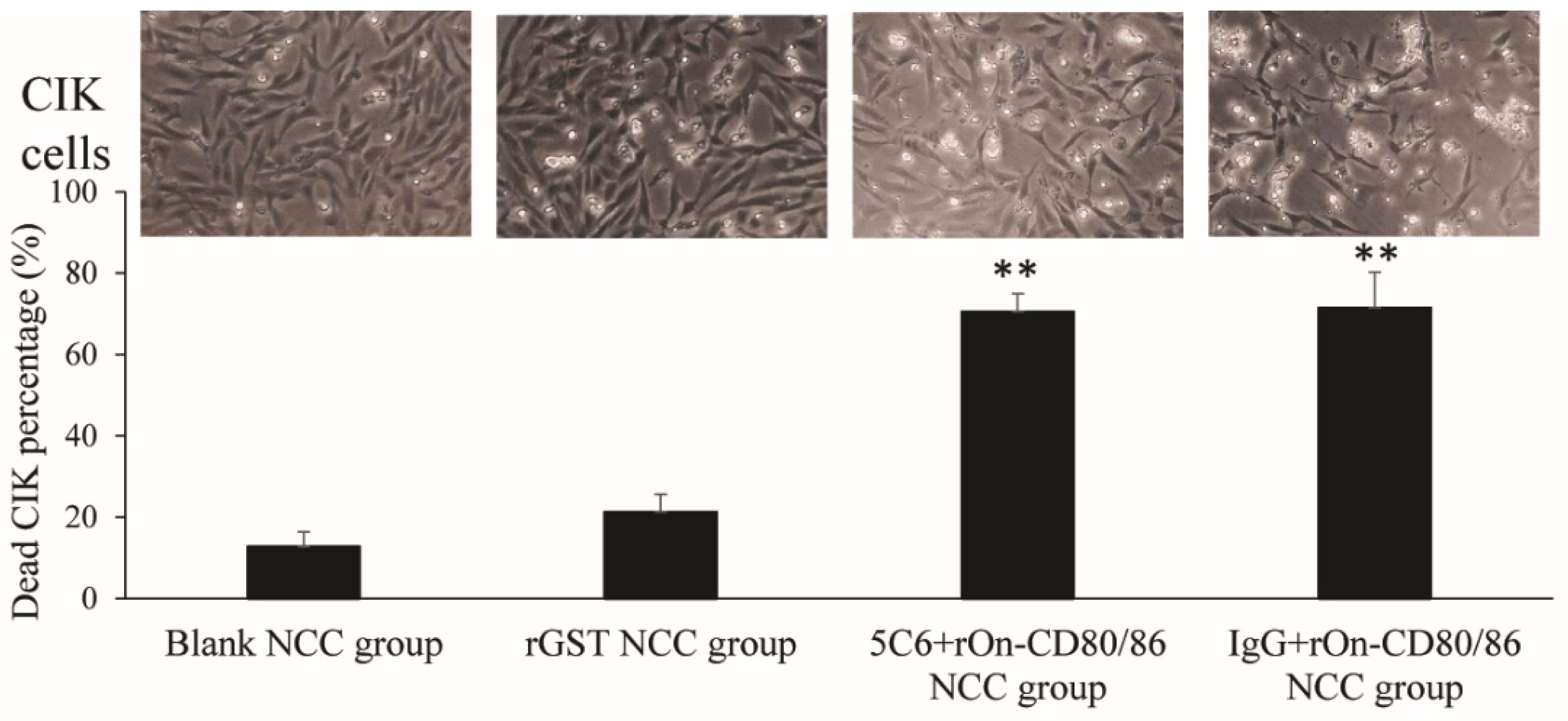
In order to determine whether the rOn-CD80/86 protein enhances the toxicity of NCC at the protein level, we first induced NCC with rOn-CD80/86, and then co-incubated NCC with a certain proportion of CIK cells. And the cell death was observed under a microscope and the CIK cell death rate was counted. The results showed that the cell death rate of the blank group and rGST NCC group was significantly lower than that of the latter two groups by microscope. Then, we calculated the percentage of dead cells in each group, and the results were as follows: 12.8% in the blank NCC group, 21.3% in the rGST NCC group, 70.5% in the 5C6+rOn-CD80/86 NCC group, and 71.5% in the IgG+rOn-CD80/86 NCC group (Figure 3).

3.4. Activity of Caspase-1, 3, and 9 on CIK

In this part, the killing effect of NCC on CIK cells was determined by detecting the activity of various caspases. The results are shown in Figure 4. There was no significant difference in caspase-1, caspase-3, or caspase-9 activity between the blank NCC+CIK group and rGST NCC+CIK group, indicating that treatment with rGST did not increase the cytotoxicity of NCC against the target cells. The activities of caspase-1 and caspase-3 in the rOn-CD80/86 treatment group were significantly higher than those in the control group (blank NCC+CIK group and rGST NCC+CIK group), indicating that rOn-CD80/86 could significantly enhance the killing activity of NCC. In addition, if the 5C6 antibody is added to block the receptor binding site of On-NCCRP-1 on the surface of the NCC cells, caspase-1 and caspase-3 are not significantly inhibited. This result indicates that rOn-CD80/86 does not activate the killing effect of NCC through the On-NCCRP-1 receptor. However, the activity of caspase-9 did not change significantly among the groups.

3.5. Interaction of On-NCCRP-1 with On-CD80/86

In this study, the interaction between these two proteins were checked by the colors of the hybridized yeasts. The result is shown in Figure 5. Five yeast groups were all grown in SD/-Leu/-Trp medium. For self-activation detection, two groups of hybridized yeasts (Figure 5 lanes 4 and 5), could not grow in medium without SD/-Leu/-Trp/-His/-Ade and SD/-Leu/-Trp/-His/-Ade/X-a-Gal, which indicates that On-CD80/86 and On-NCCRP-1 themselves had no transcriptional activity and could not activate reporter genes. In addition, the negative control group (line 2) did not grow in medium without SD/-Leu/-Trp/-His/-Ade nor SD/-Leu/-Trp/-His/-Ade/X-a-Gal. The yeasts containing pGBKT7-On-NCCRP-1/pGADT7-On-CD80/86 and pGBKT7-53/pGADT7-T1 plasmids that could grow on SD/-Leu/-Trp/-His/-Ade plates were white, whereas those grown on SD/-Leu/-Trp/-His/-Ade/X-a-Gal plates were blue. The results suggested no direct interaction between On-NCCRP-1 and On-CD80/86.

3.6. Expression Characteristics of On-CD28 and On-NCCRP-1 Genes in Nile tilapia HKLs scRNA-Seq

HKLs cell subpopulations were displayed visually via t-distributed stochastic neighbor embedding (tSNE) (Figure 6). The four cell populations were represented by different colors (B-cell: orange, T-cell: green, Mo/M: red, NCC: blue). The expression levels of On-CD28 and On-NCCRP-1 gene in HKL scRNA-Seq were analyzed. The results showed that the On-NCCRP-1 gene was only expressed in NCC cell subsets, while the On-CD28 gene was mainly expressed in T-cell subsets, a small amount in B and c subsets, but not in NCC cell subsets.

4. Discussion

The phenomenon wherein costimulatory molecules or ligands, such as CD40, CD80, and CD86, trigger NK cell-mediated cytotoxicity has been reported in mammals [12,13]. Non-specific cytotoxic cells (NCC) are the first identified cytotoxic cell population and kinds of NK cell equivalents in teleost [26]. However, whether these costimulatory molecules could mediate NCC killing activity in bony fish is unclear. In this work, we first obtained soluble rOn-CD80/86 protein using a prokaryotic expression system according to the previously reported On-CD80/86 sequence [15]. NCCs are similar to the killing pathways of mammalian NK cells. The main pathways utilized by killer cells consist of granule exocytosis and those mediated by members of the TNF superfamily. Activated-NCCs can release cytotoxic factors, such as granzyme, perforin, and NK-lysin through immune synapses, to kill target cells. Meanwhile, some genes, such CAS, FADD, CAS, TNF-α, FasL, and NCCRP-1 also take part in this process of immune response [27].
In this study, after incubating NCCs with rOn-CD80/86 protein in vitro, the results showed that, compared to the two control groups, some effector genes, such as TNF-α, CAS, and NK-lysin, were significantly up-regulated in the rOn-CD80/86 stimulation group at 3 and 6 h after incubation, and the expression of NCCRP-1 was also somewhat increased. Tilapia TNF-α can promote the expression of granzyme in NCCs and then cause cytotoxic effects. Meanwhile, activated NCCs could constitutively express TNF-α, which binds to its surface receptor TNFR-1 to exert NCCs cytotoxic effect, and kill cancer and virus-infected cells [7]. After the expression of a large amount of pro-apoptotic protease granzyme, which can enter the target cells and cause the death of the target cells, or enter the cells infected by the virus and restrict the replication of the virus [28,29]. CAS, as an apoptosis regulatory factor, is participated in multiple cellular mechanisms associated with cell proliferation and cell death [30]. NK-lysin, an effector of cytotoxic T-cells, natural killer cells, and NCCs, is a potent antimicrobial peptide widely distributed in mammals and teleosts [22,31]. Therefore, according to the above results, NCCs may exhibit stronger killing activity at 3 h compared to 6 h.
Thus, NCC may respond to extracellular signaling stimuli, which is similar to our previous results. On-NKEF and transferrin protein can effectively activate NCC and significantly up-regulate the expression of its related effectors [23]. Next, we tested the NCC killing effect on target cells by incubating CIK with rOn-CD80/86-activated NCC. The results showed that, although a small number of CIK cells died in the two control groups, CIK cells died in the rOn-CD80/86-activated NCC group when significantly compared to the control group. While binding of the NCC to target cells immediately exerts cytotoxic effects [1], this observation suggests that treatment of NCC with rOn-CD80/86 protein can significantly enhance the ability of NCC to kill other target cells.
Cysteine-requiring aspartate protease (caspase) is a protease family that plays an important role in programmed cell death (including apoptosis) [32]. The activity of caspase family members in caspase in the system will increase significantly in the course of apoptosis [33]. In this study, we detected the activity of three caspases after rOn-CD80/86 treatment. The results showed that the activities of caspase-1 and caspase-3 were significantly higher than those of the two control groups, indicating that the rOn-CD80/86 protein induced significantly increased killing activity of NCCs on CIK cells, thus activating CIK apoptosis signals.
However, it is unknown which receptor is connected to CD80/86 on the NCC surface. CD80, as a costimulatory molecule, is known to activate T-cells through CD28 receptor molecules [34]. In this study, we found that NCC did not express the CD28 gene on the surface; thus, CD80/86 could not activate NCC through this receptor molecule. In addition, NCCRP-1, as the most classical and studied receptor on the surface of NCC, were contemplated as to whether it could play a role through this receptor. The Y2H system is a powerful and commonly used genetic tool to investigate the interactions between artificial fusion proteins inside the nucleus of yeast [35]. The result of the Y2H experiment showed that CD80/86 and NCCRP-1 could not interact with each other, and the blocking of NCCRP-1 with antibody 5C6 of NCCRP-1 did not affect the regulation of CD80/86 activity on NCC. These results suggest that CD80/86 neither regulates NCC activity through CD28 nor through NCCRP-1. Hence, CD80/86 must be acting through other receptor molecules, which need to be further studied.

5. Conclusions

This study obtained Soluble rOn-CD80/86 protein from O. niloticus, and rOn-CD80/86 could up-regulate the expression of the killer effector molecule of NCC, and On-CD80/86-activated NCC could significantly improve the ability of killing CIK cells. Costimulatory Molecules rOn-CD80/86 is independent of On-NCCRP-1 and On-CD28 receptors to modulate NCC killing activity in vitro of Nile tilapia. The results provide a more theoretical basis for understanding the regulation mechanism of NCC activity.

Author Contributions

Conceptualization, Y.H.; methodology, Y.H.; formal analysis, Y.H. and Z.C.; investigation, Z.C. and R.X.; resources, Z.C., R.X. and B.W.; writing—original draft preparation, Y.H. and K.A.; writing—review and editing, J.C., Y.L. and B.W.; supervision, J.J.; funding acquisition, Y.H. All authors have read and agreed to the published version of the manuscript.

Funding

This research was funded by the National Natural Science Foundation of China (No. 32002426) and the Natural Science Foundation of Guangdong Province (No. 2022A1515010553).

Institutional Review Board Statement

The study was conducted in accordance with the Declaration of Helsinki, and approved by the Ethics Committee of the Guangdong Ocean University (Approval Code: (2019)1, Approval Date: 10 May 2019).

Informed Consent Statement

Not applicable.

Data Availability Statement

Data is contained within the article.

Acknowledgments

We extend our thanks to associate Zhe Zheng for reviewing this manuscript.

Conflicts of Interest

The authors declare no conflict of interest.

References

  1. Jaso-Friedmann, L.; Leary, J.H., 3rd; Evans, D.L. The non-specific cytotoxic cell receptor (NCCRP-1): Molecular organization and signaling properties. Dev. Comp. Immunol. 2001, 25, 701–711. [Google Scholar] [CrossRef]
  2. Praveen, K.; Leary, J.H., 3rd; Evans, D.L.; Jaso-Friedmann, L. Nonspecific cytotoxic cells of teleosts are armed with multiple granzymes and other components of the granule exocytosis pathway. Mol. Immunol. 2006, 43, 1152–1162. [Google Scholar] [CrossRef]
  3. Evans, D.L.; Leary, J.H., 3rd; Jaso-Friedmann, L. Nonspecific cytotoxic cells and innate immunity: Regulation by programmed cell death. Dev. Comp. Immunol. 2001, 25, 791–805. [Google Scholar] [CrossRef]
  4. Cuesta, A.; Esteban, M.; Meseguer, J. Molecular characterization of the nonspecific cytotoxic cell receptor (NCCRP-1) demonstrates gilthead seabream NCC heterogeneity. Dev. Comp. Immunol. 2005, 29, 637–650. [Google Scholar] [CrossRef]
  5. Greenlee, A.R.; Brown, R.A.; Ristow, S.S. Nonspecific cytotoxic cells of rainbow trout (Oncorhynchus mykiss) kill YAC-1 targets by both necrotic and apoptic mechanisms. Dev. Comp. Immunol. 1991, 15, 153–164. [Google Scholar] [CrossRef]
  6. Jaso-Friedmann, L.; Leary, J.H., 3rd; Evans, D.L. Role of nonspecific cytotoxic cells in the induction of programmed cell death of pathogenic protozoans: Participation of the Fas ligand-Fas receptor system. Exp. Parasitol. 2000, 96, 75–88. [Google Scholar] [CrossRef]
  7. Praveen, K.; Evans, D.L.; Jaso-Friedmann, L. Constitutive expression of tumor necrosis factor-alpha in cytotoxic cells of teleosts and its role in regulation of cell-mediated cytotoxicity. Mol. Immunol. 2006, 43, 279–291. [Google Scholar] [CrossRef]
  8. Meseguer, J.; Esteban, M.A.; Mulero, V. Nonspecific cell-mediated cytotoxicity in the seawater teleosts (Sparus aurata and Dicentrarchus labrax): Ultrastructural study of target cell death mechanisms. Anat. Rec. 1996, 244, 499–505. [Google Scholar] [CrossRef]
  9. Reimers, K.; Abu Qarn, M.; Allmeling, C.; Bucan, V.; Vogt, P.M. Identification of the non-specific cytotoxic cell receptor protein 1 (NCCRP1) in regenerating axolotl limbs. J. Comp. Physiol. B 2006, 176, 599–605. [Google Scholar] [CrossRef]
  10. Bachmann, M.F.; Sebzda, E.; Kündig, T.M.; Shahinian, A.; Speiser, D.; Mak, T.W.; Ohashi, P.S. T cell responses are governed by avidity and co-stimulatory thresholds. Eur. J. Immunol. 1996, 26, 2017–2022. [Google Scholar] [CrossRef]
  11. Boomer, J.S.; Green, J.M. An enigmatic tail of CD28 signaling. Cold Spring Harb. Perspect. Biol. 2010, 2, a002436. [Google Scholar] [CrossRef] [Green Version]
  12. Martín-Fontecha, A.; Assarsson, E.; Carbone, E.; Kärre, K.; Ljunggren, H.G. Triggering of murine NK cells by CD40 and CD86 (B7-2). J. Immunol. 1999, 162, 5910–5916. [Google Scholar]
  13. Wilson, J.L.; Charo, J.; Martín-Fontecha, A.; Dellabona, P.; Casorati, G.; Chambers, B.J.; Kiessling, R.; Bejarano, M.T.; Ljunggren, H.G. NK cell triggering by the human costimulatory molecules CD80 and CD. J. Immunol. 1999, 163, 4207–4212. [Google Scholar]
  14. Zhang, X.J.; Zhang, X.Y.; Wang, P.; Zhang, Y.A. Identification of another primordial CD80/86 molecule in rainbow trout: Insights into the origin and evolution of CD80 and CD86 in vertebrates. Dev. Comp. Immunol. 2018, 89, 73–82. [Google Scholar] [CrossRef]
  15. Huang, Y.; Wang, Z.; Zheng, Q.; Tang, J.; Cai, J.; Lu, Y.; Jian, J. Conservation of structural and interactional features of CD28 and CD80/86 molecules from Nile tilapia (Oreochromis niloticus). Fish Shellfish Immunol. 2018, 72, 95–103. [Google Scholar] [CrossRef]
  16. Lu, T.Z.; Liu, X.; Wu, C.S.; Ma, Z.Y.; Wang, Y.; Zhang, Y.A.; Zhang, X.J. Molecular and Functional Analyses of the Primordial Costimulatory Molecule CD80/86 and Its Receptors CD28 and CD152 (CTLA-4) in a Teleost Fish. Front. Immunol. 2022, 13, 885005. [Google Scholar] [CrossRef]
  17. Teng, J.; Cui, M.Y.; Zhao, Y.; Chen, H.J.; Du, W.J.; Xue, L.Y.; Ji, X.S. Expression changes of non-specific cytotoxic cell receptor (NCCRP1) and proliferation and migration of NCCs post-Nocardia seriolae infection in Northern Snakehead. Dev. Comp. Immunol. 2022, 139, 104576. [Google Scholar] [CrossRef]
  18. Huang, X.Z.; Li, Y.W.; Mai, Y.Z.; Luo, X.C.; Dan, X.M.; Li, A.X. Molecular cloning of NCCRP-1 gene from orange-spotted grouper (Epinephelus coioides) and characterization of NCCRP-1(+) cells post Cryptocaryon irritans infection. Dev. Comp. Immunol. 2014, 46, 267–278. [Google Scholar] [CrossRef]
  19. Zhao, X.L.; Han, Y.; Ren, S.T.; Ma, Y.M.; Li, H.; Peng, X.X. L-proline increases survival of tilapias infected by Streptococcus agalactiae in higher water temperature. Fish Shellfish Immunol. 2015, 44, 33–42. [Google Scholar] [CrossRef]
  20. Maulu, S.; Hasimuna, O.J.; Mphande, J.; Munang’Andu, H.M. Prevention and Control of Streptococcosis in Tilapia Culture: A Systematic Review. J. Aquat. Anim. Health 2021, 33, 162–177. [Google Scholar] [CrossRef]
  21. Li, Q.; Jiang, B.; Zhang, Z.; Huang, Y.; Xu, Z.; Chen, X.; Huang, Y.; Jian, J. SP protects Nile tilapia (Oreochromis niloticus) against acute Streptococcus agalatiae infection. Fish Shellfish Immunol. 2022, 123, 218–228. [Google Scholar] [CrossRef] [PubMed]
  22. Huang, Y.; Zheng, Q.; Niu, J.; Tang, J.; Wang, B.; Abarike, E.D.; Lu, Y.; Cai, J.; Jian, J. NK-lysin from Oreochromis niloticus improves antimicrobial defence against bacterial pathogens. Fish Shellfish Immunol. 2018, 72, 259–265. [Google Scholar] [CrossRef] [PubMed]
  23. Huang, Y.; Zheng, Q.; Wang, Z.; Tang, J.; Lu, Y.; Qin, Q.; Cai, J.; Jian, J. Fish natural killer enhancing factor-A (NKEF-A) enhance cytotoxicity of nonspecific cytotoxic cells against bacterial infection. Mol. Immunol. 2021, 133, 184–193. [Google Scholar] [CrossRef] [PubMed]
  24. Huang, Y.; Chen, Z.; Xie, R.; Wang, P.; Zhang, Z.; Cai, J.; Wang, B.; Jian, J. Transferrin Mediated NCC Killing Activity through NCCRP-1 in Nile tilapia (Oreochromis niloticus). Fishes 2022, 7, 253. [Google Scholar] [CrossRef]
  25. Niu, J.; Huang, Y.; Liu, X.; Zhang, Z.; Tang, J.; Wang, B.; Lu, Y.; Cai, J.; Jian, J. Single-cell RNA-seq reveals different subsets of non-specific cytotoxic cells in teleost. Genomics 2020, 112, 5170–5179. [Google Scholar] [CrossRef] [PubMed]
  26. Ishimoto, Y.; Savan, R.; Endo, M.; Sakai, M.; Immunology, J.F. Non-specific cytotoxic cell receptor (NCCRP)-1 type gene in tilapia (Oreochromis niloticus): Its cloning and analysis. Fish Shellfish Immunol. 2004, 16, 163–172. [Google Scholar] [CrossRef] [PubMed]
  27. Parida, S.; Mohapatra, A.; Sahoo, P.K. Cloning and functional characterisation of natural killer enhancing factor-B (NKEF-B) gene of Labeo rohita: Anti-oxidant and antimicrobial activities of its recombinant protein. Mol. Immunol. 2020, 126, 73–86. [Google Scholar] [CrossRef] [PubMed]
  28. Bots, M.; Medema, J.P. Granzymes at a glance. J. Cell Sci. 2006, 119, 5011–5014. [Google Scholar] [CrossRef] [PubMed] [Green Version]
  29. Walch, M.; Dotiwala, F.; Mulik, S.; Thiery, J.; Kirchhausen, T.; Clayberger, C.; Krensky, A.M.; Martinvalet, D.; Lieberman, J. Cytotoxic cells kill intracellular bacteria through granulysin-mediated delivery of granzymes. Cell 2014, 157, 1309–1323. [Google Scholar] [CrossRef] [PubMed] [Green Version]
  30. Praveen, K.; Leary, J.H.; Evans, D.L.; Jaso-Friedmann, L. Molecular cloning of cellular apoptosis susceptibility (CAS) gene in Oreochromis niloticus and its proposed role in regulation of non-specific cytotoxic cell (NCC) functions. Fish Shellfish Immunol. 2006, 20, 647–655. [Google Scholar] [CrossRef] [PubMed]
  31. Andersson, M.; Gunne, H.; Agerberth, B.; Boman, A.; Bergman, T.; Olsson, B.; Dagerlind, A.; Wigzell, H.; Boman, H.; Gudmundsson, G. NK-lysin, structure and function of a novel effector molecule of porcine T and NK cells. Vet. Immunol. Immunopathol. 1996, 54, 123–126. [Google Scholar] [CrossRef] [PubMed]
  32. Wen, R.; Liu, Y.P.; Tong, X.X.; Zhang, T.N.; Yang, N. Molecular mechanisms and functions of pyroptosis in sepsis and sepsis-associated organ dysfunction. Front. Cell. Infect. Microbiol. 2022, 12, 962139. [Google Scholar] [CrossRef] [PubMed]
  33. Choudhary, N.; Collignon, T.E.; Tewari, D.; Bishayee, A. Hypericin and its anticancer effects: From mechanism of action to potential therapeutic application. Phytomedicine 2022, 105, 154356. [Google Scholar] [CrossRef] [PubMed]
  34. Bluestone, J.A. New perspectives of CD28-B7-mediated T cell costimulation. Immunity 1995, 2, 555–559. [Google Scholar] [CrossRef] [PubMed] [Green Version]
  35. Paiano, A.; Margiotta, A.; De Luca, M.; Bucci, C. Yeast Two-Hybrid Assay to Identify Interacting Proteins. Curr. Protoc. Protein Sci. 2019, 95, e70. [Google Scholar] [CrossRef] [PubMed]
Figure 1. Identification of protein expression and purification of rOn-CD80/86 by SDS-PAGE (lane 1–5) and western blot (lanes 6 and 7). The black arrow refers to rOn-CD80/86. Lane M: marker; lane 1: E. coli with pGEX-4T-CD80/86 without IPTG induction; lane 2: E. coli with IPTG induction; lane 3: E. coli soluble protein with IPTG induction; lane 4: E. coli inclusion body protein with IPTG induction; lane 5: purified rOn-CD80/86; lane 6: experiment group (use mouse anti-his IgG); lane 7: control group (use mouse IgG).
Fishes 07 00353 g001
Figure 2. Expression of NCC-related genes after rOn-CD80/86 stimulation by qRT-PCR. All values are the mean ± SD, n = 3. Significant difference was indicated by asterisks, *: 0.05 > p > 0.01, **: p < 0.01.
Fishes 07 00353 g002
Figure 3. Killing activity of NCC on CIK. Photographs of CIK (upper) and corresponding dead cell percentage of CIK (lower). Data are shown as mean ± SE, and the significant difference was indicated by asterisks, **: p < 0.01.
Fishes 07 00353 g003
Figure 4. Effect of caspase-1, 3, and 9 activities in CIK by rOn-CD80/86-activated NCC. Value represents the mean ± S.E, n = 3. Significant difference was expressed by *: 0.05 > p > 0.01.
Fishes 07 00353 g004
Figure 5. Interaction between On-NCCRP-1 and On-CD80/86 by Y2H. pGBKT7-On-NCCRP-1/pGADT7-On-CD80/86 is the experimental group; pGBKT7-Lam/pGADT7-T is the negative control group; pGBKT7-53/pGADT7-T is the positive control group; and pGBKT7-On-NCCRP-1/pGADT7 and pGADT7-On-CD80/86+pGBKT7 are the self-activation groups. Column (AC): Yeasts grown on plates with double dropout medium (SD/–Leu/–Trp), quadruple dropout medium (SD/-Leu/-Trp/-His/-Ade), and quadruple dropout medium supplemented with X-a-Gal (SD/-Leu/-Trp/-His/-Ade/X-a-Gal), respectively.
Fishes 07 00353 g005
Figure 6. (A) The visualization of the Nile tilapia HKL population via tSNE nonlinear clustering is referred to from our previous study [25]. (B) Expression analysis of On-CD28 and On-NCCRP-1 in HKL subsets.
Fishes 07 00353 g006
Table 1. Primers used in this research.
PrimersSequences (5′→3′)Purpose
point-to-point Y2HOn-NCCRP-1-SGGAATTCATGTCTGCTGCCGAGTGGAAG
point-to-point Y2HOn-NCCRP-1-AGGGATCCCGCGGGCTGCTTTTGCTTGGTC
point-to-point Y2HpGADT7-On-CD80/86-S CGGGATCCATACAGGAAGTGCTAAATTTCTC
point-to-point Y2HpGADT7-On-CD80/86-ACCGCTCGAGCTAATCAGTGCTGTCTTGTATCTG
Protein expressionCD80/86-EcoRI-FTCCGAATTCTTCACCGTTACCGGTTCTGC
Protein expressionCD80/86-XhoI-RTGCTCGAGAGAACCACCGGTCTGACGACG
qRT-PCRqβ-actin-SAGATGAAATCGCCGCACTGG
qRT-PCRqβ-actin-ATCTGACCCATACCCACCATCA
qRT-PCRRT-TNFα-FCTCGTCGTCGTGGCTCTTT
qRT-PCRRT-TNFα-RCCTTGGCTTTGCTGCTGAT
qRT-PCRRT-NCCRP1-FCACCACCTGAACCCGAACT
qRT-PCRRT-NCCRP1-RGGTCCACAACCTGCTCCAT
qRT-PCRRT-granzyme-FATACAACTGGCAAGGAAGGAG
qRT-PCRRT-granzyme-RTACCCATCTCAGCACATCAAC
qRT-PCRRT-FasL-FCTTCTCCAAGGGCGATTCTA
qRT-PCRRT-FasL-RATCTCCCTGAGTGGCTGTGC
qRT-PCRRT-CAS-FCAGCAGTTTCGAGGAAGCAC
qRT-PCRRT-CAS-RTCCAAGCAAGCCAGGTATTT
qRT-PCRRT-FADD-FACTGGCAGAAGATAACACGG
qRT-PCRRT-FADD-RTTTGCTTTCTCCTCCTCACT
qRT-PCRRT-NK-lysin-FATTTGCGGCACAGTGATTT
qRT-PCRRT-NK-lysin-RATGGAAGTCTTGATGGGGCT
Table 2. GenBank accession numbers of relative genes of tilapia used in this study.
ProteinAccession No.
CD80/86MF150103.1
TNFαAY428948.1
NCCRP-1MF162296
GranzymeAY918866.1
FasLKM008610.1
CASAF547173
FADDXM_003456561.5
NK-lysinATW66454.1
Publisher’s Note: MDPI stays neutral with regard to jurisdictional claims in published maps and institutional affiliations.