Article

The Effects of Different LED Lights on the Main Nutritional Compositions of Isochrysis zhanjiangensis

1
School of Marine Sciences, Ningbo University, Ningbo 315211, China
2
Fujian Dalai Seed Science and Technology Co., Ltd., Ningde 352101, China
3
Ningbo Institute of Oceanography, Ningbo 315832, China
*
Author to whom correspondence should be addressed.
These authors contributed equally to this work.
Fishes 2023, 8(3), 124; https://doi.org/10.3390/fishes8030124
Submission received: 2 January 2023 / Revised: 12 February 2023 / Accepted: 21 February 2023 / Published: 22 February 2023
(This article belongs to the Special Issue Feeding and Nutrition of Bivalves)

Abstract

:
Light irradiation plays an important role in the growth of microalgae and their nutrient composition. To elucidate the effect of different LED lights (red, orange, green, blue, purple, simulated sunlight, and white) on the main nutritional compositions of Isochrysis zhanjiangensis, the growth, fatty acid composition, soluble carbohydrate, and soluble protein contents were studied. The results show that I. zhanjiangensis grew the fastest under blue light (p = 0.044). In terms of fatty acid composition, the percentage of myristic acid (C14:0) decreased significantly (p = 0.021) under blue light and simulated sunlight, but the percentages of palmitic acid (C16:0) (p = 0.032) and stearic acid (C18:0) (p = 0.037) significantly increased. The percentage of docosahexaenoic acid (C22:6n-3) increased under orange light (p = 0.021), and the percentage of total unsaturated fatty acids increased under blue light (p = 0.008). The protein content significantly increased under simulated sunlight (p = 0.025), while the carbohydrate content did not vary much (p > 0.05) under different lights. These findings provide useful guidance for optimizing the cultivation conditions of I. zhanjiangensis to improve its nutritional value and yield.

1. Introduction

Aquaculture is a rapidly growing and efficient source of sustainable food, which has become increasingly important due to the growing global demand for seafood and the decline of wild fish stocks [1]. It produces most marine bivalves, such as clams, mussels, and oysters, which account for 89% of total aquaculture production [2]. As the vital feed source, microalgae provide the necessary nutrients, such as proteins, lipids, and fatty acids, for the healthy growth of bivalves [3]. Moreover, the use of microalgae as a feed source reduces the cost of feed. Microalgae production is much more sustainable and environmentally friendly when compared with other traditional feed production methods and is relatively inexpensive, making it a cost-effective solution for bivalve shellfish farming [4]. Currently, intensive bivalve cultivation has relied on the production of live algae, which accounts for an average of 30% of the operating costs of bivalve hatcheries [5]. Thus, it is important to enhance biomass accumulation to reduce the costs further and to increase key nutrients in the microalgae.
Photoautotrophic microalgae absorb light (photons) and convert it to chemical energy. Therefore, the light source plays a key role in the process of photosynthesis in microalgae. Light emitting diodes (LEDs) have specific narrow-band light and high spectral purity, making them a more suitable light source for microalgae cultivation as compared with common light sources that have high cost and low wattage irradiance [6]. Different lights have varied effects on the physiological functions and nutrient synthesis of microalgae, emphasizing the importance of selecting an appropriate light source for microalgae cultivation. The use of LED lights in microalgae cultivation can help optimize the growth conditions, leading to improved yields and quality of the microalgae product. For example, blue light can promote astaxanthin accumulation in Haematococcus pluvialis and phycocyanin accumulation in Arthrospira [7,8]; it also facilitates the accumulation of carotenoids in Chlorella, Eustigmatos, and Dunaliella salina [9,10,11]. Red light can increase the pigment content and antioxidant capacity of Cyanobacteria [12]. Green light can help Nannochloropsis oculata and Phaeodactylum tricornutum accumulate lipids to up to 60.6% of dry weight [13,14]. Compared with blue light, yellow light can increase the growth rate of Isochrysis galbana, and it facilitates lipid accumulation in I. galbana and Pavlova lutheri [15,16]. In conclusion, the type of light source used can greatly impact the growth and nutritional compositions of various microalgae, making it an important consideration in aquaculture and microalgal cultivation.
Isochrysis zhanjiangensis is widely used for the cultivation of aquatic food, such as mollusks, shrimps, and fishes, due to its fast growth and high concentration of poly-unsaturated fatty acids (PUFA) [17]. Bivalves are unable to synthesize enough essential fatty acids such as arachidonic acid (ARA), eicosapentaenoic acid (EPA), and docosahexaenoic acid (DHA), and need to obtain these essential fatty acids from dietary intake [18]. Selecting the right light source can maximize the biomass gain of I. zhanjiangensis and increase the accumulation of nutrients that are essential to aquatic organisms. Although similar studies have investigated the effect of several LED lights on I. zhanjiangensis, the selected light sources were limited [19]. In this study, the effects of seven LED lights (red, orange, green, blue, purple, simulated sunlight, and white) on the growth and nutritional composition of I. zhanjiangensis were examined. The results provide the guidance required for selecting the right light source, which will ultimately determine the yield and quality of the algae cultivation. This information is crucial for ensuring the sustainability and efficiency of large-scale algae cultivation operations.

2. Materials and Methods

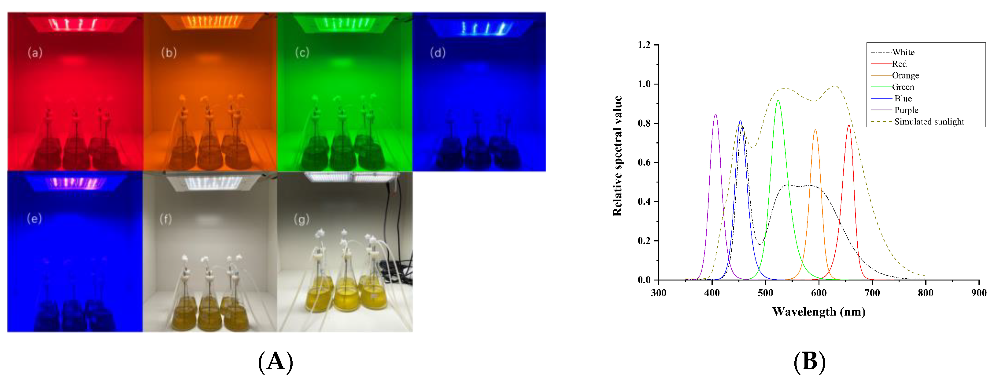
2.1. Instruments and Equipment

The experiment was carried out in 7 independent culture chambers (Figure 1A). The LED light was placed directly above the culture chamber, and the distance between the different light sources and the cultures was 60 cm. In this experiment, 7 groups of LED treatments were set up and white light used as the control (CK): white (400–700 nm), blue (450 nm), green (520 nm), orange (590 nm), red (660 nm), purple (405 nm), and simulated sunlight (400–730 nm) (San’an Optoelectronics Co., Xiamen, China). The photoperiod was controlled by an electronic timer (GND-1, Gongniu Group Co., Ningbo, China), and the light intensity was controlled by changing the brightness of each LED light. The spectrum and intensity of the lights were measured by a plant lighting analyzer (PLA-30, Everfine Co., Hangzhou, China) (Figure 1B). The cultures were mixed by continuous aeration with ultraviolet sterilized air at a flow rate of 0.3 L·min−1, which was controlled by a glass tube flowmeter (LZB-3WB, Zhenxing Flowmeter Instrument Factory, Yuyao, China).

2.2. Microalgae Cultivation and Harvesting

I. zhanjiangensis was provided by the Germplasm Bank of Aquatic Organisms of the Chinese Academy of Sciences, and the preservation number is FACHB-1750. The strain was cultured at a light intensity of 100 ± 5 μmol·m−2·s−1 under a photoperiod cycle of 12:12 h light/dark, and the temperature was controlled at 25 ± 1 °C. Seawater (pH = 7.89) of 25 PSU (practical salinity units) was filtered through a 0.22 μm filter membrane and sterilized by autoclaving. NMB3# medium was used as the culture medium [19], and 600 mL of sterile culture medium was prepared in a 1 L flask for culturing microalgae. Each culture treatment was biologically triplicated.
During the cultivation, the cell density was counted using a hemocytometer (XB-K-25, QiuJing Co., Shanghai, China) under a CX-23 optical microscope (Olympus Japan Co., Tokyo, Japan). The initial cell density was 1 × 10⁶ cells·mL−1. After culturing for 12 days, the algae at stationary phase were collected by centrifugation at 12,450× g for 10 min at 4 °C using a high-speed refrigerated centrifuge (GL-10000C, Anke Instrument Co., Shanghai, China). The collected culture was freeze-dried for 48 h with a freeze dryer (SCIENTZ -10Z, Scientz Co., Ningbo, China) and stored at −80 °C for further analysis.

2.3. Determination of Soluble Protein Content

Soluble protein content was determined using a BCA Protein Assay Kit (Solarbio Co., Beijing, China). In brief, the soluble protein was extracted from 10 mg freeze-dried microalgae by resuspending the biomass in 1.5 mL of 0.01 mol·L−1 phosphate buffered saline (PBS) and homogenized for 3 cycles of 60 s. The samples were centrifuged at 860× g for 10 min at 4 °C and the supernatants were collected; the biomass was extracted 3 times, and the supernatants were combined for determination according to the BCA Protein Assay Kit manual at a wavelength of 562 nm. The bovine serum albumin (BSA) of 500 mg·mL−1 was used as a protein standard. All samples were measured 3 times. A set of blank solutions were prepared in the same way as the samples to obtain the corrected absorbance. Finally, the percentages of proteins were calculated using the following formula:
Protein content (%) = (C × V × D) × 100/m
In the formula, C represents the protein concentration obtained from the standard curve (mg·mL−1); V represents the volume of PBS used to resuspend the biomass (mL); D is the dilution factor; and m represents the biomass (mg).

2.4. Determination of Soluble Carbohydrate Content

Soluble carbohydrate content was determined using the anthrone-sulfuric acid method [20]. First, 10 mg of freeze-dried microalgae was transferred into a 2 mL centrifuge tube containing ceramic beads, and 1 mL of absolute ethanol was added to each tube until fully dissolved. The tubes were vortexed for 10 min, ultrasonically extracted for 10 min at 40 kHz, and then homogenized for 9 cycles of 60 s. Then, the solutions were washed with 4 mL of deionized water and transferred to a new 15 mL centrifuge tube, vortexed, and centrifuged at 6500× g for 10 min at 4 °C to collect the supernatant. A standard curve between carbohydrate content and OD620 was established using anhydrous glucose as the standard. Next, 200 μL of the sample extract was added to 2 mL centrifuge tubes as well as the glucose standard. Then, 1 mL of anthrone-sulfuric acid reagent was added to each tube, mixed well, and heated in boiling water for 15 min. After the samples had cooled to room temperature, the OD620 of each sample was measured. The following formula was used to calculate the percentage of soluble carbohydrate:
Carbohydrate content (%) = (C × V × D) × 100/m
In the formula, C represents the soluble carbohydrate concentration (mg·mL−1) obtained from the standard curve; V represents the total volume (mL) used for the extraction solution; D is the dilution factor; and m represents the biomass (mg).

2.5. Fatty Acid Analysis

Fatty acid methyl esters were extracted according to Wang et al. [21]. Briefly, 20 mg of freeze-dried microalgae was placed into a screw-top glass tube. Then, 1 mL of n-hexane, 1.5 mL of acetyl chloride–methanol (1:10, v/v), and 30 μL of nonadecanoic acid (C19:0) internal standard solution (1 μg·μL−1) were added. After vortexing and mixing, the tube was placed in a 60 °C water bath for 2 h. After cooling to room temperature, 2.5 mL of 6% K2CO3 and 1 mL of n-hexane were added to the tube and vortexed for 30 s. Then, the n-hexane layer containing fatty acid methyl esters was transferred into a 2 mL centrifuge tube, followed by centrifugation at 860× g for 10 min. The supernatant was filtered using a 0.22 μm organic syringe filter for GC-MS analysis. The fatty acid methyl esters were analyzed using the GC-MS platform (7890B-7000C, Agilent Technologies Inc., CA, PaloAlto, USA), and the GC and MS conditions were as in Wang et al. [21].
The fatty acids were identified by searching the NIST14 library and referring to the relative retention time of each component compared to C19:0. Each fatty acid was quantified using the peak area normalization method.

2.6. Statistical Analysis

Statistically significant differences (p < 0.05) among the variables were determined by one-way ANOVA with Tukey’s post hoc test (SPSS 26.0, SPSS Inc., Chicago, USA). The values are represented as mean ± SD.

3. Results

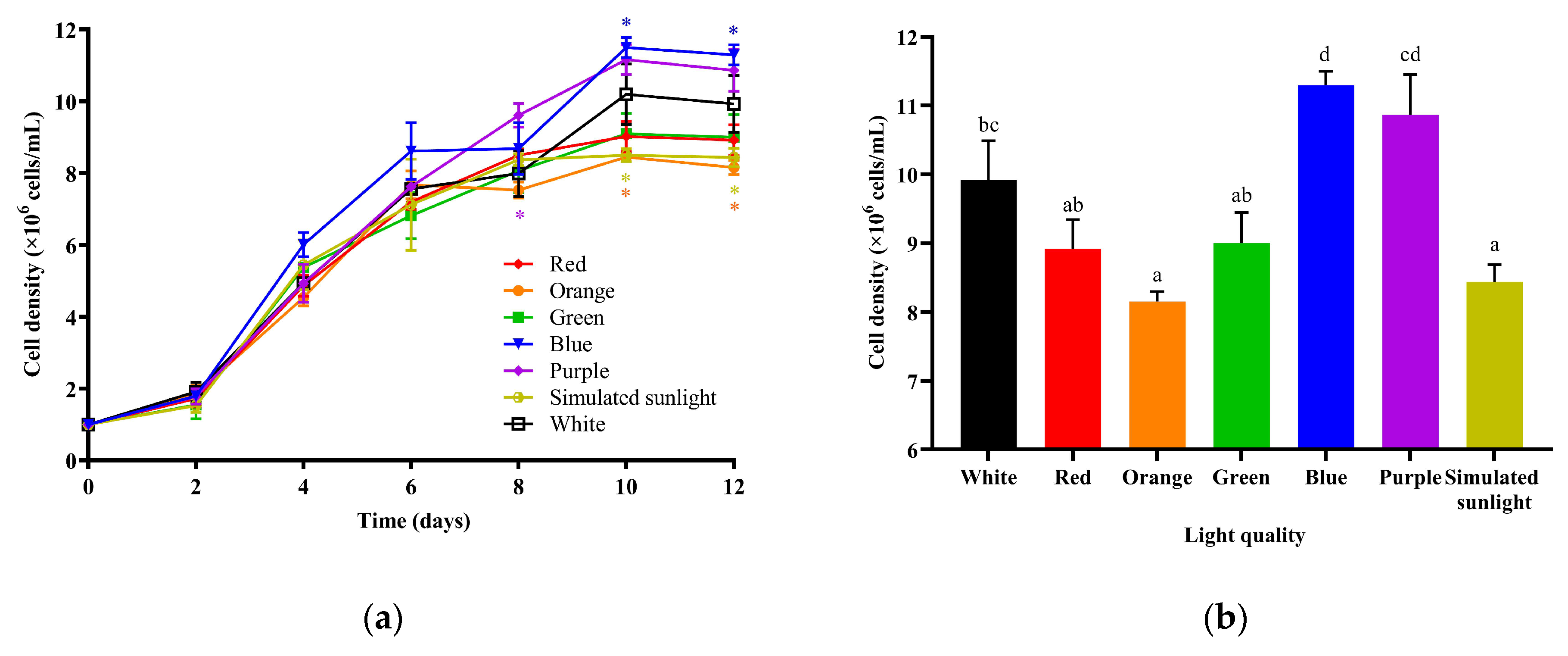
3.1. The Effects of Different LED Lights on the Growth of I. zhanjiangensis

As shown in Figure 2a, the I. zhanjiangensis algal cell density gradually increased with the growth process under each LED light. On the 12th day of cultivation, the algae density under blue light was significantly higher than the other light conditions except purple, while the algae densities under orange light (p = 0.047) and simulated sunlight (p = 0.049) were significantly lower than under white, blue, and purple light (Figure 2b), suggesting that blue light had the best growth-promoting effect and orange light had a significant inhibitory effect on the growth of I. zhanjiangensis.
The algae density under white light was significantly lower than under blue light and significantly higher than under orange light or simulated sunlight. There was no significant difference in algae density between the white, red, green, and purple lights. White light is widely used for the indoor cultivation of microalgae, so the following analysis uses white light as the control.

3.2. The Effects of Light Source on the Fatty Acid Composition of I. zhanjiangensis

Table 1 shows fatty acid composition under the seven light conditions. For saturated fatty acids (SFA), the myristic acid (C14:0) content of I. zhanjiangensis decreased significantly (p = 0.021) under simulated sunlight and blue light, and the content was the lowest under blue light; palmitic acid (C16:0) (p = 0.032), stearic acid (C18:0) (p = 0.037), and docosanoic acid (C22:0) (p = 0.018) were all significantly increased under simulated sunlight. For monounsaturated fatty acids (MUFA), the erucic acid (C22:1n-9) content of I. zhanjiangensis decreased significantly under blue light (p = 0.021) and simulated sunlight (p = 0.036). For PUFA, the content of linoleic acid (C18:2n-6) significantly increased under simulated sunlight (p = 0.000) and blue light (p = 0.000), and significantly decreased under orange light (p = 0.002), while the content of docosahexaenoic acid (DHA, C22:6n-3) significantly increased under orange light (p = 0.021) and significantly decreased under simulated sunlight (p = 0.008). For total unsaturated fatty acids (TUFA), the content increased significantly (p = 0.008) under blue light. These results suggest that light source has a certain influence on the composition of fatty acids in I. zhanjiangensis.

3.3. The effects of Light Source on Soluble Protein and Soluble Carbohydrate in I. zhanjiangensis

The levels of soluble protein and soluble carbohydrate content of I. zhanjiangensis cultured under seven different LED lights are shown in Table 2. Compared with white light, soluble protein in I. zhanjiangensis significantly increased (p = 0.025) under simulated sunlight (Table 2), but there was no significant difference in soluble carbohydrate content under the different light sources (Table 2). These results demonstrate that light source has little effect on soluble carbohydrate content in I. zhanjiangensis and simulated sunlight could increase the content of soluble protein.

4. Discussion

4.1. The Effects of Light Source on the Growth of I. zhanjiangensis

Light source has a significant effect on microalgal growth. In this study, blue light significantly promoted the growth of I. zhanjiangensis. It has been reported that the cell division and growth rate of Chlorella increase under blue LED light [22], which is consistent with our results. Similarly, Sirisuk et al. [23] found that mixed red and blue light could promote the growth of Isochrysis galbana when compared with red or white light. This is because chlorophyll a, chlorophyll c1 + c2, and some carotenoids, such as fucoxanthin, dinoxanthin, and phycoxanthin, are the main pigments in I. galbana [24], and the absorption peak of carotenoids is near blue light meaning they can absorb blue light more efficiently [25]. I. zhanjiangensis and I. galbana belong to the same genus, Isochrysis, and they have similar pigment compositions [26]. The present results show that I. zhanjiangensis grows faster under blue LED conditions, suggesting that its carotenoids absorb blue light more effectively. This finding provides a new strategy for the cultivation of I. zhanjiangensis in terms of using blue LEDs as the growth light source to enhance biomass accumulation, which could potentially improve the efficiency of this aquaculture and address the increasing demand for sustainable food sources.

4.2. The Effects of Different Lights on the Fatty Acids of I. zhanjiangensis

I. zhanjiangensis is rich in DHA and other PUFAs. It has been demonstrated that dietary intake of DHA is one of the most critical factors in the growth and development of bivalves [27]. The fatty acid composition of microalgae greatly influences the nutritional value of bivalve diets. Several studies have shown that fatty acid accumulation in microalgae is significantly different under different light sources [28,29,30]. Therefore, analyzing the effects of different lights on fatty acid accumulation and determining the optimal accumulation condition is the key to efficiently utilizing I. zhanjiangensis.
In this study, it was found that under blue light, the TUFA content increased while C14:0 content decreased, which is consistent with results from Chai et al. [31]. They showed that I. zhanjiangensis cultured under blue light has increased TUFA content and decreased C14:0 content as compared to algae cultured under red or green light; C14:0 is the synthetic precursor of C16:0. Algae cells can synthesize long-chain polyunsaturated fatty acids (LC-PUFAs) through elongation and desaturation [32]. Under blue light, C14:0 content in microalgae cells is reduced, TUFA content is high, and cell density is optimal, suggesting that blue light can increase the percentage of unsaturated fatty acids in I. zhanjiangensis to increase the fluidity of cell membranes, which is needed for rapid algae growth.
Studies have shown that light of the same color as the microalgae is likely to enhance lipid production and promote the accumulation of DHA. For example, I. galbana grown under yellow LEDs obtained higher lipid content (27.94%) [16]. Ra et al. [14] found that the lipid and the DHA content of Nannochloropsis under green LED light significantly increased compared to the control group under red and blue light. Kim et al. [16] found that the DHA content of Chlorella and Porphyridum significantly increased under green and red LEDs with similar wavelengths to the respective algae, showing 30% and 52% higher levels compared to the control group under white light. In this study, the DHA content of I. zhanjiangensis significantly increased under orange light, which has a similar color to the algae. Microalgae culture solutions have the lowest absorption rate for light that is similar in color to the solution, and algae density is also lowest. For example, in this study, cell density was lowest under orange light (Figure 2), indicating that microalgae growth was inhibited under this condition, and the accumulation of lipids and DHA was the response of the microalgae to the stress of lower light absorption [16]. In order to maximize both lipid production and cell growth, a two-stage culture strategy is envisioned in commercial cultivation. In the first stage, blue light is recommended for the cultivation of I. zhanjiangensis to obtain higher cell growth. In the second stage, orange light can be applied to accumulate lipids and DHA. By utilizing different light sources at different stages of the cultivation, this strategy allows the balancing of these competing objectives, ultimately leading to more efficient and profitable microalgae cultivation.

4.3. The Effects of Different Lights on Soluble Protein and Soluble Carbohydrate of I. zhanjiangensis

Compared to the effects of light source on biomass and fatty acid accumulation, the effect of light on protein and carbohydrate synthesis in microalgae is less studied. Our results show that, compared with white light, soluble protein content in I. zhanjiangensis significantly increases under simulated sunlight, while there is no significant difference in soluble carbohydrate between different light sources.
It has been reported that relatively slow growth rates can induce cells to synthesize and accumulate more proteins [33]. Li et al. [34] show that when blue light inhibits the cell division of Chlamydomonas reinhardtii, it also increases protein content. The present results are consistent with these studies. Compared with white light, the cell density of I. zhanjiangensis was lowest under simulated sunlight (Figure 2), and the protein content increased the most (Table 2). These findings suggest that a suitable light source can induce protein synthesis and accumulation in I. zhanjiangensis, hence it can be concluded that cultivating I. zhanjiangensis under simulated sunlight can promote the optimal utilization of microalgae protein, which has potential applications in various fields, such as food, animal feed, and biotechnology.
In the late stage of microalgal growth, the nitrogen and phosphorus nutrients have been used up, protein synthesis is inhibited, and the microalgae begin to store energy substances, such as carbohydrates and lipids [35]. Studies have shown that when there is temperature or nutrient stress, the influence of light source on the ability of the microalgae to synthesize carbohydrates is reduced, and the difference in carbohydrate content of microalgae cultured under different lights is decreased [33]. In this study, biomass was collected at the stationary stage when almost all of the nutrients in the microalgae culture solution have been completely consumed, resulting in nutrient stress [36,37]. Therefore, no dsignificant difference was observed in carbohydrate content between the different light conditions. This highlights the importance of considering growth stage and the presence of stressors when evaluating the impact of light source on microalgae growth and metabolism. In the face of nutrient stress, the ability of light source to affect the carbohydrate content of microalgae may be limited.

5. Conclusions

This study explored the effects of seven light sources on the growth and nutrient accumulation of I. zhanjiangensis. It was found that blue light could promote algae growth and the accumulation of unsaturated fatty acids, orange light could promote the accumulation of DHA, and simulated sunlight could promote the accumulation of proteins and C18:2n-6. Light source had no significant impact on carbohydrate synthesis in I. zhanjiangensis. Overall, blue light is beneficial for microalgae cell growth, while orange light is beneficial for DHA accumulation to provide more valuable nutrition for bivalves. In addition, I. zhanjiangensis cultured under simulated sunlight could be used as a source of high-value proteins for aquatic feed. These results provide useful guidance for optimizing the cultivation conditions of I. zhanjiangensis to improve its economic value and yield in the aquaculture industry.

Author Contributions

Conceptualization and funding acquisition, J.X.; investigation, data curation, writing—original draft preparation, Y.S. and J.Z.; writing—review and editing, Y.S., J.Z. and J.C.; visualization and validation, Y.L.; supervision, J.X. All authors have read and agreed to the published version of the manuscript.

Funding

This research was supported by Ningbo Science and Technology Research Projects, China (2019B10006), the National Key Research and Development Program of China (2019YFD0900400), and the earmarked fund for CARS-49.

Institutional Review Board Statement

Not applicable.

Data Availability Statement

The data presented in this study are available on request from the corresponding author.

Acknowledgments

Thanks to all the teachers and students in the lab for their support and help in my experiment.

Conflicts of Interest

The authors declare no conflict of interest.

References

  1. Little, D.C.; Newton, R.W.; Beveridge, M.C.M. Aquaculture: A rapidly growing and significant source of sustainable food? Status, transitions and potential. Proc. Nutr. Soc. 2016, 75, 274–286. [Google Scholar] [CrossRef]
  2. Wijsman, J.W.M.; Troost, K.; Fang, J.; Roncarati, A. Global Production of Marine Bivalves. Trends and Challenges. In Goods and Services of Marine Bivalves; Springer: Berlin, Germany, 2019; pp. 7–26. [Google Scholar]
  3. Mishra, B.; Tiwari, A.; Mahmoud, A.E.D. Microalgal potential for sustainable aquaculture applications: Bioremediation, biocontrol, aquafeed. Clean Technol. Environ. Policy 2022, 25, 675–687. [Google Scholar] [CrossRef]
  4. Dineshbabu, G.; Goswami, G.; Kumar, R.; Ankan, S.; Debasish, D. Microalgae–nutritious, sustainable aqua-and animal feed source. J. Funct. Foods 2019, 62, 103545. [Google Scholar] [CrossRef]
  5. Hemaiswarya, S.; Raja, R.; Ravi Kumar, R.; Ganesan, V.; Anbazhagan, C. Microalgae: A sustainable feed source for aquaculture. World J. Microbiol. Biotechnol. 2011, 27, 1737–1746. [Google Scholar] [CrossRef]
  6. Da Fontoura Prates, D.; Duarte, J.H.; Vendruscolo, R.G.; Wagner, R.; Ballus, C.A.; da Silva Oliveira, W.; Godoy, H.T.; Barcia, M.T.; Morais, M.G.; Radmann, E.M.; et al. Role of light emitting diode (LED) wavelengths on increase of protein productivity and free amino acid profile of Spirulina sp. cultures. Bioresour. Technol. 2020, 306, 123184. [Google Scholar] [CrossRef]
  7. Pereira, S.; Ptero, A. Haematococcus pluvialis bioprocess optimization: Effect of light quality, temperature and irradiance on growth, pigment content and photosynthetic response. Algal Res. 2020, 51, 102027. [Google Scholar] [CrossRef]
  8. Chen, H.B.; Wu, J.Y.; Wang, C.F.; Fu, C.C.; Shieh, C.J.; Chen, C.I.; Wang, C.Y.; Liu, Y.C. Modeling on chlorophyll a and phycocyanin production by Spirulina platensis under various light-emitting diodes. Biochem. Eng. J. 2010, 53, 52–56. [Google Scholar] [CrossRef]
  9. Duarte, J.H.; Costa, J.A.V. Blue light emitting diodes (LEDs) as an energy source in Chlorella fusca and Synechococcus nidulans cultures. Bioresour. Technol. 2017, 247, 1242–1245. [Google Scholar] [CrossRef]
  10. Zhang, H.; Chen, A.; Huang, L.; Zhang, C.; Gao, B. Transcriptomic Analysis Unravels the Modulating Mechanisms of the Biomass and Value-Added Bioproducts Accumulation by Light Spectrum in Eustigmatos cf. polyphem (Eustigmatophyceae). Bioresour. Technol. 2021, 338, 125523. [Google Scholar] [CrossRef]
  11. Li, Y.X.; Cai, X.; Gu, W.; Wang, G. Transcriptome analysis of carotenoid biosynthesis in Dunaliella salina under red and blue light. J. Oceanol. Limnol. 2020, 38, 179–187. [Google Scholar] [CrossRef]
  12. Pagels, F.; Lopes, G.; Vasconcelos, V.; Guedesa, A.C. White and red LEDs as two-phase batch for cyanobacterial pigments production. Bioresour. Technol. 2020, 307, 123105. [Google Scholar] [CrossRef]
  13. Ra, C.H.; Kang, C.H.; Jung, J.H.; Jeong, G.T.; Kim, S.K. Effects of light-emitting diodes (LEDs) on the accumulation of lipid content using a two-phase culture process with three microalgae. Bioresour. Technol. 2016, 212, 254–261. [Google Scholar] [CrossRef]
  14. Jung, J.H.; Sirisuk, P.; Ra, C.H.; Kim, J.M.; Jeong, G.T.; Kim, S.K. Effects of green LED light and three stresses on biomass and lipid accumulation with two-phase culture of microalgae. Process Biochem. 2019, 77, 93–99. [Google Scholar] [CrossRef]
  15. Kim, S.H.; Sunwoo, I.Y.; Hong, H.J.; Awah, C.C.; Jeong, G.T.; Kim, S.K. Lipid and unsaturated fatty acid productions from three microalgae using nitrate and light-emitting diodes with complementary LED wavelength in a two-phase culture system. Bioprocess Biosyst. Eng. 2019, 42, 1517–1526. [Google Scholar] [CrossRef]
  16. Suhaimi, N.B.; Kwan, P.P.; Banerje, S.; Shariff, M. Influence of blue and yellow light-emitting diodes on the lipid and carbohydrate composition of Isochrysis galbana. Aquac. Res. 2021, 52, 3226–3232. [Google Scholar] [CrossRef]
  17. Chen, K.; Wang, Y.Y.; Yang, Y.; Cao, J.Y.; Cao, X.P.; Xu, J.L. Analysis of relationship between bacterial community and high temperature tolerance of Isochrysis zhangjiangensis. J. Nucl. Agric. Sci. 2021, 35, 807–814. [Google Scholar]
  18. Soudant, P.; Chu, F.L.E.; Samain, J.F. Lipids requirements in some economically important marine bivalves. J. Shellfish. Res. 2000, 19, 605. [Google Scholar]
  19. Lv, B.; Liu, Z.; Chen, Y.; Lan, S.; Mao, J.; Gu, Z.; Wang, A.; Yu, F.; Zheng, X.; Vasquez, H.E. Effect of Different Colored LED Lighting on the Growth and Pigment Content of Isochrysis zhanjiangensis under Laboratory Conditions. J. Mar. Sci. Eng. 2022, 10, 1752. [Google Scholar] [CrossRef]
  20. Li, S.; Chen, X.; Wong, M.H.; Chen, H.; Tao, L.; Liufu, G.; Cheng, J.J.; Yang, X. Mechanism study on the regulation of metabolite flux for producing promising bioactive substances in microalgae Desmodesmus sp.YT through salinity stress. Algal Res. 2022, 64, 102721. [Google Scholar] [CrossRef]
  21. Wang, Y.Y.; Xu, S.M.; Cao, J.Y.; Wu, M.N.; Lin, J.H.; Zhou, C.-X.; Zhang, L.; Zhou, H.-B.; Li, Y.-R.; Xu, J.-L.; et al. Co-cultivation of Isochrysis galbana and Marinobacter sp. can enhance algal growth and docosahexaenoic acid production. Aquaculture 2022, 556, 738248. [Google Scholar] [CrossRef]
  22. Liu, X.; Hong, Y.; Liu, Y. Cultivation of Chlorella sp. HQ in inland saline-alkaline water under different light qualities. Front. Environ. Sci. Eng. 2022, 16, 1–10. [Google Scholar] [CrossRef]
  23. Sirisuk, P.; Ra, C.H.; Jeong, G.T.; Kim, S.K. Effects of wavelength mixing ratio and photoperiod on microalgal biomass and lipid production in a two-phase culture system using LED illumination. Bioresour. Technol. 2018, 253, 175–181. [Google Scholar] [CrossRef]
  24. Peraman, M.; Nachimuthu, S. Identification and quantification of fucoxanthin in selected carotenoid-producing marine microalgae and evaluation for their chemotherapeutic potential. Pharmacogn. Mag. 2019, 15, 243–249. [Google Scholar] [CrossRef]
  25. Ra, C.H.; Sirisuk, P.; Jung, J.H.; Jeong, G.T.; Kim, S.K. Effects of light-emitting diode (LED) with a mixture of wavelengths on the growth and lipid content of microalgae. Bioprocess Biosyst. Eng. 2018, 41, 457–465. [Google Scholar] [CrossRef]
  26. Huang, J.J.; Lin, S.L.; Xu, W.W.; Cheung, P.C.K. Enhancement of the production of bioactive microalgal metabolites by ultraviolet radiation (uva 365 nm). J. Agric. Food Chem. 2018, 66, 10215–10224. [Google Scholar] [CrossRef]
  27. Ran, Z.; Kong, F.; Xu, J.; Liao, K.; Xu, X.; Shi, P.; Chen, K.; Zhou, C.; Yan, X. Fad and Elovl expressions, fatty acid compositions, and feed effects of three representative microalgae in Sinonovacula constricta (Lamarck 1818) at early developmental stages. Aquaculture 2020, 521, 735101. [Google Scholar] [CrossRef]
  28. Ma, R.; Thomas-Hall, S.R.; Chua, E.T.; Eltanahy, E.; Netzel, M.E.; Netzel, Z.; Lu, Y.; Schenk, P.M. LED power efficiency of biomass, fatty acid, and carotenoid production in Nannochloropsis microalgae. Bioresour. Technol. 2018, 252, 118–126. [Google Scholar] [CrossRef]
  29. Amaro, H.M.; Pagels, F.; Azevedo, I.C.; Azevedo, J.; Pinto, I.S.; Malcata, F.X.; Guedes, A.C. Light-emitting diodes—A plus on microalgae biomass and high-value metabolite production. J. Appl. Phycol. 2020, 32, 3605–3618. [Google Scholar] [CrossRef]
  30. Pagels, F.; Bonomi-Barufi, J.; Vega, J.; Abdala-Díaz, R.; Vasconcelos, V.; Guedes, A.C.; Figueroa, F.L. Light quality triggers biochemical modulation of Cyanobium sp.—Photobiology as tool for biotechnological optimization. J. Appl. Phycol. 2020, 32, 2851–2861. [Google Scholar] [CrossRef]
  31. Chai, Y.; Wu, Y.; Zhao, H.H.; Yu, D. Effect of light qualities on growth and fatty acid composition of Isochrysis zhanjiangensis Hu & Liu. Plant Physiol. Commun. 2009, 45, 571–574. [Google Scholar]
  32. Ratledge, C. Fatty acid biosynthesis in microorganisms being used for single cell oil production. Biochimie 2004, 86, 807–815. [Google Scholar] [CrossRef]
  33. Bišová, K.; Zachleder, V. Cell-cycle regulation in green algae dividing by multiple fission. J. Exp. Bot. 2014, 65, 2585–2602. [Google Scholar] [CrossRef]
  34. Li, X.P.; Manuel, J.; Slavens, S.; Crunkleton, D.W.; Johannes, T.W. Interactive effects of light quality and culturing temperature on algal cell size, biomass doubling time, protein content, and carbohydrate content. Appl. Microbiol. Biotechnol. 2021, 105, 587–597. [Google Scholar] [CrossRef]
  35. Kaur, M.; Saini, K.C.; Ojah, H.; Sahoo, R.; Gupta, K.; Kumar, A.; Bast, F. Abiotic stress in algae: Response, signaling and transgenic approaches. J. Appl. Phycol. 2022, 34, 1843–1869. [Google Scholar] [CrossRef]
  36. Yaakob, M.A.; Mohamed, R.M.S.R.; Al-Gheethi, A.; Gokare, R.A.; Ambati, R.R. Influence of nitrogen and phosphorus on microalgal growth, biomass, lipid, and fatty acid production: An overview. Cells 2021, 10, 393. [Google Scholar] [CrossRef]
  37. Mirizadeh, S.; Nosrati, M.; Shojaosadati, S.A. Synergistic effect of nutrient and salt stress on lipid productivity of Chlorella vulgaris through two-stage cultivation. BioEnergy Res. 2020, 13, 507–517. [Google Scholar] [CrossRef]
Figure 1. Cultures of I. zhanjiangensis under different LED sources (A) and the spectrum of different LED light sources (B). (a) red, (b) orange, (c) green, (d,e) purple, (f) simulated sunlight, (g) white.
Fishes 08 00124 g001
Figure 2. Growth curves of I. zhanjiangensis (a) and algae densities on the 12th day under seven LED light conditions (b). * indicates that the algae density under the corresponding light was significantly different from under white light (p < 0.05); lowercase letters indicate significant difference in algae density (p < 0.05).
Fishes 08 00124 g002
Table 1. Fatty acid composition (percentage of total fatty acids) in I. zhanjiangensis under seven LED lights.
Fatty AcidWhiteRedOrangeGreenBluePurpleSimulated Sunlight
C14:017.23 ± 0.11 c16.65 ± 0.85 bc16.70 ± 0.56 c16.85 ± 0.44 c14.13 ± 0.47 a16.14 ± 0.44 bc15.68 ± 0.33 b
C15:00.69 ± 0.030.64 ± 0.140.66 ± 0.040.69 ± 0.030.63 ± 0.020.75 ± 0.020.61 ± 0.01
C16:013.17 ± 0.10 a13.23 ± 0.39 ab13.49 ± 0.56 abc13.21 ± 0.23 ab13.21 ± 0.56 ab14.15 ± 0.14 bc14.24 ± 0.15 c
C17:00.27 ± 0.030.24 ± 0.02ND0.27 ± 0.010.22 ± 0.010.25 ± 0.000.23 ± 0.01
C18:01.30 ± 0.28 a1.28 ± 0.17 a1.02 ± 0.19 a1.34 ± 0.07 a1.77 ± 0.22 b1.20 ± 0.06 a1.93 ± 0.15 b
C20:00.52 ± 0.18 b0.50 ± 0.08 b0.29 ± 0.00 ab0.37 ± 0.18 ab0.23 ± 0.12 a0.44 ± 0.04 ab0.45 ± 0.44 ab
C22:01.19 ± 0.09 ab1.14 ± 0.02 a0.98 ± 0.10 a1.22 ± 0.07 ab1.42 ± 0.19 bc1.28 ± 0.05 abc1.51 ± 0.00 c
SFA34.37 ± 0.67 b33.68 ± 1.84 ab33.15 ± 2.02 ab33.95 ± 0.88 b31.62 ± 1.17 a34.21 ± 1.03 b34.66 ± 0.47 b
C14:1n-50.20 ± 0.030.22 ± 0.030.19 ± 0.000.20 ± 0.010.12 ± 0.000.17 ± 0.010.17 ± 0.01
C16:1n-73.82 ± 0.26 ab3.78 ± 0.68 ab4.05 ± 0.58 b3.90 ± 0.06 ab3.50 ± 0.26 ab4.00 ± 0.09 b3.24 ± 0.28 a
C17:1n-70.44 ± 0.050.43 ± 0.090.37 ± 0.010.42 ± 0.040.37 ± 0.020.42 ± 0.030.36 ± 0.06
C18:1n-9t20.19 ± 0.62 ab19.29 ± 1.71 ab18.32 ± 1.27 a19.55 ± 0.47 ab20.60 ± 1.28 b19.75 ± 0.51 ab21.25 ± 0.50 b
C18:1n-9c3.45 ± 0.503.82 ± 0.783.33 ± 0.583.39 ± 0.144.10 ± 0.463.76 ± 0.363.55 ± 0.10
C22:1n-90.46 ± 0.03 b0.38 ± 0.00 ab0.34 ± 0.08 ab0.43 ± 0.06 ab0.32 ± 0.03 a0.46 ± 0.01 b0.33 ± 0.05 a
MUFA28.57 ± 0.4627.91 ± 1.8126.59 ± 1.0727.88 ± 0.4529.00 ± 0.9528.55 ± 0.8128.91 ± 0.21
C16:2n-60.65 ± 0.11 b0.51 ± 0.06 ab0.64 ± 0.09 b0.62 ± 0.02 b0.49 ± 0.10 ab0.60 ± 0.05 b0.40 ± 0.05 a
C18:2n-63.39 ± 0.07 bc3.63 ± 0.19 bc2.75 ± 0.16 a3.27 ± 0.12 bc4.85 ± 0.17 c3.10 ± 0.03 ab5.17 ± 0.21 c
C18:3n-36.04 ± 1.058.08 ± 2.466.06 ± 0.656.93 ± 1.217.20 ± 0.676.10 ± 0.506.05 ± 0.32
C18:4n-315.37 ± 1.03 abc14.55 ± 1.66 abc16.51 ± 1.52 c15.08 ± 0.49 abc16.17 ± 1.03 bc14.18 ± 0.38 ab14.00 ± 0.24 a
C20:2n-60.51 ± 0.06 a0.54 ± 0.07 a0.38 ± 0.02 ab0.49 ± 0.07 a0.32 ± 0.11 b0.42 ± 0.13 ab0.31 ± 0.02 b
C20:5n-30.24 ± 0.02 ab0.10 ± 0.05 a0.18 ± 0.18 ab0.36 ± 0.03 b0.18 ± 0.05 ab0.32 ± 0.04 b0.20 ± 0.07 ab
C22:5n-61.42 ± 0.10 ab1.55 ± 0.11 ab1.59 ± 0.16 b1.49 ± 0.03 ab1.38 ± 0.06 a1.58 ± 0.06 ab1.41 ± 0.01 ab
C22:6n-38.56 ± 0.64 bc7.55 ± 0.71 ab9.66 ± 0.78 d8.67 ± 0.16 bcd7.45 ± 0.22 ab9.56 ± 0.35 cd6.67 ± 0.45 a
PUFA36.18 ± 1.03 ab36.51 ± 3.68 ab39.56 ± 1.95 b36.91 ± 1.61 ab38.56 ± 2.15 ab35.86 ± 0.81 ab35.21 ± 1.02 a
TUFA64.59 ± 0.64 a64.42 ± 2.05 a65.92 ± 0.99 ab64.79 ± 0.79 a67.04 ± 1.32 b64.41 ± 0.79 a63.12 ± 0.67 a
SFA: saturated fatty acids; MUFA: monounsaturated fatty acids; PUFA: polyunsaturated fatty acids; TUFA: total unsaturated fatty acids. Values in the table are expressed as mean ± standard deviation (n = 3). Values in the same row with different superscripts are significantly different (p < 0.05). ND: not detected.
Table 2. Soluble protein (as BSA equivalent) and soluble carbohydrate (as glucose equivalent) in I. zhanjiangensis cultured under seven LED lights.
LightMaximum Soluble Protein Content (mg/106 cell)Maximum Soluble Carbohydrate Content (mg/106 cell)
White0.91 ± 0.16 a0.99 ± 0.21 a
Red0.85 ± 0.03 a1.09 ± 0.10 a
Orange1.04 ± 0.13 a0.95 ± 0.08 a
Green0.94 ± 0.04 a1.10 ± 0.13 a
Blue0.83 ± 0.00 a0.99 ± 0.66 a
Purple0.82 ± 0.03 a0.94 ± 0.11 a
Simulated sunlight1.37 ± 0.07 b1.26 ± 0.09 a
Different lowercase letters in the same column represent significant differences between different LED lights (p < 0.05).
Disclaimer/Publisher’s Note: The statements, opinions and data contained in all publications are solely those of the individual author(s) and contributor(s) and not of MDPI and/or the editor(s). MDPI and/or the editor(s) disclaim responsibility for any injury to people or property resulting from any ideas, methods, instructions or products referred to in the content.

