Article

In Vitro Culture of Glochidia and Morphological Changes in Juveniles of the Endangered Freshwater Mussel Solenaia oleivora

1
Key Laboratory of Integrated Rice-Fish Farming Ecology, Ministry of Agriculture and Rural Affairs, Freshwater Fisheries Research Center, Chinese Academy of Fishery Sciences, Wuxi 214081, China
2
Sino-US Cooperative Laboratory for Germplasm Conservation and Utilization of Freshwater Mollusks, Freshwater Fisheries Research Center, Chinese Academy of Fishery Sciences, Wuxi 214081, China
3
Wuxi Fisheries College, Nanjing Agricultural University, Wuxi 214081, China
*
Author to whom correspondence should be addressed.
These authors contributed equally to this work.
Fishes 2024, 9(2), 49; https://doi.org/10.3390/fishes9020049
Submission received: 25 December 2023 / Revised: 22 January 2024 / Accepted: 24 January 2024 / Published: 27 January 2024
(This article belongs to the Section Aquatic Invertebrates)

Abstract

:
The artificial propagation of the endangered Solenaia oleivora, with unknown fish hosts, was performed via in vitro culture with bighead carp, grass carp, common carp, bovine, and rabbit sera. The effects of glochidium density on transformation rates were evaluated, and the development of juveniles that metamorphosed successfully was documented. The control group had a transformation rate of 0 and a contamination rate of 0. No significant differences were found in the transformation and contamination rates of the bighead carp, grass carp, and common carp serum groups, and their transformation rates were significantly higher, and contamination rates were significantly lower than those of the bovine and rabbit serum groups. Moreover, no significant differences were observed in the transformation rates of glochidia (culture density, 2000–5000 glochidia/dish) in contamination-free conditions. Specimen shell length/height increased from 1.08 ± 0.01 on the first day to 3.08 ± 0.29 during the 13th week. On the basis of anterior and posterior growth differences, juvenile growth was divided into the following three phases: the rapid anterior growth period, distinct anterior and posterior idiophase, and rapid posterior growth period. This study not only provides technological support for the artificial propagation of S. oleivora but also lays a foundation for resource recovery.
Key Contribution: We used an in vitro culture to conduct artificial propagation experiments on the endangered freshwater mussel Solenaia oleivora, whose fish hosts are unknown, and achieved good results. This is of great importance for the artificial proliferation and conservation of Unionidae mussels, especially those with unknown host fish.

1. Introduction

Among all freshwater bivalves, mussels (Bivalvia: Unionoida) are statistically the most abundant species worldwide [1,2,3]. Brusca & Brusca [4] reported that 1200 freshwater mussel species exist; according to the classification of Graf & Cummings [5], 958 valid species of freshwater mussels that belong to six families exist worldwide. Since the 1990s, a trend of sharp decline has been observed in the wild populations of freshwater mussels worldwide, making them one of the most threatened taxa [2]. China has abundant freshwater mussel richness; 16 genera and 57 species are endemic to China [6]. However, China has not paid much attention to the conservation of freshwater mussel resources. The protection of freshwater mussel resources also varies from province to province, with S. oleivora being designated as a Grade I protected animal in Hubei Province [7], and a national protected area for S. oleivora was established in Anhui Province. S. oleivora is endangered because of habitat fragmentation, water pollution, and overfishing, so it is urgently necessary to improve its artificial protection and propagation.
S. oleivora mainly consumes algae, bacteria, organic detritus, etc., in waters through filter feeding, which has an important effect on the self-purification of natural water and material energy cycles. S. oleivora has fresh and tender meat, a delicious taste, and an abundant nutrient content [8]. In Fuyang, Anhui Province, Tianmen, Hubei Province, and other places, S. oleivora is a famous delicacy with high economic value, and it is called the ‘abalone in Huaihe River’. To date, studies on S. oleivora have mainly focused on morphology [9], nutrient content [10,11], hereditary character [12,13,14], reproductive biology [15,16,17], and resource surveys [7,18]; to the best of our knowledge, the artificial propagation of S. oleivora has not yet been reported.
The life history of S. oleivora is typical of that of freshwater mussels. Its glochidium (the parasitic larva of certain freshwater mussels of the family Unionidae) is parasitic on the gills of specific fish that act as hosts, and the glochidium becomes a juvenile only through transformation after short-term parasitism. We previously selected common mussel host fish (Tachysurus fulvidraco, Cyprinus carpio, Aristichys nobilis, Oreochromis nilotica) to artificially screen for hosts [19]. All glochidia fell off from the gill filament of the hosts within 2–3 days and failed to become juveniles through transformation. The experimental results indicate that S. oleivora shows host fish selectivity. Because of limited information on S. oleivora hosts, there has been no breakthrough in artificial propagation. This limits the development and utilization of freshwater mussels that are regarded to have significant economic value.
The in vitro culture of glochidium has become one of the most important methods for the protection and artificial propagation of freshwater mussels and resource recovery. Lima et al. [20] achieved success in cultivating 42 species of freshwater mussels. In our laboratory, Hyriopsis cumingii [21], Cristaria plicata [22], and Potamilus alatus [23] were successfully cultivated via an in vitro culture. On the basis of the method of in vitro culture [24], in this study, three common fish species were selected, and bovine and rabbit sera were collected as potentially important nutrition resources for the in vitro culture of S. oleivora glochidium. Then, the in vitro transformation success and effects of glochidium density on the transformation rates were evaluated. An indoor culture experiment was performed using the juveniles, and their growth and development were observed and recorded to accumulate first-hand data for basic research on the transformation of juvenile S. oleivora and provide technical support for the protection, artificial propagation, and exploitation of S. oleivora.

2. Materials and Methods

2.1. Experimental Materials for In Vitro Culture of S. oleivora Glochidia

S. oleivora specimens were selected from the national-level protection zone of the aquatic germplasm resource of S. oleivora in the Huaihe River in Fuyang Division, and they were provisionally cultivated in the Nanquan Base of Freshwater Fisheries Research Center at the Chinese Academy of Fishery Sciences. From the middle of March, the development of glochidium in the gill marsupia of female S. oleivora was observed, and mature glochidia were collected for in vitro culture. The criteria for maturity were egg-membrane ruptures and the opening and closing motion of the larval double shell [21]. We used tools such as a shell opener and syringes to obtain mature glochidia and cleaned the glochidia in the same way as Ma et al. [24]. The density and viability of cleaned glochidia were assessed through a microscope. They were placed in small containers, 0.1 mL was aspirated, and their density and viability were determined; this was repeated three times to obtain the mean values. If the shells were periodically closed, they were considered strong and suitable for culturing in artificial media. The glochidia were moved into the culture medium as soon as possible to avoid any influence on their vitality.
The fish used in this study were obtained from the Nanquan Base of Freshwater Fisheries Research Center at the Chinese Academy of Fishery Sciences. The fish selected for serum collection included bighead carp (A. nobilis), grass carp (Ctenopharyngodon idella), and common carp (Cyprinus carpio) with a robust body and appearance, among which the weight of bighead carp was 500 ± 44 g, grass carp was 250 ± 90 g, and common carp was 600 ± 150 g. Before blood sampling, the fish were narcotized with 200 mg/L of 2-methyl quinoline; the injector was washed with heparin sodium, and the blood was sampled from the caudal vein [25]. The blood sample was centrifuged at 3000 r/min for 10 min, and the supernatant was obtained; the supernatant was filtered (sterile filter, 0.22 µm) and stored at −80 °C until further use. Bovine serum, rabbit serum, carbenicillin, gentamycin sulfate, rifampin, and amphotericin were purchased from Sangon Biotech Bioengineering (Shanghai, China) LLC. The antibiotic solution mixture contained carbenicillin (100 µg/mL), gentamycin sulfate (100 µg/mL), rifampin (100 µg/mL), and amphotericin B (5 µg/mL) [26].

2.2. Influence of Five Nutritional Resources on the Transformation of Glochidium

The plasma of bighead carp, grass carp, and common carp, as well as bovine serum and rabbit serum, were selected as potential nutritional resources for the in vitro culture of S. oleivora glochidia, and a group in which serum was not added was used as the control. Constituents of the nutrient solution are provided in Table 1. An L-15 basic nutrient solution was purchased from Sangon Biotech Bioengineering (Shanghai). The culture medium and glochidia were placed in a sterilized culture dish (15 × 60 mm), with 2000 ± 100 glochidia in each dish. Fifteen repetitions were set for each group, and the medium was refreshed every 3 days. The 15 repetitions in each group were randomly divided into three parts, with five dishes in each part used to calculate the contamination rate. In the event of contamination, in most cases, the medium became turbid in 1–2 days, the pH decreased, and the shells of the glochidia were partially or completely open under the microscope. The culture dishes were placed in a biochemical incubator (SPX-100B-Z; Medical Equipment Plant of Shanghai Boxun Industrial Co. Ltd., Sahanghai, China); the temperature was controlled at 24 ± 0.5 °C. After complete transformation, which took 10 days, early juveniles were removed from the culture medium, rinsed in sterilized water, and placed in a beaker containing 200–300 mL of dechlorinated and aerated water. A foot extended outside the shell could be observed as an indicator of this transformation [26].
Ratio of transformation = number of metamorphic juvenile S. oleivora/total number of glochidia × 100%
Ratio of contamination = number of dishes with contamination/total number of boxes × 100%

2.3. Influence of Cultivation Density on the Transformation of Glochidium

The serum of common carp was used to test the influence of cultivation density on the transformation of glochidia. The ratio of the basic nutrient solution, serum, and antibiotics was 2:1:0.5. The cultivation density of glochidia in each dish was B1 (2000 ± 100), B2 (3000 ± 100), B3 (4000 ± 100), or B4 (5000 ± 100), and 15 repetitions were set for each group, with other conditions the same as those in the previous experiment. Upon the successful transformation of glochidia, the ratio of transformation in each cultivation dish without contamination in each group was recorded, and the number of contaminated cultivation dishes was recorded to calculate the ratio of contamination.

2.4. Cultivation and Growth Determination and Observation of the Juveniles

The plasma of common carp was used as the nutrient resource, and the cultivation density was 2000 juveniles who experienced successful transformation. The juveniles were placed in incubators (45 cm × 32 cm × 16 cm), with 20 L of cultivation water and 4000 ± 200 juveniles in each incubator. The incubators were placed in a circulating insulation system, with the water temperature at 24 ± 1 °C and an aeration stone for oxygen aeration; the water was stirred each morning and replaced through a spray system for 10 min after being static for 1–2 min. The juvenile mussels were fed a commercially prepared algal diet (concentrated Nannochloropsis oculata, purchased from Reed Mariculture, Inc., CA. USA) at mean concentrations of 2 × 10⁶ cells/mL. Three grams of fine mud substrate was added to the plastic culture unit daily. To prepare fine mud, the soil from an unfertilized garden was suspended in water, filtered through a 200-mesh sieve, and deposited naturally. The duration of cultivation was 13 weeks. The dissolved oxygen in river water for aquaculture was >5 mg/L; ammonia nitrogen was <0.030 mg/L and nitrite nitrogen was <0.008 mg/L.
Thirty juveniles were randomly selected every week via filtration through 200 Mu bolting silk to observe morphological development, and photographs were obtained using an optical microscope (Olympus CX41). According to the method proposed by Qian et al. [27], we used ScopePhoto (3.0) to measure the morphological indexes, namely, the shell length (SL), shell height (SH), the distance between the umbo and anterior shell (OA), the distance between the umbo and posterior shell (OB), and to determine SL/SH and OB/OA.

2.5. Statistical Approach

Experimental data were analyzed using Microsoft Excel 2007 and SPSS v20 statistical software. Single-factor one-way analysis of variance (ANOVA) with multiple comparisons was used to evaluate the statistical differences between groups. The least significant difference method was used for multiple comparisons. Figures and Tables were drawn using Microsoft Excel 2007.

3. Results

3.1. Influence of Different Sera on the Transformation of Glochidium

The transformation and contamination rates of the six groups of glochidia after in vitro culture for 10 days are indicated in Table 2. The transformation rate of the glochidia in the control group was zero. The ratio of transformation values in the groups in which the sera of bighead carp, grass carp, and common carp were added were not significantly different and were significantly higher than those in the groups in which bovine serum and rabbit serum were used (p < 0.05). The ratio of transformation was significantly lower in the group in which rabbit serum was added compared to the other serum groups (p < 0.05).
The contamination rate of the control group was 0, with no significant differences among the bighead carp, grass carp, and common carp addition groups; these rates were significantly lower than the contamination rates in the groups in which bovine serum or rabbit serum was used (p < 0.05). The ratio of contamination values in the groups in which the sera of cow and rabbit were added was not significantly different.

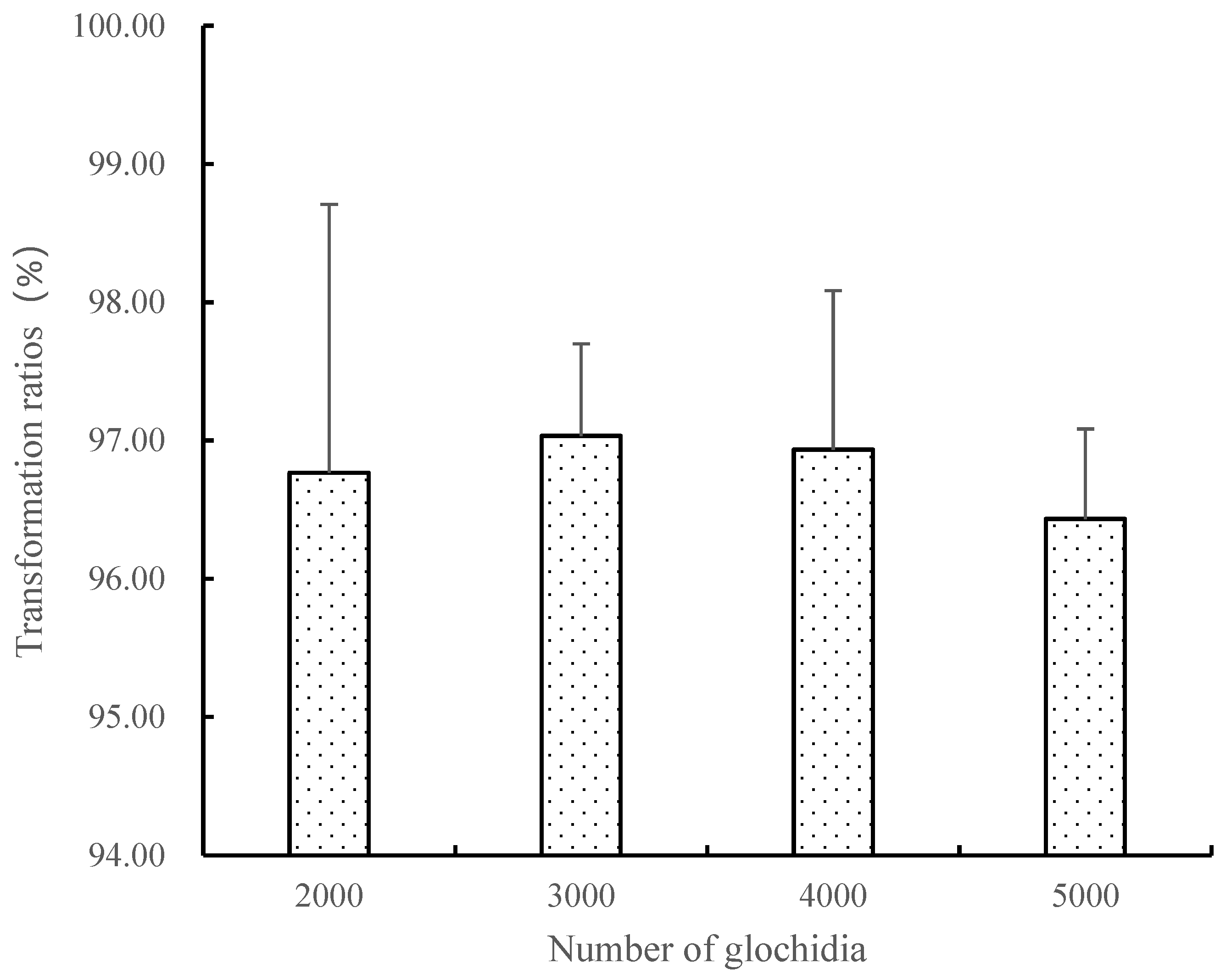
3.2. Influence of Cultivation Density on the Transformation of Glochidium

The ratio of transformation in the groups without contamination or a cultivation density of 2000–5000 glochidia/dish is shown in Figure 1. One-way ANOVA indicated that the ratio of transformation in the four groups with different cultivation densities was not significantly different in the absence of contamination.
The ratio of transformation ranged from 0% to 75.38% in the groups with a contamination and cultivation density of 2000–5000 glochidia/dish. Moreover, the higher the number of glochidia, the more serious the contamination and the lower the ratio of transformation. One-way ANOVA showed no significant differences in the ratio of contamination values in the four groups (Figure 2).

3.3. Growth Characteristics

The growth of S. oleivora juveniles (day 0 to 13 weeks) was recorded (Table 3). The ratio of the length to height of the shell indicated that the length increased faster than the height.
As the juveniles grew, the ratio of the length to height greatly increased from 1.08 ± 0.01 at 1 day of age to 3.08 ± 0.29 at 13 weeks of age, and the juveniles gradually showed the appearance features of mature S. oleivora. The analysis indicated that the length and height of the juveniles presented clear positive allometry.
The values of OB/OA indicated that the growth of the anterior and posterior juvenile shell presented clear phases. The distance between the OA and OB of the juvenile just achieving successful transformation was approximately equal, presenting symmetry between anterior and posterior growth. Anterior growth was faster than posterior growth in 1-week-old and 2-week-old juveniles, and OB/OA was 0.74 and 078, respectively. The growth of the posterior juvenile shell at 3 weeks of age was faster than that of the anterior juvenile; OB/OA increased to 1.04, almost reaching symmetry. After 3 weeks, OB/OA gradually increased, indicating that posterior growth was faster than anterior growth.
The length and height of the juvenile at 1–3 weeks of age slightly increased (Figure 3); the length increase was faster at 4 weeks, 7 weeks, 9 weeks and 11 weeks, and the same increasing trend was observed for the height of the shell.

3.4. Morphological Development of the Juvenile

The morphological development of the juvenile is shown in Figure 4. On the basis of anterior and posterior growth differences, the growth of the juvenile was divided into the following three phases: the rapid anterior growth period, distinct anterior and posterior idiophase, and rapid posterior growth period.

3.4.1. Rapid Anterior Growth Period

The length of the glochidium shell was about 90 μm, and the height was about 84 μm; it had no hook. Mature glochidium (Figure 4, 1) showed opening and closing movements and was shaped like a walnut after closing (Figure 4, 2).
The size of the juvenile that just experienced a successful transformation was similar to that of the glochidium. The shells of the juvenile became significantly thicker, and a new and transparent shell appeared at the rim. When the shell opened, the foot stretched from the middle and started to creep by attaching to the bottom (Figure 4, 3). In the 1st week, the crawling ability of the juvenile gradually became stronger. The brown stomach was visible from the shell dorsal. The vitality of the juvenile was closely related to the turning frequency of the crystal rods.
When the juvenile was 1 week of age, a new shell appeared at the rim, and a hinge line, instead of a straight line, gradually formed the umbo because of the growth of the juvenile. The ventral surface appeared rough under a light microscope and presented as an irregular polygon; asymmetric growth was observed for the anterior and posterior juvenile, and anterior shell growth was clearly faster than posterior shell growth (Figure 4, 4). When the juvenile was 2 weeks of age, the growth difference was greater. The anterior formed a circular arc shape, and the posterior was relatively flat with a slight radian. A clear growth line appeared on the ventral shell surface; shell transparency increased, and the internal organs were clearly observed, especially the primordium of the gill filament (Figure 4, 5).

3.4.2. Distinct Anterior and Posterior Idiophase

When the juvenile was 2–3 weeks of age, posterior growth greatly increased. When it was 3 weeks of age, the distance between the umbo and anterior was almost the same as that between the umbo and posterior (Figure 4, 6), and the shell was transparent. The juvenile used its foot to creep in this phase; the internal organs were clearly observed, and the hepatopancreas was dark brown.

3.4.3. Rapid Posterior Growth Period

When the juvenile was 4 weeks of age, the shell was wedge-shaped (Figure 4, 7), with the short anterior in the shape of an ellipse and a relatively wide, big, and straight rear end; a clear water orifice and water inlet successively appeared at the posterior, and adductor muscles and became more distinct, with the posterior bigger than the anterior. The gill filament was almost formed, and the distribution was compact; the dark brown hepatopancreas became more distinct, and the foot appeared milk-white with more power. Food in the rectum and midgut was clearly observed, and the anus was located at the posterior of the adductor muscle.
From the 5th to 13th week (Figure 4, 8–15), the shell of the juvenile was faintly yellow, the surface was smooth, the anterior was blunt, and the posterior expanded into a wedge shape. The dorsal was flat and straight with the extension of the hinge line, and the growth line was clear and extruded. The form gradually became similar to that of mature S. oleivora.

4. Discussion

4.1. In Vitro Culture of S. oleivora Glochidium

In vitro culture has undergone a series of technical optimizations, and it is able to provide a relatively high ratio of transformation. In this study, the ratio of S. oleivora cultivated with the serum of common carp was >95%, indicating that using the serum of a fish that does not act as the natural host met the nutritional requirements for the transformation of glochidium. The experimental results are similar to those in the study by Ma [22], in which the ratio of transformation of C. plicata glochidium cultivated with the serum of the fish host and other fish was >96%.
In this study, there was no significant difference in the ratio of the transformation of S. oleivora glochidium without contamination at a cultivation density of 2000–5000/dish, showing that this cultivation method can meet the nutritional requirements of the transformation of S. oleivora glochidium. Uthaiwan et al. [28] researched the cultivation density of five different glochidia, such as that of Hyriopsis myersiana, and reported that the ratio of transformation of the glochidia was highest at a cultivation density of 150–200/dish. Ma et al. [24] showed no significant differences in the ratio of transformation of C. plicata glochidia at a density of 300–1100/dish, and the values were all >95%. For in vitro culture, such a high cultivation density has never been used to date; the highest density was only 1100/dish in the study of Ma [22]. According to the index for glochidium (Gin: shell length × shell height), Davis [29] divided the glochidium into three types: small (Gin < 0.036), medium (Gin = 0.047), and large (Gin = 0.100). On the basis of this standard, the glochidium of S. oleivora belongs to the small type. Because the glochidium of S. oleivora is small, 4000–5000 glochidia only occupied a small space in the cultivation dish, and they were not hard to cultivate. In a practical production, 4000–5000/dish is the density at which the highest yield can be achieved based on comprehensive considerations of the cultivation density and contamination.
Fungal and bacteria contamination is an important factor that affects the transformation of glochidium during in vitro culture. In this study, contamination was detected in the low-density groups. The higher the number of glochidia, the higher the ratio of contamination, indicating that the antibiotic concentration cannot completely restrict the growth of bacteria. Owen et al. [30] studied the effects of eight antibacterial agents on transformation during in vitro culture and found that none of the antibacterial agents could completely control the growth of fungus during the cultivation process, and a high concentration of antibiotics had a negative influence on the transformation of some mussels. It is uncertain why employing serum from mammalian sources leads to contamination at a higher rate than fish plasma. We speculate that fish are the natural hosts and source of nutrition for mussel larvae during the parasitic phase of their life cycle, and fish serum constitutes the most logical protein source for artificial mussel cultures.

4.2. Morphological Development of the Juvenile

A glochidium that completes its transformation is called a juvenile mussel, and it is called a young mussel when the shell length is >1 cm; it has a body similar to that of a mature mussel [31]. In this study, the average shell length of S. oleivora reached 1.2 cm after cultivation for 13 weeks, and S. oleivora entered the young mussel phase. Anodonta woodiana enters the young mussel phase when it is 40 days of age [32]. H. cumingii entered the juvenile mussel phase after cultivation for 60 days [33], and H. (Limnoscapha) myersiana grew from 0.13 mm to 1.4 cm after cultivation for 60 days [34]. This may be because the glochidium of S. oleivora is significantly smaller than those of H. cumingii and A. woodiana, and it took a longer time for the glochidium to grow to 1 cm.
The juvenile barely changed in size in the early stages of transformation. It was hard to distinguish the anterior from the posterior; anterior growth was faster than posterior growth in the early phase. Liu et al. [32] divided the morphological development of A. woodiana into four stages as follows: an expanding stage, an umbo-extruding stage, a wing-forming phase, and horn growth stage; they also divided the morphological development of H. cumingii into four stages, a width-expanding stage, umbo-extruding stage, wing-forming stage, and sail growth stage [33]. During the growth process of S. oleivora, the posterior grew faster than the anterior; an expanding stage and umbo-extruding stage also appeared, which was similar to A. woodiana [32], H. cumingii [33] and Lamprotula scripta [35].

5. Conclusion

We used in vitro culture to conduct artificial propagation experiments on the endangered mussel S. oleivora, whose fish hosts are unknown and achieved good results. We evaluated the effect of glochidium density on transformation rates and documented the development of juveniles that had successfully metamorphosed. The investigation of juvenile mussels derived from in vitro culture confirmed that the mussels survived and developed normally. This is of great importance for the artificial proliferation and conservation of Unionidae mussels, especially those with unknown host fish.

Author Contributions

Conceptualization, X.M. and H.W.; methodology, X.M., H.W. and D.H.; validation, W.J., P.X. and H.W.; investigation, W.J., W.C. and D.X.; data curation, X.M. and G.L.; writing—original draft preparation, X.M.; writing—review and editing, W.J. and H.W.; supervision, P.X. and D.X.; funding acquisition, P.X. and H.W. All authors have read and agreed to the published version of the manuscript.

Funding

This study was supported by the Central Public-Interest Scientific Institution Basal Research Fund (2023TD66); the Solenaia oleivora Ecological Compensation Program (2023); and the Central Public-interest Scientific Institution Basal Research Fund [grant number 2023JBFM12] which provided financial support for this project.

Institutional Review Board Statement

The study protocol was reviewed and approved by the Freshwater Fisheries Research Center of the Chinese Academy of Fishery Sciences (No. 2022AA1004020012, 4 October 2022).

Data Availability Statement

The data are contained within the article.

Acknowledgments

We would like to thank the native English-speaking scientists of Elixigen Company (Huntington Beach, CA, USA) for editing our manuscript. We also thank the insightful comments from anonymous reviewers.

Conflicts of Interest

The authors declare no conflicts of interest. The data provider and scholarship funders had no role in the study design; in the collection, analysis, or interpretation of data; in the writing of the manuscript; or in the decision to publish the results.

References

  1. Porter, T.A.; Moskalenko, I.V.; Strong, A.W.; Orlando, E.; Bouchet, L. Inverse compton origin of the hard X-ray and soft gamma-ray emission from the galactic ridge. Astrophys. J. 2008, 682, 400–407. [Google Scholar] [CrossRef]
  2. Bogan, A.E. Global diversity of freshwater mussels (mollusca, bivalvia) in freshwater. Hydrobiologia 2008, 595, 139–147. [Google Scholar] [CrossRef]
  3. Graf, D.L. Patterns of freshwater bivalve global diversity and the state of phylogenetic studies on the unionoida, sphaeriidae and cyrenidae. Am. Malacol. Bull. 2013, 31, 135–153. [Google Scholar] [CrossRef]
  4. Brusea, R.C.; Brusca, G.J. Phylum Porifera: The Sponge; Invertebrates; Sinauer Press: Sunderland, MA, USA, 1990; pp. 181–210. [Google Scholar]
  5. Graf, D.L.; Cummings, K.S. A ‘big data’ approach to global freshwater mussel diversity (Bivalvia: Unionoida), with an updated checklist of genera and species. J. Molluscan Stud. 2021, 87, eyab015. [Google Scholar] [CrossRef]
  6. Hu, Z.Q. Geographical distribution of endemic species of Chinese freshwater bivalves. Chin. J. Zool. 2005, 40, 80–83. [Google Scholar]
  7. Wen, H.B. Study of Germplasm of Major Economic Freshwater Mollusks of China; Nanjing Agricultural University: Nanjing, China, 2009. [Google Scholar]
  8. Ma, X.Y.; Sun, G.X.; Wang, L.; Xu, D.P.; Jin, W.; Lv, G.H.; Xu, P.; Ding, T.Q.; Wen, H.B.; Gu, R.B. Seasonal variations of nutrients and mineral elements in Solenaia oleivora from Huaihe River. J. Agron. 2021, 11, 90–94+119. [Google Scholar]
  9. Liu, Y.Y.; Zhang, W.Z.; Wang, Y.X. Economic Fauna of China: Freshwater Mollusks; Beijing Science Press: Beijing, China, 1979. [Google Scholar]
  10. Chen, Z.F.; Chen, P. Analysis of Nutritional Components of Solenaia oleivora. In Proceedings of the 65th Annual Meeting of Chinese Animal Society, Beijing, China, 25 April 1999; Available online: https://xueshu.baidu.com/usercenter/paper/show?paperid=84413ec88bbeae5a82266a19a787bcc2&site=xueshu_se (accessed on 21 January 2024).
  11. Xu, Q.Q.; Liu, J.; He, L.R. Analysis of meat content and muscle nutritional components of Solenaia oleivora. Freshw. Fish. 2003, 33, 28–29. [Google Scholar]
  12. Huang, Y.Y.; OuYang, S.; Wu, X.P.; Liu, H.Z. Phylogeny of the unionidae based on partial mitochondrial 16S rRNA sequences. Acta Hydrobiol. Sin. 2003, 27, 258–263. [Google Scholar]
  13. Huang, X.C. Complete Paternal and Maternal Mitochondrial Genomes of the Freshwater Mussel Genus Solenaia (Unionidae: Gonideinae) in China and Mitochondrial Phylogenomics of Unionoida; Nanchang University: Nanchang, China, 2014. [Google Scholar]
  14. Xu, Y. Isolation of Microsatellite Markersand Population Genetic Diversity Analysis in Solenaia oleivora; Huazhong Agriculture University: Wuhan, China, 2004. [Google Scholar]
  15. Yang, X.L.; Li, H.C.; Song, L. Gonadal development and growth of freshwater mussel Solenaia oleivora. Fish. Sci. 2011, 30, 580–582. [Google Scholar]
  16. Wang, Y.N. Productive Traits of the Freshwater Mussel Solenaia oleivora (Bivalvia: Unionidae) in the Tianmen River; Huazhong Agriculture University: Wuhan, China, 2013. [Google Scholar]
  17. Xia, Z.Q.; Han, X.Z.; Zhu, W.; Zhang, Y.F.; Ye, C.Y.; Liu, J. Preliminary study on reproductive biology of Solenaia oleivora. Jiangxi Fish. Sci. Technol. 2014, 21, 17–19. [Google Scholar]
  18. Xiong, L.F.; OuYang, S.; Chen, T.H.; Qi, T.; Wu, X.P. The resource status and spatio-temporal variation of freshwater mussels in Qinglan Lake of Jiangxi Province. Acta Agric. Univ. Jiangxiensis 2010, 32, 1257–1264. [Google Scholar]
  19. Bai, Z.Y.; Li, J.L.; Pan, B.B. Comparison of the parasitism effects of glochidia of triangle mussel (Hyriopsis cumingii) in five fishes. Freshw. Fish. 2008, 38, 3–5+43. [Google Scholar]
  20. Lima, P.; Lima, M.L.; Kovitvadhi, U.; Kovitvadhi, S.; Owen, C.; Machado, J. A review on the “in vitro culture of freshwater mussel (Unionoida). Hydrobiologia 2012, 691, 21–33. [Google Scholar] [CrossRef]
  21. Wen, H.B.; Gu, R.B.; Hua, D.; Qiu, L.H.; Xu, G.C.; Xu, P. Culture of glochidia of Hyriopsis cumingii in artificial media and morphological variation of transformed juvenile. J. Wuhan Univ. 2011, 57, 57–62. [Google Scholar]
  22. Ma, X.Y. In Vitro Culture Glochidia and the Development of Juveniles of Cristaria plicata; Nanjing Agricultural University: Nanjing, China, 2015. [Google Scholar]
  23. Wen, H.B. Study on Basic Biological Characteristics and Transformation and Development of Potamilu salatus; Nanjing Agricultural University: Nanjing, China, 2016. [Google Scholar]
  24. Ma, X.Y.; Wen, H.B.; Zou, J.; Jin, W.; Hua, D.; Gu, R.B.; Xu, P. An improved method for in vitro culture of glochidia in freshwater mussel Cristaria plicata (Mollusca, Bivalvia). Hydrobiologia 2018, 810, 133–144. [Google Scholar] [CrossRef]
  25. Liu, B.; Wang, M.Y.; Xie, J.; Xu, P.; Ge, X.P.; He, Y.J.; Miao, L.H.; Pan, L.K. Effects of acute cold stress onserum biochemical and immune parameters and liver HSP70 gene expression in GIFT strain of Nile tilapia (Oreochromis niloticus). Acta Ecol. Sin. 2011, 37, 4866–4873. [Google Scholar]
  26. Uthaiwan, K.; Noparatnaraporn, N.; Machado, J. Culture of glochidia of the freshwater pearl mussel Hyriopsis myersiana (Lea, 1856) in artificial media. Aquaculture 2001, 195, 61–69. [Google Scholar] [CrossRef]
  27. Qian, R.H.; Li, J.L.; Dong, Z.G.; Zheng, H.F.; Li, Y.S.; Yan, W.K. Morphological variations analysis among populations of Hyriopsis cumingii in five large lakes of China. Oceanol. Limnol. Sin. 2003, 34, 436–443. [Google Scholar]
  28. Uthaiwan, K.; Pakkong, P.; Noparatnaraporn, N.; Vilarinho, L.; Machado, J. Study of a suitable fish plasma for in vitro culture of glochidia Hyriopsis myersiana. Aquaculture 2002, 209, 197–208. [Google Scholar] [CrossRef]
  29. Davis, G.M.; Fuller, S. Genetic relationships among recent Unionscea (Bivalvia) of North America. Malacologia 1981, 20, 217–253. [Google Scholar]
  30. Owen, C.T.; Alexander, J.E.; Gregor, M. Control of microbial contamination during in vitro culture of larval unionid mussels. Invertebr. Reprod. Dev. 2010, 54, 187–193. [Google Scholar] [CrossRef]
  31. Chang, Y.Q. Shellfish Culture; Chinese Agriculture Press: Beijing, China, 2007. [Google Scholar]
  32. Liu, S.L.; Li, J.L.; Zhang, G.F.; Wang, G.L.; Bai, Z.Y.; Pan, B.B. Morphological development and growth characteristics of juvenile of Hyriopsis cumingii. J. Fish. China 2009, 33, 604–609. [Google Scholar]
  33. Liu, S.L.; Li, J.L.; Zhang, G.F.; Xu, S.J.; Wang, G.L.; Bai, Z.Y. Morphological development and growth characteristics of juvenile of Sinanodonta woodiana. J. Shanghai Ocean. Univ. 2009, 18, 269–274. [Google Scholar]
  34. Kovitvadhi, S.; Kovitvadhi, U.; Sawangwong, P.; Thongpan, A.; Machado, J. Optimization of diet and culture environment for larvae and juvenile freshwater pearl mussels, Hyriopsis (Limnoscapha) myersiana Lea, 1856. Invertebr. Reprod. Dev. 2006, 49, 61–70. [Google Scholar] [CrossRef]
  35. Wang, Y.F.; Wei, Q.S. On reproductive biology of lamprotula scripta (Heude). J. Huazhong Agric. Univ. 1994, 13, 170–174. [Google Scholar]
Figure 1. Influence of density on the transformation of S. oleivora glochidia.
Fishes 09 00049 g001
Figure 2. Contamination ratios at different culture densities.
Fishes 09 00049 g002
Figure 3. Growth and development of S. oleivora juveniles. (A) Shell length Shell height, (B) Absolute growth rate.
Fishes 09 00049 g003
Figure 4. Morphological development of S. oleivora juveniles. (1,2) Mature glochidium. (3): the juvenile that just experienced successful transformation. (415): the juvenilewas 1–12 weeks-of-age, respectively.
Fishes 09 00049 g004
Table 1. Composition of artificial medium for the in vitro culture of S. oleivora glochidia.
GroupL-15 (mL)Antibiotic Mixture (mL)Bighead Carp Plasma (mL)Grass Carp Plasma (mL)Common Carp Plasma (mL)Bovine Serum (mL)Rabbit Serum (mL)Deionized Water (mL)
Control group20.5 1
A120.51
A220.5 1
A320.5 1
A420.5 1
A520.5 1
Table 2. Effects of different nutrient resources on the transformation of S. oleivora glochidia.
GroupUncontaminated Transformation RateContamination Rate
No nutritional resources0.00 ± 0.00 a0.00 ± 0.00 a
Bighead carp plasma97.47 ± 0.93 b0.00 ± 0.00 a
Grass carp plasma98.03 ± 0.71 b6.67 ± 11.54 a
Common carp plasma97.96 ± 0.76 b6.67 ± 11.54 a
Bovine serum 60.09 ± 5.96 c26.67 ± 11.54 b
Rabbit serum 39.69 ± 3.75 d40.00 ± 0.00 b
Note: Bars in the same column with different letters are significantly different (p < 0.05).
Table 3. Growth and development of S. oleivora juveniles.
Growth PeriodAverage Shell Length (mm)Shell Length Range (mm)Average Shell Height (mm)Shell Height Range (mm)Shell Length/Shell Height Ratio RangeOB/OA
1 d0.090 ± 0.040.82–0.100.08 ± 0.050.08–0.091.08 ± 0.010.94–1.201.07 ± 0.03
1 W0.15 ± 0.020.13–0.170.13 ± 0.090.12–0.151.10 ± 0.110.95–1.250.74 ± 0.11
2 W0.41 ± 0.050.22–0.470.34 ± 0.040.28–0.401.19 ± 0.051.14–1.290.78 ± 0.06
3 W0.76 ± 0.100.62–0.940.60 ± 0.050.55–0.711.26 ± 0.071.13–1.361.04 ± 0.09
4 W1.27 ± 0.320.79–1.750.83 ± 0.150.6–1.061.51 ± 0.141.27–1.711.20 ± 0.12
5 W1.69 ± 0.371.10–2.751.01 ± 0.150.71–1.461.66 ± 0.151.40–2.031.35 ± 0.07
6 W2.12 ± 0.441.48–3.021.19 ± 0.180.90–1.521.77 ± 0.131.55–2.031.49 ± 0.10
7 W3.79 ± 0.832.25–5.261.71 ± 0.331.16–2.202.22 ± 0.241.90–2.781.61 ± 0.05
8 W4.80 ± 0.543.66–5.892.06 ± 0.151.81–2.462.32 ± 0.162.01–2.801.67 ± 0.11
9 W5.86 ± 0.604.92–7.002.36 ± 0.202.08–2.752.48 ± 0.132.23–2.741.70 ± 0.09
10 W6.20 ± 0.784.83–7.702.50 ± 0.232.17–2.972.48 ± 0.142.21–2.761.75 ± 0.08
11 W7.85 ± 0.527.12–8.692.76 ± 0.172.41–3.082.84 ± 0.142.59–3.121.81 ± 0.08
12 W8.97 ± 1.537.12–8.693.07 ± 0.412.04–4.322.91 ± 0.182.57–3.321.84 ± 0.10
13 W12.38 ± 2.338.39–18.244.00 ± 0.543.15–5.073.08 ± 0.292.19–3.751.88 ± 0.09
Disclaimer/Publisher’s Note: The statements, opinions and data contained in all publications are solely those of the individual author(s) and contributor(s) and not of MDPI and/or the editor(s). MDPI and/or the editor(s) disclaim responsibility for any injury to people or property resulting from any ideas, methods, instructions or products referred to in the content.

