Article

Vitamin C Alleviates Intestinal Inflammation Caused by Aeromonas hydrophila in Juvenile Blunt Snout Bream (Megalobrama amblycephala)

1
Wuxi Fisheries College, Nanjing Agricultural University, Wuxi 214081, China
2
Taizhou Aohua Agricultural and Animal Husbandry Technology Co., Ltd., Taizhou 225300, China
3
Key Laboratory of Aquatic Animal Nutrition and Health, Freshwater Fisheries Research Center, Chinese Academy of Fishery Science, Wuxi 214081, China
*
Author to whom correspondence should be addressed.
Fishes 2024, 9(4), 129; https://doi.org/10.3390/fishes9040129
Submission received: 4 March 2024 / Revised: 1 April 2024 / Accepted: 5 April 2024 / Published: 8 April 2024

Abstract

:
Vitamin C (VC) can be used to increase disease resistance in practice in intensive aquaculture. But it is still unconfirmed whether VC could alleviate inflammation and what dosage is suitable. This study investigated the effects of dietary VC on the immunity and enteritis of juvenile blunt snout bream (Megalobrama amblycephala) challenged with Aeromonas hydrophila. The fish were fed with VC levels ranging from 25.35 to 2231.98 mg/kg for 60 days. After that, fish fed with 150.65 mg/kg, 573.79 mg/kg, and 2231.98 mg/kg VC were challenged with A. hydrophila orally to simulate enteritis. The results showed that 285.39 and 573.79 mg/kg dietary VC significant improved growth performance and feed utilization. Dietary VC (573.79 and 1133.79 mg/kg) significantly enhanced the serum immune parameters, antioxidant enzymes’ activities, and relative gene expressions of ikbα and cat in the liver in the 60-day feeding trail. The survival rate was significantly higher in 573.79 mg/kg dietary VC group than the other two treatments. Cytokines were activated after the bacteria challenge. Dietary 573.79 mg/kg VC caused significantly higher TNF-α level at 12 hpi and maintained a high level of IL-8 at 48 to 72 h compared to other treatments. In conclusion, more than 500 mg/kg VC was needed to maintain the health of blunt snout bream juveniles. Suitable VC would activate cytokines to increase disease resistance along with antioxidant enzymes.
Key Contribution: Dietary VC supplementation from 500–1000 mg/kg could improve the immunity of blunt snout bream. Enteritis could be alleviated by suitable dietary VC, mainly because VC stimulates cytokines of TNF-α and IL-8. Excessive dietary VC of about 2000 mg/kg would have some negative effects on the health of blunt snout bream.

1. Introduction

Safeguarding fish against infections and diseases is a strategy aimed at boosting production and ensuring food security [1]. Fish employ a combination of innate and adaptive immune responses to combat diseases and parasites [2]. In intensive aquaculture systems, fish are particularly vulnerable to diseases and pathogens. Aeromonas hydrophila, a bacterium that thrives in freshwater environments, poses a significant threat to fish health, causing diseases such as gastroenteritis septicaemia and necrotizing fasciitis [3]. In China, bacterial haemorrhagic disease resulting from A. hydrophila infection is a major concern in the cultivation of blunt snout bream, leading to high mortality rates and economic losses [4]. Fish pathogens typically enter the fish through the intestine, which serves as a crucial site for digestion, absorption of nutrients, and immunity. Various fish species, including grass carp (Ctenopharyngodon idella) [5] and silver carp (Hypophthalmichthys molitrix) [6], have been reported to experience intestinal damage and dysfunction due to A. hydrophila infection. Enhancing the intestinal health of fish through improved dietary formulations and immunostimulants has emerged as a promising strategy to bolster their resistance to A. hydrophila infections, thereby reducing the incidence of inflammation and diseases [7].
Vitamin C (VC) is a vital micronutrient for fish, playing a key role in maintaining epithelial barriers, supporting immune cells, and regulating inflammatory responses [8]. However, fish are unable to synthesize vitamin C on their own due to the lack of a specific enzyme (L-gulonolactone oxidase), necessitating its inclusion in their diet for optimal health and immunity [9].
The blunt snout bream (Megalobrama amblycephala) is a herbivorous freshwater fish native to China [4] and is found in various regions such as Africa, North America, and Eurasia [10]. Known for their rapid growth and economic value, these fish have specific VC requirements, particularly in the juvenile stages. Previous research suggests that a 150 mg/kg VC level is optimal for growth when provided in a purified diet [11]. Dietary 133.7–251.5 mg/kg VC could enhance anti-pH stress ability in M. amblycephala [12]. However, the adequacy of this VC level for inflammatory response in blunt snout bream remains uncertain and warrants further investigation.
Studies on other fish species, like Asian catfish (Clarias batrachus) [13] and juvenile grouper (Epinephelus malabaricus) [14], have shown increased immunity response with higher dietary VC supplementation above 150 mg/kg. Yet, the impact of elevated VC levels on the inflammation of juvenile blunt snout bream has not been extensively explored. Previous research by Wan et al. [11] focused on a purified VC diet, prompting the current study to assess the effects of dietary VC (at higher and excessive levels) on non-specific immunity and intestinal inflammation in juvenile blunt snout bream when exposed to A. hydrophila. The hypothesis being tested is that the VC requirement for immunity and inflammatory response may differ from that needed for growth in this fish species. Therefore, the present study was undertaken to evaluate the effect of dietary VC (higher and too high) on the non-specific immunity and intestinal inflammation in juvenile blunt snout bream against A. hydrophila.

2. Materials and Methods

2.1. Experimental Diet Formulation

Six experimental diets with iso-nitrogenic (34.69%) and iso-lipidic (9.31%) ingredients were formulated using fishmeal, soybean meal, cottonseed meal, and canola meal as protein sources and soybean oil and soybean phospholipids as lipid sources. Coated VC (95%, Hangzhou Minsheng Pharmaceutical Co., Ltd., Hangzhou, China) was supplemented from 150 to 2400 mg/kg in the experimental treatment beside the contrast group without VC supplementation. The composition and main nutrients in the basal feed are listed in Table 1. First, fishmeal, soybean meal, cottonseed meal, and canola meal were ground into a fine powder and passed through a 60 mm mesh sieve. Then, VC and the additives were mixed and amplified step by step. Soybean oil and water were then added, stirred, and mixed. Finally, an SLP-45 granulator (Institute of Fishery Machinery and Instruments, Chinese Academy of Fishery Sciences, Shanghai, China) was used to prepare a 1.5 mm granular diet. Pelleted diets were placed in a cool place to dry naturally. After air drying, the diet was stored at −20 °C for later use. The actual content of VC in each diet was measured using an ascorbic acid kit (Nanjing Jiancheng Bioengineering Institute, Nanjing, China) as follows: 25.35, 150.65, 285.39, 573.79, 1133.79, and 2231.98 mg/kg, respectively.

2.2. Proximate Composition of the Diets

The proximate composition (crude protein, crude fat, ash, and moisture) of the diets was determined according to the AOAC [15] method. Briefly, for moisture determination, the diet samples were weighed and dried to a constant weight in an oven at 105 °C. Crude protein content (N × 6.25) was determined using a semi-automatic Kjeldahl system (1030 Auto-analyzer, Tecator, Hoganos, Sweden) after acid digestion. The Soxhlet extraction system HT6 (Soxhlet System, Tecator, Sweden) was employed to measure the crude lipid content, whereas the ash content was measured by burning in a muffle furnace at 560 °C for approximately 5 h.

2.3. Management of the Experimental Fish

Blunt snout bream juveniles were obtained from the Freshwater Fisheries Research Center of the Chinese Academy of Fishery Sciences (Wuxi, China). The fish were acclimatized to the experimental environment for two weeks, during which they were fed a commercial diet (Tongwei Group, Wuxi, China). After acclimatization, 360 healthy juveniles of uniform size (initial average weight of approximately 12 g) were selected and randomly divided into 18 round polyvinyl chloride tanks (Φ 820 mm and 1000 mm in height, with water about 370 L). Each tank was randomly assigned to one of the six experimental diets. Each diet had three replicates, with 20 fish per tank. The 18 tanks were in a recirculating system with activated carbon and coral stones as filters. The fish were hand-fed three times daily at 8:00, 12:00, and 16:00 to apparent satiation. The feeding trial lasted 60 days. During the feeding period, the number and weight of dead fish and feed consumption were recorded daily. The control water temperature was 27–30 °C and was measured using a mercury-in-glass thermometer. The pH was 7.0~8.0, and dissolved oxygen (DO) >5 mg/L, ammonia nitrogen <0.02 mg/L, and nitrite nitrogen concentration <0.05 mg/L were determined using corresponding Sunpu Test kits (Beijing Sunpu Biochem. Tech. Co., Ltd., Beijing, China).

2.4. Sample Collection

At the end of the 60-day feeding trail, the experimental fish were sampled. The fish were starved for 24 h before sample collection. Fish in each tank were weighed, and the growth index was calculated as follow.
Coefficient of variation (CV, %) = Standard deviation/Mean × 100
Weight gain (WG, %) = (Wt − W0)/W0 × 100
Specific growth rate (SGR, % day-1) = (LnWt − LnW0)/d × 100
Feed conversion ratio (FCR) = Weight of dry feed intake/Weight gain
Survival rate (SR, %) = Final number of fish per tank/Initial number of fish per tank × 100
Wt is the final average fish body weight per tank, W0 is the initial average fish body weight per tank, and d is the experimental duration.
For serum collection, three fish per tank were deeply anaesthetized with MS-222 (100 mg/L), and blood was collected from the caudal vein using a disposable syringe (rinsed with heparin sodium salt) and centrifuged at 4000 rpm at 4 °C for 10 min. Serum was obtained and stored at −20 °C for analysis of serum biochemical indices. After blood collection, liver samples were collected and preserved in RNAiso Plus (Takara, Dalian, China), which was maintained at −80 °C for gene expression analysis.

2.5. Challenge Test

An artificial infection experiment was conducted two days later after sample collection. Fifteen fish per tank were randomly selected from the 150.65, 573.79, and 2231.98 mg/kg VC groups based on blunt snout bream’s optimal VC requirement for growth, immunity, and excessive VC. A. hydrophila (NJ-35) was kept in the laboratory of FFRC, CAFS (Wuxi, China). It was cultured in liquid LB medium at 28 °C for 18 h, then the concentration was detected using a turbidity meter (Shanghai, China). According to the pre-experiment protocol, A. hydrophila was diluted with sterile saline solution (0.85%) to 5 × 10⁸ CFU/mL. Bacteria were orally administered from the mouth of the fish using a syringe with a feeding needle at 1 mL per 100 g of fish body weight, and then returned to the culture tank for observation [16]. Sufficient DO (>5 mg/L) and a stable temperature (28 ± 2 °C) were maintained. Dead fish were checked, removed, and recorded at 0, 12, 24, 48, and 72 h post-infection (hpi). Meanwhile, liver and intestinal samples from three fish per tank were collected before infection (0 h) and at 12 hpi, 24 hpi, 48 hpi, and 72 hpi, respectively. Fish were deeply anaesthetized and liver samples were collected as mentioned above for gene expression. At the same time, the intestinal samples were collected. Parts were put into tubes and kept at −20 °C for antioxidant enzyme and cytokine detection, and parts were immersed in Bouin’s solution for histopathology.

2.6. Intestinal Histology

The intestinal histological examination was carried out according to Yusuf et al. [17]. Briefly, the intestines of blunt snout bream challenged by A. hydrophila were excised at 0 h before infection and at 12, 24, 48, and 72 h after infection. A fragment of the intestinal tissue was immersed in Bouin’s solution immediately after dissection. They were then dehydrated, immersed in wax, embedded, and sectioned at a thickness of 4 µm. The sectioned tissues were mounted on glass slides and stained with haematoxylin and eosin. Finally, the slides were examined under a light microscope (Olympus IX71, Tokyo, Japan) reinforced with Image-Pro Plus 7.0C software, and photomicrographs of sectioned intestinal tissue were taken.

2.7. Serum and Intestinal Immune-Related Indicator Determination

Intestinal samples were homogenated with sterile saline solution (0.85%), and then the intestinal homogenate was used to detect the antioxidant enzymes and cytokines after the detection of the protein concentration. Serum alkaline phosphatase (AKP), albumin (ALB), and complement 3 (C3) levels were determined using a Mindray BS-400 automatic biochemical analyser (Shenzhen, China) and commercial kits from Mindray Biomedical Electronics Co., Ltd. (Shenzhen, China), respectively. The malondialdehyde (MDA) content and antioxidant activities of catalase (CAT), superoxide dismutase (SOD), and glutathione peroxidase (GPx) were determined using a commercial kit (Nanjing Jiancheng Bioengineering Institute, Nanjing, China) [18].
Tumour necrosis factor α (TNF-α), transforming growth factor β (TGF-β), interleukin 8 (IL-8), and interleukin 10 (IL-10) levels were determined using commercial kits (Shanghai Enzyme-linked Biotechnology Co., Ltd., Shanghai, China) based on enzyme-linked immunoassay (ELISA). In brief, cytokines in the serum were combined with the corresponding purified antibody for carps and HRP labelled to form an antibody–antigen–enzyme–antibody complex, which was then detected spectrophotometrically at 450 nm.

2.8. Quantitative Real-Time PCR (qRT-PCR) Analysis

The relative gene expression of cu/zn-sod, mn-sod, cat, gpx-1, ikbα, p65, and hsp70 was determined before infection and after artificial infection with A. hydrophila at 12, 24, 48, and 72 h. Ribonucleic acid (RNA) was extracted from the liver using RNAiso Plus (Dalian Takara Co. Ltd., Dalian, China). The quantity and quality of the extracted RNA were estimated based on the A260/A280 ratio using NanoDrop (DN-1000, Thermo Scientific, Waltham, MA, USA), and RNA integrity was verified on a 1.2% agarose gel. cDNAs were synthesized using the ExScriptTM RT-PCR kit following the manufacturer’s instructions (Dalian Takara Co. Ltd., Dalian, China) and amplified using a TB Green® Premix Ex TaqTM II (Tli RNaseH Plus) Kit (Dalian Takara Co. Ltd., Dalian, China) in a CFX96 RT-PCR detection system (Bio-Rad, Hercules, CA, USA). Primers were synthesized by Shanghai Gene-ray Biotech Co. Ltd. (Shanghai, China) (Table 2). The reaction system (final volume 20 μL) contained 2 μL cDNA; 10 μL TB Green Premix Ex Taq II (Tli RNasePlus) (2×); 0.8 μL upstream and downstream primers (10 μM), respectively; and 6.4 μL ddH2O. The conditions were as follows: pre-denaturation at 95 °C for 30 s; 45 cycles at 95 °C for 5 s and 62 °C for 30 s; smelt curve analysis, including 95 °C for 10 s and 62 °C for 5 s; 60 s at 62 °C; and 95 °C for 15 s. Gene expression was corrected using β-actin as the reference gene. Gene expression data were calculated using the 2−ΔΔCt method [19].

2.9. Statistical Analysis

All statistical analyses were performed using SPSS v23 (SPSS Inc., Michigan Avenue, Chicago, IL, USA), and GraphPad Prism 9 was used to draw the figures. One-way analysis of variance (ANOVA) was used to evaluate the data on growth performance serum parameters and gene expression in the feeding trail, and the mean significant differences among the different groups were compared using Duncan’s multiple range test (p < 0.05). Orthogonal polynomial contrast was adopted to obtain a follow-up trend to determine whether the effect was linear, quadratic, or cubic. The survival rate after challenge was drawn using GraphPad Prism 9 with the long-rank (Mantel–Cox) test. Two-way analysis of variance was used to evaluate the intestinal parameters and gene expression after bacterial challenge to identify the effect and interaction effect of the VC level and infection time. ANOVA was used to identify the significant differences among different groups at the same time and among the different times in the same group. Significant differences were ranked using Duncan’s multiple range test (p < 0.05).

3. Results

3.1. Growth Performance, Feed Utilization, and Survival Rate of Juvenile Blunt Snout Bream

The growth performance, feed utilization, and survival rate of juvenile blunt snout bream fed VC diets are presented in Table 3. Dietary VC had no significant effects on the survival rate of blunt snout bream (p > 0.05). FBW, WG, and SGR were significantly higher (p < 0.05) in fish fed 285.39 mg/kg and 573.79 mg/kg VC diets than in the control group. However, they showed a decreasing trend when dietary VC exceeded 1000 mg/kg. The FCR of juvenile blunt snout bream was significantly lower (p < 0.05) with dietary VC inclusion than in the control group. Moreover, the lowest FCR was observed in bream fed a 285.39 mg/kg VC diet. Orthogonal polynomial contrast further showed that feed intake and survival rate were not significantly affected by dietary VC supplementation (p > 0.05). FBW cubically increased with increasing dietary VC levels (cubic, p < 0.05). FCR was cubically reduced (cubic, p < 0.01). The initial body weight coefficient of variation was lowest in the 1133.79 mg/kg VC group and highest in the 150.65 mg/kg VC group. The final body weight coefficient of variation was relatively highest in the 2231.98 mg/kg VC group and lowest in the 573.79 mg/kg VC group.

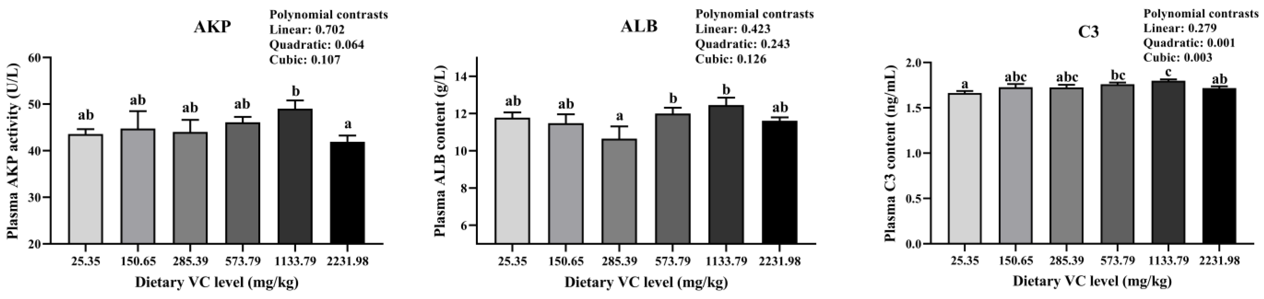
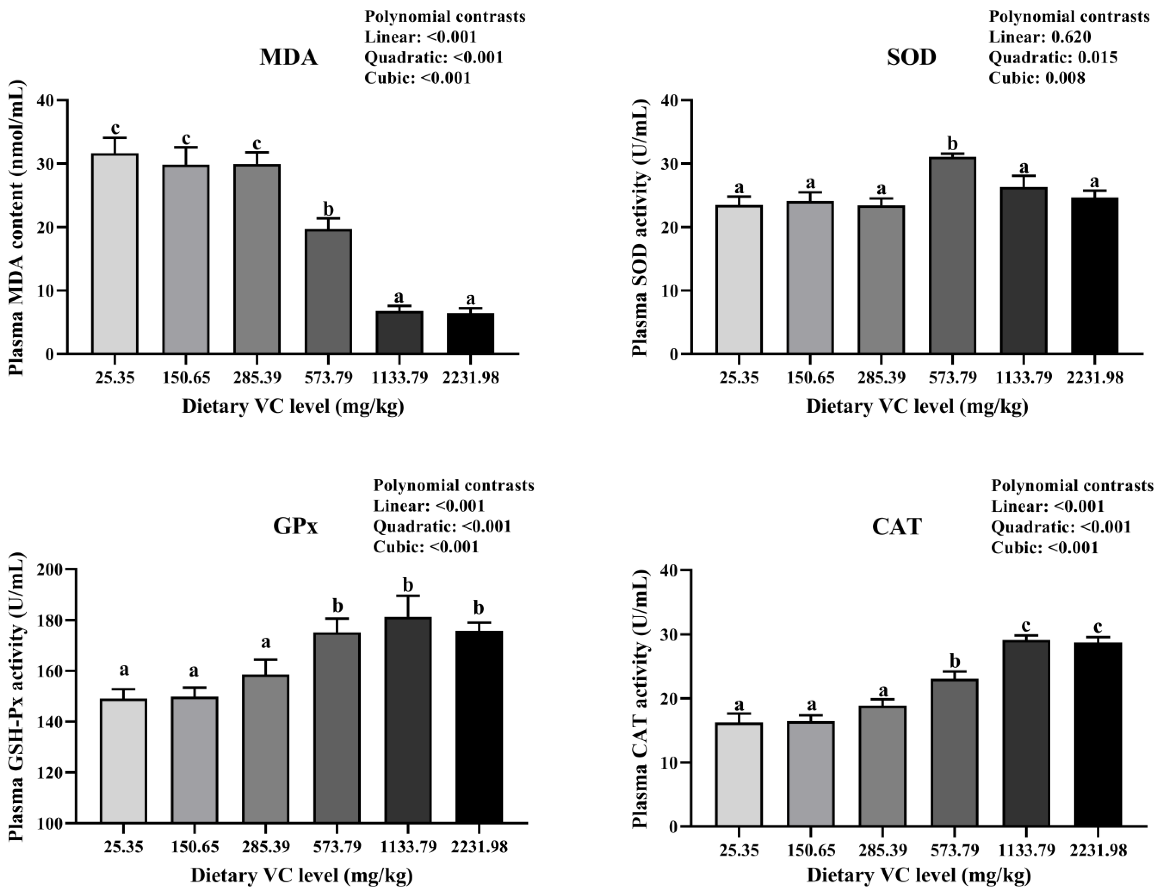
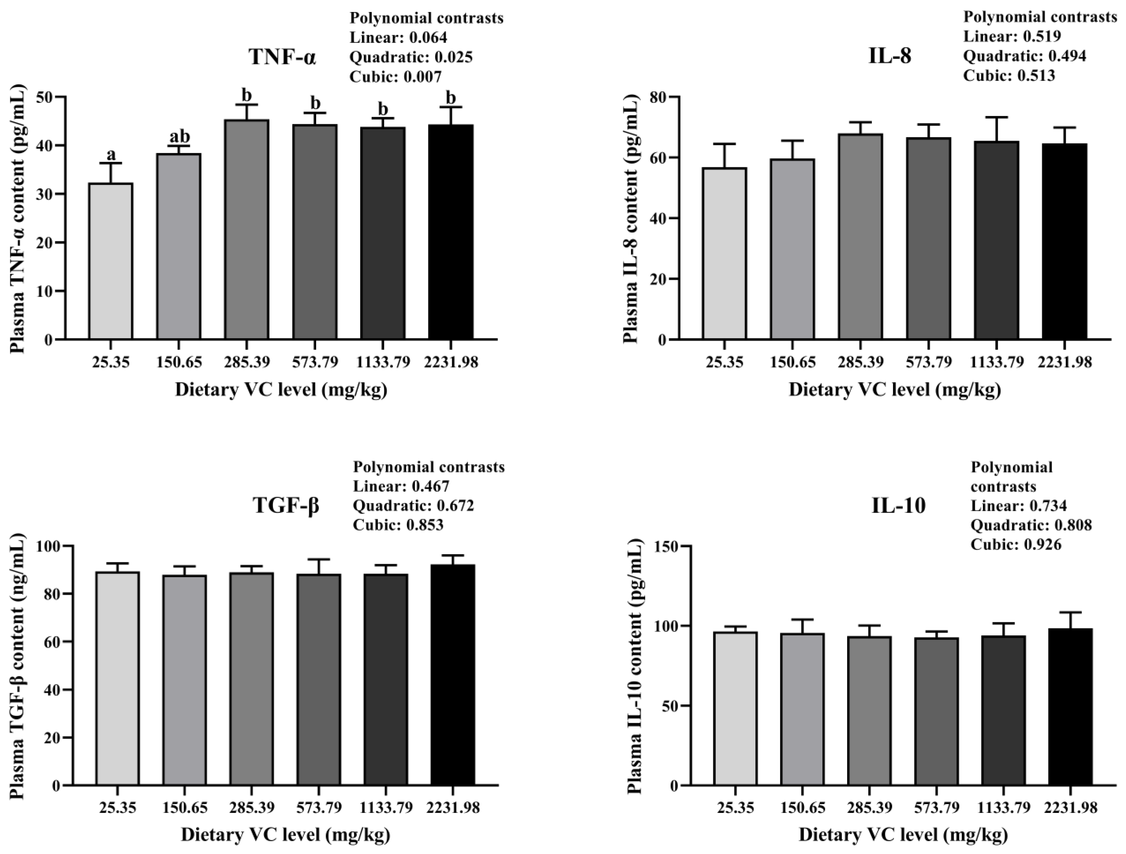
3.2. Serum Immune-Related Indices, Antioxidant Capacity, and Cytokines of Juvenile Blunt Snout Bream in the Feeding Trail

The effects of VC levels on serum immune-related indices in juvenile blunt snout bream are shown in Figure 1. A significantly higher AKP (p < 0.05) was observed in the fish fed the 1133.79 mg/kg VC diet than those fed the 2231.98 mg/kg VC diet, while no significant differences existed between the control group and the fish fed with different levels of dietary VC (p > 0.05). The ALB content in fish fed 573.79 and 1133.79 mg/kg VC was significantly higher (p < 0.05) than in fish fed a 285.39 mg/kg VC diet. Moreover, the C3 content in the 573.79 mg/kg and 1133.79 mg/kg VC groups was significantly higher (p < 0.05) than that in the control group. Polynomial contrasts showed that only serum C3 was significantly both quadratically and cubically (p < 0.01) affected by dietary VC supplementation.
The effects of VC concentrations on the serum antioxidant capacity of juvenile bream are shown in Figure 2. Serum MDA content significantly decreased with increased dietary VC supplementation. It was significantly decreased (p < 0.05) in the high dietary VC group (1133.79 mg/kg and 2231.98 mg/kg) compared with the other groups. Serum SOD activity in the group fed a 573.79 mg/kg VC diet was significantly higher (p < 0.05) than that in the other groups. Moreover, serum CAT and GPx activities significantly increased with increasing dietary VC supplementation (p < 0.05) from 573.79 mg/kg to 2231.98 mg/kg compared with the group with VC content lower than 285.39 mg/kg. The polynomial contrasts showed that the MDA content, GPx, and CAT activities were linearly, quadratically, and cubically (p < 0.001) influenced by dietary VC supplementation, whereas SOD activity was quadratically (p < 0.05) and cubically (p < 0.01) influenced by dietary VC supplementation.
The effects of VC levels on serum cytokine levels in juvenile bream are shown in Figure 3. The results showed that TNF-α levels were significantly affected by VC supplementation (p < 0.05). The levels of other cytokines, such as IL-8, TGF-β, and IL-10, were not significantly different (p > 0.05) among the treatment groups. Serum TNF-α levels in the groups supplemented with dietary VC ranged from 285.39 to 2231.98 mg/kg, significantly higher than those in the control group (p < 0.05). From the polynomial contrasts, TNF-α was quadratically (p < 0.05) and cubically (p < 0.01) influenced by dietary VC supplementation.

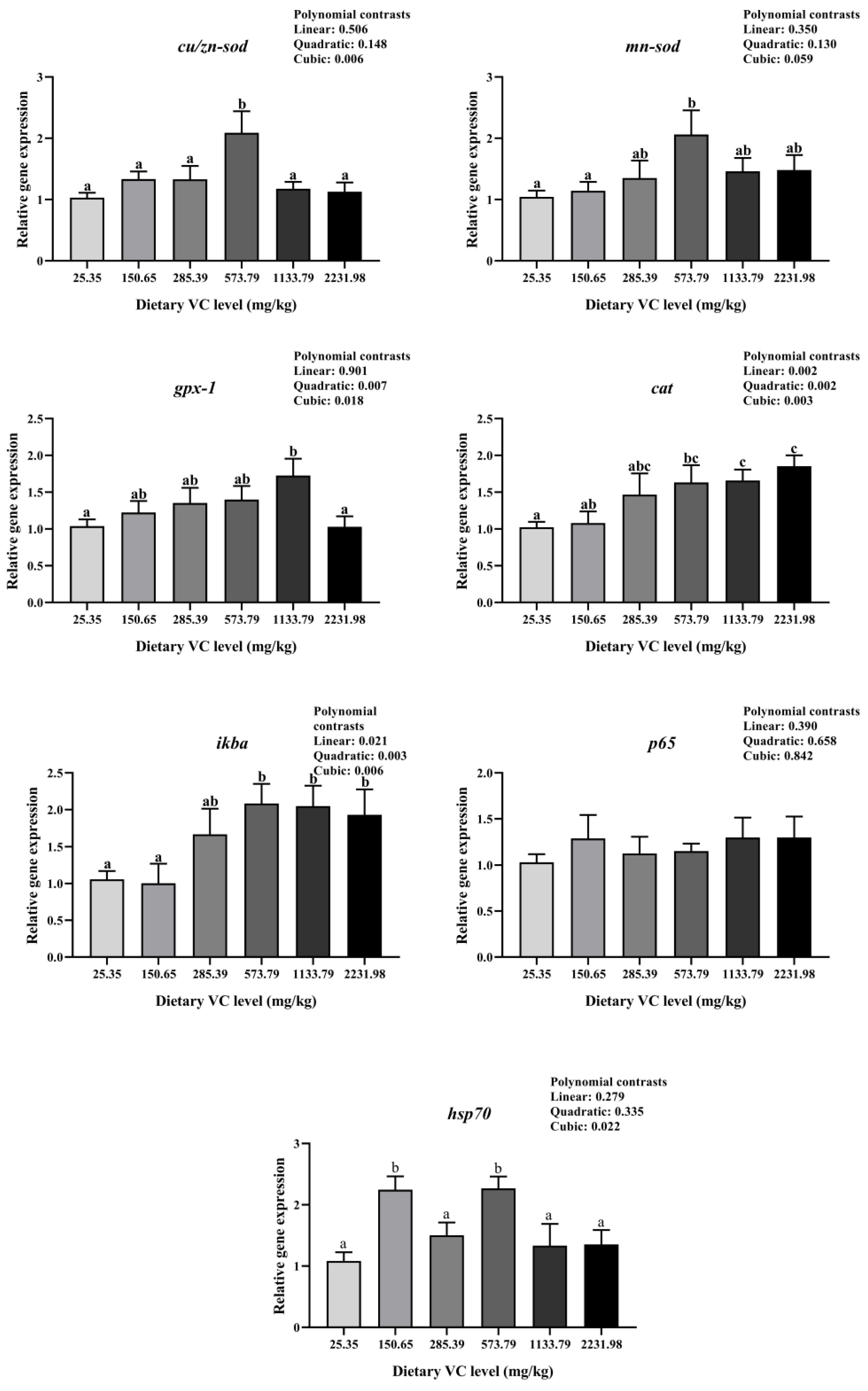
3.3. Immune-Related Gene Expression of Juvenile Blunt Snout Bream in the Feeding Trail

The expression of genes such as cu/zn-sod, mn-sod, gpx1, cat, ikbα, p65, and hsp70 in the livers of blunt snout bream was evaluated (Figure 4). The mRNA levels of cu/zn-sod, mn-sod, and ikbα were significantly upregulated when dietary VC reached 573.79 mg/kg (p < 0.05) compared with the 25.35 mg/kg to 150.65 mg/kg groups. mRNA levels of cu/zn-sod and mn-sod were downregulated with excessive dietary VC supplementation, except ikbα, which was stable with excessive dietary VC. The mRNA levels of cat increased with the increasing dietary VC levels, with the highest levels in the excessive VC groups (1133.79 mg/kg and 2231.98 mg/kg). The gpx1 mRNA level in the 1133.79 mg/kg group was significantly higher (p < 0.05) than that in the 25.35 mg/kg and 2231.98 mg/kg groups. No significant difference was found in the mRNA level of p65 among the treatments (p > 0.05). The hsp70 expression was significantly upregulated in fish fed with 150.65 mg/kg and 573.79 mg/kg than in fish fed with other dietary VC levels (p > 0.05). Significant cubic relationships between relative gene expression and dietary VC levels were observed for cu/zn-sod and hsp70 (p < 0.05). The relative expression of gpx1 was quadratically (p < 0.01) and cubically (p < 0.05) affected by dietary VC supplementation. Moreover, linear (p < 0.05), quadratic (p < 0.01), and cubic (p < 0.01) relationships were observed in cat and ikbα.

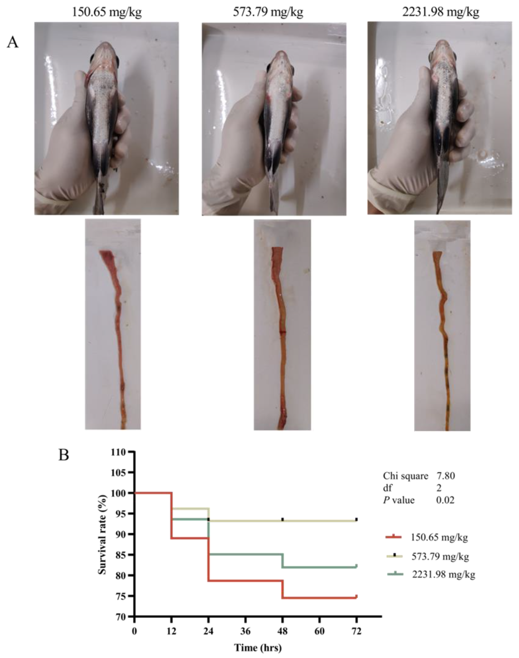
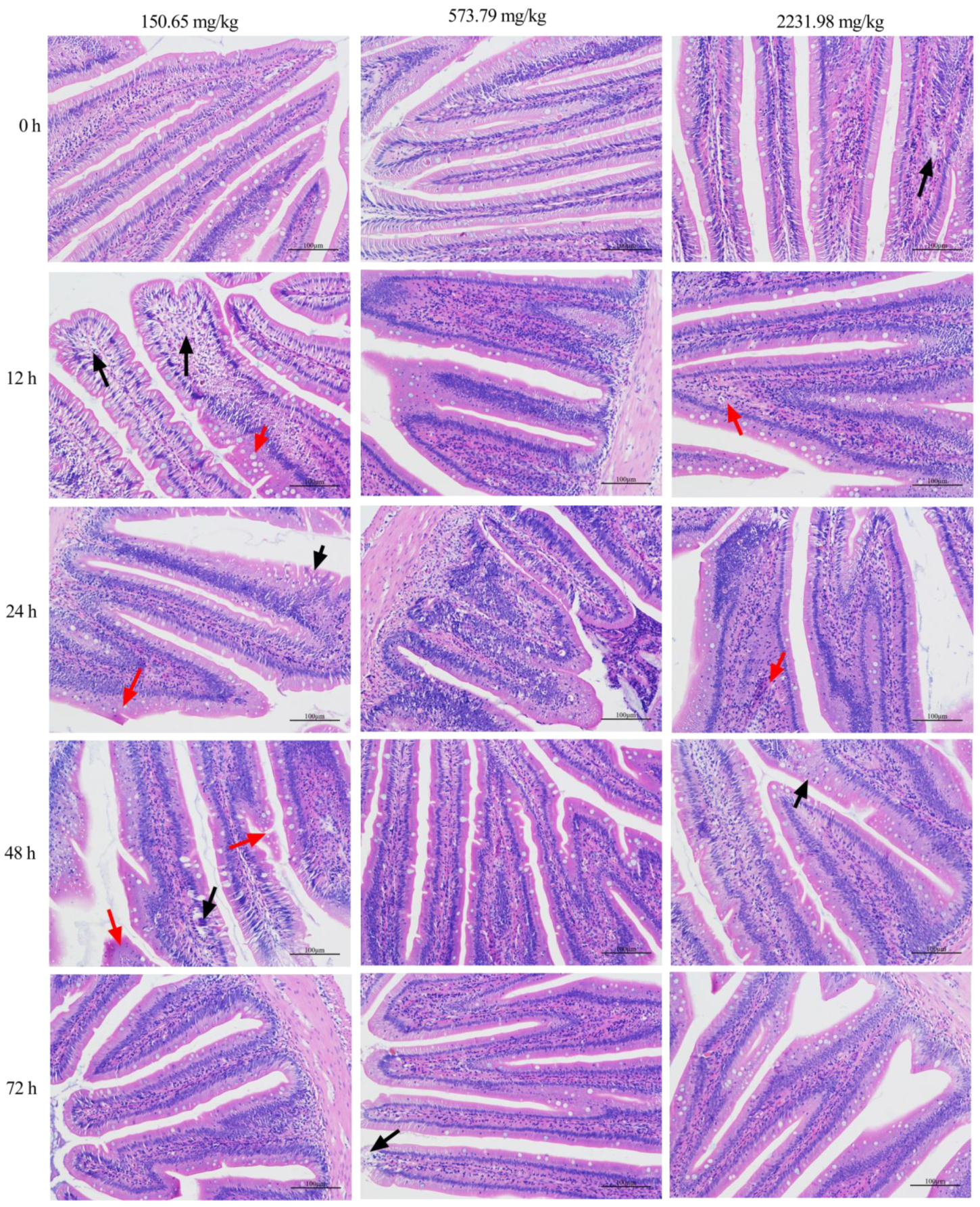
3.4. Survival Curve, Inflammatory Response, and Intestinal Histopathology after A. hydrophila Challenging

To evaluate the immunity function of VC, blunt snout breams fed with three different VC levels (150.65, 573.79, and 2231.98 mg/kg VC) were randomly selected to orally administer A. hydrophila. Inflammation statuses were observed and recorded at 24 hpi. As shown in Figure 5A, each group of breams showed typical bacterial sepsis to varying degrees. Among them, the inflammatory response of the 150.65 mg/kg VC group was the most obvious; the abdomen, fin base, and the back of the gill slit of the fish had clinical signs of hyperaemia or bleeding, bilateral eyeballs were protruding, and some fish with dying clinical signs had swollen abdomens, redness, and swelling protrusions. Autopsy found that there was water accumulation in the abdominal cavity; congestion and inflammation of the intestinal wall; and many yellowish, viscous fluids.
Survival rates were recorded (Figure 5B). It can be seen that, with the extension of the challenge time, the survival rate of the blunt snout bream showed a decreasing trend at 12 hpi and then stabilized at a different time interval after infection. Blunt snout bream fed the 573.79 mg/kg VC diet had significantly higher survival rates, which stabilized 24 hpi (p < 0.05) compared to breams fed the 160.65 and 2231.98 mg/kg VC diets, respectively. Compared to other experimental groups, the 573.79 mg/kg VC group had the best disease resistance effect and the lowest number of deaths.
As shown in Figure 6, after the challenge with A. hydrophila, some clinical signs of acute inflammatory reaction occurred in the intestine over time. The enteritis sign in the lower VC group (150.65 mg/kg) was the most obvious compared to the 573.79 and 2231.98 mg/kg VC groups. At 12 hpi and 24 hpi, there was shedding of the mucosal epithelial cell in the 150.65 mg/kg VC group, which gradually intensified at 48 hpi with manifestations of large-scale detachment of mucosal epithelial cells; separation of the lamina propria and mucosal layer; and increased inflammatory cells in the mucosal layer, submucosa, and lamina propria, with only minor epithelial cell damage. However, the clinical signs of enteritis were significantly reduced in the 573.79 and 2231.98 mg/kg VC groups at that time. At 72 hpi, the clinical signs of enteritis in each test group basically disappeared, and the fish returned to normal.

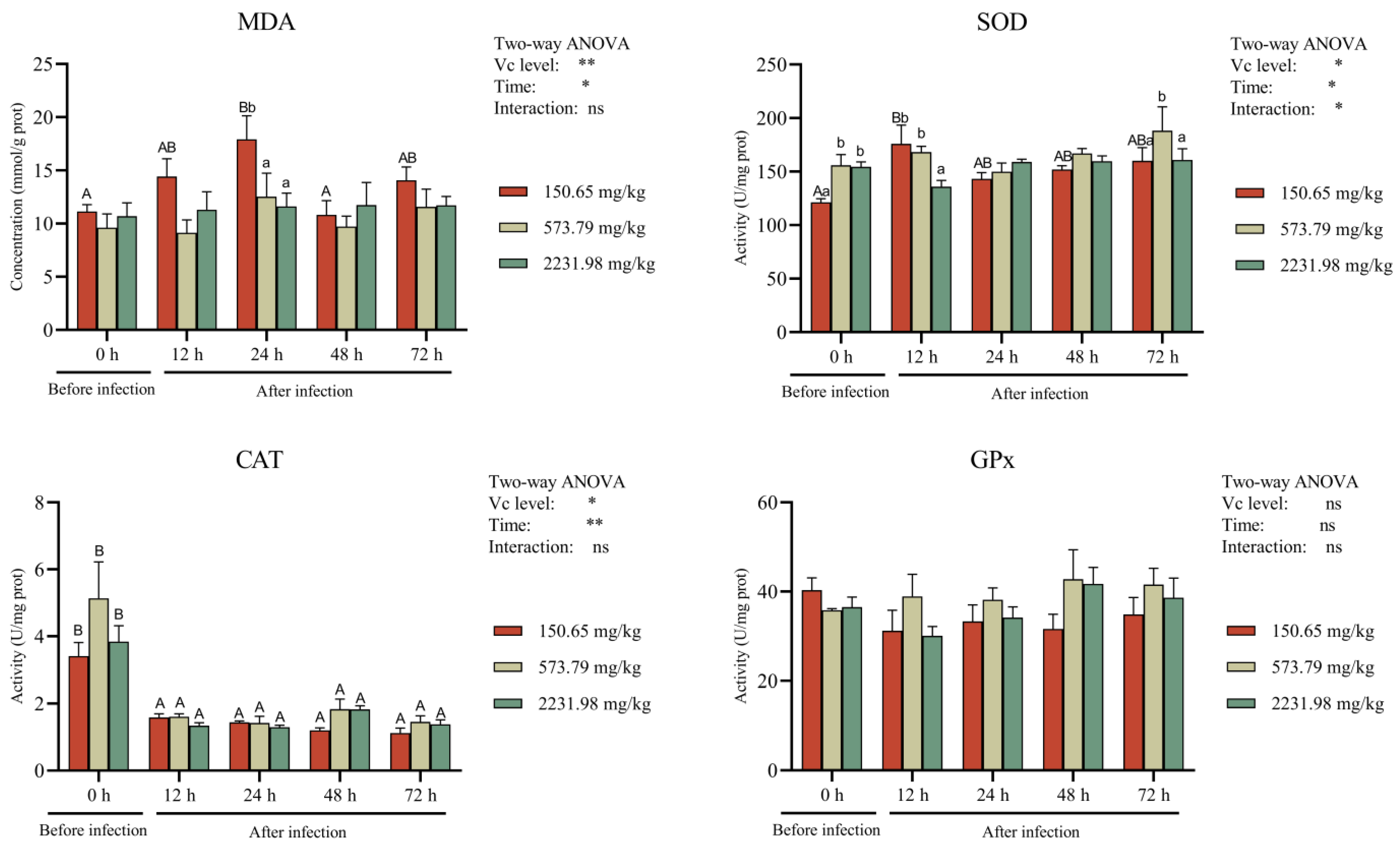
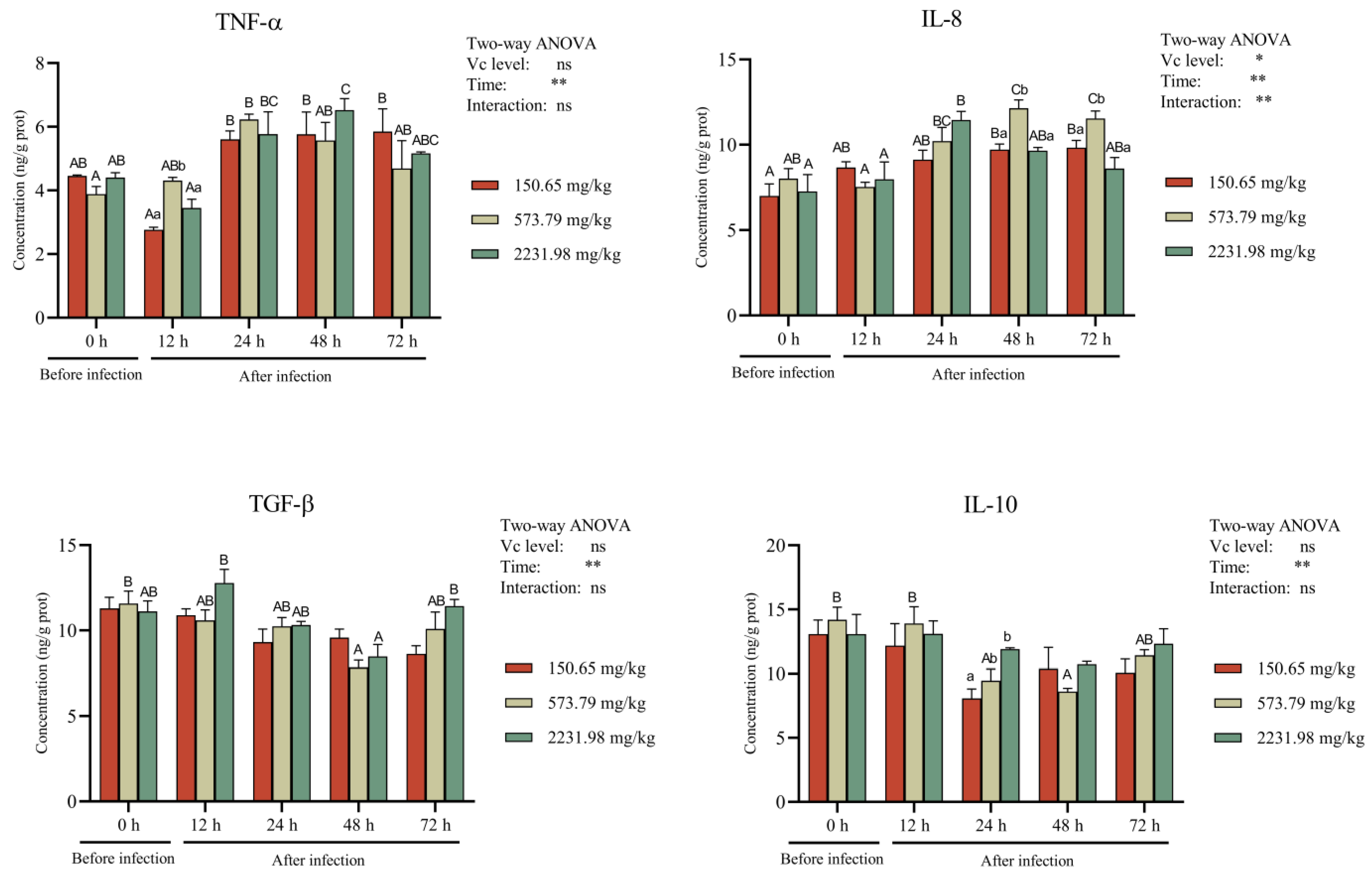
3.5. Intestinal Antioxidant Activity and Cytokines after A. hydrophila Challenge

As shown in Figure 7, the intestinal MDA content, SOD, and CAT activities were significantly affected by the dietary VC level and time (p < 0.05). However, the interaction between VC levels and infection time did not significantly affect the MDA content, CAT, or GPx activities (p > 0.05). Compared to the lower VC group, the intestinal MDA of the blunt snout bream fed the 573.79 mg/kg VC diet was significantly lower before and after infection with A. hydrophila (p < 0.05). The SOD activity of the blunt snout bream fed higher VC diets first increased significantly before infection, then decreased at 12 hpi, and then normalized 24 hpi and 48 hpi. At 72 hpi, the SOD activity of the blunt snout bream fed the 573.79 mg/kg VC diet was significantly increased compared to the group fed the lower-VC diet. Meanwhile, the CAT activity was significantly higher before the challenge, and the highest level was observed in bream fed the 573.79 mg/kg VC diet. With the extension of the infection time, the intestinal CAT activity decreased significantly and became normalized throughout the infection time. The intestinal GPx levels were not significantly affected by the VC level, time, or their interaction (p > 0.05).
Intestinal cytokine concentrations are shown in Figure 8. The proinflammatory cytokine TNF-α was significantly affected by time, while IL-8 was significantly affected by VC levels, time, and its interaction in response to the A. hydrophila challenge (p < 0.05). The TNF-α concentration first decreased at 12 hpi, increased at 24 hpi, peaked at 48 h, and then decreased gradually by 72 hpi in both the 150.65 mg/kg and 2231.98 mg/kg groups. However, no decrease was observed at 12 hpi, and there was a peak at 24 hpi in the 573.79 mg/kg group. At 12 hpi, the TNF-α concentration in the 573.79 mg/kg group was significantly higher than that in the lower and excessive dietary VC groups. As for IL-8, it was slightly decreased at 12 hpi and then increased, and maintained a relative higher level at 48 hpi and 72 hpi in the 573.79 mg/kg group. Lower dietary VC (150.65 mg/kg) could not induce high levels of IL-8 as the 573.79 mg/kg diet did, while excessive dietary VC (2231.98 mg/kg) caused an early peak (24 hpi) and then a decrease. At 48 hpi and 72 hpi, the IL-8 concentration in the 573.79 mg/kg group was significantly higher than that in the lower and excessive dietary VC groups.
Anti-inflammatory cytokines (TGF-β and IL-10) was significantly affected by the infection time only (p < 0.05). Dietary 150.65 mg/kg VC could not significantly change the TGF-β concentration (p > 0.05). The TGF-β concentration was significantly decreased at 48 hpi compared to before the challenge in the dietary 573.79 mg/kg VC group (p < 0.05), while it was increased at 12 hpi and 72 hpi and decreased at 48 hpi in the dietary 2231.98 mg/kg VC group (p < 0.05). As for IL-10, lower and excessive dietary VC (150.65 mg/kg and 2231.98 mg/kg) did not significantly change its concentration (p > 0.05). The IL-10 concentration was significantly decreased at 24 hpi and 48 hpi compared to before challenge and at 12 hpi (p < 0.05). Moreover, at 24 hpi, the IL-10 concentration in the 150.65 mg/kg group was significantly lower than that in the other groups (p < 0.05).

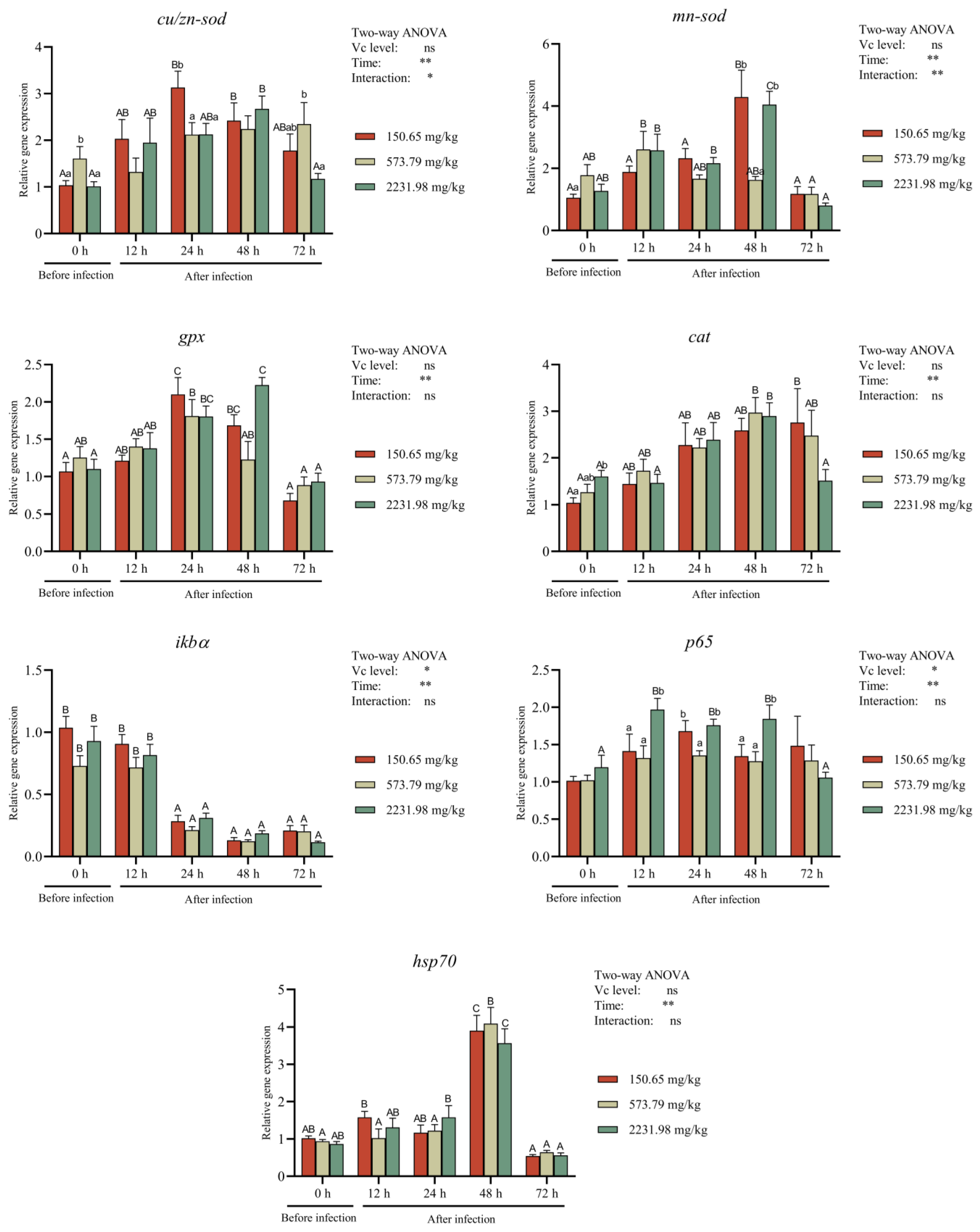
3.6. Immune-Related Gene Expressions after A. hydrophila Challenging

The expression levels of liver cu/zn-sod, mn-sod, cat, and gpx were upregulated at first and then downregulated with the extension of the challenge time (Figure 9). The expressions of cu/zn-sod and mn-sod were significantly affected by the challenge time and the interaction of VC and challenge time (p < 0.01), while the expressions of cat and gpx were significantly affected only by the challenge time (p < 0.01). In the 573.79 mg/kg VC group, no significant variation in cu/zn-sod expression was observed (p > 0.05), and slight variation in the mn-sod expression was found, with a peak at 12 hpi. Similar trends were observed for cat and gpx expression, with different peak times. Low or excessive dietary VC caused significant variations in cu/zn-sod and mn-sod expression (p < 0.05). Similar trends were found in the low and excessive dietary VC groups than in the 573.79 mg/kg VC group regarding cat and gpx expression. The cu/zn-sod expression at 24 hpi was significantly lower in the 573.79 mg/kg and 2231.98 mg/kg VC groups than that in the 150.65 mg/kg VC group (p < 0.05). The mn-sod expression at 48 hpi was significantly lower in the 573.79 mg/kg VC group than that in the 150.65 mg/kg and 2231.98 mg/kg VC groups (p < 0.05).
The mRNA levels of p65 and ikbα were significantly affected by the VC level and challenge time (p < 0.05). The gene expression of ikbα was significantly inhibited from 24 hpi to 72 hpi compared to before and at 12 hpi in all groups (p < 0.05). The mRNA level of ikbα was lower in the 573.79 mg/kg group than in the other two groups, but without significant differences (p > 0.05). The expression of p65 was significantly increased after challenge and then decreased at 72 hpi in the 2231.98 mg/kg VC group (p < 0.05), and it varied without significant differences in the other two groups (p > 0.05). Moreover, the p65 mRNA expression level in the 2231.98 mg/kg VC group was significantly higher than that in the other two groups at 12 hpi and 48 hpi (p < 0.05), and it was significantly higher than that in the 573.79 mg/kg group at 24 hpi (p < 0.05). The expression levels of hsp70 mRNA were significantly affected by the challenge time (p < 0.05). The expression level at 48 hpi was significantly higher than that at other times (p < 0.05).

4. Discussion

Vitamin C plays a crucial role in maintaining normal physiological functions in fish. When added to fish diets in appropriate amounts, it can enhance their growth performance [25]. The findings of this study indicate that the growth (measured by WG and SGR) of juvenile blunt snout bream significantly improved when they were fed diets containing 285.39 mg/kg and 573.79 mg/kg of VC for 60 days. Additionally, their feed conversion ratio (FCR) showed a significant decrease, suggesting that the inclusion of the right amount of VC in the diet can enhance growth performance by improving feed efficiency.
Previous research has shown that VC supplementation improves the growth performance of various fish species, such as Chu’s croaker (Nibea coibor) [26] and coral trout (Plectropomus leopardus) [27]. The optimal dietary VC requirement for growth in blunt snout bream has been determined to be 150 mg/kg, which differs from values reported for other species like striped catfish [28] and grass carp [29]. These variations in VC requirements can be attributed to factors such as species differences; size variations; experimental conditions; feed nutritional content; metabolic rates of fish; and the forms, purity, and bioavailability of the VC used in the studies [30].
Aeromonas hydrophila, a common aquaculture pathogen, colonizes and multiplies in the fish intestine following infection, typically causing intestinal inflammation and acute enteritis, resulting in significant mortality rates [31,32]. Outbreaks of A. hydrophila have been linked to alterations in the susceptibility of fish, attributed to changes in environmental factors such as temperature, which are associated with the production of virulence factors [33]. These virulence factors include secretion systems, motility and adhesins, toxins, enzymes, haemolysins, and increased nitrite levels in farmed fish [34]. A study on snakehead fish (Channa striata) revealed that A. hydrophila infection resulted in mortality rates ranging from 60% to 100% [35]. The blunt snout bream, being herbivorous with a unique digestive system—lacking a stomach but having a wide oesophagus and long intestine—emphasizes the importance of gut health for its growth and development. In this study, the blunt snout bream fed a diet containing 573.79 mg/kg VC showed no signs of bacterial sepsis and exhibited higher survival rates when challenged with A. hydrophila compared to those fed lower or excessive VC diets. This study suggests that the optimal VC level for disease resistance in blunt snout bream is 573.79 mg/kg, significantly higher than the 150 mg/kg requirement for growth. This disparity is attributed to the fact that VC requirements for stress resistance, as indicated by disease resistance, can be six times or more than those for growth [13,36]. Therefore, fish need to consume higher levels of VC to enhance their immune systems, combat diseases, and maintain overall health to withstand environmental stress and bacterial threats. Similar research has demonstrated that supplementing feed with VC can effectively reduce mortality rates in grass carp challenged with A. hydrophila [29].
During feeding processes, the intestine is exposed to various bacteria, viruses, antigens, and potentially harmful substances, leading to oxidative stress, intestinal damage, inflammation, and a threat to overall health. In this study, blunt snout breams fed a lower VC diet (150.65 mg/kg) exhibited significant intestinal inflammatory responses at 12, 24, 48, and 72 hpi. They showed clinical signs of hyperaemia and congestion of the intestines, and inflammatory infiltration and intestinal epithelial cell injury on histopathology. Similar inflammatory clinical signs have been observed in grass carp [31] and Nile tilapia [37] when challenged with Aeromonas schubertii. Conversely, blunt snout breams fed a higher-VC diet (573.79 mg/kg) displayed milder clinical signs of inflammation, likely due to lower intestinal MDA levels and increased SOD levels, which helped to protect the body cells and tissues from damage caused by excessive oxygen free radicals.
In the current study, it was observed that serum levels of AKP, ALB, and C3 significantly increased with high dietary VC supplementation for 60 days. The elevation in AKP levels can be attributed to VC’s ability to enhance lysosomal enzymes in fish, thereby improving the body’s defence against foreign pathogens. The increase in ALB levels may be linked to nutrient transport and the maintenance of blood pressure balance, while the rise in C3 levels could be due to VC’s role in regulating neutrophil phagocytosis, activating the complement system, and enhancing nonspecific immune function. The study also found a positive correlation between serum immunity and survival in challenge tests, suggesting that higher VC levels may enhance immunity in juvenile blunt snout bream. Similar findings have been reported in other fish species such as Nile tilapia [38] and grass carp [29], where increased VC levels improve immune enzyme activity and substance content. Interestingly, in the group with excessive VC intake (2231.98 mg/kg), the serum levels of AKP, ALB, and C3 decreased, potentially explaining the slightly higher mortality rate following A. hydrophila infection. Further research is needed in order to understand the mechanism by which excessive dietary VC intake may reduce immunity in fish.
The oxidative regulation systems in aquatic species can serve as indicators of their health [20]. The levels of SOD, CAT, GPx, and MDA in fish serum can accurately depict the body’s antioxidant status. In the present study, it was observed that serum MDA levels decreased significantly, while CAT, SOD, and GPx levels increased in fish fed diets containing 573.79, 1133.79, and 2231.98 mg/kg of VC for 60 days. The impact of VC on antioxidant defence in juvenile blunt snout bream was further investigated by analysing the activities and levels of these enzymes in the intestine before and after infection with A. hydrophila. The intestinal MDA content decreased with higher dietary VC levels (573.79 and 2231.98 mg/kg) and prolonged A. hydrophila infection. SOD activity showed an initial increase, followed by a decrease, and eventually stabilized 24 h post-infection. CAT activity remained lower and stable throughout the infection period with A. hydrophila compared to pre-infection levels. These findings suggest that VC may enhance the body’s immune response by scavenging free radicals, restoring GPx activities, and improving the ability to eliminate peroxide products like MDA. This, in turn, helps to prevent oxidative damage to cells or tissues and the invasion of pathogenic bacteria into the fish’s body. Dietary VC has been shown to reduce MDA content and enhance the activities of antioxidant enzymes in various species, including grass carp infected with Flavobacterium columnare [39], golden pompano [40], and gibel carp [41]. VC is a powerful nonenzymatic antioxidant and scavenger of free radicals, essential for scavenging reactive oxygen species (ROS) that could otherwise combine with nitric oxide (NO) to form the highly reactive free radical peroxynitrite [42]. Furthermore, VC safeguards cells from oxidative damage by functioning as an antioxidant and providing electrons to free radicals, effectively neutralizing them and averting potential harm [43].
Under normal circumstances, fish typically have low levels of cytokines or remain inactive. When triggered by foreign pathogens, cytokines can regulate immune responses by binding to their corresponding receptors on cell surfaces [17]. In this study, it was observed that VC did not significantly affect the levels of anti-inflammatory cytokines TGF-β and IL-10 in the serum of blunt snout bream. However, it did lead to an increase in pro-inflammatory cytokines (TNF-α). This could be attributed to the absence of inflammation during the regular feeding experiment prior to the bacterial challenge, which resulted in the anti-inflammatory cytokines not being activated. On the other hand, infection with A. hydrophila caused changes in the levels of pro- and anti-inflammatory cytokines in the intestines. TGF-β and IL-10, as important anti-inflammatory cytokines, were found to modulate with variation in TNF-α and IL-8 levels, thereby reducing inflammation. The study revealed that the infection duration directly impacted the inflammatory response in the fish intestines, with longer durations exacerbating the response. At 72 h, higher VC supplementation levels (573.75 and 2231.98 mg/kg) were associated with reduced intestinal TNF-α levels and increased TGF-β and IL-10 levels. Additionally, VC supplementation was found to decrease mRNA levels of pro-inflammatory cytokines and increase mRNA levels of anti-inflammatory cytokines in the gills of grass carp infected with Flavobacterium columnare through the NF-κB signalling pathway [39]. Furthermore, dietary VC intake was shown to elevate levels of pro-inflammatory cytokines (TNF-α and IL-1) in the serum, gut, and spleen of Nile tilapia [44].
It may be argued that, in the early and middle phases of enteritis in the blunt snout bream, the 573.79 mg/kg VC level could increase the adaptive immune response and the synthesis of a significant number of cytokines with immunomodulatory properties. Furthermore, during enteritis, 573.79 mg/kg VC increases the activity of intestinal SOD, preventing harm to fish cells or tissues from excessive oxygen-free radicals. VC may increase the activity of intestinal cytokines and antioxidant enzymes, reinforce their synergy, promote non-specific immune responses, eliminate harmful microorganisms, and alleviate intestinal inflammation. Cytokines primarily act on macrophages, causing a respiratory explosion and the production of a large number of oxygen-free radicals, which enhance macrophage killing of harmful bacteria [45]. Research suggests that VC can enhance the immune response to Th1 cells, leading to increased production of TNF-α and IL-8 by T cells. This, in turn, activates immune cells like phagocytes, T lymphocytes, and neutrophils, improving antigen recognition and killing [43,46,47,48]. As a result, VC can increase the immunomodulatory and anti-inflammatory properties of juvenile blunt snout bream by inducing an inflammatory response.
The immunological status of the blunt snout bream was assessed by examining the expression of antioxidant-related genes in the liver. The liver acts as a key immune organ, tasked with identifying and eliminating potential pathogens entering the body via the gut into the bloodstream while maintaining immune hyporesponsiveness. This immunotolerance programming prevents unnecessary inflammatory reactions against harmless substances or normal levels of microbe-derived molecules. However, changes in microbial products can prompt the liver to shift from immune hyporesponsiveness to a strong inflammatory response and adaptive immunity. In cases of acute liver injury, inflammation aids in tissue repair. The balance between immunotolerance and robust immune responses is regulated by interactions among different immune cell populations in the liver, crucial for maintaining liver tissue homeostasis [49].
Immunity and oxidative stress are interconnected and have a mutual influence [50]. In this study, during the normal feeding trial, the antioxidative genes cu/zn-sod, mn-sod, cat, and gpx1 were found to be upregulated in the liver of juvenile blunt snout bream. When comparing the dietary requirements of VC for growth (150.65 mg/kg), immunity (573.79 mg/kg), and excessive VC intake (2231.98 mg/kg) during the bacterial challenge, the mRNA expression of these antioxidative genes increased after the challenge and decreased at 72 h post-infection, with mn-sod and gpx levels dropping below the pre-infection expression levels. This indicates that VC can help to maintain intestinal integrity and reduce inflammation up to 48 h after infection by reducing oxidative stress biomarkers and increasing mRNA expression levels. This finding aligns with previous studies by Yusuf et al. [17] and Dawood et al. [51].
In most cells, iκbα serves as the primary inhibitor protein of NF-κB, a transcription factor that plays a crucial role in regulating various pathophysiological processes, including cellular survival and death. NF-κB is involved in controlling inflammatory, immunological, cellular survival, and proliferation responses [52]. The p65 subunit, a key component of NF-κB, is essential for innate defence against microbial pathogens and is constitutively expressed in multiple organs of teleosts [53]. The current study observed that prolonged infection time led to a decrease in iκbα mRNA expression and an increase in p65 mRNA expression in the liver, resulting in activating NF-κB signalling. Vitamin C was found to activate the immune response associated with decreased iκbα and increased p65 expression, thereby enhancing fish immunity and mitigating the negative effects of bacterial challenge. In this study, the iκbα/p65 classical pathway may have been activated to regulate intestinal TNF-α, TGF-β, IL-8, and IL-10, potentially impacting interferon and inflammatory signalling networks. p65 has been shown to regulate interferon signalling molecules (such as TNFα) and inflammatory cytokines (such as IL-8 and IL-10) in common carp [53] and grass carp [54].
In response to stress, hsp70, a member of the heat shock protein (HSP) family, plays a role in various cellular functions and immune responses in fish [12,55]. When subjected to pH stress, hsp70 levels increased and peaked after one day in juvenile Megalobrama amblycephala [12]. Similarly, in the current study, following a bacterial challenge, the mRNA levels of hsp70 in all VC groups rose at 12 and 24 h, reached a peak at 48 h post-infection, and then declined, with the 573.79 mg/kg VC group showing the highest expression level at 48 h. The elevated hsp70 levels during the infection period promoted cellular, tissue, and overall fish tolerance to A. hydrophila infection, suggesting the potential for developing strategies to enhance tolerance to pathogenic stresses by activating the cellular stress response. However, when the infection duration was extended to 72 h, the expression levels of hsp70 in all VC groups decreased below pre-infection levels. Qiang et al. [56] found that excessive stress intensity or prolonged stress can lead to changes in liver cell membrane structure and protein composition, inhibiting HSP transcription. This aligns with the results of the present study.
The cost-effectiveness of using VC in aquaculture can vary depending on factors such as the specific application, dosage, and the overall health and productivity of the fish being raised. Study have shown that VC supplementation can have beneficial effects on fish health, growth, and stress resistance, potentially leading to improved production outcomes [57]. However, the cost of incorporating VC into aquaculture practices should be weighed against the potential benefits in terms of fish health and overall productivity to determine its cost-effectiveness in a specific aquaculture operation.

5. Conclusions

In conclusion, intestinal inflammation was observed in juvenile blunt snout breams within the first 24 h following A. hydrophila infection. Dietary VC supplementation above 500 mg/kg could enhance antioxidant capacity and non-specific immunity and activate cytokines, hence reducing intestinal inflammation 48 h post-infection. Furthermore, excessive dietary VC may have adverse effects like impaired serum immunity, elevated mortality following A. hydrophila challenge, and lowered levels of antioxidant gene expression.

Author Contributions

Conceptualization, Q.Z. and X.G.; methodology, A.M.M., C.Y., J.W. and G.G.; data curation, Q.Z. and A.M.M.; writing—original draft, A.M.M.; revision, Q.Z.; supervision, B.L. and X.G.; funding acquisition, L.M. and B.L. All authors have read and agreed to the published version of the manuscript.

Funding

This research was funded by the earmarked fund for China Agriculture Research System (grant number: CARS-45), the National Natural Science Foundation of China (grant number: 32172990), Central Public-interest Scientific Institution Basal Research Fund, CAFS (grant number: 2023TD63), and National Key Research and Development Plan Program of China (2022YFD2400600/2022YFD2400603).

Institutional Review Board Statement

This research was approved by the scientific research protocols of the Chinese Academy of Fishery Sciences (CAFS), the Ministry of Agriculture and Rural Affairs, the People’s Republic of China, and the Animal Care and Use Committee of Nanjing Agricultural University (permit number: SYXK(Su)2017–0007). All relevant local and/or international animal welfare laws, guidelines, and policies for the care and use of animals were followed.

Informed Consent Statement

Not applicable.

Data Availability Statement

Data are available from the corresponding author upon reasonable request.

Conflicts of Interest

Jingyuan Wang is now working at Taizhou Aohua Agricultural and Animal Husbandry Technology Co., Ltd. However, this research work was conducted when he was a student at Wuxi Fisheries College, Nanjing Agricultural University. Therefore, no conflict of interest is related to Taizhou Aohua Agricultural and Animal Husbandry Technology Co., Ltd. Other authors declare no conflicts of interest.

References

  1. Dewali, S.; Sharma, N.; Melkani, D.; Arya, M.; Kathayat, N.; Panda, A.K.; Bisht, S.S. Aquaculture: Contributions to Global Food Security. In Emerging Solutions in Sustainable Food and Nutrition Security; Ghosh, S., Kumari Panda, A., Jung, C., Singh Bisht, S., Eds.; Springer: Cham, Switzerland, 2023; pp. 123–139. [Google Scholar] [CrossRef]
  2. Alvarex-Pellitero, P. Fish immunity and parasite infections: From innate immunity to immunoprophylactic prospects. Vet. Immunol. Immunopathol. 2008, 126, 171198. [Google Scholar] [CrossRef]
  3. Torres, M.J.M.; Peterson, J.M.; Wolf, S.E. Detection of infection and sepsis in burns. Surg. Infect. 2021, 22, 20–27. [Google Scholar] [CrossRef]
  4. Zhang, Y.; Li, Y.; Liang, X.; Gao, J. Effects of dietary vitamin E supplementation on growth performance, fatty acid composition, lipid peroxidation and peroxisome proliferator-activated receptors (PPAR) expressions in juvenile blunt snout bream Megalobrama amblycephala. Fish Physiol. Biochem. 2017, 43, 913–922. [Google Scholar] [CrossRef]
  5. Yuan, G.; Zhao, W.; Zhang, Y.; Jia, Z.; Chen, K.; Wang, J.; Feng, H.; Zou, J. The biological functions and intestinal inflammation regulation of IL-21 in grass carp (Ctenopharyngodon idella) during infection with Aeromonas hydrophila. Cells 2023, 12, 2276. [Google Scholar] [CrossRef]
  6. Semwal, A.; Kumar, A.; Kuma, N. A review on pathogenicity of Aeromonas hydrophila and their mitigation through medicinal herbs in aquaculture. Heliyon 2023, 9, e14088. [Google Scholar] [CrossRef]
  7. Xiong, X.M.; Chen, Y.L.; Liu, L.F.; Wang, W.; Robinson, N.A.; Gao, Z.X. Estimation of genetic parameters for resistance to Aeromonas hydrophila in blunt snout bream (Megalobrama amblycephala). Aquaculture 2017, 479, 768–773. [Google Scholar] [CrossRef]
  8. Carr, A.; Maggini, S. Vitamin C and immune function. Nutrients 2017, 9, 1211. [Google Scholar] [CrossRef]
  9. Drouin, G.; Godin, J.R.; Pagé, B. The genetics of vitamin C loss in vertebrates. Curr. Genom. 2011, 12, 371–378. [Google Scholar] [CrossRef]
  10. Habte-Tsion, H.M.; Ge, X.; Liu, B.; Xie, J.; Ren, M.; Zhou, Q.; Miao, L.; Pan, L.; Chen, R. A deficiency or an excess of dietary threonine level affects weight gain, enzyme activity, immune response and immune-related gene expression in juvenile blunt snout bream (Megalobrama amblycephala). Fish Shellfish Immunol. 2015, 42, 439–446. [Google Scholar] [CrossRef]
  11. Wan, J.J.; Liu, B.; Ge, X.; Xie, J.; Cui, S.; Zhou, M. Effect of dietary vitamin C on growth performance, hematology and muscle physicochemical indexes of juvenile Wuchang bream (Megalobrama amblycephala). J. Shanghai Ocean. Univ. 2013, 22, 112–119, (written in Chinese with English abstract). [Google Scholar]
  12. Wan, J.; Ge, X.; Liu, B.; Xie, J.; Cui, S.; Zhou, M.; Xia, S.; Chen, R. Effect of dietary vitamin C on non-specific immunity and mRNA expression of three heat shock proteins (HSPs) in juvenile Megalobrama amblycephala under pH stress. Aquaculture 2014, 434, 325–333. [Google Scholar] [CrossRef]
  13. Kumari, J.; Sahoo, P.K.; Swain, T.; Sahoo, S.K.; Sahu, A.K.; Mohanty, B.R. Seasonal variation in the innate immune parameters of the Asian catfish Clarias batrachus. Aquaculture 2006, 252, 121–127. [Google Scholar] [CrossRef]
  14. Lin, M.; Shiau, S. Dietary l-ascorbic acid affects growth, nonspecific immune responses and disease resistance in juvenile grouper, Epinephelus malabaricus. Aquaculture 2005, 244, 215–221. [Google Scholar] [CrossRef]
  15. AOAC. Official Methods of Analysis of AOAC International, 18th ed.; Association of Official Analytical Chemists: Washington, DC, USA, 2005. [Google Scholar] [CrossRef]
  16. Xie, J.; Liu, B.; Zhou, Q.L.; Su, Y.; He, Y.; Pan, L.; Ge, X.; Xu, P. Effects of anthraquinones extract from rhubarb R. officinale Bail on the crowding stress response and growth of common carp (Cyprinus carpio var. Jian). J. Aquac. 2008, 281, 5–11. [Google Scholar] [CrossRef]
  17. Yusuf, A.; Huang, X.; Chena, N.; Aprakua, A.; Wanga, W.; Cornela, A.; Rahman, M.R. Impact of dietary vitamin C on plasma metabolites, antioxidant capacity and innate immunocompetence in juvenile largemouth bass, Micropterus salmoides. Aquac. Rep. 2020, 17, 10038. [Google Scholar] [CrossRef]
  18. Zhang, X.D.; Zhu, Y.F.; Cai, L.S.; Wu, T.X. Effects of fasting on the meat quality and antioxidant defenses of market-size farmed large yellow croaker (Pseudosciaena crocea). Aquaculture 2008, 280, 136–139. [Google Scholar] [CrossRef]
  19. Livak, K.J.; Schmittgen, T.D. Analysis of relative gene expression data using real-time quantitative PCR and the 2−ΔΔCT method. Methods 2001, 25, 402–408. [Google Scholar] [CrossRef] [PubMed]
  20. Yang, X.Y.; He, Y.F.; Lin, S.; Dong, X.H.; Yang, Q.H.; Liu, H.Y.; Zhang, S.; Chi, S.Y.; Tan, B.P. Saccharomyces cerevisiae extracts improved the effects of a low fishmeal, complex plant protein diet in the orange-spotted grouper, Epinephelus coioides. Aquac. Rep. 2021, 19, 100574. [Google Scholar] [CrossRef]
  21. Yang, Q.; Liang, H.; Mokrani, A.; Ji, K.; Yu, H.; Ge, X.; Ren, M.; Xie, J.; Pan, L.; Sun, A. Dietary histidine affects intestinal antioxidant enzyme activities, antioxidant gene expressions and inflammatory factors in juvenile blunt snout bream (Megalobrama amblycephala). Aquac. Nutr. 2018, 25, 249–259. [Google Scholar] [CrossRef]
  22. Ji, K.; Liang, H.; Ren, M.; Ge, X.; Mi, H.; Pan, L.; Yu, H. The immunoreaction and antioxidant capacity of juvenile blunt snout bream (Megalobrama amblycephala) involves the PI3K/Akt/Nrf2 and NF-κB signal pathways in response to dietary methionine levels. Fish Shellfish Immunol. 2020, 105, 126–134. [Google Scholar] [CrossRef]
  23. Dai, Y.J.; Jiang, G.Z.; Yuan, X.Y.; Liu, W.B. High-fat-diet-induced inflammation depresses the appetite of blunt snout bream (Megalobrama amblycephala) through the transcriptional regulation of leptin/mammalian target of rapamycin. Br. J. Nutr. 2018, 120, 1422–1431. [Google Scholar] [CrossRef] [PubMed]
  24. Zhou, Q.L.; Xia, D.; Pan, L.; Wang, J.; Chen, Q.; Ge, X.; Sun, C.; Miao, L.; Lin, Y.; Liu, B. Molecular cloning and expression mechanism of Mnp65 in Megalobrama amblycephala response to Aeromonas hydrophilia challenge. Comp. Biochem. Physiol. Part A 2021, 261, 111046. [Google Scholar] [CrossRef] [PubMed]
  25. Cheng, C.H.; Liang, H.Y.; Guo, Z.X.; Wang, A.L.; Ye, C.X. Effect of dietary vitamin C on growth performance, antioxidant status and innate immunity of juvenile Puffer fish (Takifugu obscurus). Isr. J. Aquac. Bamidgeh IJA 2017, 69, 1434. [Google Scholar] [CrossRef]
  26. Zou, W.; Lin, Z.; Huang, Y.; Limbu, S.M.; Rong, H.; Yu, C.; Lin, F.; Wen, X. Effect of dietary vitamin C on growth performance, body composition and biochemical parameters of juvenile Chu’s croaker (Nibea coibor). Aquac. Nutr. 2019, 26, 60–73. [Google Scholar] [CrossRef]
  27. Zhu, X.; Hao, R.; Zhang, J.; Tian, C.; Hong, Y.; Zhu, C.; Li, G. Improved growth performance, digestive ability, antioxidant capacity, immunity and Vibrio harveyi resistance in coral trout (Plectropomus leopardus) with dietary vitamin. Aquac. Rep. 2022, 24, 101111. [Google Scholar] [CrossRef]
  28. Daniel, N.; Muralidhar, A.P.; Srivastava, P.P.; Jain, K.K.; Pani Prasad, K.; Manish, J.; Sivaramakrishnan, T. Dietary ascorbic acid requirement for growth of striped catfish, Pangasianodon hypophthalmus (Sauvage, 1878) juveniles. Aquac. Nutr. 2018, 24, 616–624. [Google Scholar] [CrossRef]
  29. Xu, H.J.; Jiang, W.D.; Feng, L.; Liu, Y.; Wu, P.; Jiang, J.; Kuang, S.Y.; Tang, L.; Tang, W.N.; Zhang, Y.A.; et al. Dietary vitamin C deficiency depresses the growth, head kidney and spleen immunity and structural integrity by regulating NF-kappaB, TOR, Nrf2, apoptosis and MLCK signaling in young grass carp (Ctenopharyngodon idella). Fish Shellfish Immunol. 2016, 52, 111–138. [Google Scholar] [CrossRef] [PubMed]
  30. Dabrowski, K. Primitive actimoterigian fishes can synthesize ascorbic acid. Cell. Mol. Life Sci. 1994, 50, 745–748. [Google Scholar] [CrossRef]
  31. Song, X.; Zhao, J.; Bo, Y.; Liu, Z.; Wu, K.; Gong, C. Aeromonas hydrophila induces intestinal inflammation in grass carp (Ctenopharyngodon idella): An experimental model. Aquaculture 2014, 434, 171–178. [Google Scholar] [CrossRef]
  32. Turutoglu, H.; Ercelik, S.; Corlu, M. Aeromonas hydrophila-associated skin lesions and septicaemia in a Nile crocodile (Crocodylus niloticus). J. S. Afr. Vet. Assoc. 2005, 76, 40–42. [Google Scholar] [CrossRef]
  33. Qiao, N.; Shao, S. Isolation and characterization of a novel biosurfactant produced by hydrocarbon-degrading bacterium Alcanivorax dieselolei B5. J. Appl. Microbiol. 2010, 108, 1207–1216. [Google Scholar] [CrossRef]
  34. Tomás, J.M. The main Aeromonas pathogenic factors. ISRN Microbiol. 2012, 2012, 256261. [Google Scholar] [CrossRef] [PubMed]
  35. Samayanpaulraj, V.; Velu, V.; Uthandakalaipandiyan, R. Determination of lethal dose of Aeromonas hydrophila Ah17 strain in snake head fish Channa striata. Microb. Pathog. 2019, 127, 7–11. [Google Scholar] [CrossRef]
  36. Soliman, A.K.; Jauncey, K.; Roberts, R.J. Stability of L-ascorbic acid (vitamin C) and its forms in fish feeds during processing, storage and leaching. Aquaculture 1987, 60, 73–83. [Google Scholar] [CrossRef]
  37. Ren, Z.; Wang, S.; Cai, Y.; Wu, Y.; Tian, L.; Liao, J.; Wang, S.; Jiang, L.; Guo, W.; Zhou, Y. Antioxidant capacity, non-specific immunity, histopathological analysis and immune-related genes expressions in Nile tilapia Oreochromis niloticus infected with Aeromonas schubertii. Aquaculture 2020, 529, 735642. [Google Scholar] [CrossRef]
  38. Ibrahim, R.E.; Ahmed, S.A.A.; Amer, S.A.; Al-Gabri, N.A.; Ahmed, A.I.; Abdel-Warith, A.W.A.; Younis, E.S.M.I.; Metwally, A.E. Influence of vitamin C feed supplementation on the growth, antioxidant activity, immune status, tissue histomorphology, and disease resistance in Nile tilapia, Oreochromis niloticus. Aquac. Rep. 2020, 18, 100545. [Google Scholar] [CrossRef]
  39. Xu, H.; Jiang, W.; Feng, L.; Liu, Y.; Wu, P.; Jiang, J.; Zhou, X. Dietary vitamin C deficiency depressed the gill physical barriers and immune barriers referring to Nrf2, apoptosis, MLCK, NF-κB and TOR signaling in grass carp (Ctenopharyngodon idella) under infection of Flavobacterium columnare. Fish Shellfish Immunol. 2016, 58, 177–192. [Google Scholar] [CrossRef] [PubMed]
  40. Zhang, G.R.; Wang, S.Q.; Chen, C.Y.; Ma, Y.C.; Xie, D.Z.; Wang, Y.; Sun, L.H.; You, C.H.; Li, Y.Y. Effects of dietary vitamin C on growth, flesh quality and antioxidant capacity of juvenile golden pompano Trachinotus ovatus. Aquac. Res. 2019, 50, 2856–2866. [Google Scholar] [CrossRef]
  41. Shao, L.Y.; Han, D.; Yang, Y.X.; Jin, J.Y.; Liu, H.K.; Zhu, X.M.; Xie, S.Q. Effects of dietary vitamin C on growth, gonad development and antioxidant ability of on-growing gibel carp (Carassius auratus gibelio var. CAS III). Aquac. Res. 2018, 49, 1242–1249. [Google Scholar] [CrossRef]
  42. Gordon, D.S.; Rudinsky, A.J.; Guillaumin, J.; Parker, V.J.; Creighton, K.J. Vitamin C in health and disease: A companion animal focus. Top Companion Anim. Med. 2020, 39, 100432. [Google Scholar] [CrossRef]
  43. Dawood, M.; Koshio, S. Vitamin C supplementation to optimize growth, health and stress resistance in aquatic animals. Rev. Aquac. 2018, 10, 334–350. [Google Scholar] [CrossRef]
  44. Dawood, M.A.O.; Zommara, M.; Eweedah, N.M.; Helal, A.I.; Aboel-Darag, M.A. The potential role of nano-selenium and vitamin C on the performances of Nile tilapia (Oreochromis niloticus). Environ. Sci. Pollut. Res. 2020, 27, 9843–9852. [Google Scholar] [CrossRef]
  45. Mclaren, J.E.; Michael, D.R.; Ashlin, T.G.; Ramji, D.P. Cytokines, macrophage lipid metabolism and foam cells: Implications for cardiovascular disease therapy. Prog. Lipid Res. 2011, 50, 331–347. [Google Scholar] [CrossRef]
  46. Kim, H.; Bae, S.; Yu, Y.; Kim, Y.; Kim, H.R.; Hwang, Y.; Kang, J.S.; Lee, J.L. The analysis of vitamin C concentration in organs of gulo-/-mice upon vitamin C withdrawal. Immune Netw. 2012, 12, 18–26. [Google Scholar] [CrossRef]
  47. Young, J.I.; Zuchner, S.; Wang, G. Regulation of the epigenome by vitamin C. Annu. Rev. Nutr. 2015, 35, 545–564. [Google Scholar] [CrossRef]
  48. Kubes, P.; Jenne, C. Immune Responses in the Liver. Annu. Rev. Immunol. 2018, 36, 247–277. [Google Scholar] [CrossRef]
  49. Liu, M.Y.; Miao, T.Z.; Zhu, H.X.; Symonds, A.L.J.; Li, L.; Schurich, A.; Miani, M.K.; Zhang, J.M.; Kennedy, P.T.F.; Li, S.L.; et al. IL-2-Engineered nano-APC effectively activates viral antigen-mediated T cell responses from chronic hepatitis B virus-infected patients. J. Innumon. 2012, 188, 1534–1543. [Google Scholar] [CrossRef]
  50. Abdel-Mageid, A.D.; Zaki, A.G.; El Senosi, Y.A.; Fahmy, H.A.; El Asely, A.M.; Abo-Al-Ela, H.G.; El-Kassas, S. Modulatory effect of lipopolysaccharide on immune-related gene expression and serum protein fractionation in grey mullet, Mugil cephalus. Aquac. Res. 2020, 51, 1643–1652. [Google Scholar] [CrossRef]
  51. Dawood, M.A.O.; Koshio, S.; El-Sabagh, M. Changes in the growth, humoral and mucosal immune responses following β-glucan and vitamin C administration in red sea bream, Pagrus major. Aquaculture 2017, 470, 214–222. [Google Scholar] [CrossRef]
  52. Hayden, M.S.; Ghosh, S. NF-κB, the first quarter-century: Remarkable progress and outstanding questions. Genes Dev. 2012, 226, 203–234. [Google Scholar] [CrossRef] [PubMed]
  53. Shan, S.; Liu, R.; Feng, H.; Zhang, Y.; Zhang, F.; Lv, C.; Yang, G. Identification and functional characterization of the transcription factor NF-κB subunit p65 in common carp (Cyprinus carpio L.). Fish Shellfish Immunol. 2019, 95, 25–34. [Google Scholar] [CrossRef]
  54. Wang, H.; Zhu, Y.; Xu, X.; Wang, X.; Hou, Q.; Xu, Q.; Sun, Z.; Mi, Y.; Hu, C. Ctenopharyngodon idella NF-κB subunit p65 modulates the transcription of IκBα in CIK cells. Fish Shellfish Immunol. 2016, 54, 564–572. [Google Scholar] [CrossRef]
  55. Zheng, H.; Nagaraja, G.M.; Kaur, P.; Asea, E.E.; Asea, A. Chaperokine function of recombinant Hsp72 produced in insect cells using a baculovirus expression system is retained. J. Biol. Chem. 2010, 285, 349–356. [Google Scholar] [CrossRef]
  56. Qiang, J.; Yang, H.; Wang, H.; Xu, P.; Qi, Z.L.; He, J. Studies on blood biochemical indices and expression of hepatic HSP70 mRNA of different tilapia strains artificially challenged with Streptococcus iniae. J. Fish. China 2010, 36, 958–968. [Google Scholar] [CrossRef]
  57. Zhu, C.B.; Ren, H.C.; Wu, Y.J.; Yang, S.; Fei, H. Benefits and applications of vitamin C in farmed aquatic animals: An updated review. Aquac. Int. 2023. [Google Scholar] [CrossRef]
Figure 1. Effects of VC on serum immunity of juvenile blunt snout bream. AKP, alkaline phosphatase; ALB, albumin; C3, complement 3. Different small letters indicate significant differences among different treatments after Duncan’s test (p < 0.05) (n = 9).
Fishes 09 00129 g001
Figure 2. Effects of VC on serum antioxidant enzyme activity of juvenile blunt snout bream. MDA, malondialdehyde; SOD, superoxide dismutase; GPx, glutathione peroxidase; CAT, catalase. Different small letters indicate significant differences among different treatments after Duncan’s test (p < 0.05) (n = 9).
Fishes 09 00129 g002
Figure 3. Effects of VC on serum cytokines of juvenile blunt snout bream. Note: TNF-α, tumour necrosis factor α; IL-8, interleukin 8; TGF-β, transforming growth factor β; IL-10, interleukin 10. Different small letters indicate significant differences among different treatments after Duncan’s test (p < 0.05) (n = 6).
Fishes 09 00129 g003
Figure 4. Effects of VC on immune-related gene expression in the livers of juvenile blunt snout bream. cu/zn-sod, copper zinc superoxide dismutase; mn-sod, manganese superoxide; cat, catalase; gpx-1, glutathione peroxidase 1; iκbα, inhibitor of κB alpha; p65, one of the dimeric transcription factors in the NF-κB signalling family; hsp70, heat shock protein 70. Different small letters indicate significant differences among different treatments after Duncan’s test (p < 0.05) (n = 9).
Fishes 09 00129 g004
Figure 5. Survival curve and inflammatory response of blunt snout bream after challenging with A. hydrophila. (A) Inflamed fish and intestine at 24 h after bacteria challenge; (B) survival curve after challenging.
Fishes 09 00129 g005
Figure 6. Intestinal histopathology of blunt snout bream challenged with A. hydrophila (H.E. × 100) The red arrow indicates an enlarged space between mucosal epithelial cells, and the black arrow indicates an abscission of villous epithelial cells.
Fishes 09 00129 g006
Figure 7. Intestinal antioxidant activities of blunt snout bream challenged with A. hydrophila. MDA, malondialdehyde; SOD, superoxide dismutase; CAT, catalase; GPx, glutathione peroxidase; ns, not significant. *: p < 0.05; **: p < 0.01; different small letters indicate significant differences among different groups at the same time after Duncan’s test (p < 0.05), while different capital letters indicate significant differences among different infection times in the same group after Duncan’s test (p < 0.05) (n = 6).
Fishes 09 00129 g007
Figure 8. Intestinal cytokines of blunt snout bream challenged with A. hydrophila. TNF-α, tumour necrosis factor α; IL-8, interleukin 8; TGF-β, transforming growth factor β; IL-10, interleukin 10. ns, not significant. *: p < 0.05; **: p < 0.01; different small letters indicate significant differences among different groups at the same time after Duncan’s test (p < 0.05), while different capital letters indicate significant differences among different infection times in the same group after Duncan’s test (p < 0.05) (n = 3).
Fishes 09 00129 g008
Figure 9. Immune-related genes’ expression of blunt snout bream challenged with A. hydrophila. cu/zn-sod, copper zinc superoxide dismutase; mn-sod, manganese superoxide; cat, catalase; gpx-1, glutathione peroxidase 1; iκbα, inhibitor of κB alpha; p65, one of the dimeric transcription factors in the NF-κB signalling family; hsp70, heat shock protein 70. ns, not significant; *: p < 0.05; **: p < 0.01; different small letters indicate significant differences among different groups at the same time after Duncan’s test (p < 0.05), while different capital letters indicate significant differences among different infection times in the same group (p < 0.05) (n = 9).
Fishes 09 00129 g009
Table 1. Formulation and proximate composition of the basal diet.
Ingredient (%)Nutrient Composition Content (%)
Fish meal a8.00Moisture 6.38
Soybean meal a22.00Crude protein 34.69
Cottonseed meal a12.00Ether extract 9.14
Canola meal a22.00Ash 7.82
Wheat a19.00
Rice bran a6.80
Soybean oil3.40
Lecithin b2.00
Chlorine chloride b0.10
Vitamin premix b,c1.00
Mineral premix b,c1.00
Ca(H2PO4)2 b1.80
Lysine 0.60
Methionine 0.30
a Obtained from Wuxi Tongwei feedstuffs Co., Ltd., Wuxi, China; b provided by Wuxi Hanove Animal Health Products Co., Ltd., Wuxi, China. c Vitamins and mineral premix (IU, g or mg/kg of diet): vitamin A, 25,000 IU; vitamin D3, 20,000 IU; vitamin E, 200 mg; vitamin K3, 20 mg; thiamin, 40 mg; riboflavin, 50 mg; calcium pantothenate, 100 mg; pyridoxine HCl, 40 mg; cyanocobalamin, 0.2 mg; biotin, 6 mg; folic acid, 20 mg; niacin, 200 mg; inositol, 1000 mg; choline, 2000 mg; calcium biphosphate, 20 g; sodium chloride, 2.6 g; potassium chloride, 5 g; magnesium sulphate, 2 g; ferrous sulphate, 0.9 g; zinc sulphate, 0.06 g; cupric sulphate, 0.02 g; manganese sulphate, 0.03 g; sodiumselenate, 0.02 g; cobalt chloride, 0.05 g; potassium iodide, 0.004 g.
Table 2. qRT-PCR primer sequences.
Target GenesSequence Information
Forward Primer (5′–3′)Reverse Primer (5′–3′)Reference
cu/zn-sodAGTTGCCATGTGCACTTTTCTAGGTGCTAGTCGAGTGTTAGG[20]
mn-sodAGCTGCACCACAGCAAGCACTCCTCCACCATTCGGTGACA[20]
catCAGTGCTCCTGATACCCAGCTTCTGACACAGACGCTCTCG[21]
gpx-1GAACGCCCACCCTCTGTTTGCGATGTCATTCCGGTTCACG[20]
ikbαTCTTGCCATTATTCACGAGGTGTTACCACAGTCATCCACCA[22]
p65TGGACGTGCTGAACTCCATCAGAAGCGTTTAGGTCGGGTG[23]
hsp70CGACGCCAACGGAATCCTAAATCTTTGCTCAGTCTGCCCTTGT[24]
β-actinTCGTCCACCGCAAATGCTTCTACCGTCACCTTCACCGTTCCAGT[21]
cu/zn-sod, copper zinc superioxide dismutase; mn-sod, manganese superioxide dismutase; cat, catalase; gpx1, glutathione peroxidase 1; iκbα, inhibitor of κB alpha; p65, one of the dimeric transcription factors in the NF-κB signalling family; hsp70, heat shock protein 70.
Table 3. Effect of vitamin C on growth performance and diet utilization of juvenile blunt snout bream 1.
Dietary Vitamin C Levels (mg/kg)p Values 2
Parameters 25.35
(0)
150.65
(150)
285.39
(300)
573.79
(600)
1133.79
(1200)
2231.98
(2400)
LinearQuadratic Cubic
IBW (g) 312.76 ± 0.3812.68 ± 0.4112.72 ± 0.1612.91 ± 0.1813.05 ± 0.1412.95 ± 0.250.1250.8520.283
IBW CV (%) 43.023.301.281.401.081.97
FBW (g) 543.70 ± 1.66 b50.26 ± 1.90 ab52.31 ± 0.75 a51.72 ± 0.71 a50.16 ± 4.48 a47.43 ± 6.03 ab0.8630.1450.035
FBW CV (%) 63.083.781.451.398.9412.71
WG (%) 7242.58 ± 15.76 b296.28 ± 4.52 a311.25 ± 9.70 a300.59 ± 9.25 a284.22 ± 33.44 ab266.73 ± 50.91 ab0.6110.2540.052
SGR (% day−1) 82.05 ± 0.07 b2.29 ± 0.02 a2.35 ± 0.04 a2.31 ± 0.03 a2.24 ± 0.14 ab2.15 ± 0.23 ab0.5710.2410.053
FCR 91.61 ± 0.02 a1.45 ± 0.02 b1.33 ± 0.06 c1.35 ± 0.07 c1.48 ± 0.07 b1.53 ± 0.04 ab0.4880.1020.001
SR (%) 1098.33 ± 2.88 96.97 ± 5.7791.67 ± 7.6391.67 ± 5.7793.33 ± 7.6395.00 ± 8.660.7760.4960.493
1 Values are means ± SD (n = 3). Different small letters in the same line indicate significant differences among different treatments after Duncan’s test (p < 0.05). 2 p values: significantly different (p < 0.05). 3 IBW: initial body weight. 4 IBW CV (%): initial body weight coefficient of variation. 5 FBW: final body weight. 6 FBW CV (%): final body weight coefficient of variation. 7 WG: weight gain. 8 SGR: specific growth rate. 9 FCR: feed conversion ratio. 10 SR: survival rate.
Disclaimer/Publisher’s Note: The statements, opinions and data contained in all publications are solely those of the individual author(s) and contributor(s) and not of MDPI and/or the editor(s). MDPI and/or the editor(s) disclaim responsibility for any injury to people or property resulting from any ideas, methods, instructions or products referred to in the content.

