Article

Mechanism of Ligilactobacillus salivarius GX118 in Regulating the Growth of Rainbow Trout (Oncorhynchus mykiss) and Resistance to Aeromonas salmonicida Infection

1
School of Marine Science and Engineering, Qingdao Agricultural University, Qingdao 266109, China
2
Yellow Sea Fisheries Research Institute, Chinese Academy of Fishery Sciences, Qingdao 266072, China
*
Authors to whom correspondence should be addressed.
These authors contributed equally to this work.
Fishes 2024, 9(5), 157; https://doi.org/10.3390/fishes9050157
Submission received: 4 March 2024 / Revised: 22 April 2024 / Accepted: 23 April 2024 / Published: 27 April 2024
(This article belongs to the Special Issue Fish Diseases Diagnostics and Prevention in Aquaculture)

Abstract

:
Lactic acid bacteria Ligilactobacillus salivarius has been shown to be a substitute for antibiotics in the treatment of bacterial disease in high animals. However, its beneficial mechanism in fish farming is still unclear. This study evaluated the antagonistic effects of the Ligilactobacillus salivarius GX118 strain on Aeromonas salmonicida and its regulation of rainbow trout growth in vivo and in vitro. The results found that GX118 produces an antibacterial substance that can directly destroy the cell wall of A. salmonicida. Whole-genome sequencing of GX118 revealed that Enterolysin A is a type III bacteriocin with antibacterial properties. An in vivo experiment showed that the supplementation of GX118 in diet competitively inhibited the colonization of A. salmonicida in the intestine. In addition, it was able to improve the growth performance of rainbow trout within a 21-day feeding experiment. The supplementation of GX118 increased the diversity of gut microbiota, in which the abundance of Bacteroidota, Blautia, and Rhodobacteraceae increased. In addition, the use of GX118 activated the expression of IFN-γ and NF-κB genes and reduced the expression level of IL-6 and IL-8, thus exhibiting a certain effect on activating the immunity of rainbow trout. This study provides a scientific basis for the development of antibacterial probiotics in the healthy farming of rainbow trout.
Key Contribution: L. salivarius GX118 may antagonize A. salmonicida by secreting antimicrobial peptides. The supplementation of GX118 improved the growth performance of rainbow trout by regulating gut microbiota. The use of GX118 was capable of activating immune-related genes and inhibiting inflammatory reactions. GX118 can serve as a potential alternative to antibiotics in aquacultures of rainbow trout.

1. Introduction

Intensive farming practices at high densities elevate the risk of pathogen infections among aquatic species. The overuse of antibiotics and disinfectants in aquaculture has led to environmental contamination and the presence of drug residues, raising significant concerns regarding the safety of food [1]. Currently, the application of effective approaches to mitigate these risks in the context of eco-friendly and sustainable aquaculture is critically imperative.
Probiotics, recognized as a viable alternative to antibiotic usage, have garnered considerable interest for their application in aquaculture. The precise mechanisms by which probiotics influence growth and exhibit antibacterial effects in aquatic species remain to be fully elucidated. Probiotics acting as immunostimulants have been shown to have potential in enhancing disease resilience, improving growth performance, and elevating feed conversion efficiency [2]. For example, studies have documented that Bacillus velezensis and Lactobacillus sakei play crucial roles in modulating the intestinal microbiome and its metabolites, which in turn influences growth and immunity in rainbow trout [3]. Additionally, strains of Bacillus, such as B. tequilensis GPSAK2, B. velezensis GPSAK4, and B. subtilis GPSAK9, have been identified to adjust the gut microbiota in a hybrid grouper, facilitating nutrient absorption and enhancing resistance to V. harveyi [4]. Oral administration represents the predominant mode of probiotic intervention. Upon entry into the gastrointestinal tract, probiotics facilitate the interaction between the host’s immune system and gut microbiota, thereby playing a pivotal role in disease prevention and host health enhancement. Notably, lactic acid bacteria (LABs), as a group of probiotics within the intestinal microbiota, significantly influence the growth performance of hosts [5]. Certain LABs have the capability to produce bacteriocins via ribosomal synthesis. The production of bacteriocins by probiotics presents a favorable characteristic and is effective in suppressing the proliferation of closely related bacterial strains, offering an alternative to antibiotics [6,7]. Specifically, Lactobacillus panis C-M2 has been identified to produce Lactocin C-M2, a peptide that exhibits notable efficacy in the preservation of aquatic products [8]. L. gasseri is known to modulate both innate and adaptive immune responses through its antimicrobial actions and the production of bacteriocins in sustaining human health [9]. Bacteriocins are recognized for their biosafety and have a wide range of industrial applications, notably within the food sector [10]. Despite being widely used, research into the specific antimicrobial capabilities of probiotics for therapeutic purposes in aquaculture remains limited. Consequently, it is imperative to elucidate the antimicrobial mechanisms of lactic acid bacteria in aquatic organisms, providing a scientific basis for the precise development of probiotics.
Rainbow trout (Oncorhynchus mykiss) is highly regarded in aquaculture for its exceptional quality protein and distinctive flavor, representing a species of significant economic value [11]. Nevertheless, diseases induced by bacterial pathogens, particularly Aeromonas salmonicida, A. hydrophila, and Flavobacterium psychrophilum, have emerged as significant obstacles to the sustainable advancement of rainbow trout aquacultures [12,13]. In this study, Ligilactobacillus salivarius GX118 was employed as a probiotic to investigate its impact on the growth of rainbow trout and its resistance to A. salmonicida infection. The in vitro antibacterial properties of GX118 were evaluated, and whole-genome sequencing was utilized to identify the bacteriocins involved. Moreover, the influence of GX118 on the growth and intestinal microbiota of rainbow trout was examined in vivo. This research offers innovative insights into the probiotic functions of lactic acid bacteria and their precise antimicrobial characteristics.

2. Materials and Methods

2.1. Lactic Acid Bacteria Strain and Cultivation Conditions

Lactic acid bacteria strain GX118 was isolated from the fish intestine using MRS medium, which exhibited inhibitory effect on pathogenic bacteria A. salmonicida, A. hydrophila, and Vibrio anguillarum. The strain GX118 was identified as Ligilactobacillus salivarius using 16S rRNA-coding gene sequence. Two concentrations of GX118 bacterial solution, i.e., 1 × 10⁷ cfu/mL and 1 × 10⁹ cfu/mL, were used in this experiment. The strain GX118 was incubated in MRS broth at 37 °C for a duration of 24 h. The bacteria were centrifuged at 8000 rpm at 4 °C for 15 min to collect the bacterial cells and rinse them twice with 1× PBS Buffer (B540626-0500, Sangon Biotech, Shanghai, China) and finally remix with 1× PBS Buffer for next experiments.

2.2. In Vitro Antibacterial Experiment

The antibacterial effect of GX118 on A. salmonicida was conducted using the Oxford cup method. Sterilized Oxford cups were put on LB agar plate pre-coated with A. salmonicida, with 200 μL of lactobacillus solution being added into each Oxford cup. Then, the LB agar plate was maintained at 22 °C for 12 h and antibacterial zones were monitored using vernier caliper. Different concentrations of GX118, i.e., 1 × 10⁵ cfu/mL (L group), 1 × 10⁷ cfu/mL (M group), and 1 × 10⁹ cfu/mL (H group), were cultured for 24 h. The bacterial solutions were added to the exponential growth period of A. salmonicida, in which growth curve was analyzed by measuring OD600 values using Molecular Devices SpectraMax iD (iD3, California, USA). The cells of A. salmonicida were fixed with 2.5% glutaraldehyde to make transmission electron microscopy sections after being cultured with the supernatant for 2 h, 4 h, and 6 h, respectively. The cells were observed using a transmission electron microscope digital image acquisition system (Olympus, Tokyo, Japan).

2.3. Whole-Genome Analysis of GX118

The genome of GX118 was sequenced by Illumina NovaSeq PE150 at Beijing Nova Technology Co., LTD, Beijing, China. Genomic DNA of GX118 was extracted using SDS, DNA was detected by agar gel electrophoresis, and then Qubit quantification was performed. After DNA samples passed the electrophoresis detection, fragments with a growth degree of about 350 bp were interrupted randomly by Covaris ultrasonic fragmentation instrument. The entire Library was prepared using the NEBNext®Ultra™ DNA Library Prep Kitfor Illumina (NEB, Beijing, China) kit. The raw data after sequencing were processed using readfq (version 10) to obtain clean data. They were then assembled using SOAP denovo (version 2.04), SPAdes, ABySS assembly software and finally integrated using CISA. Gapclose (Version: 1.12) software was used to optimize the filtering of fragments below 500 bp, and then related software was used for subsequent genome component analysis. The protein sequences were compared with COG, KEGG, and GO databases by Diamond comparison (evalue ≤ 1 × 10⁻⁵) and then annotated.
The whole-genome sequence of GX118 was predicted using the bacteriocin online software BAGEL4 to mine bacteriocin fragments. The bacteriocin was subjected to nucleotide and amino acid BLAST validation to analyze its homology. Its amino acid sequence was used for signal peptide prediction by SignalP-5.0, its transmembrane helical signal was analyzed by TMHMM-2.0, its secondary structure was analyzed by npsa_sopma.html, and its tertiary structure was analyzed by SWISS-MODEL.

2.4. Rainbow Trout Feeding Experiment

Healthy rainbow trout (7.49 ± 0.55 g) were obtained from a fish farm in Linqu, Shandong Province. Rainbow trout were transported to the lab within 4 h using ice bag and pure oxygen. The fish were kept in culture tanks with a volume of 820 L (95 cm high, 105 cm diameter) for two weeks to adapt to the environment. The healthy rainbow trout were randomly divided into nine tanks with thirty individuals in each tank. The rainbow trout were fed twice daily with a dietary ration of 2.0% of the fish body weight using commercial diets (Qihao Co., Ltd., Qingdao, China). Water temperature was kept at 16 ± 0.5 °C, dissolved oxygen was above 8.0 mg/L, photoperiod was 10:14 (light–dark), and daily water changes were 50% of the total water volume.
The experiment was designed as three treatment groups, i.e., 107 cfu/mL GX118 supplementation group (A), 109 cfu/mL GX118 supplementation group (B), and the control group without supplementation of GX118 (CK). The diet was prepared once a day by spraying GX118 or PBS Buffer as control on the surface of diet. After 21 days of feeding, the experimental fish were intraperitoneally injected with a concentration of 100 μL (LD50 = 5 × 10⁷ cfu/mL) A. salmonicida. The mortality rate was calculated for 7 consecutive days after challenge.

2.5. Rainbow Trout Sampling

Rainbow trout from each experimental group were sampled on days 0, 7, 14, and 21, respectively. At each sampling, nine fish were anesthetized using Ethyl 3-aminobenzoate methanesulfonate (E107465-25g, aladdin) at a concentration of 200 g/L. Intestinal samples were then collected in sterile conditions and immediately frozen at −80 °C for future analysis.

2.6. Growth and Survival of Rainbow Trout

The body weight of rainbow trout was measured using a digital scale. To evaluate fish growth and survival, the specific growth rate (SGR), weight gain (WG), and survival percentage (%) were computed using the formulas below:
Specific growth rate (%/day) = [(ln FW − ln IW)/d] × 100
Survival rate (%) = (FN/IN) × 100
where FW was the final body weight, IW was the initial body weight, d was the duration of rearing, FN was the final number of fish, and IN was the initial number of fish.

2.7. 16S rRNA Sequencing Analysis

Intestinal samples underwent 16S rRNA gene sequence analysis at NovoHozyme China Ltd. The DNA was extracted from the entire intestinal genome employing CTAB and SDS methods, amplified 16S rRNA, and constructed a genomic library. The library’s quality was evaluated using Qubit 3.0 and Q-PCR before sequencing on the NovaSeq6000 platform. Subsequently, sequencing was performed on the amplicon library. Assembly of the data was performed using Flash (v.1.2.7), and raw data filtration through Trimmomatic (v 0.33) for all specimens. Qiime software version 1.9.1 was utilized to perform quality control on the tag sequences, while species annotation databases were employed to remove chimeric sequences from the tags. For sequence clustering, we applied the Uparse algorithm (version 7.0.1001) with a similarity cutoff of 97.0%. Subsequently, these tags were grouped into operational taxonomic units (OTUs), and species identification was carried out using the SSUrRNA database from SILVA138. The Chao1 richness, Shannon’s and Simpson’s diversity, and abundance-based coverage estimator (ACE) metric were calculated using Qiime (Version 1.9.1). R software (v2.15.3) was utilized for principal coordinate analysis (PCoA) and plotting, incorporating WGCNA, stats, and ggplot2 packages. LEfSe software was used to identify potential biomarkers among treatment groups. Finally, Tax4Fun was utilized to predict the KEGG pathway.

2.8. Gene Expression Pertaining to the Immune System

The manifestation of genes associated with the immune system was examined, including IL-6, IL-8, TNF-α, IFN-γ, and NF-κB, through RT-PCR, utilizing ef1α as the internal reference gene. Total RNA was extracted from the kidneys with the SPARkeasy tissue/cell rapid extraction kit (Sparkjade, Qingdao, China) and subsequently reverse-transcribed into cDNA using the SPARKscript II RT Plus Kit (Sparkjade, Qingdao, China). Primer Premier 5 facilitated the design of the necessary primers. The quantification of fluorescence was conducted using the 2 × SYBR Green qPCR Mix (Sparkjade, Qingdao, China) in the LightCycler@96 system (Roche, Basel, Switzerland) for real-time fluorescence quantification. The qPCR cycling parameters were established as follows: an initial denaturation step at 95.0 °C for 30 s, followed by the execution of 40 cycles consisting of denaturation at 95.0 °C for 5 s and subsequent annealing at 60.0 °C for 30 s. The study was replicated three times to ensure the robustness of the results, and the data were subjected to analysis using the 2−ΔΔCT method. All the primers used are listed in Table 1.

2.9. Freezing Section DAPI Staining Experiment

Healthy rainbow trout were exposed to 1 × 10⁷ cfu/mL of GFP-labeled A. salmonicid for 12 h, with and without the addition of GX118. Intestine samples were collected at 12 h intervals and promptly subjected to rapid freezing using liquid nitrogen. These samples were then embedded in OCT (Optimal Cutting Temperature compound) and re-frozen by employing the cryogenic properties of liquid nitrogen. The embedded tissue blocks were sectioned with a cryostat (CRYOSTAR NX50) at a consistent temperature. The cryosections placed on slides were submerged in 1X PBS Buffer, oscillated, and washed thrice on a decolorizing shaker (DS-2S100) for 5 min per wash. After washing, DAPI dye was applied and the slides were incubated in darkness for 10 min and then agitated and washed three more times, each time for 5 min. Anti-fluorescence quenching sealing tablets were used. Fluorescence microscopy observations of the sections were performed using a Standing fluorescence microscope (Nikon Eclipse C1, Nikon, Tokyo, Japan), employing a DAPI excitation wavelength of 330–380 nm and an emission wavelength of 420 nm.

2.10. Statistical Analysis

SPSS statistics 23.0 (IBM, Chicago, USA) was utilized for conducting statistical analysis. One-way ANOVA was used to assess the mean variances of rates, followed by post hoc comparison using Duncan’s test. Data were presented as mean ± standard deviation (SD). Levene’s test was applied to verify the consistency of variance. Statistical significance was established at a threshold of p < 0.05.

3. Results

3.1. Bacteriostasis of GX118 In Vitro

GX118 and its supernatant both exhibited a significant inhibitory effect on A. salmonicida (Figure 1). Transmission electron microscopy showed that the cell membrane of A. salmonicida ruptured and the contents were lost after adding GX118 for 2 h, while its morphology was intact in the control group. After 4 h, the cytoplasm and the cell membrane of A. salmonicida were completely separated, and the majority of the cells showed vacuolization after 6 h (Figure 2).

3.2. Figures, Tables, and Schemes

GFP-labeled A. salmonicida was found in the intestine after 6 h and increased with time. Notably, it was widely distributed in the intestines after 48 h of soaking in the control group (Figure 3A–C). Interestingly, the green fluorescent quantity was significantly reduced in the GX118 treatment groups compared to in the control group (Figure 3E,F).

3.3. Genome-Wide Analysis of L. salivarius GX118

The GX118 whole-genome analysis is shown in Table S1. The functional annotation of different databases based on the coding gene sequence can be seen in Figure S2. The functional annotation analysis revealed a total of 3289 biological processes, 1904 molecular functions, and 1579 cellular components in the GX118 genome (Figure S2A). The COG functional classification analysis identified 188 annotations associated with translation, ribosomal structure, and biogenesis; 133 annotations related to amino acid transport and metabolism; and 125 annotations linked to carbohydrate transport and metabolism (Figure S2B). KEGG database analysis demonstrated the presence of 164 metabolic pathways in GX118, including 81 pathways involved in amino acid metabolism as well as three immune-related pathways (Figure S2C).
BAGEL4 analysis predicated that GX118 contains Enterolysin A, a class III lactobacillus bacteriocin (Figure S1A). The BLAST nucleotide and amino acid sequences showed 100% homology. The amino acid sequence of Enterolysin A was predicted using SignalP-5.0, and the results showed that 98% of Enterolysin A contained Sec/SPI signal peptides (Figure S1B). A segment of the transmembrane helix signal was found using TMHMM-2.0 analysis (Figure S1C), and the Sec/SPI signal peptide was found to be a membrane protein. The secondary structure analysis of the amino acid sequence showed that Alpha helix was 7.06%, the extended strand was 33.52, betatum was 15.77%, randomcil was 43.66%, and the sequence length was 907. The online software SWISS-MODEL was used to analyze the tertiary structure and obtain the prediction model (Figure S1D). The figure was listed in the Supplementary Materials (Figure S1).
The whole-genome sequencing of GX118 strain was analyzed and the data overview was listed in Supplementary Materials (Table S1). GX118 contains 2005 coding genes with the total length of the coding regions accounting for 88.63%, as well as a total of 10 genomics islands. The GO database showed that the gene sequences of GX118 were mainly enriched in the biological process metabolic pathway. The COG database showed that the metabolic pathways of GX118 were mainly enriched in Tranelation, ribosomal structure, and biogenesis. KEGG database showed that the metabolic pathway of GX118 was mainly enriched in carbohydrate metabolism (see in Supplementary Materials, Figure S2).

3.4. GX118 Improved Growth Performance and Survival of Rainbow Trout

The survival rates of rainbow trout were 100% in all the treatment groups. The finial body weight and specific growth rate of rainbow trout in the A and B treatment groups were significantly higher than in the control group (p < 0.05) (Table 2). Moreover, the use of GX118 significantly increased the survival rate of rainbow trout after the A. salmonicida challenge (p < 0.05) (Figure 4).

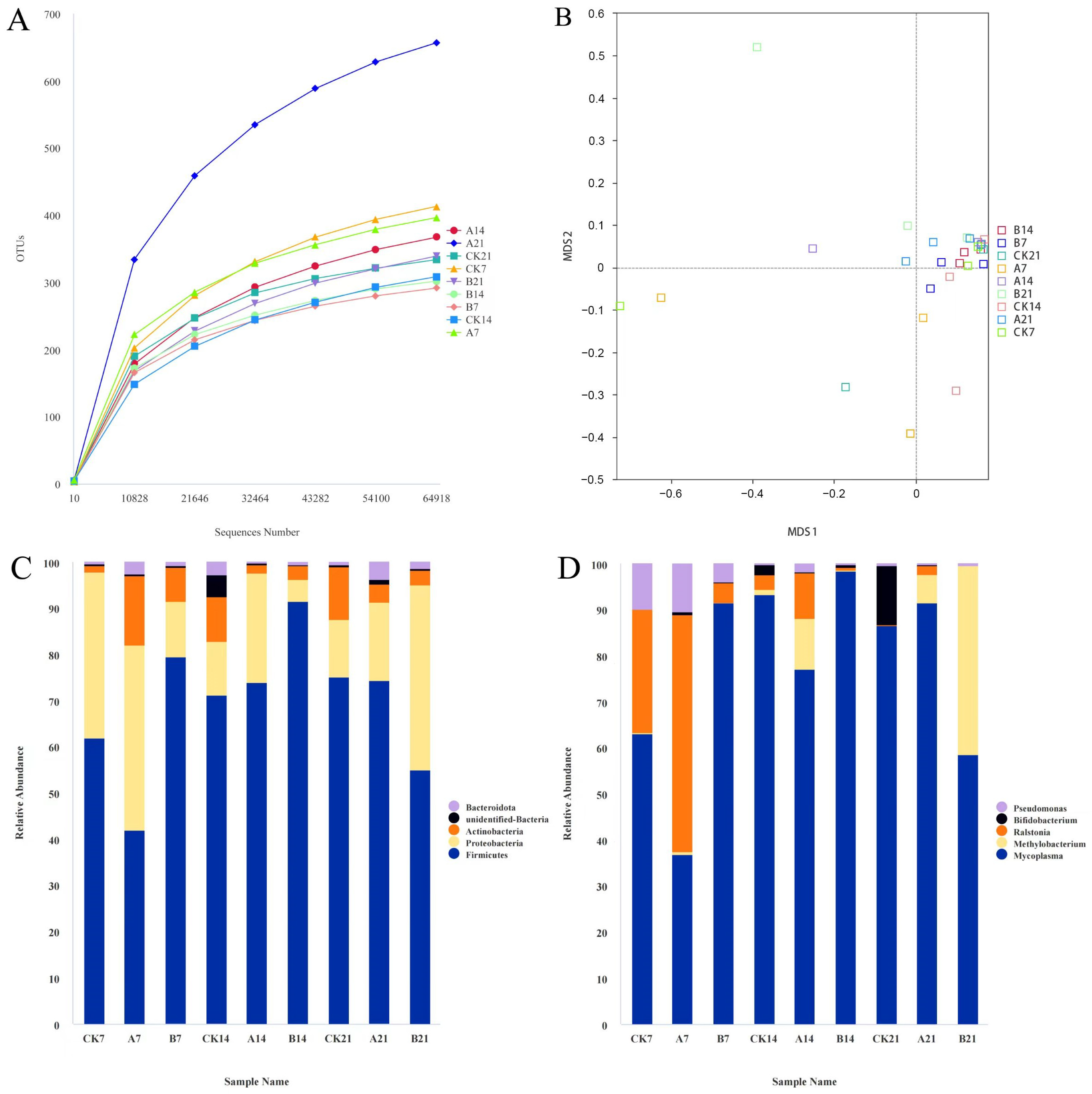
3.5. GX118 Affected Gut Microbiota Diversity and Composition

The sparsity curve indicated that the saturation of sequencing data meets the analysis requirements (Figure 5A). NMDS analysis on the basis of Bray–Curtis similarity distance indicated that the clustering of the experimental groups was much closer (Figure 5B). The number of OTUs in group A21 was much higher than that in groups CK0, CK21, and B21. The Chao1 and ACE indices of the gut microbiota in each group showed a downward trend on the 7th day, and then an increase on the 14th day; Shannon and Simpson, in all treatment groups, showed a trend of decreasing on the 7th day and then increasing on the 14th day (see in Supplementary Materials, Table S2).
The dominant phyla were Firmicutes, Proteobacteria, Actinobacteria, Bacteroidota, and Cyanobacteria. At the phylum level, the abundance of Firmicutes in the B treatment group was higher than that in the CK group on day 7 and day 14, while it was lower than that in the control group on day 21. The abundance of Firmicutes in the A treatment group was significantly more decreased than that in the CK group on day 7 and then returned to the level of the CK group on day 14 and 21. Compared to the CK group, the abundance of Proteobacteria was significantly decreased on day 7 and 14, while it significantly increased on day 21 in the B treatment group; moreover, it increased on day 14 and seems to not have changed much change on day 7 and 21 in the A treatment group. Compared to the control group, the abundance of Actinobacteria increased on day 7, while it decreased on day 14 and day 21. The abundance of Bacteroidota increased in groups A (2.36%) and B (0.72%) on the 7th day, as well as in groups A (3.67%) and B (1.35%) on the 21st day compared with the that in the control group (0.50%) (Figure 5C).
At the genus level, the dominant bacterial genera were Mycoplasma, Methylobacteria-methylorubrum, Ralstonia, Bifidobacterium, and Pseudomonas. Compared to the control group, experimental groups A and B had a higher abundance of Mycoplasma and Pseudomonas after 14 days, while the abundance of Mycoplasma and Pseudomonas decreased on day 21. The abundance of Ralstonia was observed to be comparatively reduced in group B compared to in the control group CK (Figure 5D).
LEfSe analysis identified taxonomic groups with differential abundance (LDA > 3, p < 0.05) (see in Supplementary Materials, Figure S3). Compared to the control group, bacterial communities containing more potential beneficial bacteria, e.g., Bacteroides uniformis, Bifidobacteriales, and Lactobacillales, increased their abundance in the A and B groups on day 7. The abundance of potentially harmful bacteria, e.g., Camcylobacter, Desulfovibrio, and Haemophius, decreased in the A and B groups on day 7. On day 21, bacterial communities containing more potentially beneficial bacteria, such as Anaerolineae, Blautia, Lachnospiraceae, and massllia, showed an increased abundance in the A and B groups, while the abundance of bacterial communities containing more potentially harmful bacteria, such as Sainivibrio, Rickettsiales, and Escherichia Shigella, decreased.
The metabolite pathways of gut microbiota were predicated using PICRUSt. The dietary supplement of GX118 up-regulated a serious of metabolic pathways in the gut microbiota. In particular, amino acid metabolism, the immune system, and transcription were up-regulated in the A group on day 7; nucleotide metabolism and environmental_ adaptation were up-regulated in the B group on day 14; and the metabolism of other amino acids, cell motility and transport, and catabolism were up-regulated in the B group on day 21 (see in Supplementary Materials, Figure S4).

3.6. Expression of Immune-Related Genes

The relative expression levels of immune-related genes, IL-6, IL-8, TNF-α, IFN-γ, and NF-κB, in the kidney of rainbow trout were analyzed. After a 21-day feeding period, group B exhibited a notable reduction in the expression of inflammatory cytokines IL-6 and IL-8 when compared to that of the control group. Group A exhibited a notable decrease in the expression of inflammatory factors IL-8 compared to that of the control group. A notable enhancement in the expression of immune genes was observed in group A compared to in the control group, particularly of NF-κB and IFN-γ (Figure 6).

4. Discussion

Probiotics have been shown to mitigate the effects of diseases by directly inhibiting or enhancing colonization resistance against pathogens [14]. Research has confirmed that LAB strains sourced from fish and shrimp can produce bacteriocins, thereby achieving inhibitory effects on pathogens [15,16]. Ramos et al. found that probiotics within the gut microbiota enhanced the disease resistance of fish either by stimulating immunity or competitively excluding pathogens [17]. In this study, L. salivarius GX118 exhibited varying levels of efficacy in inhibiting A. salmonicida, both in vivo and in vitro. In particular, bacteriocin-like polypeptide sequence Enterolysin A was found in GX118, belonging to class III lactobacilli bacteriocins and being a thermosensitive macromolecular protein. In a previous study, Enterolysin A was shown to be able to prevent various diseases by destroying cell walls to kill sensitive strains of bacteria in vitro, such as Enterococci, Lactococci, and Pediococci [18]. It is widely recognized that the primary function of the bacterial cell wall is to safeguard and maintain their characteristic structure. Any disruption to this vital component can lead to the release of cellular contents, ultimately resulting in bacterial lysis. In this study, the GX118-supernatant-induced cell wall damage in A. salmonicida resulted in a gradual depletion of cellular contents over time and ultimately led to a complete bacterial rupture. The amino acid sequence of Enterolysin A was analyzed indicating its potential role as a membrane-associated protein. Currently, studies on L. salivarius mainly focus on higher animals, for instance, L. salivarius 7247 has been found to have high antagonism against Salmonella enteritidis and Salmonella typhimurium in the intestine of humans, pigs, and chicken; moreover, L. salivarius UCC118 can regulate the immune function of epithelial cells by persistent colonization in the human intestine [19,20]. Based on our study, we speculated that Enterolysin A may be a key factor involved in the antimicrobial activity of GX118. GX118 may secrete Enterolysin A to antagonize A. salmonicida and gains a competitive colonization advantage in the intestine. Moreover, colonizing GX118 in the intestine may continue to secrete bacteriocins, further inhibiting the colonization of pathogenic bacteria. Interestingly, other studies found that strain L. salivarius UCC118™ secrets bacteriocins of abp118 that can alleviate colitis by affecting gut microbes in mice [21]. However, we did not find abp118 in the GX118 strain. Apparently, further validation of the action of Enterolysin A is needed through methods such as synthetic, heterologous expression, or gene knockout.
Probiotics can affect the gut microbiota balance by improving the gut of hosts [22]. For example, bacteriocin-producing bacterium Lactiplantibacillus plantarum YRL45 was found to activate immunity by increasing the abundance of beneficial bacteria in mice [23]. Enterococcus sp. MA176 and E. thalandicus MA122 are expected to become potential drugs for the prevention of Yersiniosis infection in salmonids [24]. Lactococcus lactis subsp. cremoris WA2-67 can protect rainbow trout from Lactococcus garvieae infection [25,26,27]. In this study, besides antagonizing pathogens, the use of GX118 significantly improved the growth performance of rainbow trout, indicating that GX118 improved the feed efficiency of rainbow trout, which may be related to changes in intestinal barrier function. Previous studies have shown that the “competitive exclusion” effect mechanism facilitates the effective inhibition of pathogenic or detrimental bacterial growth in the intestines by short-chain fatty acids (SCFAs) produced by the gut microbiota within the human body [28]. Ke et al. (2021) showed that the supplementation of Nisin in carp feed induced alterations in both the diversity and abundance of intestinal microbiota, leading to significant anti-inflammatory effects [29]. In this study, among the core microbial communities, Firmicutes and Bacteroidetes accounted for over 90% of the gut microbiota. It was observed that both treatment groups exhibited a significantly increased abundance of Bacteroidota in comparison to that of the control group. Previous studies have found that the ratio of Firmicutes to Bacteroidetes (F/B) is a marker of intestinal health associated with the dysbiosis of gut microbiota, and an increase in this proportion means inflammatory bowel disease and also influences changes in the production of short-chain fatty acids. In this study, the F/B ratio in the treatment groups decreased with the increased diversity and richness of gut microbiota. The Bacteroidota species present in the human gastrointestinal tract can expedite angiogenesis within the intestinal mucosa, enhance immune function, and maintain ecological equilibrium within the intestines [30]. Studying the fish found that Bacteroidota plays a pivotal role in augmenting nutrient assimilation and facilitating weight gain in largemouth bass [31]. Following a Lefse analysis, it was observed that the intestinal tracts of rainbow trout in groups A and B exhibited significantly higher levels of Blautia (belonging to Firmicutes) and Rhodobacteraceae (belonging to Proteobacteria) compared to those in the control group. Blautia is shown to produce short-chain fatty acids in European sea bass (Dicentrarchus labrax) fish intestines. The bacteria Rhodobacteraceae possess the capability to synthesize a broad-spectrum antibacterial compound such as tropodithietic acid (TDA), which plays a pivotal role in the prevention of fish diseases [32]. In addition, the use of GX118 activated pathways of the gut microbiota such as the metabolism of cofactors and vitamins, glycan biosynthesis, and amino acid metabolism biosynthesis, as well as cell growth and death and other pathways, thus improving the growth of fish. The innate immune system of fish assists in safeguarding the host against inflammatory reactions triggered by pathogen infiltration [33]. The transcription factor NF-κB, which is ubiquitously present in the nucleus, can be activated through multiple signaling pathways and plays a crucial role in diverse immune regulatory processes [34]. L. Salivarius has been demonstrated to down-regulate pro-inflammatory cells and inhibits NF-κB, thereby exerting anti-inflammatory effects in the gut of piglets [35]. Lactobacillus reuteri I5007 exhibits the potential to augment the presence of intestinal proteins and mitigate inflammation induced by lipopolysaccharide (LPS) in the gastrointestinal tract of neonatal piglets [36]. In this study, the inclusion of GX118 in diet resulted in a significant reduction in the expression of pro-inflammatory immune genes IL-6 and IL-8, while increasing levels of IFN-γ and NF-κB in the kidney of rainbow trout. Additionally, KEGG analysis revealed a notable up-regulation of immune-related pathways such as cell motility, cellular processes and signaling, and cell growth and death. Previous studies have demonstrated the potential of Lactobacillus acidophilus and Bifidobacterium animalis subsp to effectively reduce IL-8 expression within the human system, indicating their anti-inflammatory properties [37]. Previous research has indicated that the inclusion of thymol in the diet of rainbow trout leads to a decrease in IL-8 expression [38]. The incorporation of grape seed extract (GSE) into the rainbow trout’s diet resulted in enhanced growth performance and augmented mucosal immunity by up-regulating immune-related genes, particularly increasing the expression of the IFN-γ gene [39]. The administration of diverse probiotic strains can effectively mitigate pro-inflammatory factors in animal organisms, thereby yielding favorable outcomes in the reduction in inflammation [40]. It is plausible to hypothesize that the supplementation of L. salivarius GX118 has the potential to augment the innate immune defense system in fish. However, further research is imperative to authenticate and comprehend the precise mechanisms responsible for modulating immune responses.

5. Conclusions

We found that GX118 can secrete peptide Enterolysin A, which may be the key factor in disrupting the cell walls of A. salmonicida and inhibiting its colonization in the intestine of rainbow trout. Dietary supplementation of GX118 improved the growth performance of rainbow trout by altering the composition and function of gut microbiota, with an increased abundance of beneficial bacteria such as Bacteroidota, Blautia, Rhodobacteraceae and a decreased abundance of harmful bacteria such as Ralstonia, Pseudomonas, and Mycoplasma. In addition, the use of GX118 can stimulate the NF-κB immune-related pathways, thereby enhancing the ability of rainbow trout to resist pathogen infection.

Supplementary Materials

The following supporting information can be downloaded at https://www.mdpi.com/article/10.3390/fishes9050157/s1, Figure S1: GX118 Bacteriocin characteristics. Figure S2: Gene function annotation of GX1118. Figure S3: LEfSe analysis for differential abundant microbiota. Figure S4: KEGG pathway of gut microbiota predicted by Tax4Fun analysis (First 35 predicted pathways)., Table S1: Assembly of L. salivarius GX118 genome., Table S2: The statistical estimates of alpha diversity of gut microbiota.

Author Contributions

Conceptualization, Y.R. and X.C.; methodology, X.C. and B.W.; software, B.W.; formal analysis, W.R. and J.W.; investigation, X.C., B.W. and X.M.; resources, Y.R.; data curation, Q.L.; writing—original draft preparation, X.C.; writing—review and editing, X.C., B.W., Y.R. and X.M.; visualization, X.C.; supervision, Y.R. and X.M.; project administration, Y.R.; funding acquisition, Y.R. All authors have read and agreed to the published version of the manuscript.

Funding

This research was funded by Key R&D Program of Shandong Province (NO. 2021LZGC027); Shandong Provincial Natural Science Foundation (NO. ZR202102250235); Major Agricultural Application Technology Innovation Projects in Shandong Province (NO. SD2019YY006); and “First class fishery discipline” program in Shandong Province, China.

Institutional Review Board Statement

The study was approved by the Experimental Animal Ethics Committee of Qingdao Agricultural University (approval code: 2022-066), and the procedures were performed strictly according to the Management Rule of Laboratory Animals (Chinese order no. 676 of the State Council, revised on 1 March 2017).

Informed Consent Statement

Not applicable.

Data Availability Statement

Data will be made available on request.

Conflicts of Interest

The authors declare no conflicts of interest.

References

  1. Chen, J.; Sun, R.; Pan, C.; Sun, Y.; Mai, B.; Li, Q.X. Antibiotics and Food Safety in Aquaculture. J. Agric. Food Chem. 2020, 68, 11908–11919. [Google Scholar] [CrossRef]
  2. Kakakhel, M.A.; Narwal, N.; Kataria, N.; Johari, S.A.; Zaheer Ud Din, S.; Jiang, Z.; Khoo, K.S.; Xiaotao, S. Deciphering the dysbiosis caused in the fish microbiota by emerging contaminants and its mitigation strategies—A review. Environ. Res. 2023, 237, 117002. [Google Scholar] [CrossRef]
  3. Zhao, C.; Men, X.; Dang, Y.; Zhou, Y.; Ren, Y. Probiotics Mediate Intestinal Microbiome and Microbiota-Derived Metabolites Regulating the Growth and Immunity of Rainbow Trout (Oncorhynchus mykiss). Microbiol. Spectr. 2023, 11, e0398022. [Google Scholar] [CrossRef]
  4. Amoah, K.; Tan, B.; Zhang, S.; Chi, S.; Yang, Q.; Liu, H.; Yang, Y.; Zhang, H.; Dong, X. Host gut-derived Bacillus probiotics supplementation improves growth performance, serum and liver immunity, gut health, and resistive capacity against Vibrio harveyi infection in hybrid grouper (female symbol Epinephelus fuscoguttatus × male symbol Epinephelus lanceolatus). Anim. Nutr. 2023, 14, 163–184. [Google Scholar] [CrossRef]
  5. Alonso, S.; Carmen Castro, M.; Berdasco, M.; de la Banda, I.G.; Moreno-Ventas, X.; de Rojas, A.H. Isolation and Partial Characterization of Lactic Acid Bacteria from the Gut Microbiota of Marine Fishes for Potential Application as Probiotics in Aquaculture. Probiotics Antimicrob. Proteins 2019, 11, 569–579. [Google Scholar] [CrossRef]
  6. Hashim, H. Bacteriocin: The avenues of innovation towards applied microbiology. Pure Appl. Biol. 2018, 7, 460–478. [Google Scholar] [CrossRef]
  7. Juturu, V.; Wu, J.C. Microbial production of bacteriocins: Latest research development and applications. Biotechnol. Adv. 2018, 36, 2187–2200. [Google Scholar] [CrossRef]
  8. Shan, C.; Wu, H.; Zhu, Y.; Zhou, J.; Yan, W.; Jianhao, Z.; Liu, X. Preservative effects of a novel bacteriocin from Lactobacillus panis C-M2 combined with dielectric barrier discharged cold plasma (DBD-CP) on acquatic foods. Food Sci. Technol. Int. 2023, 29, 406–416. [Google Scholar] [CrossRef]
  9. Selle, K.; Klaenhammer, T.R. Genomic and phenotypic evidence for probiotic influences of Lactobacillus gasseri on human health. FEMS Microbiol. Rev. 2013, 37, 915–935. [Google Scholar] [CrossRef]
  10. Daba, G.M.; Elkhateeb, W.A. Ribosomally synthesized bacteriocins of lactic acid bacteria: Simplicity yet having wide potentials—A review. Int. J. Biol. Macromol. 2024, 256, 128325. [Google Scholar] [CrossRef] [PubMed]
  11. Xiang, F.; Xu, G.; Gu, W.; Huang, T.; Liu, C.; Wang, B. Expression of cytochrome genes during the early gonadal development in triploid female rainbow trout Oncorhynchus mykiss. J. Fish. Sci. China 2018, 25, 949. [Google Scholar] [CrossRef]
  12. Duman, M.; Altun, S.; Saticioglu, I.B.; Romalde, J.L. A review of bacterial disease outbreaks in rainbow trout (Oncorhynchus mykiss) reported from 2010 to 2022. J. Fish Dis. 2023, 00, 1–17. [Google Scholar] [CrossRef] [PubMed]
  13. Everson, J.L.; Jones, D.R.; Taylor, A.K.; Rutan, B.J.; Leeds, T.D.; Langwig, K.E.; Wargo, A.R.; Wiens, G.D. Aquaculture Reuse Water, Genetic Line, and Vaccination Affect Rainbow Trout (Oncorhynchus mykiss) Disease Susceptibility and Infection Dynamics. Front. Immunol. 2021, 12, 721048. [Google Scholar] [CrossRef] [PubMed]
  14. Ma, T.; Shen, X.; Shi, X.; Sakandar, H.A.; Quan, K.; Li, Y.; Jin, H.; Kwok, L.-Y.; Zhang, H.; Sun, Z. Targeting gut microbiota and metabolism as the major probiotic mechanism—An evidence-based review. Trends Food Sci. Technol. 2023, 138, 178–198. [Google Scholar] [CrossRef]
  15. Darbandi, A.; Asadi, A.; Mahdizade Ari, M.; Ohadi, E.; Talebi, M.; Halaj Zadeh, M.; Darb Emamie, A.; Ghanavati, R.; Kakanj, M. Bacteriocins: Properties and potential use as antimicrobials. J. Clin. Lab. Anal. 2022, 36, e24093. [Google Scholar] [CrossRef] [PubMed]
  16. Pérez-Sánchez, T.; Ruiz-Zarzuela, I.; de Blas, I.; Balcázar, J.L. Probiotics in aquaculture: A current assessment. Rev. Aquac. 2013, 6, 133–146. [Google Scholar] [CrossRef]
  17. Ramos, M.A.; Weber, B.; Goncalves, J.F.; Santos, G.A.; Rema, P.; Ozorio, R.O. Dietary probiotic supplementation modulated gut microbiota and improved growth of juvenile rainbow trout (Oncorhynchus mykiss). Comp. Biochem. Physiol. A Mol. Integr. Physiol. 2013, 166, 302–307. [Google Scholar] [CrossRef]
  18. Khan, H.; Flint, S.H.; Yu, P.L. Determination of the mode of action of enterolysin A, produced by Enterococcus faecalis B9510. J. Appl. Microbiol. 2013, 115, 484–494. [Google Scholar] [CrossRef]
  19. Abramov, V.M.; Kosarev, I.V.; Machulin, A.V.; Deryusheva, E.I.; Priputnevich, T.V.; Panin, A.N.; Chikileva, I.O.; Abashina, T.N.; Manoyan, A.M.; Ahmetzyanova, A.A.; et al. Ligilactobacillus salivarius 7247 Strain: Probiotic Properties and Anti-Salmonella Effect with Prebiotics. Antibiotics 2023, 12, 1535. [Google Scholar] [CrossRef]
  20. O’Callaghan, J.; Butto, L.F.; MacSharry, J.; Nally, K.; O’Toole, P.W. Influence of adhesion and bacteriocin production by Lactobacillus salivarius on the intestinal epithelial cell transcriptional response. Appl. Environ. Microbiol. 2012, 78, 5196–5203. [Google Scholar] [CrossRef]
  21. Iyer, N.; Williams, M.A.; O‘Callaghan, A.A.; Dempsey, E.; Cabrera-Rubio, R.; Raverdeau, M.; Crispie, F.; Cotter, P.D.; Corr, S.C. Lactobacillus salivarius UCC118 Dampens Inflammation and Promotes Microbiota Recovery to Provide Therapeutic Benefit in a DSS-Induced Colitis Model. Microorganisms 2022, 10, 1383. [Google Scholar] [CrossRef] [PubMed]
  22. Kumar, A. Probiotics: Nature′s medicine. Int. J. Nutr. Pharmacol. Neurol. Dis. 2013, 3, 219–228. [Google Scholar] [CrossRef]
  23. Bu, Y.; Liu, Y.; Zhang, T.; Liu, Y.; Zhang, Z.; Yi, H. Bacteriocin-Producing Lactiplantibacillus plantarum YRL45 Enhances Intestinal Immunity and Regulates Gut Microbiota in Mice. Nutrients 2023, 15, 3437. [Google Scholar] [CrossRef]
  24. Amin, M.; Adams, M.B.; Burke, C.M.; Bolch, C.J.S. Screening and activity of potential gastrointestinal probiotic lactic acid bacteria against Yersinia ruckeri O1b. J. Fish. Dis. 2023, 46, 369–379. [Google Scholar] [CrossRef]
  25. Wei, S.; Bahl, M.I.; Baunwall, S.M.D.; Hvas, C.L.; Licht, T.R. Determining Gut Microbial Dysbiosis: A Review of Applied Indexes for Assessment of Intestinal Microbiota Imbalances. Appl. Environ. Microbiol. 2021, 87, e00395-21. [Google Scholar] [CrossRef] [PubMed]
  26. Luo, X.; Kong, Q.; Wang, Y.; Duan, X.; Wang, P.; Li, C.; Huan, Y. Colonization of Clostridium butyricum in Rats and Its Effect on Intestinal Microbial Composition. Microorganisms 2021, 9, 1573. [Google Scholar] [CrossRef]
  27. Kapoor, B.; Gulati, M.; Gupta, R.; Singla, R.K. Microbiota dysbiosis and myasthenia gravis: Do all roads lead to Rome? Autoimmun. Rev. 2023, 22, 103313. [Google Scholar] [CrossRef] [PubMed]
  28. Zhao, L.; Zhang, F.; Ding, X.; Wu, G.; Lam, Y.Y.; Wang, X.; Fu, H.; Xue, X.; Lu, C.; Ma, J.; et al. Gut bacteria selectively promoted by dietary fibers alleviate type 2 diabetes. Science 2018, 359, 1151–1156. [Google Scholar] [CrossRef] [PubMed]
  29. Ke, F.; Xie, P.; Yang, Y.; Yan, L.; Guo, A.; Yang, J.; Zhang, J.; Liu, L.; Wang, Q.; Gao, X. Effects of Nisin, Cecropin, and Penthorum chinense Pursh on the Intestinal Microbiome of Common Carp (Cyprinus carpio). Front. Nutr. 2021, 8, 729437. [Google Scholar] [CrossRef]
  30. Cheng, J.; Hu, J.; Geng, F.; Nie, S. Bacteroides utilization for dietary polysaccharides and their beneficial effects on gut health. Food Sci. Human Wellness 2022, 11, 1101–1110. [Google Scholar] [CrossRef]
  31. Fan, Z.; Wu, D.; Li, J.; Zhang, Y.; Cui, Z.; Li, T.; Zheng, X.; Liu, H.; Wang, L.; Li, H. Assessment of Fish Protein Hydrolysates in Juvenile Largemouth Bass (Micropterus salmoides) Diets: Effect on Growth, Intestinal Antioxidant Status, Immunity, and Microflora. Front. Nutr. 2022, 9, 816341. [Google Scholar] [CrossRef] [PubMed]
  32. Henriksen, N.; Lindqvist, L.L.; Wibowo, M.; Sonnenschein, E.C.; Bentzon-Tilia, M.; Gram, L. Role is in the eye of the beholder-the multiple functions of the antibacterial compound tropodithietic acid produced by marine Rhodobacteraceae. FEMS Microbiol. Rev. 2022, 46, fuac007. [Google Scholar] [CrossRef] [PubMed]
  33. Chu, Q.; Bi, D.; Zheng, W.; Xu, T. MicroRNA negatively regulates NF-kappaB-mediated immune responses by targeting NOD1 in the teleost fish Miichthys miiuy. Sci. China Life Sci. 2021, 64, 803–815. [Google Scholar] [CrossRef] [PubMed]
  34. Barnabei, L.; Laplantine, E.; Mbongo, W.; Rieux-Laucat, F.; Weil, R. NF-kappaB: At the Borders of Autoimmunity and Inflammation. Front. Immunol. 2021, 12, 716469. [Google Scholar] [CrossRef] [PubMed]
  35. Qiao, J.; Sun, Z.; Liang, D.; Li, H. Lactobacillus salivarius alleviates inflammation via NF-kappaB signaling in ETEC K88-induced IPEC-J2 cells. J. Anim. Sci. Biotechnol. 2020, 11, 76. [Google Scholar] [CrossRef] [PubMed]
  36. Yang, F.; Wang, A.; Zeng, X.; Hou, C.; Liu, H.; Qiao, S. Lactobacillus reuteri I5007 modulates tight junction protein expression in IPEC-J2 cells with LPS stimulation and in newborn piglets under normal conditions. BMC Microbiol. 2015, 15, 32. [Google Scholar] [CrossRef] [PubMed]
  37. Li, S.C.; Hsu, W.F.; Chang, J.S.; Shih, C.K. Combination of Lactobacillus acidophilus and Bifidobacterium animalis subsp. lactis Shows a Stronger Anti-Inflammatory Effect than Individual Strains in HT-29 Cells. Nutrients 2019, 11, 969. [Google Scholar] [CrossRef]
  38. Hafsan, H.; Saleh, M.M.; Zabibah, R.S.; Obaid, R.F.; Jabbar, H.S.; Mustafa, Y.F.; Sultan, M.Q.; Gabr, G.A.; Ramirez-Coronel, A.A.; Khodadadi, M.; et al. Dietary Thymol Improved Growth, Body Composition, Digestive Enzyme Activities, Hematology, Immunity, Antioxidant Defense, and Resistance to Streptococcus iniae in the Rainbow Trout (Oncorhynchus mykiss). Aquac. Nutr. 2022, 2022, 3288139. [Google Scholar] [CrossRef] [PubMed]
  39. Mousavi, S.; Sheikhzadeh, N.; Hamidian, G.; Mardani, K.; Oushani, A.K.; Firouzamandi, M.; Esteban, M.A.; Shohreh, P. Changes in rainbow trout (Oncorhynchus mykiss) growth and mucosal immune parameters after dietary administration of grape (Vitis vinifera) seed extract. Fish. Physiol. Biochem. 2021, 47, 547–563. [Google Scholar] [CrossRef]
  40. Cristofori, F.; Dargenio, V.N.; Dargenio, C.; Miniello, V.L.; Barone, M.; Francavilla, R. Anti-Inflammatory and Immunomodulatory Effects of Probiotics in Gut Inflammation: A Door to the Body. Front. Immunol. 2021, 12, 578386. [Google Scholar] [CrossRef]
Figure 1. Antibacterial effect of GX118 on A. salmonicida. (A): antibacterial zone; (B): growth curve by OD value. CK: control group; H, M, and L: growth of A. salmonicida co-culture with GX118 at a concentration of 1 × 10⁹ cfu/mL, 1 × 10⁷ cfu/mL, and 1 × 10⁵ cfu/mL, respectively.
Fishes 09 00157 g001
Figure 2. Electron microscopy observations on cell damage of A. salmonicida. (A,B): control. (C,D): co-culture of GX118 and A. salmonicida for 2 h; (E,F): co-culture of GX118 and A. salmonicida for 4 h; (G,H): co-culture of GX118 and A. salmonicida for 6 h.
Fishes 09 00157 g002
Figure 3. Fluorescence observations of intestinal sections. (A): control; (BD): immersion infection of A. salmonicida for 6 h, 24 h, and 48 h, respectively; (E,F): the use of GX118 after immersion of A. salmonicida for 12 h and 48 h.
Fishes 09 00157 g003
Figure 4. Survival rate of rainbow trout after A. salmonicida challenge. CK: control group; A: supplemented with 1 × 10⁷ cfu/mL GX118; B: supplemented with 1 × 10⁹ cfu/mL GX118.
Fishes 09 00157 g004
Figure 5. Gut microbiota diversity and taxonomic classification of rainbow trout. (A): rarefaction curves of 16S rRNA gene sequencing; (B): sparse curve based on sequential data; (C): stacked bar chart of gut bacteria on the phylum level classification; (D): stacked bar chart of gut bacteria on the genus level classification. A7, A14 and A21: supplemented with 1 × 10⁷ cfu/mL GX118 for 7 days, 14 days, and 21 days, respectively; B7, B14 and B21: supplemented with 1 × 10⁹ cfu/mL GX118 for 7 days, 14 days, and 21 days, respectively; CK7, CK14 and CK21: control.
Fishes 09 00157 g005
Figure 6. Relative expression levels of immunological genes in the kidney of rainbow trout at 21 days of the test. Lowercase letters represent significant differences at p < 0.05.
Fishes 09 00157 g006
Table 1. Primers for gene expression.
GenesPrimer DirectionPrimer Sequences (5′–3′)
IL-6FACTCCCCTCTGTCACACACC
RGGCAGACAGGTCCTCCACTA
IL-8FAGAATGTCAGCCAGCCTTGT
RTCTCAGACTCATCCCCTCAGT
TNF-α-FGGGGACAAACTGTGGACTGA
RGAAGTTCTTGCCCTGCTCTG
IFN-γFAAGGGCTGTGATGTGTTTCTG
RTGTACTGAGCGGCATTACTCC
NF-κBFCAGCGCAGAGAACAACGAAT
RCCGAAGACAGGCTCAGGTAG
ef1αFCATGCCTGGTGACAATGTTGG
RTGGGGGCATCCTCAAGTTTC
Table 2. Growth performance of rainbow trout. BWI: initial body weight; BWF: final body weight; WGR: weight gain rate; SGR: specific growth rate. Superscript lowercase letters represent significant differences among different groups at p < 0.05.
SampleBWI (g)BWF (g)WGR (g)SGR (%/d)
CK7.02 ± 0.349.33 ± 0.802.26 ± 0.35 a1.28 ± 0.15 a
A7.53 ± 0.4810.23 ± 0.872.70 ± 0.46 b1.45 ± 0.16 b
B7.14 ± 0.379.46 ± 0.422.32 ± 0.15 a1.34 ± 0.09 a
Disclaimer/Publisher’s Note: The statements, opinions and data contained in all publications are solely those of the individual author(s) and contributor(s) and not of MDPI and/or the editor(s). MDPI and/or the editor(s) disclaim responsibility for any injury to people or property resulting from any ideas, methods, instructions or products referred to in the content.

