Communication

Morphological Development and DNA Barcoding Identification of Pholis fangi Larvae and Juveniles in the Yellow Sea

1
East China Sea Ecology Center, Ministry of Natural Resources, Shanghai 201206, China
2
Key Laboratory of Marine Ecological Monitoring and Restoration Technologies, Ministry of Natural Resources, Shanghai 201206, China
3
College of Fisheries and Life Sciences, Shanghai Ocean University, Shanghai 201306, China
4
East China Sea Investigating Center, Ministry of Natural Resources, Shanghai 200137, China
5
Taihu Basin & East China Sea Ecological Environment Supervision and Administration Authority, Ministry of Ecology and Environment, Shanghai 200125, China
*
Author to whom correspondence should be addressed.
These authors contributed equally to this work.
Fishes 2024, 9(6), 213; https://doi.org/10.3390/fishes9060213
Submission received: 26 March 2024 / Revised: 23 May 2024 / Accepted: 27 May 2024 / Published: 3 June 2024

Abstract

:
Pholis fangi is a small, bottom-dwelling fish species endemic to the Yellow Sea and Bohai Sea in China. While previous research has focused on its population biology and molecular structure, little is known about the early developmental stages of this species. In this study, larval and juvenile P. fangi specimens were collected from the Sheyang Sea Area, Jiangsu Province, in 2017. Morphological features were examined using microscopy, and DNA barcoding was conducted to confirm species identification. The research documented detailed changes in yolk sac, fin development, and melanophore distribution patterns across larval and juvenile stages of P. fangi. Comparative analysis with other Pholis species revealed that melanophore distribution is a key distinguishing characteristic, allowing effective differentiation between larval and juvenile stages, as well as between Pholis species. This study provides valuable insights into the early life history of P. fangi, contributing to a better understanding of the genus Pholis. The findings demonstrate the utility of combining traditional morphological observation and molecular techniques for accurate species identification, particularly during the critical larval and juvenile developmental phases.
Key Contribution: This study presents a comprehensive investigation into the early growth stages of Pholis fangi, providing detailed insights into its morphological development and DNA barcoding identification. A significant breakthrough is the discovery of melanophores as unique markers, facilitating effective differentiation between larval and juvenile stages and among Pholis species. These findings deepen our understanding of P. fangi’s early life history and underscore the importance of integrating traditional morphological observation with molecular techniques for accurate species identification during crucial developmental phases.

1. Introduction

Pholis fangi (Wang and Wang, 1935), classified within the genus Pholis Scopoli, 1777, belongs to the order Perciformes, suborder Zoarcoidei, and family Pholidae. It is a small-sized, bottom-dwelling species found in offshore waters, known for its tolerance to both cold and warm temperatures. P. fangi exhibits seasonal migrations along banks. Endemic to China [1], P. fangi is found exclusively in the Yellow Sea and Bohai Sea [2]. Research on P. fangi has primarily targeted its population biology [1,3,4,5,6,7] and molecular structure [8,9], with scant attention to its early developmental stages [10]. Wan and Zhang [10] were the only researchers to describe the larval and juvenile morphologies of Enedrias fangi in Rushan Bay, Yellow Sea, including details on post-larvae (4.50, 9.00, and 21.45 mm) and juveniles (24.60 and 28.00 mm). Kunihiko and Kunio [11] classified the larvae and juvenile of P. nebulosa (Temminck and Schlegel, 1845) in waters off Hokkaido’s south bank into Type I and Type II. Okiyama [12] detailed the morphologies of P. nebulosa (19.2–40.5 mm), P. crassispina (Temminck and Schlegel, 1845) (10.8–35.0 mm), and P. ornata (Girard, 1854) (27.3 and 33.7 mm) in Japan’s offshore areas.
Three traditional morphological methods—artificial insemination, dynamic research, and static research—are used to identify early developmental changes in fish [13]. Due to limited information, traditional morphological identification restricts not only species identification but also the understanding of larval and juvenile stages in many fish species. Molecular technology overcomes the bottleneck in larval and juvenile species identification [14,15,16,17,18], enabling a broader understanding of species diversity. This study combined DNA barcoding with dynamic research methods to identify species at various stages and sizes. We compared the developmental differences and relevant morphological features of P. fangi larvae and juveniles across various stages. Consequently, we conducted a comparative study of the morphological features of Pholis genus larvae and juveniles to gather data and offer insights for species identification in early developmental stages.

2. Materials and Methods

In April 2017, larvae and juvenile samples were collected from the Sheyang Sea Area (120.58°–121.45°E, 33.35°–33.92°N) in Jiangsu Province. The collection sites are depicted in Figure 1. Following the Specifications for Oceanographic Survey (GB/T 12763.6-2007) [19], we employed both a macroplankton net (280 cm in length, 80 cm inner mesh diameter, 0.5 m2 opening area, 0.505 mm mesh) and a shallow water I-type plankton net (145 cm in length, 50 cm inner mesh diameter, 0.2 m2 opening area, 0.505 mm mesh) for horizontal and vertical dragging at the stations. Samples were stored in absolute ethyl alcohol.
In the laboratory, the morphological features (e.g., yolk sac changes, development of fins, and melanophore distribution) of the collected larvae and juvenile samples were observed using a stereoscopic microscope (Nikon SMZ 25). Meanwhile, preliminary clusters and species identifications were carried out [12,13]. Photographs were captured using NIS-Elements D software, and the body length, total length, head length, eye diameter, and anterior anal distance were measured.
DNA barcode analysis: Entire larvae of small body sizes were collected, while only the tails of larger larvae were sampled. Around 100 mg of muscle tissue was harvested to extract total DNA using the Marine Animal Tissue Genome DNA Extraction Kit (Beijing Tiangen Biochemical Technology Co., Ltd., Beijing, China). Universal primers F1: 5′-TCRACYAAYCAYAAAGAYATYGGCAC-3′ and R1: 5′-TAGACTTCWGGGTGRCCRAAGAATCA-3′ facilitated the amplification of the COI sequence from fish mitochondrial DNA. The PCR reaction mixture totaled 50 μL, comprising 5 μL of 10× PCR buffer, 4 μL of dNTPs (2.5 mmol/L), 2 μL ofeach primer (10 mmol/L), 0.8 μL (5 U/μL) of Taq DNA polymerase, 1 μL of template DNA, and 35.2 μL of distilled water. Amplification was performed on an AG-22331PCR system (Eppendorf) with the following protocol: an initial denaturation at 94 °C for 5 min; 35 cycles of 94 °C for 30 s, 52 °C for 45 s, and 72 °C for 1 min; followed by a final extension at 72 °C for 10 min, and storage at 4 °C. The amplification products were verified using 1.0% agarose gel electrophoresis and subsequently sequenced by Shanghai Jieli Biotechnology Co., Ltd., Shanghai, China [14,15,16].
The sequences identified, along with those downloaded from GenBank, were aligned using DNAStar. Subsequently, all sequences were compared and aligned Clustal X, with redundant sequences at both ends being removed. Effective COI gene sequences underwent BLAST analysis in both the Barcode of Life Data System (BOLD) database and GenBank databases to facilitate species identification [15]. These sequences were further aligned and edited in MEGA7 software. A phylogenetic tree was then constructed employing the Maximum Likelihood (ML) method, utilizing the general Time Reversible +G+I model. The confidence levels for each branch point were assessed through 1000 bootstrap replicates to ensure robustness. This methodology was polished to meet the standards required for scientific publication [16].

3. Results

3.1. Molecular Identification

In using the COI sequences of 10 Pholidae species obtained from GenBank, along with a COI sequence fragment from one Pholidae species identified in this study, a ML phylogenetic tree was constructed (Figure 2). Lateolabrax japonicas Cuvier, 1828 and Lateolabrax Cuvier, 1828 (Lateolabracidae) served as the out-group. The analysis revealed that the samples clustered with P. fangi with a 100% confidence level.

3.2. Morphological Description of P. fangi

Yolk-sac larva: The body length of the larvae ranges from 5.53 to 5.82 mm (n = 6), with an average total length of 5.73 mm. The yolk sac, not yet fully absorbed, is slender and rod-like, measuring 2.61 mm in length. The head length is 0.44 mm, constituting 8.0% of the body length, while the eye diameter is 0.22 mm, representing 50% of the head length. The lips are blunt and rounded, featuring a small oral opening. The eyes are distinct and contain melanin. A linear arrangement of melanophores stretches from the yolk sac’s abdomen to the anus, totaling approximately 26 melanophores. Additionally, a disordered row of melanophores extends laterally from the center of the yolk sac to the anus. The distance from the anterior end to the anus is 4.14 mm, accounting for 74.9% of the body length. The dorsal fin membrane runs continuously from the head to the tail fin membrane, with the ventral fin membrane positioned slightly lower than the dorsal fin membrane (Figure 3a and Figure 4a).
Preflexion larva: The body length of these stages ranges from 6.22 to 13.74 mm (n = 67). Initially, the total length averages 6.67 mm with a body length of 6.48 mm. At this stage, the yolk has been completely absorbed. The head length measures 0.71 mm, constituting 11.0% of the body length, while the eye diameter is 0.22 mm, making up 31.0% of the head length. The eyes turn black. The mouth, throat, and digestive tract are fully connected, with the digestive tract appearing slender. A linear arrangement of melanophores extends from the digestive tract to the anus. Melanophores on the sides, extending from the center of the former yolk sac to the anus, have disappeared. The anus is located slightly behind the body’s center, with the anterior anal distance being 4.85 mm, accounting for 74.8% of the body length. A dotted black melanophore is observed at the posterior anal margin (Figure 3b and Figure 4b). As the larvae grow, the total length reaches 7.33 mm, and the body length increases to 7.10 mm. The head length is now 0.92 mm, representing 13.0% of the body length. The eye diameter expands to 0.24 mm, constituting 26.1% of the head length. The oral opening becomes larger. The anterior anal distance extends to 5.26 mm, representing 74.1% of the body length. The notochord end remains straight, and fin primordia appear on the tail fin membrane, which forms on the hypural bone. The melanophore distribution is similar to the previous stage (Figure 3c and Figure 4c).
Flexion larva: The total length is 10.65 mm, with the body length at 10.31 mm. The head length is 1.28 mm, representing 12.4% of the body length. The eye diameter is 0.29 mm, making up 22.7% of the head length. The lower jawbone extends slightly beyond the upper jawbone and curves upward. The distance from the anterior end to the anus is 7.87 mm, accounting for 76.3% of the body length. The anus is positioned just behind the midpoint of the body. The notochord end remains straight, and several short fins are visible on the tail fin membrane, which develops on the hypural bone. The distribution of melanophores remains consistent with the previous stage (Figure 3d,e and Figure 4d).
Juvenile: Body length varies from 32.64 to 43.86 mm (n = 182), with a total length of 43.45 mm and a body length of 39.82 mm. The head length measures 3.92 mm, constituting 9.8% of the body length. The eye diameter is 1.41 mm, representing 36% of the head length. The head is proportionate, with fully developed upper and lower jaws. The pectoral fins are fan-shaped, and there is an increase in the number of dorsal and anal fins. Additionally, fins have developed on the superior and anterior lobes of the tail fins. Several small dotted melanophores are present at the lower thoracic margin. A line of melanophores extends from the rear of the digestive tract to the anus, while melanophores at the abdomen of the digestive tract have disappeared. The distance from the anterior end to the anus is 17.53 mm, making up 44.0% of the body length, with the anus shifting slightly forward. Small groups of melanophores are arranged from above the anus to the midline of the body side, extending to the notochord end. A small star-shaped melanophore is located between the two anal fins. Melanophores are also present on the hypural bone, with radiating filaments developed on the lower caudal fin membrane. The visible features are largely similar to those of adult fish, marking the transition into the juvenile developmental stage (Figure 3f and Figure 4e).

3.3. Comparative Study of Pholis Larvae and Juveniles

In drawing on the outcomes of this study and previously documented morphological characteristics of Pholis larvae and juveniles (P. crassispina, P. nebulosa, and P. ornata) [10,12], it is evident that the melanophore distribution and arrangement patterns on the posterior anal fin, abdominal back, and vertebrae exhibit significant variations across different Pholis species during both the larval and juvenile stages. These distinctions allow for the effective differentiation of species within the genus. Notably, the pattern of the melanophore distribution within the same species also shows marked changes from the larval to the juvenile stage. Consequently, the larval and juvenile stages can be categorized as two distinct groups in the identification key. Based on these observations, the following detailed classification feature retrieval was developed.
Features in the Pholis larval stage include the following:
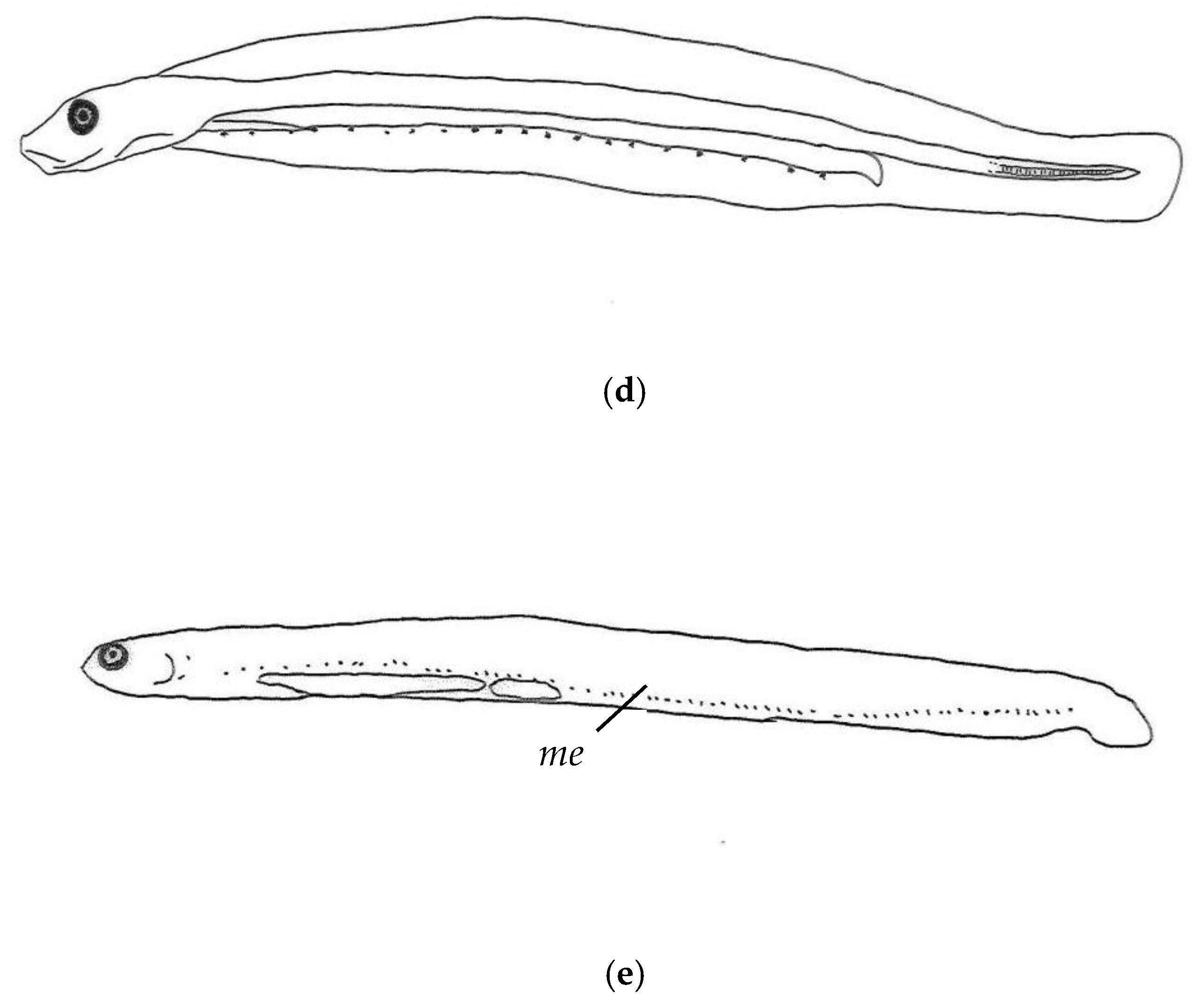
  • (2) Absence of melanophores at the posterior anal fin································Pholis fangi;
  • (1) Presence of melanophores at the posterior anal fin;
  • (4) Sparse melanophores on the abdominal back, not connected·······Pholis crassispina;
  • (3) Dense distribution of melanophores on the abdominal back·······Pholis nebulosa.
Features in the Pholis juvenile stage include the following:
  • (2) Presence of melanophores on the abdomen··········································Pholis fangi;
  • (1) Melanophores present on the abdomen;
  • (4) Dense distribution of melanophores on the abdominal back·······Pholis nebulosa;
  • (3) Sparse melanophores on the abdominal back, and a few melanophores on the abdominal back that are not connected;
  • (6) Presence of melanophores on vertebrae················································Pholis ornate;
  • (5) Absence of melanophores on vertebrae···········································Pholis crassispina.

4. Discussion

The classification of the Pholidae family has been subject to some debate and revisions [20,21,22,23,24,25]. Radchenko et al. [26] ultimately incorporated Enedrias Jordan and Gilbert, 1898, and Allopholis Yastu, 1981 into the genus Pholis, drawing on molecular (e.g., COI, cytochrome b, and 16S rRNA) and morphological evidence. The family Pholidae is divided into two subfamilies, Pholinae and Apodichthyinae, encompassing five genera: Pholis; Rhodymenichthys Jordan and Evermann, 1896; Apodichthys Girard, 1854; Ulvicola Gilbert and Starks, 1897; and Xererpes Jordan and Gilbert, 1880. Within these, the genus Pholis includes 11 species, which exhibit distinct regional distributions. Nine of these species are found in the Pacific Ocean, while two are located in the Atlantic Ocean. The West Pacific hosts five species: P. crassispina; P. fangi; P. nea Peden and Hughes, 1984; P. nebulosa; and P. picta (Kner, 1868) [27]. In China, the Pholis genus is represented by P. nebulosa and P. fangi, with P. fangi being unique to China [1]. According to ichthyographic records, P. fangi is found along the coast from Liaoning to Jiangsu, whereas P. nebulosa’s range extends further south, from Liaoning to Zhejiang [28,29,30,31,32,33,34,35]. There are no records of P. fangi or P. nebulosa from Fujian to the southern regions [36,37,38,39,40,41,42]. This distribution pattern corroborates that these two small-sized offshore fish species are exclusive to the northern frigid and temperate zones [1].
The genetic results in this study mostly rely on sequences taken from GenBank. Although GenBank is a very useful data source, it is important to use these data with caution due to the possibility of misidentifications. Misidentifications in GenBank can arise from various factors, including incorrect species identification at the time of sequence submission. Therefore, when using data from GenBank, it is crucial to discuss the potential drawbacks and risks. In our study, we cross-verified the genetic data with morphological characteristics to mitigate these risks. However, researchers should remain vigilant and consider these limitations in future studies.
During melanophore development, the shapes and distribution patterns of melanophores are key characteristics for the classification and identification of larval and juvenile stages [13]. In this study, melanophores were observed in the abdominal region of the digestive duct in both the larval and juvenile stages of P. fangi. However, melanophores on the back of the digestive duct and along the side abdominal margins near the posterior anal tail developed only during the juvenile stage. The distribution patterns of melanophores in P. fangi from Rushan Bay show slight variations from the findings of this study. From the post-larval to the juvenile stage (body length range: 4.5–28.00 mm), melanophores were present along the side abdominal margins of the posterior anal tail, and they developed in the larval stage. In larger individuals (body length: 21.45 mm) during the post-larval stage, melanophores were also developed in the abdominal region of the digestive duct and persisted into the juvenile stage [10]. The melanophore distribution in P. fangi differs from that in other Pholis species, with distinct stages of melanophore development. For P. crassispina and P. nebulosa, melanophores are found in the abdominal region of the digestive duct, as well as on the back and side abdominal margins near the posterior anal tail, across both larval and juvenile stages. This pattern of melanophore distribution does not alter with the transition from larval to juvenile stages [12]. The four Pholis species can be differentiated based on the melanophore distribution on the vertebrae and the developmental stages of melanophores on the abdominal front, back, and side margins near the posterior anal tail. Additionally, the quantity of melanophores on the abdominal back varies. These classification features evolve with the developmental stages of the fish, allowing species to be distinguished by combining developmental stages with classification characteristics.
Body shape transformations, including modifications to the yolk sac, anal positioning, and the ratio of anterior anal distance to body length, serve as significant indicators of morphological evolution through the larval and juvenile phases [13]. In this research, the initial body length of P. fangi was recorded at 5.53 mm, with the yolk not fully absorbed during the pre-larval stage. Contrarily, for specimens measuring 4.5 mm in Rushan Bay, the complete absorption of the yolk sac was observed [10]. As individuals growing, the ratio of anterior anal distance to body length in P. fangi exhibits variations. This study found that the ratios during the larval stages (pre-larvae and post-larvae) of P. fangi remained relatively stable. However, these ratios significantly dropped from approximately 75% during larval stages to 45% in the juvenile phase, a discrepancy from findings related to P. fangi in Rushan Bay [10]. Notably, shifts in anal positioning from larval to juvenile stages were exclusively documented in P. nebulosa. Additionally, there have been no accounts of anal position alterations in other species throughout their larval development [12].

5. Conclusions

This study provides a comprehensive analysis of the morphological development and melanophore distribution patterns of Pholis fangi larvae and juveniles in the Yellow Sea. By combining traditional morphological examination with DNA barcoding, we successfully identified and described the key developmental stages of P. fangi, from yolk-sac larvae to juveniles. Our findings highlight the significance of melanophore patterns in distinguishing P. fangi from other Pholis species and emphasize the importance of considering ontogenetic changes in melanophore arrangement for accurate species identification. Comparative analysis revealed distinct melanophore distribution patterns in P. fangi, setting it apart from other Pholis species. Moreover, we observed notable differences in melanophore development between larval and juvenile stages, suggesting the potential for stage-specific classification criteria. These results contribute to a better understanding of the early life history and taxonomy of Pholis species, providing valuable insights for further research in ichthyology and marine ecology. Our study also demonstrates the effectiveness of integrating morphological and molecular approaches in the identification and classification of fish larvae and juveniles.

Author Contributions

S.L.: methodology, writing—original draft preparation, data curation, and funding acquisition. H.Z.: writing—reviewing and editing. X.J.: data curation and software. X.P. and Y.Q.: data curation and methodology. W.Y.: conceptualization, methodology, and writing—reviewing and editing. All authors have read and agreed to the published version of the manuscript.

Funding

This research was funded by the National Key Research and Development Program of China (2022YFC3106002), the Open Fund of Key Laboratory of Marine Ecological Monitoring and Restoration Technologies, Ministry of Natural Resources (202010).

Institutional Review Board Statement

This study was conducted in strict adherence to ethical guidelines, ensuring no harm came to the fish involved. Consequently, ethical approval was not deemed necessary.

Informed Consent Statement

Not applicable.

Data Availability Statement

Data are contained within the article.

Acknowledgments

We extend our gratitude to Chen Lianfang for her thorough review of the manuscript and her insightful feedback. Additionally, we would like to express our sincere thanks to the three anonymous reviewers for their valuable comments and suggestions.

Conflicts of Interest

The authors declare no conflicts of interest.

References

  1. Liu, J. Fauna Sinica: Scleroichthyes, Perciformes (IV); Science Press: Beijing, China, 2016. [Google Scholar]
  2. Meng, Q.W.; Su, J.X.; Miao, X.Z. Systematics of Fishes; Chinese Agricultural Press: Beijing, China, 1995. [Google Scholar]
  3. Kim, J.M.; Kim, D.Y.; Yoo, J.M.; Huh, H.T. Food of the Larval Gunnel Enedrias fangi. Bull. Korean Fish. Soc. 1985, 18, 484–490. [Google Scholar]
  4. Bi, Y.B. Biology and Its Fishery of Fang’s Blenny Enedrias fangi Wang et Wang in Offshore Liaoning Province. Fish. Sci. 2005, 24, 27–28. [Google Scholar]
  5. Hwang, S.D.; Lee, T.W.; Hwang, S.W. Age, Growth, and Life History of Gunnel, Pholis fangi, in the Yellow Sea. Fish. Res. 2008, 93, 72–76. [Google Scholar] [CrossRef]
  6. Huang, X.X.; Zeng, X.Q.; Zhang, C. The Reproductive Biology of Enedrias fangi in the Inshore Waters of Qingdao. Period. Ocean Univ. China 2010, 40, 55–59. [Google Scholar]
  7. Li, S.Y. Feed Habits of Enedrias fangi in Jiaozhou Bay Based on Stomach Contents Analysis and Stable Isotope Analysis. Ph.D. Thesis, Ocean University of China, Qingdao, China, 2015. [Google Scholar]
  8. Li, L.; Zhang, H.; Sun, D.R.; Gao, T.X. Structure of Mitochondrial DNA Control Region of Pholis fangi and Its Phylogenetic Implication. J. Ocean Univ. China 2014, 13, 491–496. [Google Scholar] [CrossRef]
  9. Gao, T.X.; Li, L.; Fang, R.D.; Liu, G.H.; Wang, L.; Xu, H.X.; Song, N. Shallow Genetic Structure of Pholis fangi in Bohai Sea and Yellow Sea Inferred from mtDNA Control Region. J. Ocean Univ. China 2019, 18, 947–952. [Google Scholar] [CrossRef]
  10. Wan, R.J.; Zhang, R.Z. (Eds.) Fish Eggs, Larvae and Juveniles in the Offshore Waters of China and Their Adjacent Waters; Shanghai Science and Technology Press: Shanghai, China, 2016. [Google Scholar]
  11. Kunihiko, T.; Kunio, A. Studies on larval and juvenile blennies in the coastal waters of the southern Hokkaido (Pisces: Blennioidei). Bull. Fac. Fish. 1980, 31, 16–49. [Google Scholar]
  12. Okiyama, M. An Atlas of the Early Stage Fishes in Japan, 2nd ed.; Takai University Press: Tokyo, Japan, 2014. [Google Scholar]
  13. Zhang, R.Z.; Lu, S.F.; Zhao, C.Y.; Chen, L.F.; Zang, Z.J. (Eds.) Fish Eggs and Larvae in the Offshore Waters of China; Shanghai Science and Technology Press: Shanghai, China, 1985. [Google Scholar]
  14. Zhou, M.Y.; Chen, X.; Yang, S.Y. Identification of several fish eggs and larvae by DNA barcoding in Xiamen water. Mar. Environ. Sci. 2015, 34, 120–125, 135. [Google Scholar]
  15. Liu, S.H.; Qin, Y.T.; Liu, C.C.; Ji, X.; Zhang, H.F. Molecular identification of fish larvae and juveniles based on DNA barcoding. Ocean Dev. Manag. 2017, 34, 92–95. [Google Scholar]
  16. Liu, S.H.; Yang, Y.Y.; He, Y.L.; Ji, X.; Wang, Y.T.; Zhang, H.J.; Mao, R.Y.; Jiang, X.S.; Cheng, X.S. Morphological classification of ichthyoplankton in the Changjiang River Estuary based on DNA barcoding. Haiyang Xuebao 2021, 43, 93–104. [Google Scholar]
  17. Hanahara, N.; Miyamoto, K.; Oka, S. Morphological and genetic identification of formalin–fixed gobioid larvae and description of postflexion larvae of Paragunnellichthys sp. and Ctenogobiops feroculus. Ichthyol. Res. 2021, 68, 182–190. [Google Scholar] [CrossRef]
  18. Endo, S.; Hibino, Y.; Mochioka, N. Identification of first recorded ophichthid larvae of Ophichthus celebicus and O. macrochir (Anguilliformes; Ophichthidae) from Japan, based on morphometric and genetic evidence. Ichthyol. Res. 2022, 69, 393–398. [Google Scholar] [CrossRef]
  19. GB/T 12763.6-2007; Specification for Oceanographic Survey Part 6: Marine Biology Survey. China Standards Press: Beijing, China, 2007; pp. 38–41.
  20. Makushok, V.M. Morphological Foundations of Stichaeoidae and Closely Related to Them Fish Families (Blennioidei, Pisces). Tr. Zool. Inst. Akad. Nauk SSSR 1958, 25, 3–129. [Google Scholar]
  21. Yatsu, A. A Revision of the Gunnel Family Pholididae (Pisces, Blennioidei). Bull. Nat. Sci. Mus. Tokyo Ser. A 1981, 7, 165–190. [Google Scholar]
  22. Yatsu, A. Phylogeny of the Family Pholididae (Blenniodei) with a Redescription of Pholis scopoli. J. Ichthyol. 1985, 12, 273–282. [Google Scholar] [CrossRef]
  23. Nelson, J.S. Fishes of the World, 3rd ed.; Wiley: NewYork, NY, USA, 1994. [Google Scholar]
  24. Nelson, J.S. Fishes of the World, 4th ed.; Wiley: NewYork, NY, USA, 2006. [Google Scholar]
  25. Mecklenburg, C.W. Family Pholidae Gill, 1893 Gunnels. Calif. Acad. Sci. Annot. Checkl. Fish. 2003, 6, 15–20. [Google Scholar]
  26. Fedorov, V.V. Annotated Checklist of Fishes and Fishlike Organisms Living in Seas of Russia and Adjacent Countries. Part 6. Suborder Zoarcoidei. J. Ichthyol. 2004, 44, 73–128. [Google Scholar]
  27. Radchenko, O.A.; Chereshnev, I.A.; Petrovskaya, A.V. Phylogenetic relations in the family Pholidae (Perciformes: Zoarcoidei) based on genetic and morphological data. J. Ichthyol. 2010, 50, 728–739. [Google Scholar] [CrossRef]
  28. Wu, H.L.; Shao, G.Z.; Lai, C.F.; Zhuang, D.H.; Lin, P.L. (Eds.) Latin–Chinese Dictionary of Fishes Names by Classification System; The Shuichan Press: Keelung, China, 1999. [Google Scholar]
  29. Zhu, Y.D.; Zhang, C.L.; Cheng, Q.T. The Fishes of the East China Sea; Science Press: Beijing, China, 1963. [Google Scholar]
  30. Cheng, Q.T.; Zheng, B.S. Systematic Synopsis of Chinese Fishes; Science Press: Beijing, China, 1987. [Google Scholar]
  31. Liu, C.X.; Qin, K.J.; Ding, G.W.; Wang, X.Y.; Mou, X.L.; Piao, X.L.; Chen, J.K.; Shi, Y.R.; Qin, Y.J.; Qin, K.J. (Eds.) Fauna in Liaoning Province (Fish); Liaoning Science and Technology Press: Shenyang, China, 1987. [Google Scholar]
  32. Cheng, Q.T.; Zhou, C.W. The Fishes of Shandong Province; Shandong Science and Technology Press: Jinan, China, 1997. [Google Scholar]
  33. Wang, S.A.; Wang, Z.M.; Li, G.L.; Cao, Y.P.; Zhang, Z.W.; Xu, X.J.; Sun, J.H. Fauna in Hebei Province (Fish); Hebei Science and Technology Press: Shijiazhuang, China, 2001. [Google Scholar]
  34. Ni, Y.; Wu, H.L. Fishes of Jiangsu; Chinese Agriculture Press: Beijing, China, 2006. [Google Scholar]
  35. Zhao, S.L.; Xu, H.X.; Zhong, J.S.; Chen, J. Fauna of Marine Fishes in Zhejiang; Zhejiang Science and Technology Press: Hangzhou, China, 2016. [Google Scholar]
  36. Zhuang, P.; Zhang, T.; Li, S.F.; Ni, Y.; Wang, Y.H.; Deng, S.M.; Zhang, L.Z.; Ling, J.Z.; Hu, F.; Yang, G. Fishes of the Yangtze Estuary, 2nd ed.; Shanghai Science and Technology Press: Shanghai, China, 2019. [Google Scholar]
  37. Fishes of Fujian Province Editorial Subcommittee. The Fishes of Fujian Province; Fujian Science and Technology Press: Fuzhou, China, 1984. [Google Scholar]
  38. Liu, M.; Chen, X.; Yang, S.Y. Marine Fishes of Southern Fujian, China; China Ocean Press: Beijing, China, 2014; Volume 2. [Google Scholar]
  39. Shao, K.T. Taiwan Fish Database. WWW Web Electronic Publication. 2023. Available online: http://fishdb.sinica.edu.tw (accessed on 26 January 2024).
  40. Zheng, C.Y. Fishes of the Zhujiang River; Science Press: Beijing, China, 1989. [Google Scholar]
  41. Institute of Zoology, Chinese Academy of Sciences; Institute of Oceanography, Chinese Academy of Sciences; Shanghai Fisheries University. The Fishes of the South China Sea; Science Press: Beijing, China, 1962. [Google Scholar]
  42. South China Sea Fisheries Research Institute, Chinese Academy of Fishery Sciences. The Fishes of the Islands in the South China Sea; Science Press: Beijing, China, 1979. [Google Scholar]
Figure 1. Research area map.
Fishes 09 00213 g001
Figure 2. M–L phylogenetic tree of Pholis based on the mtCOⅠ sequence.
Fishes 09 00213 g002
Figure 3. Photographs of larval and juvenile stages of P. fangi: (a) 5.53 mm BL yolk-sac larva; (b) 6.48 mm BL preflexion larva; (c) 7.10 mm BL preflexion larva; (d,e) 10.31 mm BL flexion larva; (f) 39.82 mm BL juvenile.
Fishes 09 00213 g003aFishes 09 00213 g003bFishes 09 00213 g003c
Figure 4. Linedrawings of larval and juvenile stages of P. fangi: (a) 5.53 mm BL yolk-sac larva; (b) 6.48 mm BL preflexion larva; (c) 7.10 mm BL preflexion larva; (d) 10.31 mm BL flexion larva; (e) 39.82 mm BL juvenile. me: melanophore; mdda: melanophores on digestive duct abdomen.
Fishes 09 00213 g004aFishes 09 00213 g004b
Disclaimer/Publisher’s Note: The statements, opinions and data contained in all publications are solely those of the individual author(s) and contributor(s) and not of MDPI and/or the editor(s). MDPI and/or the editor(s) disclaim responsibility for any injury to people or property resulting from any ideas, methods, instructions or products referred to in the content.

