Review

Role of Peripheral Immune Cells in Multiple Sclerosis and Experimental Autoimmune Encephalomyelitis

1
College of Medicine, and Immuno-Oncology Group, The Sharjah Institute for Medical Research, University of Sharjah, Sharjah 27272, United Arab Emirates
2
Department of Clinical Sciences, College of Medicine, University of Sharjah, Sharjah 27272, United Arab Emirates
*
Author to whom correspondence should be addressed.
Submission received: 16 November 2020 / Revised: 20 January 2021 / Accepted: 27 January 2021 / Published: 1 February 2021
(This article belongs to the Special Issue Novel Therapeutic Modalities for Multiple Sclerosis)

Abstract

:
Multiple sclerosis (MS) is a chronic autoimmune disease that affects the myelination of the neurons present in the central nervous system (CNS). The exact etiology of MS development is unclear, but various environmental and genetic factors might play a role in initiating the disease. Experimental autoimmune encephalomyelitis (EAE) is a mouse model that is used to study the pathophysiology of MS disease as well as the effects of possible therapeutic agents. In addition, autoreactive immune cells trigger an inflammatory process upon the recognition of CNS antigens, which leads to destruction of the neurons. These include innate immune cells such as macrophages, dendritic cells, and natural killer cells. Additionally, the activation and extravasation of adaptive immune cells such as CD4+ T cells into the CNS may lead to further exacerbation of the disease. However, many studies revealed that immune cells could have either a protective or pathological role in MS. In this review, we highlight the roles of innate and adaptive immune cellular and soluble players that contribute to the pathogenesis of MS and EAE, which may be used as potential targets for therapy.

1. Introduction

Multiple sclerosis (MS) is chronic autoimmune disabling diseases of the central nervous system (CNS) characterized by varying degrees of demyelination of uncertain etiology that is thought to be associated with specific environmental and genetic factors [1]. The pathological characteristics of MS and its progression include an interplay between specific mechanisms mainly inflammation, which is thought to be the triggering point in the course of the disease, demyelination, axonal damage, and gliosis [2].
Multiple sclerosis has four clinically defined subtypes, with relapsing-remitting multiple sclerosis (RRMS) being the most common. RRMS is characterized by periods of acute neurological deterioration known as relapses, which are followed by recovery phases of varying degrees known as remissions. Remission could vary from full recovery to a sequela with a residual CNS deficit. However, RRMS is distinguished by its stable course between the attacks, but this could shift to a progressive stage, which is known as secondary-progressive multiple sclerosis (SPMS) [3]. On the other hand, the primary-progressive multiple sclerosis (PPMS) subtype is associated with neurological deterioration in function from the onset of the disease and is characterized by a continuously worsening baseline with no defined relapses. The other form of MS is the progressive-relapsing multiple sclerosis (PRMS), which shows distinct relapses throughout its course [3]. A different classification was suggested, which removed the PRMS from the subtypes of MS and relocated it to the PPMS group [3]. Interestingly, some patients display a series of events indicating an inflammatory demyelinating disorder of the CNS called “clinically isolated syndrome (CIS)”, which lasts for at least 24 h and is not caused by fever or infection. This is usually considered as an early indicator of development of MS [4].
Although the exact etiology of MS is unknown, the infiltration of lymphocytes and macrophages into the CNS in addition to pre-existing environmental and genetic factors could lead to the development of the disease [5,6]. As shown in Figure 1, activated T helper 1 (Th1) cells are known to release inflammatory cytokines such as interferon (IFN)-γ and tumor necrosis factor (TNF)-α, which increase the expression of surface receptors on nearby antigen-presenting cells (APCs) and lymphocytes. As a result, the presence of genetically susceptible MS antigens such as myelin basic protein (MBP) and myelin oligodendrocyte glycoprotein (MOG) bind activated immune cells in the CNS, leading to a variable and continuous inflammatory process of destruction and deterioration in the CNS [7]. In order to study MS, researchers have developed a number of models in the animal laboratory, including experimental autoimmune encephalomyelitis (EAE) [8]. It is the most commonly used animal model that is developed by immunizing the mice with myelin peptides/antigens, thus inducing autoimmune disease that usually resembles MS [9]. It has been widely used to study the pathophysiology of MS [8] and potential MS therapies [10,11,12,13].

Diagnosis of MS Disorder

There are no specific tests to diagnose MS [14]. As symptoms vary widely, the disease may not be recognized in its early stage. Ultimately, MS is diagnosed based on clinical measures and tests such as magnetic resonance imaging (MRI) of the brain. An MRI scan can detect brain and spinal damages identifying myelin loss. Meanwhile, the examination of the cerebrospinal fluid (CSF), withdrawn from the spinal canal, may indicate a defect or a specific problem related to MS, such as abnormal levels of white blood cells or the presence of proteins. Additionally, blood tests can be used to exclude the presence of viral and other infectious agents that may elicit neurological symptoms similar to those observed in MS patients [15,16].

2. Innate Immunity in MS

The innate immunity is the first defense system in vertebrates. It generates a fast, non-specific, inflammatory response to all types of pathogens and tissue damage. Cells of the innate immune system recognize foreign antigens by means of pathogen-associated molecular patterns (PAMPs), as well as danger-associated molecular patterns (DAMPs), released from damaged or dying cells during injury, via pattern recognition receptors (PRRs) such as Toll-like receptors (TLRs) and nucleotide-binding oligomerization domain NOD-like receptors (NLRs). Cells of myeloid and lymphoid origins are activated in response to PRR signaling [17]. The innate immune responses are mediated through cell-dependent mechanisms such as phagocytosis and cytotoxicity or through secreted factors such as antimicrobial peptides, complement proteins, cytokines, and chemokines [18].
The perturbation of PRRs induces the activation of macrophages, dendritic cells (DCs), neutrophils, and other innate immune cells. However, PRRs can also be expressed on non-immune CNS-resident cells. Many PRRs show increased gene expression in MS patients where genome-wide association studies (GWAS) described some PRRs variants to be associated with increased risk of the disease [19].

2.1. Role of TLRs in MS

TLRs represent a group of receptors located either on the surface of the cell or in endosomes of both nonimmune and immune cells. Activation of the immune system via TLRs is chiefly mediated by APCs such as macrophages, DCs, and B cells [20]. TLRs are involved in recognizing and binding to PAMPs and DAMPs, resulting in transcriptional activation of pro-inflammatory cytokines, chemokines, co-stimulatory and major histocompatibility complex (MHC) molecules [21,22]. In the brain, DAMPs released after a spinal cord injury induce the activation of TLR2 and TLR4 [23].
TLRs have crucial roles in MS or EAE brain lesions. For instance, a study by Sloane et al. has shown that TLR2 is expressed in oligodendrocytes, and it is upregulated in MS lesions [24]. Additionally, hyaluronan, a low-molecular-weight glycosaminoglycan, a major component of extracellular matrix, is broken down by hyaluronidases into hyaluronan oligomers that block oligodendrocyte precursor cell maturation and re-myelination through TLR2–MyD88 signaling [24]. Elevated levels of hyaluronan are observed in both EAE lesions and areas of complete demyelination in MS patients. This could be attributed to an increased stimulation of TLR2 on oligodendrocytes, altered hyaluronan synthesis, or partial hyaluronan degradation, resulting in the remyelination blockade [25,26].
Likewise, MyD88-dependent signaling through TLR2 and other TLRs such as TLR4, TLR7, and TLR9 was found to be associated with MS progression [27,28]. In contrast, TLR3 signaling induced neuroprotective responses and was found to suppress demyelination in EAE mice [29]. Furthermore, TLR7 induces the maturation and differentiation of B lymphocytes into immunoglobulin-secreting plasma cells. Studies have shown that TLR7 deficiency stimulated immunoglobin M (IgM) and immunoglobin IgG production in MS patients, and this could be associated with worsening the disease state corroborated with compromised immune responses against TLR7-recognized RNA viruses and infections [30,31].

2.2. NOD-Like Receptors in MS

NOD-like receptors (NLRs, nucleotide-binding domain, leucine-rich repeat-containing family) are essential sensors of cellular stress induced by infection, tissue damage, or cell death [32]. NLR proteins such as NLRP1, NLRP3, NLRC4, as well as the absent in melanoma 2 protein (AIM2) are activated by PAMPs and DAMPs. This activation results in the recruitment of the inflammasome-adaptor protein apoptosis-associated speck-like protein containing a caspase recruiting domain (ASC or PYCARD) and procaspase-1 [33]. Other NLR proteins in addition to ASC and procaspase-1 form the inflammasome multiprotein complex; the proximity of these proteins induces the autoactivation of caspase-1, which subsequently cleaves pro-IL-1β and pro-IL-18 to the pro-inflammatory mature IL-1β and IL-18 cytokines [34].
The NLRs are expressed on the nuclear membrane or in the cytoplasm of immune cells such as monocytes, DCs, macrophages, neutrophils, and cerebral endothelial cells. NLRs are also expressed as a secreted form in granulocytes, monocytes, B, and T cells [35,36]. In the CNS, the NLRP1 inflammasome is expressed mainly by pyramidal neurons and oligodendrocytes [37], while the NLRP3 inflammasome is expressed by microglia [38]. Ghrelin is an octanoylated peptide that has been shown to have neuroprotective effects. A study by Liu et al. showed that treatment using Ghrelin was effective in EAE Sprague–Dawley rats. Gherlin-treated EAE rats had less inflammatory infiltration and demyelination as shown by the histologic analysis of the brain and spinal cord [39]. Interestingly, the Ghrelin-treated EAE rats had reduced expression of proteins involved in the NLRP3 signaling pathway and pyroptosis as well as decreased expression of inflammatory cytokines [39].

2.3. Role of Innate Immune Cells in MS Disease

Macrophages and dendritic cells are the main constituents of the innate immunity. Foreign antigens are phagocytosed by these immune cells and then presented on their surfaces to be recognized by T cells of the appropriate specificity. This leads to the activation and proliferation of T cells, and a consequent cytokine secretion [40]. In MS, the recognized antigen is believed to be an epitope on myelin proteins. Activated T cells infiltrate through the BBB into the nervous system, where cytokines are secreted. For instance, Th1 cells secrete inflammatory cytokines such as IFN-γ, whereas Th17 cells secrete IL-17, which plays an important role in host defenses [41]. These cytokines mobilize macrophages and phagocytic cells in the periphery and microglia in the CNS, which consequently induce the MS attacks [42]. In MS, macrophages induce tissue damage by producing pro-inflammatory mediators. Paradoxically, these cells may also have anti-inflammatory and neuroprotective functions. Macrophages polarization into pro-inflammatory or neuroprotective depends on the mechanism through which they are activated. For instance, monocyte-derived macrophages stimulated with IFN-γ and lipopolysaccharide differentiate into the classical phenotype M1 macrophages, whereas upon stimulation with IL-4 and IL-33, they differentiate into M2 macrophages [43]. Macrophages in active MS lesions mainly exhibit M1 characteristics and markers, including CD40, CD86, CD64, and CD32, which are abundantly expressed by microglia in the healthy white matter and activated microglia and macrophages in the active MS lesions [43]. Vainchtein et al. found that microglia were weakly immune-activated, with a concomitant reduced expression of MHC II, co-stimulatory molecules, and pro-inflammatory cytokines such as IL-1β. However, infiltrated macrophages were highly immune-activated and expressed high levels of pro-inflammatory molecules such as IL-1β and TNF-α as well as being more abundant during the high clinical score phase of EAE [44].
On the other hand, M2 markers such as mannose receptor and CD163 were expressed by myelin-laden macrophages in demyelinating lesions and perivascular macrophages [43]. Another study demonstrated that in the early phases of MS and EAE, macrophages of the M1 phenotype produce pro-inflammatory cytokines that contribute to the destruction of the CNS. However, in the later phase of the disease, more macrophages of the anti-inflammatory M2 phenotype are present in the CNS, leading to the neuroprotective effects and re-establishing homeostasis [45].
It was noted that the number of circulating myeloid DCs isolated from SPMS patients were higher in comparison with RRMS patients and control subjects, suggesting that DCs are highly recruited during the transition into the progressive phase of the disease. In addition, DCs isolated from SPMS patients were found to produce IL-12 and TNF-α, while DCs from RRMS patients produced higher levels of Th1 cytokines IFN-γ and TNF-α, or Th2 cytokines IL-4 and IL-13 compared to control subjects. DCs from SPMS patients only promoted a polarized Th1 response [46].
Interestingly, a neuro-steroid and progesterone derivative, allopregnanolone (ALLO), was highly investigated in different neurological diseases, including MS [47]. Moreover, it was shown to affect macrophages and DCs activity by interaction through gamma aminobutyric acid-A (GABA-A) receptors. Such receptors are expressed by various cells including neuron and glial cells, affecting their physiological activities such as their gene expression, proliferation, and survival. In addition, macrophages express GABA-A receptors, where upon the activation of these receptors, the ability to produce pro-inflammatory cytokines was reduced [47,48]. Another way of activation of GABA-A receptors using agonists caused a modification in the responses of antigen-presenting cells such as macrophages and DCs, affecting the subsequent T cell activities [49].

2.3.1. Role of Neutrophils in MS/EAE

The activation of CNS-specific autoreactive CD4+ T cells in the periphery can result in a dysregulation of the innate immune system [50]. For example, neutrophils and monocytes are expanded in the bone marrow and accumulated in the circulation in response to systemic upregulation of the CXCL1 and granulocyte-colony stimulating factor (G-CSF) in EAE. In MS patients, it was observed that there is an increased number and priming of circulating neutrophils. Furthermore, a correlation between MS relapse phases and G-CSF, CXCL1, CXCL5, or neutrophil elastases supports the role of neutrophils in CNS damage and the pathogenesis of MS. It was also demonstrated that peripheral neutrophils can affect autoreactive T cell activation, which contributes to MS pathogenesis [51].
NETosis is a process mediated by neutrophils through which these cells form neutrophils extracellular traps (NETs), which are composed of decondensed chromatin and histones, forming a large web-like structure. NETs contain factors such as myeloperoxidase (MPO), neutrophil elastase, and proteinase 3, which are released into the surrounding environment, allowing NETs to capture and kill foreign pathogens. This has been previously discussed in studies that showed the significant roles for NETs in MS and EAE, among other autoimmune diseases [52].
In EAE, the depletion of MPO was found to decrease the disease severity, reducing the levels of reactive oxygen species (ROS), and leading to improved BBB functions [53,54,55]. Additionally, the inhibition of neutrophil elastase reduced the infiltration of neutrophils into the optic nerves in EAE mice [56]. Neutrophil elastase and MPO in addition to other neutrophil related factors were increased in the plasma of MS patients and correlated with MS lesion burden [51,57].

2.3.2. Role of NK Cells in MS/EAE

Human CD56+ CD3 NK cell subsets are defined based on the expression of the cell surface molecules CD16 and CD56 [58]. CD56dim CD16+ NK cells are present mainly in the peripheral blood and can attack and lyse targeted tumor cells, whereas CD56bright CD16 NK cells are found mostly in the lymphoid organs and can produce large amounts of cytokines but have little ability to kill tumor target cells [58]. Murine NK cells lack the CD56 marker but express other markers that identify these cells obtained from various mice strains [59].
The role of NK cells in MS is not yet resolved [60,61]. It was reported that the CSF of MS patients harbor a distinct subset of NK cells expressing CCR4, releasing IL-17 and IFN-γ, and hence, they are designated as NK17/NK1 cells [62]. Intriguingly, it was reported that most drugs used to treat MS patients activate NK cell lysis of DCs. These include glatiramer acetate (GA, Copaxone) [63], vitamin D3, calcipotriol, and FTY720 [64]. In addition, monomethyl fumarate (MMF) and dimethyl fumarate (DMF, Tecfidera) enhance NK cell lysis of DCs [65,66]. Furthermore, MMF and vitamin D3 ameliorated the EAE clinical score in mice [67]. Previously, Munger et al. reported a negative association between the intake of vitamin D and the risk of developing MS in a large female cohort. Women with a high intake of dietary vitamin D (about 700 IU/day) had a 33% lower incidence of MS compared to those with lower intake. In addition, women who used vitamin D supplements (more than or equal to 400 IU) had a 41% reduced risk of developing MS [68]. This was further supported by another study where subjects with serum levels of the circulating form of vitamin D3, 25-hydroxyvitamin D, greater than 100 nmol/L (40 ng/mL) had a 62% lower chance of developing MS [69].
These results suggest that a possible mechanism of action for these drugs is potentiating the cytolytic activity of NK cells against DCs, which impede the latter from presenting autoantigens to autoreactive T cells. In this regard, it was reported that NK cells isolated from MS patients treated with GA express high cytolytic activity against DCs [70]. Collectively, these findings support the notion that drugs used to treat MS patients promote NK cell lysis of DCs, and consequently, impede the activation of autoreactive T cells.

2.3.3. Role of NKT Cells in MS/EAE

Natural killer T (NKT) cells are lymphocytes that are a part of the innate immune system. They express T cell receptor (TCR) α/β as well as NK cell markers such as CD161 and CD94. However, the TCR of NKT cells does not interact with peptide antigens presented by MHC-class I or class II molecules such as the conventional T cells, but instead their TCR recognizes glycolipids presented by CD1d, which is a non-classical antigen-presenting molecule expressed by APCs [71]. The NKT cells bind to their cognate antigens, resulting in the polarization of IFN-γ and/or IL-4, resulting in regulation of the adaptive immune responses [71]. NKT cells have important regulatory effects in autoimmunity and immune responses to infections and tumors through secreting high levels of IL-4 and IFN-γ. The role of NKT cells in MS was investigated in studies of EAE mice. As an analog of the synthetic glycolipid, α-galactosylceramide (GalCer) binds to CD1d on APCs and induces NKT cells to produce IL-4, thus ameliorating EAE severity [72].

2.3.4. Role of γδ T Cells in MS/EAE

γδ T cells mediate host defense and immunoregulatory functions. These cells represent a small population of T cells that express a unique TCR composed of γδ chains instead of the αβ chains. In addition, they lack a precise MHC restriction, acting as a bridge between the innate and adaptive immune systems. γδ T cells are present mainly in peripheral blood, lymph nodes, spleen, intestine, and skin [73]. Previous findings using EAE mice discussed the role of these cells in MS pathogenesis. In EAE mice, γδ T cells were found in low numbers in the spleen, but their concentration in the CNS rises to above 10% at the peak of the relapse phase and falls back during the remission phase [74]. Moreover, the depletion of γδ T cells during different stages of EAE resulted in a decline of disease worsening, suggesting that these cells play a vital role in the EAE pathogenesis [75].

2.3.5. Role of MAIT Cells in MS/EAE

Mucosal-associated invariant T (MAIT) cells are a subset of innate-like lymphocytes that represent about 25% of CD8+ T cells in healthy subjects. These cells recognize conserved microbial ligands and act against bacterial and fungal pathogens [76]. Furthermore, MAIT cells display a semi-invariant TCR and are restricted by the nonpolymorphic MHC-related molecule-1 (MR1). In order for these cells to accumulate in the periphery, B cells, commensal flora, and the MR1 molecule are needed. In humans, the MAIT cells expand quickly after birth and acquire a memory phenotype, whereas in mice, they remain few in number and stay in a naive state [77].
Recent studies suggested their potential role in chronic inflammatory diseases, such as MS, through IFN-γ and/or IL-17 production [76]. However, contradictory observations have been reported, indicating an immunoregulatory behavior of MAIT cells in MS through inhibiting Th1 pathogenic responses [78].

2.4. Role of Complement System in MS

The complement system has an important role in the antimicrobial defense. The brain is considered an immune-privileged site and separated from the periphery by the BBB. Yet, all major CNS cells produce most of the complement proteins, where astrocytes are the main source of CNS complement, indicating their involvement in immune defenses against pathogens and their contribution to tissue destruction [79].
The demyelination process results from an autoreactive immune response against myelin through the complement classical pathway, in addition to the direct activation of complement upon binding to myelin. It is well known that myelin purified from the CNS can activate the complement classical pathway [80]. Additionally, mature oligodendrocytes from rats were found to be lysed in vitro by complement in the absence of anti-myelin antibodies [80]. MOG may be able to bind and activate the C1q component of complement because it has a similar domain to the C1q-binding sequence of IgG antibodies. These interactions between myelin-specific protein and Clq have consequential effects in inflammatory diseases that affect the CNS such as MS [81].

3. Role of Adaptive Immune Cells in MS

3.1. Role of CD4+ T Cells in MS and EAE

Adaptive immunity is evident in acute and active chronic lesions of MS. This has been reported and confirmed by various studies [82]. The pathology of MS disease is initiated by the activation of CNS antigen-specific CD4+ T cells in the periphery [83]. This stems from the similarities amomg MS and the EAE model, as EAE can be induced by immunization with CNS-derived peptides or myelin and is mainly driven by autoreactive CD4+ T cells specific for CNS peptides [84].
The theory of molecular mimicry indicates a similarity between a peptide from a foreign agent and an autoantigen. This was introduced by Fujinami and Oldstone by immunizing rabbits with hepatitis B virus peptides that shared six amino acids with the myelin basic protein (MBP), after which EAE was induced and T cells were involved with cross-reactivity against the peptides [85]. Another hypothesis for the development of MS disease is that autoreactive CD4+ T cells are activated in the circulation after cross-recognition of peptides derived from a foreign antigen, such as that of a virus [86].
Studies are still investigating the antigens responsible for activating autoreactive CD4+ T cells in the periphery. Reports from studies in animal models demonstrated that immunization with MBP results in the peripheral activation of MBP-specific CD4+ T cells in the draining lymph nodes as one of the initial events in the development of EAE [87]. After activation, these CD4+ T cells produce inflammatory cytokines, which may differentiate into T helper 1 (Th1) or T helper 17 (Th17) [88], which will be further discussed in this section.
Activated CD4+ T cells upregulate integrins, including the lymphocyte function-associated antigen (LFA-1) and the very late antigen-4 (VLA-4), which enable them to cross the BBB. Upon encountering the antigen, autoreactive T cells are reactivated and differentiated, leading to cytokine production, which consequently activates neighboring immune and neural cells, further attracting inflammatory cells into the CNS (Figure 1). This activation was observed in macrophages that are thought to indirectly or directly damage the CNS [89]. In addition, these CNS antigen-specific CD4+ T cells are the only immune cells that upon being transferred to immunocompetent recipient animals induce the EAE disease. Furthermore, transgenic mice that expressed TCRs derived from human CD4+ myelin-specific T cells and the appropriate human leukocyte antigen (HLA) class II molecule developed EAE disease [8].
As mentioned above, the main two subsets of CD4+ T cells that contribute to the pathology of MS and EAE are Th1 and Th17 [88]. CD4+ Th1 cells secrete the pro-inflammatory cytokines IFN-γ, IL-2, and TNF. MS disease severity was correlated with the expression of IFN-γ and IL-12 in the CSF and the CNS [90]. In addition, the administration of IFN-γ increased the severity of the disease [91]. This could be due to the finding that IFN-γ induces the expression of MHC class II on cells residing in the CNS, which triggers the production of chemokines and trafficking of immune cells toward the CNS.
Th17 cells secrete the pro-inflammatory cytokines such as IL-17 and IL-22 [92]. Aside from that, these cells induce other cell types to produce pro-inflammatory factors including granulocyte/macrophage colony-stimulating factor (GM-CSF), cytokines such as IL-6, and several chemokines such as CXCL8, indicating the important role of Th17 cells in inducing the inflammation in the CNS [93]. Moreover, a study by Langrish et al. showed that the transfer of Th17 cells resulted in a highly severe form of EAE compared to the transfer of Th1 cells [94]. Several other studies were reviewed regarding the roles of T cells and their subsets in MS and EAE [95,96].

3.2. Role of CD8+ Cells in MS

CNS antigen-specific CD8+ T cells are involved in CNS damage during the relapse phases and in the chronic phase of MS. These cells are activated by APCs presenting CNS-derived peptides [97] and are more abundant in MS lesions [98]. Moreover, axons and neurons express MHC-class I and not class II molecules, allowing CD8+ T cells to recognize their cognate antigen on axons/neurons and directly attack and damage them [99]. Several studies demonstrated that CD8+ T cells play a role in the pathophysiology of MS. It was reported that there is a higher count of CD8+ T cells compared to CD4+ T cells in MS brain lesions, especially in the parenchyma [100]. Another study analyzed brain tissues from MS patients and found that CD8+ T cells were present in about 77% of the cortical plaques, which are associated with MS progression and cognitive loss [101]. As mentioned previously, T lymphocytes migrate from the periphery, adhere to the endothelium of the CNS via adhesion molecules and ligands such as P-selectin, and then cross the BBB. A study by Battistini L et al. showed that CD8+ but not CD4+ T cells from RRMS patients exhibited increased rolling on P-selectin and higher recruitment in the inflamed brain venules during the acute phase of the disease [102]. In another study, adoptive transfer of CD8+ MOG-specific T cells induced a considerably more severe and lasting disease compared to mice actively immunized with MOG (35–55) only. Furthermore, the CNS lesions in MOG-specific CD8+ T cell-induced EAE model were progressive and showed more damage [103].
On the other hand, studies showed that CD8+ T cells could have immunoregulatory roles [104,105]. For instance, the depletion of CD8+ T cells caused CD28-deficient mice to be more prone to EAE, whereas the adoptive transfer of CD8+CD28 T cells into CD8−/− mice suppressed the disease severity [104]. Moreover, in vitro experiments demonstrated that co-culturing APCs with CD8+CD28 T cells caused these APCs to be less efficient in stimulating T cell-dependent immune reactions. This led to an inhibition of the co-stimulatory molecules on APCs, reducing these cells’ ability to deliver the activation signals to CD4+ T cells. Therefore, such findings indicate a potential protective role of CD8+ T cells against EAE [104].
Additionally, a study has shown that the transfer of MOG-specific CD8+ T cells suppressed the induction of EAE, thus acting as immune-regulatory cells through the production of IFN-γ and perforin as well as the direct killing of effector MOG-loaded CD4+ T-cells. Moreover, this resulted in reduced APCs responses and MOG-specific CD4+ T cell function and activity [105]. A review by Sinha et al. further highlights examples of the role of CD8+ T cells as immune regulators of MS [106].

3.3. Role of Regulatory T Cells (Tregs) in MS or EAE

Regulatory T cells (Tregs) play crucial roles in maintaining self-tolerance, immune balance in the periphery, and immune privilege in the CNS [107]. The dysregulation of suppressive and migratory markers on Tregs has been associated with MS disease. For example, genetic abnormalities in Treg suppressive markers CTLA-4 and CD25 have been found in some MS patients, whereas others had a decreased FoxP3 and IL-10 levels [108]. However, studies in animal models indicated that the transfer of isolated Tregs from naive mice into CNS injured immune-competent Balb/c mice decreased their neuroprotective effect [109]. Conversely, the exogenous transfer of the same Treg cells resulted in a neuroprotective phenotype in C57BL/6 mice. These findings indicate that Tregs might lead to either neuro-destruction or neuro-protection effects in mice with different genetic backgrounds [110].
On another note, Treg cells are affected by various host factors such as the gut microbiota, which was shown to play a vital role in the pathogenesis of MS through altering the host’s immune system, and the BBB functions, thus leading to autoimmune demyelination [111]. This is supported by analysis of the microbiomes of 71 MS patients, which showed that the presence of specific bacterial populations was highly associated with MS. For instance, Akkermansia muciniphila and Acinetobacter calcoaceticus increased in MS patients where they induced pro-inflammatory effects in mono-colonized mice and human peripheral blood mononuclear cells. On the other hand, Parabacteroides distasonis was reported to be lower in MS patients. In mice models, the presence of this species was found to enhance the anti-inflammatory IL-10-expressing human CD4+CD25+ T cells and IL-10+FoxP3+ Tregs activities. Furthermore, transplanting microbiota from MS patients into germ-free mice induced more severe EAE and reduced proportions of IL-10+ Tregs compared with mice transplanted with microbiota from healthy subjects [112].
Another host-related factor is melatonin, the hormone of sleep, which was found to have protective effects against EAE and MS through inducing the protective effects of IL-10 producing T regulatory cells in addition to blocking the pathogenic Th17 cells and inhibiting their production of IL-17 [113].

3.4. Role of B Cells in MS/EAE

B cells arise from the stem cells present in the bone marrow, where they develop to an immature naïve B cell. In order to mature, further development occurs in the spleen or lymph nodes to give rise to either memory B cells or to plasma cells [114]. Some B cells tend to be autoreactive by possessing the capacity to recognize and react to self-antigens. Therefore, the developmental process includes a checkpoint to limit the production of autoreactive B cells. In MS, some autoreactive B cells are able to bypass this checkpoint and escape to the maturity stage. This has been supported by the findings where autoreactive B cells reach the BBB and infiltrate into the CNS, thus contributing to MS pathology through different inflammatory mechanisms.
B cells play important roles in both human MS and mouse EAE models [115], and they are currently considered therapeutic targets for this purpose. For instance, B cells, plasma cells, and excess immunoglobulins are known to be present in both the lesions and CSF of patients with MS [116]. Studies in animal models demonstrate a complicated role for B cells [117]. Current theories suggest that two probable independent inflammatory processes induce the CNS injury in MS, which possibly involve B cells. It was suggested that the de novo infiltration of immune cells from the periphery into the CNS correlate with focal inflammation, MRI-detectable lesions, and relapse periods. Other studies indicate that chronic progression supposedly driven by CNS-intrinsic inflammation is induced by CNS-resident immune cells, in addition to the CNS-trapped B cells [118].
There are several possible mechanisms of B cells association with the pathophysiology of MS. One of these mechanisms is antigen presentation, where B cells play an important role in the immune response by recognizing and internalizing specific antigens, which is followed by intercellular processing to generate fragments of antigens that are eventually expressed on B cell surface. Studies suggest that antigen presentation and the co-stimulation of autoreactive B cells in the CNS may activate T cells toward a pro-inflammatory response, where reciprocal inflammatory signals and interactions cause the further activation of B cells [115]. On the other hand, autoreactive B cells can differentiate into plasma cells, producing antibodies that bind to myelin sheath and oligodendrocyte proteins. These bound antibodies result in the induction and activation of the complement proteins on tissue surfaces, promoting injury. Antibodies may activate other immune cells such as NK cells to destroy tissues via antibody-dependent cell-mediated cytotoxicity [119].
B cells could also promote inflammation in the CNS via cytokine secretion. It has been previously reported that B cells in MS patients tend to produce more pro-inflammatory cytokines and less protective cytokines compared with healthy controls [120]. In the meninges of patients with progressive MS, B cells may form ectopic lymphoid structures or germinal centers, which contain activated B cells and follicular DCs in addition to T cells and thus promote ongoing T cell activation within the brain [121]. These ectopic lymphoid structures may be possibly linked to microglial activation, local inflammation, and neuronal loss [121].
Other studies have shown that B cells may have immunoregulatory roles in MS disease [122]. For example, EAE mice deficient in B cells had less recovery and low opportunity of returning to normal state compared to control EAE mice, suggesting that B cells may contribute to the immune modulation of acute phase of the disease [123]. Additionally, another study demonstrated that B cells from recovered mice produced IL-10 in response to autoantigen. Furthermore, the lack of IL-10 secretion by B cells in EAE led to a lasting pro-inflammatory response corroborated with reduced recovery, indicating the importance of B cells in regulating the immune responses during EAE [124].

4. Conclusions

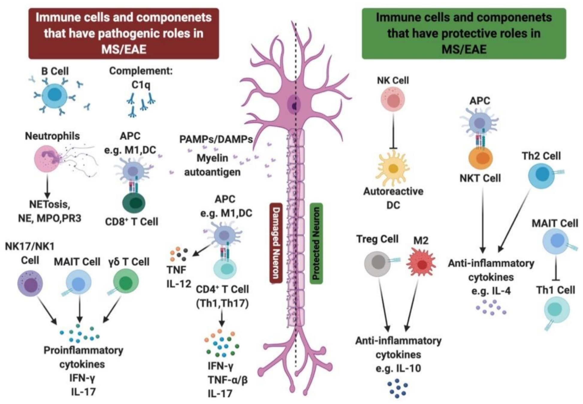
In MS and EAE, several immune cells are involved in the pathogenesis and remission of MS. In this review, we highlighted the role of the innate immune cellular players including macrophages, neutrophils, NK, NKT, γδ T, and MAIT cells as well as the complement system in the inflammatory processes associated with MS and EAE. On the other hand, T cells including CD4+, CD8+, and Tregs are suggested to be crucial contributors to MS, either exacerbating or ameliorating the disease. This is related to their autoreactivity, inflammatory cytokine secretion, and their recruitment into CNS inflammatory sites. Furthermore, the adaptive immune B cells are known to be one of the primary causes of MS development, and hence are currently targets for therapeutic agents in MS. The potential protective and pathological roles of the innate and adaptive immune systems in MS/EAE are summarized in Figure 2.

Author Contributions

S.D., M.A.-A., N.M.E. and A.A.M. wrote the manuscript. M.H.A.-A., and Z.A.-R. assisted in writing the manuscript. All authors have read and agreed to the published version of the manuscript.

Funding

This work was funded by the Terry Fox Foundation MISC051. M.A. is a recipient of L’Oréal-UNESCO for Women in Science Middle East Young Talents Programme.

Institutional Review Board Statement

Not applicable.

Informed Consent Statement

Not applicable.

Conflicts of Interest

The authors declare that the research was conducted in the absence of any commercial or financial relationships that could be construed as a potential conflict of interest.

Abbreviations

AIM2Absent in melanoma 2
APCsAntigen-presenting cells
ALLOAllopregnanolone
ASCApoptosis-associated speck-like protein
BBBBlood–brain barrier
CISClinical isolated syndrome
CNSCentral nervous system
CSFCerebrospinal fluid
CTLA-4Cytotoxic T-lymphocyte-associated protein 4
DAMPsDanger-associated molecular patterns
DCsDendritic cells
DMFDimethyl fumarate
EAEExperimental autoimmune encephalitis
G-CSFGranulocyte-colony stimulating factor
GAGlatiramer acetate
GABA-AGamma aminobutyric acid-A
GalCerGalactosylceramide
GWASGenome-wide association studies
HLAHuman leukocyte antigen
IFNInterferon
IL-Interleukin
IgImmunoglobulin
LFA-1Lymphocyte function-associated antigen 1
MAITMucosal-associated invariant T
MBPMyelin basic protein
MHCMajor histocompatibility complex
MMFMonomethyl fumarate
MOGMyelin oligodendrocyte glycoprotein
MPOMyeloperoxidase
MR1MHC-related molecule 1
MRIMagnetic resonance imaging
MSMultiple sclerosis
NETsNeutrophils’ extracellular traps
NKNatural killer
NKTNatural killer T
NLRsNOD-like receptors
NODNucleotide binding oligomerization domain
PAMPsPathogen-associated molecular patterns
PPMSPrimary progressive multiple sclerosis
PRMSProgressive relapsing multiple sclerosis
PRRsPattern recognition receptors
ROSReactive oxygen species
RRMSRelapsing remitting multiple sclerosis
SPMSSecondary progressive multiple sclerosis
TCRT cell receptor
ThT helper
TLRToll-like receptor
TNFTumor necrosis factor
TregsRegulatory T cells
VLA-4Very late antigen-4.

References

  1. Kobelt, G.; Thompson, A.; Berg, J.; Gannedahl, M.; Eriksson, J. New insights into the burden and costs of multiple sclerosis in Europe. Mult. Scler. 2017, 23, 1123–1136. [Google Scholar] [CrossRef] [PubMed]
  2. Popescu, B.F.; Pirko, I.; Lucchinetti, C.F. Pathology of multiple sclerosis. Continuum 2013, 19, 901–921. [Google Scholar] [PubMed]
  3. Lublin, F.D.; Reingold, S.C.; Cohen, J.A.; Cutter, G.R.; Sorensen, P.S.; Thompson, A.J.; Wolinsky, J.S.; Balcer, L.J.; Banwell, B.; Barkhof, F.; et al. Defining the clinical course of multiple sclerosis: The 2013 revisions. Neurology 2014, 83, 278–286. [Google Scholar] [CrossRef] [PubMed] [Green Version]
  4. Miller, D.H.; Chard, D.T.; Ciccarelli, O. Clinically isolated syndromes. Lancet Neurol. 2012, 11, 157–169. [Google Scholar] [CrossRef]
  5. Gourraud, P.; Harbo, H.F.; Hauser, S.L.; Baranzini, S.E. The genetics of multiple sclerosis: An up-to-date review. Immunol. Rev. 2012, 248, 87–103. [Google Scholar] [CrossRef] [Green Version]
  6. Holmøy, T.; Hestvik, A.L. Multiple sclerosis: Immunopathogenesis and controversies in defining the cause. Curr. Opin. Infect. Dis. 2008, 21, 271–278. [Google Scholar] [CrossRef]
  7. Egg, R.; Reindl, M.; Deisenhammer, F.; Linington, C.; Berger, T. Anti-MOG and anti-MBP antibody subclasses in multiple sclerosis. Mult. Scler. 2001, 7, 285–289. [Google Scholar] [CrossRef]
  8. Robinson, A.P.; Harp, C.T.; Noronha, A.; Miller, S.D. The experimental autoimmune encephalomyelitis (EAE) model of MS: Utility for understanding disease pathophysiology and treatment. Handb. Clin. Neurol. 2014, 122, 173–189. [Google Scholar]
  9. Giralt, M.; Molinero, A.; Hidalgo, J. Active induction of experimental autoimmune encephalomyelitis (EAE) with MOG35-55 in the mouse. Methods Mol. Biol. 2018, 1791, 227–232. [Google Scholar]
  10. Farooqi, N.; Gran, B.; Constantinescu, C.S. Are current disease-modifying therapeutics in multiple sclerosis justified on the basis of studies in experimental autoimmune encephalomyelitis? J. Neurochem. 2010, 115, 829–844. [Google Scholar] [CrossRef]
  11. Polman, C.H.; O’Connor, P.W.; Havrdova, E.; Hutchinson, M.; Kappos, L.; Miller, D.H.; Phillips, J.T.; Lublin, F.D.; Giovannoni, G.; Sandrock, A.W.; et al. A randomized, placebo-controlled trial of natalizumab for relapsing multiple sclerosis. N. Engl. J. Med. 2006, 354, 899–910. [Google Scholar] [CrossRef] [PubMed] [Green Version]
  12. Yednock, T.A.; Cannon, C.; Fritz, L.C.; Sanchez-Madrid, F.; Steinman, L.; Karin, N. Prevention of experimental autoimmune encephalomyelitis by antibodies against alpha 4 beta 1 integrin. Nature 1992, 356, 63–66. [Google Scholar] [CrossRef] [PubMed]
  13. Ridge, S.C.; Sloboda, A.E.; McReynolds, R.A.; Levine, S.; Oronsky, A.L.; Kerwar, S.S. Suppression of experimental allergic encephalomyelitis by mitoxantrone. J. Clin. Immunol. Immunopathol. Res. 1985, 35, 35–42. [Google Scholar] [CrossRef]
  14. Huang, W.; Chen, W.; Zhang, X. Multiple sclerosis: Pathology, diagnosis and treatments. Exp. Ther. Med. 2017, 13, 3163–3166. [Google Scholar] [CrossRef] [PubMed] [Green Version]
  15. Ghasemi, N.; Razavi, S.; Nikzad, E. Multiple sclerosis: Pathogenesis, symptoms, diagnoses and cell-based therapy. Cell J. 2016, 19, 1–10. [Google Scholar]
  16. Calabresi, P.A. Diagnosis and management of multiple sclerosis. Am. Fam. Physician 2004, 70, 1935–1944. [Google Scholar]
  17. Olson, J.K.; Miller, S.D. The innate immune response affects the development of the autoimmune response in Theiler’s virus-induced demyelinating disease. J. Immunol. 2009, 182, 5712–5722. [Google Scholar] [CrossRef]
  18. Tosi, M.F. Innate immune responses to infection. J. Allergy Clin. Immunol. 2005, 116, 241–250. [Google Scholar] [CrossRef]
  19. Deerhake, M.E.; Biswas, D.D.; Barclay, W.E.; Shinohara, M.L. Pattern recognition receptors in multiple sclerosis and its animal models. Front. Immunol. 2019, 10, 2644. [Google Scholar] [CrossRef] [Green Version]
  20. Pone, E.J.; Zan, H.; Zhang, J.; Al-Qahtani, A.; Xu, Z.; Casali, P. Toll-like receptors and B-cell receptors synergize to induce immunoglobulin class-switch DNA recombination: Relevance to microbial antibody responses. Crit. Rev. Immunol. 2010, 30, 1–29. [Google Scholar] [CrossRef] [Green Version]
  21. Wu, J.; Meng, Z.; Jiang, M.; Zhang, E.; Trippler, M.; Broering, R.; Bucchi, A.; Krux, F.; Dittmer, U.; Yang, D.; et al. Toll-like receptor-induced innate immune responses in non-parenchymal liver cells are cell type-specific. Immunology 2010, 129, 363–374. [Google Scholar] [CrossRef] [PubMed]
  22. Suresh, R.; Mosser, D.M. Pattern recognition receptors in innate immunity, host defense, and immunopathology. Adv. Physiol. Educ. 2013, 37, 284–291. [Google Scholar] [CrossRef] [PubMed]
  23. Kigerl, K.A.; Lai, W.; Rivest, S.; Hart, R.P.; Satoskar, A.R.; Popovich, P.G. Toll-like receptor (TLR)-2 and TLR-4 regulate inflammation, gliosis, and myelin sparing after spinal cord injury. J. Neurochem. 2007, 102, 37–50. [Google Scholar] [CrossRef] [PubMed]
  24. Sloane, J.A.; Batt, C.; Ma, Y.; Harris, Z.; Trapp, B.; Vartanian, T. Hyaluronan blocks oligodendrocyte progenitor maturation and remyelination through TLR2. Proc. Natl. Acad. Sci. USA 2010, 107, 11555–11560. [Google Scholar] [CrossRef] [PubMed] [Green Version]
  25. Hanafy, K.H.; Sloane, J.A. Regulation of remyelination in multiple sclerosis. FEBS Lett. 2011, 585, 3821–3828. [Google Scholar] [CrossRef] [PubMed] [Green Version]
  26. Back, S.A.; Tuohy, T.M.; Chen, H.; Wallingford, N.; Craig, A.; Struve, J.; Luo, N.; Banine, F.; Liu, Y.; Chang, A.; et al. Hyaluronan accumulates in demyelinated lesions and inhibits oligodendrocyte progenitor maturation. Nat. Med. 2005, 11, 966–972. [Google Scholar] [CrossRef]
  27. Zheng, C.; Chen, J.; Chu, F.; Zhu, J.; Jin, T. Inflammatory role of TLR-Myd88 signaling in multiple sclerosis. Front. Mol. Neurosci. 2020, 12, 314. [Google Scholar] [CrossRef]
  28. Marta, M. Toll-like receptors in multiple sclerosis mouse experimental models. Ann. N. Y. Acad. Sci. 2009, 1173, 458–462. [Google Scholar] [CrossRef]
  29. Touil, T.; Fitzgerald, D.; Zhang, G.; Rostami, A.; Gran, B. Cutting Edge: TLR3 stimulation suppresses experimental autoimmune encephalomyelitis by inducing endogenous IFN-β. J. Immunol. 2006, 177, 7505–7509. [Google Scholar] [CrossRef] [Green Version]
  30. Giacomini, E.; Severa, M.; Rizzo, F.; Mechelli, R.; Annibali, V.; Ristori, G.; Riccieri, V.; Salvetti, M.; Coccia, E.M. IFN-β therapy modulates B-cell and monocyte crosstalk via TLR7 in multiple sclerosis patients. Eur. J. Immunol. 2013, 43, 1963–1972. [Google Scholar] [CrossRef]
  31. Zhang, X.; Jin, J.; Tang, Y.; Speer, D.; Sujkowska, D.; Markovic-Plese, S. IFN-β1a inhibits the secretion of Th17-polarizing cytokines in human dendritic cells via TLR7 up-regulation. J. Immunol. 2009, 182, 3928–3936. [Google Scholar] [CrossRef] [PubMed] [Green Version]
  32. Kim, Y.; Shin, J.; Nahm, M.H. NOD-like receptors in infection, immunity, and diseases. Yonsei Med. J. 2016, 57, 5. [Google Scholar] [CrossRef] [PubMed] [Green Version]
  33. Hachim, M.Y.; Khalil, B.A.; Elemam, N.M.; Maghazachi, A.A. Pyroptosis: The missing puzzle among innate and adaptive immunity crosstalk. J. Leukoc. Biol. 2020, 108, 323–338. [Google Scholar] [CrossRef] [PubMed]
  34. Franchi, L.; Eigenbrod, T.; Muñoz-Planillo, R.; Nuñez, G. The inflammasome: A caspase-1-activation platform that regulates immune responses and disease pathogenesis. Nat. Immunol. 2009, 10, 241–247. [Google Scholar] [CrossRef]
  35. Hernández-Pedro, N.Y.; Espinosa-Ramirez, G.; de la Cruz, V.; Pineda, B.; Sotelo, J. Initial immunopathogenesis of multiple sclerosis: Innate immune response. Clin. Dev. Immunol. 2013, 2013, 1–15. [Google Scholar] [CrossRef] [Green Version]
  36. Muhammad, J.S.; Jayakumar, M.N.; Elemam, N.M.; Venkatachalam, T.; Raju, T.K.; Hamoudi, R.A.; Maghazachi, A.A. Gasdermin D hypermethylation inhibits pyroptosis and LPS-induced IL-1β release from NK92 cells. ImmunoTargets Ther. 2019, 8, 29–41. [Google Scholar] [CrossRef] [Green Version]
  37. Yap, J.K.; Pickard, B.S.; Chan, E.W.; Gan, S. The role of neuronal NLRP1 inflammasome in Alzheimer’s disease: Bringing neurons into the neuroinflammation game. Mol. Neurobiol. 2019, 56, 7741–7753. [Google Scholar] [CrossRef]
  38. Tan, M.; Yu, J.; Jiang, T.; Zhu, X.; Tan, L. The NLRP3 inflammasome in alzheimer’s disease. Mol. Neurobiol. 2013, 48, 875–882. [Google Scholar] [CrossRef]
  39. Liu, F.; Li, Z.; He, X.; Yu, H.; Feng, J. Ghrelin attenuates neuroinflammation and demyelination in experimental autoimmune encephalomyelitis involving NLRP3 inflammasome signaling pathway and pyroptosis. Front. Pharmacol. 2019, 10, 1320. [Google Scholar] [CrossRef] [Green Version]
  40. Gaudino, S.J.; Kumar, P. Cross-talk between antigen presenting cells and T cells impacts intestinal homeostasis, bacterial infections, and tumorigenesis. Front. Immunol. 2019, 10, 360. [Google Scholar] [CrossRef] [Green Version]
  41. Raphael, I.; Nalawade, S.; Eagar, T.N.; Forsthuber, T.G. T cell subsets and their signature cytokines in autoimmune and inflammatory diseases. Cytokine 2015, 74, 5–17. [Google Scholar] [CrossRef] [PubMed] [Green Version]
  42. Mayo, L.; Quintana, F.J.; Weiner, H.L. The innate immune system in demyelinating disease. Immunol. Rev. 2012, 248, 170–187. [Google Scholar] [CrossRef] [PubMed]
  43. Vogel, D.Y.; Vereyken, E.J.; Glim, J.E.; Heijnen, P.D.; Moeton, M.; van der Valk, P.; Amor, S.; Teunissen, C.E.; van Horssen, J.; Dijkstra, C.D. Macrophages in inflammatory multiple sclerosis lesions have an intermediate activation status. J. Neuroinflamm. 2013, 10, 809. [Google Scholar] [CrossRef] [Green Version]
  44. Vainchtein, I.D.; Vinet, J.; Brouwer, N.; Brendecke, S.; Biagini, G.; Biber, K.; Boddeke, H.W.; Eggen, B.J. In acute experimental autoimmune encephalomyelitis, infiltrating macrophages are immune activated, whereas microglia remain immune suppressed. Glia 2014, 62, 1724–1735. [Google Scholar] [CrossRef] [PubMed]
  45. Chu, F.; Shi, M.; Zheng, C.; Shen, D.; Zhu, J.; Zheng, X.; Cui, L. The roles of macrophages and microglia in multiple sclerosis and experimental autoimmune encephalomyelitis. J. Neuroimmunol. 2018, 318, 1–7. [Google Scholar] [CrossRef]
  46. Karni, A.; Abraham, M.; Monsonego, A.; Cai, G.; Freeman, G.J.; Hafler, D.; Khoury, S.J.; Weiner, H.L. Innate immunity in multiple sclerosis: Myeloid dendritic cells in secondary progressive multiple sclerosis are activated and drive a proinflammatory immune response. J. Immunol. 2006, 177, 4196–4202. [Google Scholar] [CrossRef] [Green Version]
  47. Noorbakhsh, F.; Baker, G.B.; Power, C. Allopregnanolone and neuroinflammation: A focus on multiple sclerosis. Front. Cell Neurosci. 2014, 8, 134. [Google Scholar] [CrossRef] [Green Version]
  48. Reyes-García, M.G.; Hernández-Hernández, F.; Hernández-Téllez, B.; García-Tamayo, F. GABA receptor subunits RNA expression in mice peritoneal macrophages modulate their IL-6/IL-12 production. J. Neuroimmunol. 2007, 188, 64–68. [Google Scholar] [CrossRef]
  49. Bhat, R.; Axtell, R.; Mitra, A.; Miranda, M.; Lock, C.; Tsien, R.W.; Steinman, L. Inhibitory role for GABA in autoimmune inflammation. Proc. Natl. Acad. Sci. USA 2010, 107, 2580–2585. [Google Scholar] [CrossRef] [Green Version]
  50. Legroux, L.; Arbour, N. Multiple sclerosis and T lymphocytes: An entangled story. J. Neuroimmune Pharmacol. 2015, 10, 528–546. [Google Scholar] [CrossRef] [Green Version]
  51. Rumble, J.M.; Huber, A.K.; Krishnamoorthy, G.; Srinivasan, A.; Giles, D.A.; Zhang, X.; Wang, L.; Segal, B.M. Neutrophil-related factors as biomarkers in EAE and MS. J. Exp. Med. 2015, 212, 23–35. [Google Scholar] [CrossRef] [PubMed] [Green Version]
  52. He, Y.; Yang, F.; Sun, E. Neutrophil extracellular traps in autoimmune diseases. Chin. Med. J. 2018, 131, 1513–1519. [Google Scholar] [CrossRef] [PubMed]
  53. Strzepa, A.; Dittel, B.N. Inflammatory disease severity is ameliorated by inhibition of neutrophil-derived MPO that supports endothelial/epithelial integrity. J. Immunol. 2017, 198, 127. [Google Scholar]
  54. Yu, G.; Zheng, S.; Zhang, H. Inhibition of myeloperoxidase by N-acetyl lysyltyrosylcysteine amide reduces experimental autoimmune encephalomyelitis-induced injury and promotes oligodendrocyte regeneration and neurogenesis in a murine model of progressive multiple sclerosis. NeuroReport 2018, 29, 208–213. [Google Scholar] [CrossRef] [PubMed] [Green Version]
  55. Zhang, H.; Ray, A.; Miller, N.M.; Hartwig, D.; Pritchard, K.A.; Dittel, B.N. Inhibition of myeloperoxidase at the peak of experimental autoimmune encephalomyelitis restores blood-brain barrier integrity and ameliorates disease severity. J. Neurochem. 2015, 136, 826–836. [Google Scholar] [CrossRef] [PubMed] [Green Version]
  56. Herges, K.; de Jong, B.A.; Kolkowitz, I.; Dunn, C.; Mandelbaum, G.; Ko, R.M.; Maini, A.; Han, M.H.; Killestein, J.; Polman, C.; et al. Protective effect of an elastase inhibitor in a neuromyelitis optica-like disease driven by a peptide of myelin oligodendroglial glycoprotein. Mult. Scler. 2012, 18, 398–408. [Google Scholar] [CrossRef] [PubMed] [Green Version]
  57. Minohara, M.; Matsuoka, T.; Li, W.; Osoegawa, M.; Ishizu, T.; Ohyagi, Y.; Kira, J. Upregulation of myeloperoxidase in patients with opticospinal multiple sclerosis: Positive correlation with disease severity. J. Neuroimmunol. 2006, 178, 156–160. [Google Scholar] [CrossRef]
  58. Maghazachi, A.A. Compartmentalization of human natural killer cells. Mol. Immunol. 2005, 42, 523–529. [Google Scholar] [CrossRef]
  59. Glimcher, L.; Shen, F.W.; Cantor, H. Identification of a cell-surface antigen selectively expressed on the natural killer cell. J. Exp. Med. 1977, 145, 1–9. [Google Scholar] [CrossRef] [Green Version]
  60. Maghazachi, A.A. Role of natural killer cells in multiple sclerosis. ISRN Immunol. 2012, 2012, 1–14. [Google Scholar] [CrossRef]
  61. Høglund, R.A.; Maghazachi, A.A. Multiple sclerosis and the role of immune cells. World J. Exp. Med. 2014, 4, 27. [Google Scholar] [CrossRef] [PubMed]
  62. Pandya, A.D.; Al-Jaderi, Z.; Høglund, R.A.; Holmøy, T.; Harbo, H.F.; Norgauer, J.; Maghazachi, A.A. Identification of human NK17/NK1 cells. PLoS ONE 2011, 6, e26780. [Google Scholar] [CrossRef] [PubMed] [Green Version]
  63. Sand, K.L.; Knudsen, E.; Rolin, J.; Al-Falahi, Y.; Maghazachi, A.A. Modulation of natural killer cell cytotoxicity and cytokine release by the drug glatiramer acetate. Cell. Mol. Life Sci. 2009, 66, 1446–1456. [Google Scholar] [CrossRef] [PubMed]
  64. Al-Jaderi, Z.; Maghazachi, A.A. Effects of vitamin D3, calcipotriol and FTY720 on the expression of surface molecules and cytolytic activities of human natural killer cells and dendritic cells. Toxins 2013, 5, 1932–1947. [Google Scholar] [CrossRef] [PubMed] [Green Version]
  65. Maghazachi, A.A.; Sand, K.L.; Al-Jaderi, Z. Glatiramer acetate, dimethyl fumarate, and monomethyl fumarate upregulate the expression of CCR10 on the surface of natural killer cells and enhance their chemotaxis and cytotoxicity. Front. Immunol. 2016, 7, 437. [Google Scholar] [CrossRef] [PubMed] [Green Version]
  66. Vego, H.; Sand, K.L.; Høglund, R.A.; Fallang, L.; Gundersen, G.; Holmøy, T.; Maghazachi, A.A. Monomethyl fumarate augments NK cell lysis of tumor cells through degranulation and the upregulation of Nkp46 and CD107a. Cell. Mol. Immunol. 2016, 13, 57–64. [Google Scholar] [CrossRef]
  67. Al-Jaderi, Z.; Maghazachi, A.A. Vitamin D3 and monomethyl fumarate enhance natural killer cell lysis of dendritic cells and ameliorate the clinical score in mice suffering from experimental autoimmune encephalomyelitis. Toxins 2015, 7, 4730–4744. [Google Scholar] [CrossRef]
  68. Munger, K.L.; Zhang, S.M.; O’Reilly, E.; Hernán, M.A.; Olek, M.J.; Willett, W.C.; Ascherio, A. Vitamin D intake and incidence of multiple sclerosis. Neurology 2004, 62, 60–65. [Google Scholar] [CrossRef]
  69. Munger, K.L.; Levin, L.I.; Hollis, B.W.; Howard, N.S.; Ascherio, A. Serum 25-hydroxyvitamin D levels and risk of multiple sclerosis. JAMA 2006, 296, 2832–2838. [Google Scholar] [CrossRef] [Green Version]
  70. Høglund, R.A.; Holmøy, T.; Harbo, H.F.; Maghazachi, A.A. A one year follow-up study of natural killer and dendritic cells activities in multiple sclerosis patients receiving glatiramer acetate (GA). PLoS ONE 2013, 8, e62237. [Google Scholar]
  71. Balato, A.; Unutmaz, D.; Gaspari, A.A. Natural killer T cells: An unconventional T-cell subset with diverse effector and regulatory functions. J. Investig. Dermatol. 2009, 129, 1628–1642. [Google Scholar] [CrossRef] [PubMed] [Green Version]
  72. Cui, Y.; Wan, Q. NKT cells in neurological diseases. Front. Cell. Neurosci. 2019, 13, 245. [Google Scholar] [CrossRef] [PubMed]
  73. Latha, T.S.; Reddy, M.C.; Durbaka, P.V.; Rachamallu, A.; Pallu, R.; Lomada, D. γδ T cell-mediated immune responses in disease and therapy. Front. Immunol. 2014, 5, 571. [Google Scholar] [CrossRef] [PubMed] [Green Version]
  74. Rajan, A.J.; Gao, Y.L.; Raine, C.S.; Brosnan, C.F. A pathogenic role for gamma delta T cells in relapsing-remitting experimental allergic encephalomyelitis in the SJL mouse. J. Immunol. 1996, 157, 941–949. [Google Scholar] [PubMed]
  75. Spahn, T.W.; Issazadah, S.; Salvin, A.J.; Weiner, H.L. Decreased severity of myelin oligodendrocyte glycoprotein peptide 33–35-induced experimental autoimmune encephalomyelitis in mice with a disrupted TCR δ chain gene. Eur. J. Immunol. 1999, 29, 4060–4071. [Google Scholar] [CrossRef]
  76. Treiner, E.; Liblau, R.S. Mucosal-associated invariant T cells in multiple sclerosis: The jury is still out. Front. Immunol. 2015, 6, 503. [Google Scholar] [CrossRef] [PubMed] [Green Version]
  77. Le Bourhis, L.; Guerri, L.; Dusseaux, M.; Martin, E.; Soudais, C.; Lantz, O. Mucosal-associated invariant T cells: Unconventional development and function. Trends Immunol. 2011, 32, 212–218. [Google Scholar] [CrossRef] [PubMed]
  78. Miyazaki, Y.; Miyake, S.; Chiba, A.; Lantz, O.; Yamamura, T. Mucosal-associated invariant T cells regulate Th1 response in multiple sclerosis. Int. Immunol. 2011, 23, 529–535. [Google Scholar] [CrossRef] [PubMed]
  79. Barnum, S.R. Complement biosynthesis in the central nervous system. Crit. Rev. Oral Biol. Med. 1995, 6, 132–146. [Google Scholar] [CrossRef]
  80. Wren, D.R.; Noble, M. Oligodendrocytes and oligodendrocyte/type-2 astrocyte progenitor cells of adult rats are specifically susceptible to the lytic effects of complement in absence of antibody. Proc. Natl. Acad. Sci. USA 1989, 86, 9025–9029. [Google Scholar] [CrossRef] [Green Version]
  81. Johns, T.G.; Bernard, C.C. Binding of complement component Clq to myelin oligodendrocyte glycoprotein: A novel mechanism for regulating CNS inflammation. Mol. Immunol. 1997, 34, 33–38. [Google Scholar] [CrossRef]
  82. Bhat, R.; Steinman, L. Innate and adaptive autoimmunity directed to the central nervous system. Neuron 2009, 64, 123–132. [Google Scholar] [CrossRef] [PubMed] [Green Version]
  83. Goverman, J. Autoimmune T cell responses in the central nervous system. Nat. Rev. Immunol. 2009, 9, 393–407. [Google Scholar] [CrossRef] [PubMed] [Green Version]
  84. Constantinescu, C.S.; Farooqi, N.; O’Brien, K.; Gran, B. Experimental autoimmune encephalomyelitis (EAE) as a model for multiple sclerosis (MS). Br. J. Pharmacol. 2011, 164, 1079–1106. [Google Scholar] [CrossRef] [PubMed]
  85. Fujinami, R.S.; Oldstone, M.B. Amino acid homology between the encephalitogenic site of myelin basic protein and virus: Mechanism for autoimmunity. Science 1985, 230, 1043–1045. [Google Scholar] [CrossRef] [PubMed]
  86. Getts, D.R.; Chastain, E.M.; Terry, R.L.; Miller, S.D. Virus infection, antiviral immunity, and autoimmunity. Immunol. Rev. 2013, 255, 197–209. [Google Scholar] [CrossRef] [Green Version]
  87. Keller, C.W.; Sina, C.; Kotur, M.B.; Ramelli, G.; Mundt, S.; Quast, I.; Ligeon, L.; Weber, P.; Becher, B.; Münz, C.; et al. ATG-dependent phagocytosis in dendritic cells drives myelin-specific CD4+ T cell pathogenicity during CNS inflammation. Proc. Natl. Acad. Sci. USA 2017, 114, e11228–e11237. [Google Scholar] [CrossRef] [Green Version]
  88. Lovett-Racke, A.E.; Yang, Y.; Racke, M.K. Th1 versus Th17: Are T cell cytokines relevant in multiple sclerosis? Biochim. Biophys. Acta 2011, 1812, 246–251. [Google Scholar] [CrossRef] [Green Version]
  89. Takeshita, Y.; Ransohoff, R.M. Inflammatory cell trafficking across the blood-brain barrier: Chemokine regulation and in vitro models. Immunol. Rev. 2012, 248, 228–239. [Google Scholar] [CrossRef] [Green Version]
  90. Gutcher, I.; Becher, B. APC-derived cytokines and T cell polarization in autoimmune inflammation. J. Clin. Investig. 2007, 117, 1119–1127. [Google Scholar] [CrossRef] [Green Version]
  91. Panitch, H.S.; Hirsch, R.L.; Haley, A.S.; Johnson, K.P. Exacerbations of multiple sclerosis in patients treated with gamma interferon. Lancet 1987, 1, 893–895. [Google Scholar] [CrossRef]
  92. Korn, T.; Bettelli, E.; Oukka, M.; Kuchroo, V.K. IL-17 and Th17 cells. Annu. Rev. Immunol. 2009, 27, 485–517. [Google Scholar] [CrossRef] [PubMed]
  93. Lock, C.; Hermans, G.; Pedotti, R.; Brendolan, A.; Schadt, E.; Garren, H.; Langer-Gould, A.; Strober, S.; Cannella, B.; Allard, J.; et al. Gene-microarray analysis of multiple sclerosis lesions yields new targets validated in autoimmune encephalomyelitis. Nat. Med. 2002, 8, 500–508. [Google Scholar] [CrossRef]
  94. Langrish, C.L.; Chen, Y.; Blumenschein, W.M.; Mattson, J.; Basham, B.; Sedgwick, J.D.; McClanahan, T.; Kastelein, R.A.; Cua, D.J. IL-23 drives a pathogenic T cell population that induces autoimmune inflammation. J. Exp. Med. 2005, 201, 233–240. [Google Scholar] [CrossRef] [PubMed] [Green Version]
  95. Fletcher, J.M.; Lalor, S.J.; Sweeney, C.M.; Tubridy, N.; Mills, K.H. T cells in multiple sclerosis and experimental autoimmune encephalomyelitis. Clin. Exp. Immunol. 2010, 162, 1–11. [Google Scholar] [CrossRef]
  96. Segal, B.M. The diversity of encephalitogenic CD4+ T cells in multiple sclerosis and its animal models. J. Clin. Med. 2019, 8, 120. [Google Scholar] [CrossRef] [Green Version]
  97. Salou, M.; Nicol, B.; Garcia, A.; Laplaud, D.A. Involvement of CD8+ T cells in multiple sclerosis. Front. Immunol. 2015, 6, 604. [Google Scholar] [CrossRef] [Green Version]
  98. Babbe, H.; Roers, A.; Waisman, A.; Lassmann, H.; Goebels, N.; Hohlfeld, R.; Friese, M.; Schröder, R.; Deckert, M.; Schmidt, S.; et al. Clonal expansions of Cd8+ T cells dominate the T cell infiltrate in active multiple sclerosis lesions as shown by micromanipulation and single cell polymerase chain reaction. J. Exp. Med. 2000, 192, 393–404. [Google Scholar] [CrossRef] [Green Version]
  99. Chevalier, G.; Suberbielle, E.; Monnet, C.; Duplan, V.; Martin-Blondel, G.; Farrugia, F.; Le Masson, G.; Liblau, R.; Gonzalez-Dunia, D. Neurons are MHC class I-dependent targets for CD8 T cells upon neurotropic viral infection. PLoS Pathog 2011, 7, e1002393. [Google Scholar] [CrossRef]
  100. Booss, J.; Esiri, M.M.; Tourtellotte, W.W.; Mason, D.Y. Immunohistological analysis of T lymphocyte subsets in the central nervous system in chronic progressive multiple sclerosis. J. Neurol Sci. 1983, 62, 219–232. [Google Scholar] [CrossRef]
  101. Lucchinetti, C.F.; Popescu, B.F.; Bunyan, R.F.; Moll, N.M.; Roemer, S.F.; Lassmann, H.; Brück, W.; Parisi, J.E.; Scheithauer, B.W.; Weigand, S.D.; et al. Inflammatory cortical demyelination in early multiple sclerosis. N. Engl. J. Med. 2011, 365, 2188–2197. [Google Scholar] [CrossRef] [PubMed] [Green Version]
  102. Battistini, L.; Piccio, L.; Rossi, B.; Bach, S.; Galgani, S.; Gasperini, C.; Ottoboni, L.; Ciabini, D.; Caramia, M.D.; Bernardi, G.; et al. CD8+ T cells from patients with acute multiple sclerosis display selective increase of adhesiveness in brain venules: A critical role for P-selectin glycoprotein ligand-1. Blood 2003, 101, 4775–4782. [Google Scholar] [CrossRef] [PubMed] [Green Version]
  103. Sun, D.; Whitaker, J.N.; Huang, Z.; Liu, D.; Coleclough, C.; Wekerle, H.; Raine, C.S. Myelin antigen-specific CD8+ T cells are encephalitogenic and produce severe disease in C57BL/6 mice. J. Immunol. 2001, 166, 7579–7587. [Google Scholar]
  104. Najafian, N.; Chitnis, T.; Salama, A.D.; Zhu, B.; Benou, C.; Yuan, X.; Clarkson, M.R.; Sayegh, M.H.; Khoury, S.J. Regulatory functions of CD8+CD28-T cells in an autoimmune disease model. J. Clin. Investig. 2003, 112, 1037–1048. [Google Scholar] [CrossRef] [PubMed]
  105. York, N.R.; Mendoza, J.P.; Ortega, S.B.; Benagh, A.; Tyler, A.F.; Firan, M.; Karandikar, N.J. Immune regulatory CNS-reactive CD8+T cells in experimental autoimmune encephalomyelitis. J. Autoimmun. 2010, 35, 33–44. [Google Scholar] [CrossRef] [PubMed] [Green Version]
  106. Sinha, S.; Boyden, A.W.; Itani, F.R.; Crawford, M.P.; Karandikar, N.J. CD8(+) T-cells as immune regulators of multiple sclerosis. Front. Immunol. 2015, 6, 619. [Google Scholar] [CrossRef] [Green Version]
  107. He, F.; Balling, R. The role of regulatory T cells in neurodegenerative diseases. Wiley Interdiscip. Rev. Syst. Biol. Med. 2013, 5, 153–180. [Google Scholar] [CrossRef]
  108. Danikowski, K.M.; Jayaraman, S.; Prabhakar, B.S. Regulatory T cells in multiple sclerosis and myasthenia gravis. J. Neuroinflamm. 2017, 14, 117. [Google Scholar] [CrossRef] [Green Version]
  109. Kipnis, J.; Avidan, H.; Caspi, R.R.; Schwartz, M. Dual effect of CD4+CD25+ regulatory T cells in neurodegeneration: A dialogue with microglia. Proc. Natl. Acad. Sci. USA 2004, 101, 14663–14669. [Google Scholar] [CrossRef] [Green Version]
  110. Walsh, J.T.; Kipnis, J. Regulatory T cells in CNS injury: The simple, the complex and the confused. Trends Mol. Med. 2011, 17, 541–547. [Google Scholar] [CrossRef] [Green Version]
  111. Calvo-Barreiro, L.; Eixarch, H.; Montalban, X.; Espejo, C. Combined therapies to treat complex diseases: The role of the gut microbiota in multiple sclerosis. Autoimmun. Rev. 2018, 17, 165–174. [Google Scholar] [CrossRef] [PubMed]
  112. Cekanaviciute, E.; Yoo, B.B.; Runia, T.F.; Debelius, J.W.; Singh, S.; Nelson, C.A.; Kanner, R.; Bencosme, Y.; Lee, Y.K.; Hauser, S.L.; et al. Gut bacteria from multiple sclerosis patients modulate human T cells and exacerbate symptoms in mouse models. Proc. Natl. Acad. Sci. USA 2017, 114, 10713–10718. [Google Scholar] [CrossRef] [PubMed] [Green Version]
  113. Wurtman, R. Multiple sclerosis, melatonin, and neurobehavioral diseases. Front. Endocrinol. 2017, 8, 280. [Google Scholar] [CrossRef] [PubMed] [Green Version]
  114. Hardy, R.R.; Carmack, C.E.; Shinton, S.A.; Kemp, J.D.; Hayakawa, K. Resolution and characterization of pro-B and pre–pro-B cell stages in normal mouse bone marrow. J. Exp. Med. 1991, 173, 1213–1225. [Google Scholar] [CrossRef] [PubMed] [Green Version]
  115. Michel, L.; Touil, H.; Pikor, N.B.; Gommerman, J.L.; Prat, A.; Bar-Or, A. B cells in the multiple sclerosis central nervous system: Trafficking and contribution to CNS-compartmentalized inflammation. Front. Immunol. 2015, 6, 636. [Google Scholar] [CrossRef] [Green Version]
  116. Al-ani, M.R.; Raju, T.K.; Hachim, M.Y.; Hachim, I.Y.; Elemam, N.M.; Guimei, M.; Bendardaf, R.; Maghazachi, A.A. Rituximab prevents the development of experimental autoimmune encephalomyelitis (EAE): Comparison with prophylactic, therapeutic or combinational regimens. J. Inflamm. Res. 2020, 13, 151–164. [Google Scholar] [CrossRef] [Green Version]
  117. Lin, M.; Wang, Z.; Han, X. B cells with regulatory function in animal models of autoimmune and non-autoimmune diseases. Open J. Immunol. 2015, 5, 9–17. [Google Scholar] [CrossRef] [Green Version]
  118. Kowarik, M.C.; Cepok, S.; Sellner, J.; Grummel, V.; Weber, M.S.; Korn, T.; Berthele, A.; Hemmer, B. CXCL13 is the major determinant for B cell recruitment to the CSF during neuroinflammation. J. Neuroinflamm. 2012, 9, 93. [Google Scholar] [CrossRef] [Green Version]
  119. Häusser-Kinzel, S.; Weber, M.S. The role of B Cells and antibodies in multiple sclerosis, neuromyelitis optica, and related disorders. Front. Immunol. 2019, 10, 201. [Google Scholar] [CrossRef]
  120. Duddy, M.; Niino, M.; Adatia, F.; Hebert, S.; Freedman, M.; Atkins, H.; Kim, H.J.; Bar-Or, A. Distinct effector cytokine profiles of memory and naive human B cell subsets and implication in multiple sclerosis. J. Immunol. 2007, 178, 6092–6099. [Google Scholar] [CrossRef] [Green Version]
  121. Pikor, N.B.; Prat, A.; Bar-Or, A.; Gommerman, J.L. Meningeal tertiary lymphoid tissues and multiple sclerosis: A gathering place for diverse types of immune cells during CNS autoimmunity. Front. Immunol. 2016, 6, 657. [Google Scholar] [CrossRef] [PubMed] [Green Version]
  122. Negron, A.; Robinson, R.R.; Stüve, O.; Forsthuber, T.G. The role of B cells in multiple sclerosis: Current and future therapies. Cell Immunol. 2019, 339, 10–23. [Google Scholar] [CrossRef] [PubMed]
  123. Wolf, S.D.; Dittel, B.N.; Hardardottir, F.; Janeway, C.A., Jr. Experimental autoimmune encephalomyelitis induction in genetically B cell-deficient mice. J. Exp. Med. 1996, 184, 2271–2278. [Google Scholar] [CrossRef] [PubMed] [Green Version]
  124. Fillatreau, S.; Sweenie, C.H.; McGeachy, M.J.; Gray, D.; Anderton, S.M. B cells regulate autoimmunity by provision of IL-10. Nat. Immunol. 2002, 3, 944–950. [Google Scholar] [CrossRef] [PubMed]
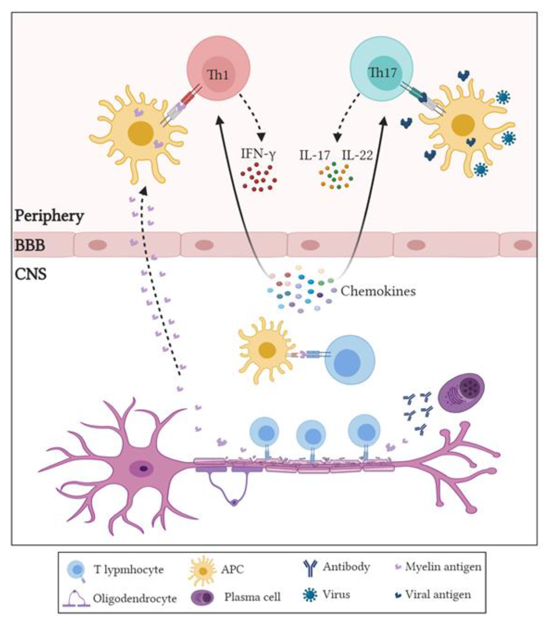
Figure 1. Involvement of various immune players in the pathogenesis of multiple sclerosis. There are two main hypotheses for the initial triggering of MS autoimmune response: either infectious antigens (mostly viral) that have molecular similarity (mimicry) with CNS antigens are engulfed by dendritic cells (DCs) or macrophages and presented to CD4+ T cells in the periphery, or stress such as inflammation, genetics, and some environmental factors cause damage to the myelin protein of the axons, releasing myelin antigens that bypass the blood–brain barrier (BBB) and induce an autoimmune response in the periphery. These events activate CD4+ T cells, which produce inflammatory cytokines and differentiate into Th1 that produce IFN-γ, or T helper 17 (Th17) cells producing interleukin 17 (IL-17) or interleukin 22 (IL-22), which permeabilize the BBB. Released chemokines inside the brain recruit immune cells including B cells, T cells, and monocytes/macrophages from the periphery into the CNS through the permeabilized BBB. Migrating immune cells encounter the cognate myelin antigen, which is presented by CNS resident cells or migrating antigen-presenting cells. Activated autoreactive T cells induce direct and indirect damage to the CNS, while B cells differentiate into plasma cells, producing autoantibodies against myelin proteins, which can activate complement or induce antibody-dependent cytotoxicity.
Sci 03 00012 g001
Figure 2. Pathogenic and protective roles of innate and adaptive immune cells and components in multiple sclerosis (MS) and experimental autoimmune encephalomyelitis (EAE). Innate and adaptive immune cells have important pathogenic roles that lead to the development and worsening of MS and EAE. Antigen-presenting cells such as macrophages (in the classical inflammatory phenotype, M1) and DCs phagocytose the DAMPs or PAMPs and the myelin autoantigens released from the damaged myelin in CNS. This leads to the activation of CD4+ T cells such as Th1 and Th17 to secrete pro-inflammatory factors such as IFN-γ, TNF-α/β, and IL-17, in addition to the inflammatory factors secreted by macrophages and DCs, including TNF and IL-12. Other immune cells (NK17/NK1, γδ T cells, and MAIT cells) release inflammatory cytokines, while B cells secrete antibodies against the myelin autoantigens, which bind the C1q complement, resulting in the activation of the complement classical pathway. Neutrophils start the “NETosis” process and form NETs, releasing factors such as neutrophil elastase (NE) and MPO. All these inflammatory mediators further exacerbate the inflammatory responses in the CNS of MS patients or EAE animal models. On the other hand, studies have shown that there are several immune cells that may have protective effects against MS and EAE through the secretion of anti-inflammatory cytokines. These include Treg, M2, NKT, and Th2 cells, or possibly through the lysis of autoreactive DCs by NK cells, thus preventing them from acting as antigen-presenting cells and inhibiting the inflammatory responses, or through inhibiting pathogenic Th1 cells responses by MAIT cells. APC: antigen-presenting cell, MAIT: mucosal-associated invariant T, MPO: myeloperoxidase, NETs: neutrophils’ extracellular traps, NE: neutrophil elastase.
Sci 03 00012 g002
Publisher’s Note: MDPI stays neutral with regard to jurisdictional claims in published maps and institutional affiliations.