Article

The Influence of Ionizing Radiation on the Morphological Structure of the Fluoride-Releasing Restorative Materials in Cancer Patients: An In Vitro Study

1
Department of Endodontics and Restorative Dental Medicine, School of Dental Medicine, 10000 Zagreb, Croatia
2
Department of Oral Medicine, School of Dental Medicine, 10000 Zagreb, Croatia
3
Department of Orthodontics, School of Dental Medicine, 10000 Zagreb, Croatia
4
Department of Oncology, Radiotherapy Unit, University Hospital Centre Zagreb, 10000 Zagreb, Croatia
*
Author to whom correspondence should be addressed.
Submission received: 21 May 2024 / Revised: 29 July 2024 / Accepted: 5 August 2024 / Published: 7 August 2024

Abstract

:
Radiotherapy plays a key role in the treatment of the early and advanced stages of head and neck cancer. To date, there is still no consensus on the effects of radiotherapy on the mechanical properties of fluoride-releasing restorative materials which can be used in patients undergoing radiotherapy with increased incidence of caries. The fluoride-releasing materials Equia Forte HT and Cention N were compared to the resin-based materials Tetric EvoCeram and Tetric Power Fill. Standard irradiation was performed with a linear accelerator. Vickers microhardness, mass, surface roughness and color were measured before and after irradiation. Cention N and Tetric PowerFill showed stability in the mass, while the surface roughness did not change in any of the examined groups. Resistance to microhardness change was shown by Cention N, Tetric PowerFill and Tetric EvoCeram, and the color change was significant in all groups (p < 0.05). It should be remembered that patients receiving head and neck radiation therapy may experience adverse effects from the treatment, including changes in the mechanical properties of the restorative materials. The obtained results suggest that Cention N can be used as a material in patients undergoing head and neck radiotherapy due to the mechanical stability and depo effect of fluoride release.

1. Introduction

Radiotherapy plays a key role in the treatment of early and advanced stages of head and neck cancer in a dose range of 54 to 70 Gray (Gy), administered with a standard fractionation schedule of 2 Gy per 5-fraction per week [1]. Gray serves as the unit of ionizing radiation dose in the International System of Units. Although technological advances have led to the spread of radiotherapy indications in everyday clinical practice, the application of radiotherapy in the head and neck area is still marked by a series of acute and late side effects affecting cells of numerous anatomical structures in the oral cavity, head and neck [2,3,4,5]. In addition, radiation alone can also affect restorative materials and cause clinically significant changes depending on the properties of the material [6,7]. Composite materials and glass ionomer cements have largely found their use as restorative materials due to their corresponding clinical efficacy [8]. However, a number of factors, including ionizing radiation, can affect material properties and contribute to early structural deformations [9,10]. Doctors often recommend dental treatment to patients immediately before head and neck radiotherapy [11,12,13]. Such treatment usually requires replacement of amalgam fillings with composite materials and glass ionomer cements, since in the presence of amalgam, ionizing radiation interacts with the amalgam filling, which in turn enhances radiation and can lead to localized mucositis [10,12,14]. This secondary radiation mainly depends on the atomic number of the material components [14], which is why this effect should be reduced by the use of composites and glass ionomer cements, since they have the ability to absorb radiation.
Currently, the choice of restorative materials in patients undergoing head and neck radiotherapy is primarily based on personal clinical preferences rather than scientific evidence [15,16]. Due to the fact that restorative fillings are also within the area of the primary field of the irradiated tumor, the question of their sensitivity to the direct effects of radiotherapy arises. Some in vitro studies show a negative interaction between ionizing radiation and amalgam fillings, increasing the original radiation dose due to high density, atomic number and conductivity [17,18]. Furthermore, some authors state that the mechanical properties and clinical life of restorative dental materials, such as conventional glass ionomer cements and resin-modified glass ionomer cements, are severely affected by the indirect hyposalivation effect closely associated with radiation damage to the salivary glands [19,20].
In view of the above, composite materials seem to be the material of choice: in addition to excellent optical properties, they also have an enamel and dentin-like elastic modulus that allows more even distribution of masticatory load and show greater biocompatibility compared to amalgam fillings [21]. These issues are relevant during restorative treatment of patients with head and neck cancer who have undergone radiotherapy. One of the foundations of today’s restorative dentistry is the desire to preserve healthy hard dental tissues to the greatest extent possible with adequate bond strength, avoiding the replacement of restorative fillings after the start of radiotherapy. However, as previously shown in a number of studies, radiotherapy of tumors in the maxillofacial region has a direct and indirect impact on restorative fillings in the irradiated area [18,19,22].
Unfortunately, there is still no consensus on the effects of radiotherapy on the mechanical properties of fluoride-releasing materials. Also, there are still discussions about which is the most suitable restorative material for use in patients treated with head and neck radiotherapy. The aim of this study was to demonstrate the potential effects of ionizing radiation on four different modern restorative materials. The hypothesis of the study was that different types of ionizing radiation have no effect on changing the morphological properties and color of restorative materials.

2. Materials and Methods

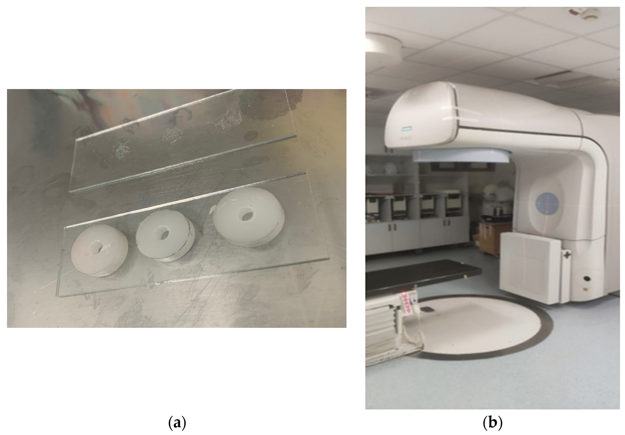
2.1. Sample Preparation

In this study, two materials with fluoride release, Equia Forte HT, referred to in the later text as EQ (GC, Tokyo, Japan), and Cention N, referred to in later text as CEN (Ivoclar Vivadent, Schaan, Liechtenstein), and two composite materials, Tetric EvoCeram, referred to in later text as TC and Tetric PowerFill, referred to in later text as PF (Ivoclar Vivadent, Schaan, Liechtenstein), were used (Table 1). Equia Forte HT is a strong biocompatible long-term bulk-fill restorative glass hybrid system with enhanced mechanical properties, superior fluoride release, excellent handling and improved translucency. Cention N is an alkasite bioactive ion-release material with especially long-term fluoride release, which is claimed to be significantly higher even after 180 days when compared to glass ionomer [23]. On the other hand, Tetric EvoCeram is a sculptable universal composite for the incremental layering technique, while Tetric PowerFill is a sculptable 4 mm composite for esthetic results in the posterior region, and both are inert resin composite materials without any fluoride-release activity.
It was necessary to make 30 samples of each material (total n = 120), to have 10 samples (n = 10) in each category. Category 1 was samples irradiated for 35 days at 2 Gy/day; Category 2 was samples irradiated with an impact dose of 70 Gy; and Category 3 was the control group without irradiation (Figure 1). The materials were prepared in Teflon molds (4 × 4 × 3 mm), under which the glass was placed; then, the materials were mixed in an automatic mixer (Septodont, France) for 10 s, placed in the mold and pressed again with glass to expel the excess material (Figure 2a). The samples were then polymerized for 20 s from both sides using an LED curing lamp (Ivoclar Vivadent, Liechtenstein) with a power of 1100 mW/cm2. Specimens were fine furbishings with disks (Water Proof Silicon Carbide Paper, 4000 grit; Buehler, Dusseldorf, Germany) and 1.0 μm, 0.3 and 0.05 μm professional polishing silicon granules (Buehler, Dusseldorf, Germany). The polishing procedure was carried out using a polishing appliance (Minitech 250, Presi, France). After that, samples were washed in distilled water. The samples were randomly divided into groups of 10 samples (n = 10).

2.2. Irradiation Procedure

To simulate oral cancer radiotherapy, the materials in Category 1 were exposed to 2 Gray (Gy) fractions, 5 days a week for 7 weeks for a total of 35 fractions equal to 70 Gy (frequent oral cancer dose). One gray (Gy) is the international system of units (SI) equivalent of 100 rads, which is equal to an absorbed dose of 1 Joule/kilogram. An absorbed dose of 0.01 Gy means that 1 g of material absorbed 100 ergs of energy (a small but measurable amount) as a result of exposure to radiation. Over the weekend, the samples were not irradiated. The materials in Category 2 were exposed to one experimental 70 Gy irradiation dose. Radiation was performed at the Department of Oncology, University Hospital Centre Zagreb, with a linear accelerator Siemens Primus (Siemens Healtheneers AG, Erlangen, Germany) radiotherapy unit (Figure 2b). A 6 MV radiation beam was used with a SSD (source to surface distance) of 100 cm setup for sample irradiation. Two centimeters of buildup material was placed above and below samples to ensure sufficient buildup and scatter conditions. Between the radiation cycles, samples were stored in deionized water, which was renewed daily, in an incubator at 37 °C (INEL, Zagreb, Croatia). Also, the impact of a shock dose of ionizing radiation with one single dose of 70 Gy on the materials was also investigated.

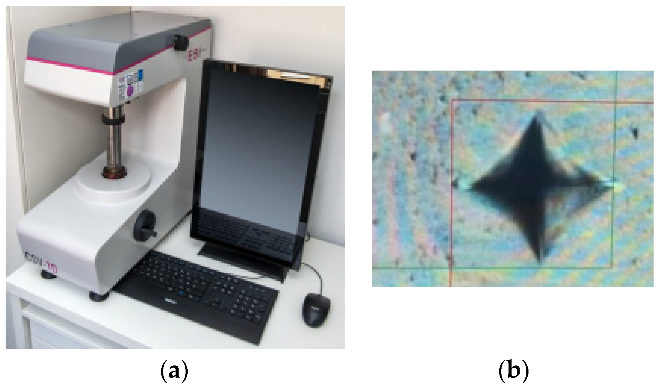
2.3. Surface Microhardness Analysis
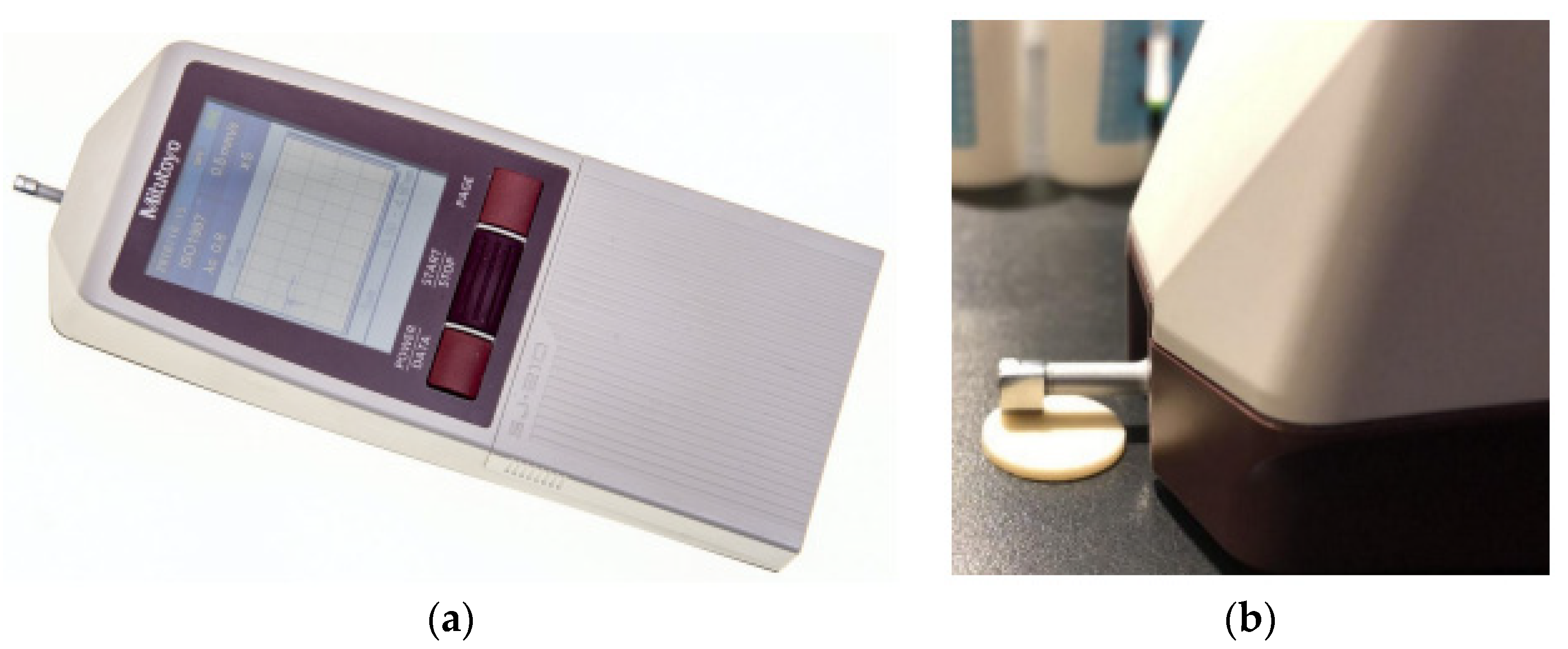
The microhardness of the enamel was measured at three different time points: after polishing, the initial measurement and after irradiation. The Vickers microhardness (HV) of the material surface was determined with a microhardness tester (CSV-10; ESI Prüftechnik GmbH, Wendlingen, Germany) using a load of 100 g and a dwell time of 10 s (Figure 3a). A diamond pyramid applied to the sample surface was used for measurement. Measurements were performed at three different points on the enamel: the surface, middle, and deep part (at 50 μm intervals) (Figure 3b). The average of all three Vickers hardness values obtained from the enamel was recorded as the total value of enamel hardness [24].

2.4. Prophylometry

The measurement of the surface roughness of the sample was performed with a high-precision profilometer (Mitutoyo, Japan) (Figure 4a). The following measurement parameters were used: stylus speed: 0.1 mm/s, stylus force: 4 mN, cut-off length: 0.25 mm, sampling length: 0.8 mm, and number of sampling lengths: 5. Evaluations were carried out at three different sites of each specimen within a radius of 3 mm from the specimen center and the mean value was calculated (Figure 4b). These tests determined the roughness parameter Ra after polishing, before irradiation and after ionizing radiation of all groups of materials in order to determine the effect of radiation on the exposed material’s surface. A common unit of measurement of surface roughness is by measuring the “average roughness”, which is often communicated as “Ra”. Ra is the calculated average between peaks and valleys on a surface. The lower the Ra value, the less variation between the peaks and troughs on a surface, making the surface smoother. In addition to surface roughness, they can also present other deviations such as waveforms and surface defects in the form of pores.

2.5. Measurement of Color Change

The color of restorative materials was measured by a VITA Easyshade (Vita Zahnfabrik, Bad Säckingen, Germany) spectrometer (Figure 5a). The materials were measured in such a way that the peak of the spectrometer, which was coated with a protective cap to defend the optical elements of the instrument against damage, was directed to the center of the sample surface. Measurements were repeated three times and the instrument automatically averaged the measurements for each sample, which was then used for the overall data analysis. All measurements were performed by the same researcher, under the same conditions. The color difference before and after radiation (ΔE) was calculated (Figure 5b).

2.6. Mass Measurement

The measurement of the mass of the sample was performed using a precision Mettler Toledo scale (Columbus, OH, USA) (Figure 6). Samples were weighed before and after ionizing radiation. Each sample was measured three times, from which the arithmetic mean was calculated.

2.7. Statistical Analysis

The results of the research are presented descriptively in the form of arithmetic means, standard deviations and 95% confidence intervals. Initial measurements and measurements after different types of irradiations were compared separately for each parameter. Changes caused by irradiation were calculated by the Wilcoxon rank test, since the measured parameters did not follow the normal distribution, which was confirmed by the Shapiro–Wilk normality test and Kolmogorov test for each parameter. Also, indicators of asymmetry and roundness indicated an abnormal distribution. Changes in color parameters showed normal distribution and the selection test was the t-test for dependent samples. The results are considered statistically significant at a significance level of 0.05. The analysis was performed using the Software package statistics (TIBICO Statistica Version 13.5.0.17).

3. Results

CEN showed the greatest resistance to mass change during a shock dose of ionizing radiation. With long-term exposure to gamma rays for 35 days, CEN and PF did not show statistically significant changes in mass (Table 2). By comparing the measurement of the Ra parameter before and after exposure of the material to radiation, no statistically significant change was found in any group. A statistically significant decrease in microhardness after irradiation for 35 days with 2 Gy/day was recorded only in the EQ material p = 0.006911. The change in Vickers microhardness (HV.01) after impact radiation with 70 Gy was recorded only with PF p = 0.006911 (Table 3). By including the measured parameters in the formula ΔE * = [(ΔL*)2 + (Δa*)2 + (Δb*)2]½, experimental results were obtained (CEN 7.25, EQ 5.99, PF 5.28 and EC 5.30), as well as impact samples (CEN 9.88, EQ 7.97, PF 8.18 and EC 10.30) in the interval 2–10, which means that the color change is “perceptible at a glance”. The exception is the shock EC E* = 10.30, so it does not belong to the same interval. It is defined as “Colors more similar than opposite”. Comparing parameters L, a and b individually, we obtained the following: For parameter L (luminance), in the group that was irradiated for 35 days with 2 Gy/day (experimental group), there was no statistically significant change in any material. While in the group that received a shock dose of radiation of 70 Gy, there was a statistically significant change in all materials except PF. For parameter a (red-green axis), in the experimental group, a statistically significant difference was found with EQ and PF, while in the impact group it was present with all materials. And for Parameter b (blue-yellow axis), in the experimental group, a statistically significant difference was found with EQ and PF, while in the impact group it was present with all materials (Table 4 and Table 5).

4. Discussion

In this research, the effects of radiation on several properties of dental restorative materials were examined using two radiation modes: 35 days at 2 Gy per day and a 70 Gy shock dose in a single day. For clinical practice, long-term radiation data (first mode) are particularly relevant, as they mimic the conditions experienced by patients undergoing head and neck radiotherapy. The shock dose, while not typical in clinical scenarios, was included to evaluate the impact of intense, short-duration radiation on the materials. Table 1 and Table 2 show mass changes during both radiation modes. The CEN samples exhibited the highest stability or resistance to mass change under both 1-day and 35-day irradiation. The PF samples showed stability only with long-term radiation. This stability supports the use of these materials in areas subjected to higher occlusion loads, reducing the risk of microleakage and edge cracks. Both materials contain a germanium-based photoinitiator (Ivocerin) that enhances polymerization depth, contributing to their stability [25,26,27,28,29,30,31,32]. Table 3 and Table 4 indicate no statistically significant changes in surface roughness (Ra parameter) for any test groups post-irradiation. Surface roughness is a critical factor influencing the clinical life of restorative fillings, as increased roughness can lead to rapid colonization by microorganisms, plaque accumulation, secondary caries, gingivitis, and loss of periodontal attachment [28,29]. Proper polishing of restorations is crucial for durability and esthetics [33,34,35,36], and this study suggests that radiation did not adversely affect surface roughness. The EQ material initially showed the highest strength but exhibited a significant decrease in microhardness after prolonged radiation. The PF showed a significant decrease after the shock dose. Patients undergoing head and neck radiotherapy are particularly exposed to the mentioned changes due to damage to the salivary glands, which reduces saliva secretion [1]. This results in sticky mucinous saliva with a diminished capacity to wash away harmful microorganisms and their metabolites. Previous research by Billingham [37] suggests that ion radiation can alter hardness and elasticity by forming free radicals, which may affect cross-linking in the materials. The TC material showed an increase in microhardness after shock dose irradiation, potentially due to the better chain connection post-radiation [14,38,39]. A recent analysis by the Radiation Therapy Oncology Group, a nonprofit organization dedicated to improving outcomes for cancer patients through the conduct of practice changing clinical trials, suggests that patients treated with more than 63 Gy in 1.8 to 2 Gy fractions had significantly better outcomes. A study by Kong at al. suggests that a higher dose (>70 Gy) through a personalized prescription can further improve local regional tumor control and overall survival, especially in patients treated concurrently with chemotherapy [40]. However, no correlation between surface roughness and microhardness was found, which is consistent with the findings of Bala et al. [41]. Color changes, measured by ΔE, ranged from 2 to 10, indicating visible changes. While brightness (L parameter) remained stable after 35 days of radiation, chromaticity (a and b parameters) showed significant changes in EQ and PF materials. The impact doses caused more pronounced color changes, decreasing brightness in all materials except PF and increasing chromaticity. Meena et al. [42] found that nanohybrid composites are less prone to color changes in different liquids, which aligns with our findings for EQ [43,44]. This research confirmed that the ΔE value for EC (nanohybrid composite) exceeds 10, indicating that the color change is classified as “Colors more similar than the opposite”. Given that the purpose of this paper was to become acquainted with the changes in the mentioned properties of restorative materials after long-term irradiation with a dose of 2 Gy, in order to obtain knowledge related to the material of choice for head and neck radiotherapy patients, we believe that it is necessary to summarize the results obtained after 35 days of radiation in next few sentences:
(1)
After 35 days of radiation at 2 Gy per day, the CEN and PF materials demonstrated mass stability.
(2)
The surface roughness remained unchanged across all groups, while CEN, PF and EQ showed resistance to microhardness changes.
(3)
Significant color changes were observed in all groups, but brightness remained stable.
(4)
The CEN material showed the best overall stability and had an anti-caries effect, releasing ions under low pH conditions, which is beneficial for patients with xerostomia due to radiotherapy [25].
This is particularly important for patients who experience irreversible damage to the salivary glands, resulting in xerostomia. Serous glands are more radiosensitive than mucous glands, making the radiation volume of the parotid gland a key factor in xerostomia development. Radiation doses of 60 Gy can reduce saliva secretion by 80%, leading to qualitative and quantitative changes in saliva. These changes include decreased pH due to lower bicarbonate ion concentration and altered bacterial microflora with increased acidogenic bacteria. These conditions contribute to chronic side effects of head and neck radiotherapy, such as radiation caries [44,45,46]. Contrary findings in the literature, such as those of Lima et al. [47], reported increased roughness in glass ionomer materials post-radiation, though these studies used different materials. However, our previous research supports the findings of this study, indicating no significant impact of radiation on the microhardness of glass hybrid, resin composite, and alkasite materials [48]. Atalay et al. [48] found varied effects on roughness depending on the material, with the giomer Beautifl II and Cention roughness values being negatively affected by radiotherapy, while the roughness of the rest of the Equia and Activa Bioactive Restorative was not altered. However, this research has certain limitations. Firstly, in vitro conditions do not fully replicate the oral environment. Secondly, samples were stored in distilled water and were not exposed to saliva’s buffering effects, temperature changes or cyclic mechanical loads. Therefore, future research should aim to mimic in vivo conditions more closely and consider extended preservation in artificial saliva. Furthermore, variations in material composition necessitate further studies to generalize findings and resolve the conflicting literature on radiation’s effects on restorative materials. Future studies should also assess other mechanical properties in clinical contexts.

5. Conclusions

In the context of this in vitro research and despite the limitations mentioned, it is possible to draw the following conclusions: ionizing radiation has a significant effect on the change in material mass of Equia Forte HT and Tetric Evo Ceram; ionizing radiation has no significant effect on the change in surface roughness of all materials tested; ionizing radiation has a significant effect on the change in microhardness of the Equia Forte HT material; and ionizing radiation has a significant effect on the change in color of all materials tested. Cention N should be the material of choice for patients undergoing radiotherapy in the head and neck region, as the morphological properties of the material itself are the most stable and it is known as a modern ion-release bioactive material.

Author Contributions

Conceptualization, E.K., L.Š. and B.Š.; data curation, E.K., L.Š. and B.Š.; formal analysis, E.K., L.Š. and B.Š.; funding acquisition, T.G., M.S. and A.I.; investigation, E.K., L.Š. and B.Š.; methodology, E.K., L.Š. and B.Š.; project administration, E.K., M.S. and A.I.; resources, E.K. and A.I.; software, L.Š. and B.Š.; supervision, E.K.; validation, E.K., L.Š. and B.Š.; visualization, E.K., L.Š. and B.Š.; writing—original draft, E.K.; writing—review and editing, E.K., L.Š. and B.Š. All authors have read and agreed to the published version of the manuscript.

Funding

This research was funded by University of Zagreb research support “Testing of The Properties of Modern Restorative Dental Materials Under the Influence of Ionizing Radiation and Alternative Polymerization Techniques” for the academic year 2024/25, leader professor Eva Klarić.

Institutional Review Board Statement

The study was conducted in accordance with the Declaration of Helsinki and approved by the Institutional Review Board (or Ethics Committee) of School of Dental Medicine University of Zagreb (05-PA-30-XXI-10/2020).

Informed Consent Statement

Not applicable.

Data Availability Statement

The data presented in this study are available on request from the corresponding author due to privacy or ethical restrictions.

Conflicts of Interest

The authors declare no conflicts of interest.

References

  1. National Comprehensive Cancer Center Network. NCCN Guidelines for Head and Neck Cancers. J. Natl. Compr. Canc. Netw. 2018, 16, 479–490. [Google Scholar]
  2. Gernhardt, C.R.; Kielbassa, A.M.; Hahn, P.; Schaller, H.G. Tensile bond strengths of four different dentin adhesives on irradiated and non-irradiated human dentin in vitro. J. Oral Rehabil. 2001, 28, 814–820. [Google Scholar] [PubMed]
  3. Soares, C.J.; Castro, C.G.; Neiva, N.A.; Soares, P.V.; Santos-Filho, P.C.; Naves, L.Z. Effect of gamma irradiation on ultimate tensile strength of enamel and dentin. J. Dent. Res. 2010, 89, 159–164. [Google Scholar] [CrossRef] [PubMed]
  4. Kielbassa, A.M.; Hinkelbein, W.; Hellwig, E.; Meyer-Luckel, H. Radiation-related damage to dentition. Lancet Oncol. 2006, 7, 326–335. [Google Scholar] [CrossRef] [PubMed]
  5. De Sanctis, V.; Bossi, P.; Sanguineti, G.; Trippa, F.; Ferrari, D.; Bacigalupo, A. Mucositis in head and neck cancer patients treated with radiotherapy and systemic therapies: Literature review and consensus statements. Crit. Rev. Oncol. Hematol. 2016, 100, 147–166. [Google Scholar] [CrossRef] [PubMed]
  6. Naves, L.Z.; Novais, V.R.; Armstrong, S.R.; Correr-Sobrinho, L.; Soares, C.J. Effect of gamma radiation on bonding to human enamel and dentin. Support. Care Cancer 2012, 20, 2873–2878. [Google Scholar] [CrossRef] [PubMed]
  7. Yesilyurt, C.; Bulucu, B.; Sezen, O.; Bulut, G.; Celik, D. Bond strengths of two conventional glass ionomer cements to irradiated and non-irradiated dentin. Dent. Mater. J. 2008, 27, 695–701. [Google Scholar] [CrossRef] [PubMed]
  8. Da Rosa Rodolpho, P.A.; Donassollo, T.A.; Cenci, M.S.; Loguercio, A.D.; Moraes, R.R.; Bronkhorst, E.M. 22-year clinical evaluation of the performance of two posterior composites with different filler characteristics. Dent. Mater. 2011, 27, 955–963. [Google Scholar] [CrossRef] [PubMed]
  9. Bernardo, M.; Luis, H.; Martin, M.D.; Leroux, B.G.; Rue, T.; Leitao, J. Survival and reasons for failure of amalgam versus composite posterior restorations placed in a randomized clinical trial. J. Am. Dent. Assoc. 2007, 138, 775–783. [Google Scholar] [CrossRef]
  10. Silva, A.R.; Alves, F.A.; Berger, S.B.; Giannini, M.; Goes, M.F.; Lopes, M.A. Radiation-related Caries and early failure in head and neck cancer patients. A polarized light microscopy and scanning electron microscopy study. Support. Care Cancer 2010, 18, 83–87. [Google Scholar] [CrossRef]
  11. Andrews, N.; Griffiths, C. Dental complications of head and neck radiotherapy: Part 2. Aust. Dent. J. 2001, 46, 174–182. [Google Scholar] [CrossRef] [PubMed]
  12. Hancock, P.J.; Epstein, J.B.; Sadler, G.R. Oral and dental management related to radiation therapy for head and neck cancer. J. Can. Dent. Assoc. 2003, 69, 585–590. [Google Scholar] [PubMed]
  13. Shaw, M.J.; Kumar, N.D.; Duggal, M.; Fiske, J.; Lewis, D.A.; Kinsella, T. Oral management of patients following oncology treatment: Literature review. Br. J. Oral Maxillofac. Surg. 2000, 38, 519–524. [Google Scholar] [CrossRef] [PubMed]
  14. Cruz, A.; Sinhoreti, M.; Ambrosano, G.; Rastelli, A.; Bagnato, V.; Bóscolo, F. Effect of therapeutic dose X rays on mechanical and chemical properties of esthetic dental materials. Mater. Res. 2008, 11, 313–318. [Google Scholar] [CrossRef]
  15. Beech, N.; Robinson, S.; Porcedds, S.; Batstone, M. Dental management of patients irradiated for head and neck cancer. Aust. Dent. J. 2014, 59, 20–28. [Google Scholar] [CrossRef] [PubMed]
  16. Galetti, R.; Santos-Silva, A.R.; Antunes, A.N.; Alves Fde, A.; Lopes, M.A.; de Goes, M.F. Radiotherapy does not impair dentin adhesive properties in head and neck cancer patients. Clin. Oral Investig. 2014, 18, 1771–1778. [Google Scholar] [CrossRef] [PubMed]
  17. Chin, D.W.; Treister, N.; Friedland, B.; Cormack, R.A.; Tishler, R.B.; Makrigiorgos, G.M. Effect of dental restorations and prostheses on radiotherapy dose distribution: A Monte Carlo study. J. Appl. Clin. Med. Phys. 2009, 10, 80–89. [Google Scholar] [CrossRef] [PubMed]
  18. Reitemeier, B.; Reitemeier, G.; Schmidt, A.; Schaal, W.; Blochberger, P.; Lehmann, D. Evaluation of a device for attenuation of electron release from dental restorations in a therapeutic radiation field. J. Prosthet. Dent. 2002, 87, 323–327. [Google Scholar] [CrossRef] [PubMed]
  19. De Moor, R.J.; Stassen, I.G.; van’t Veldt, Y.; Torbeyns, D.; Hommez, G.M. Two-year clinical performance of glass ionomer and resin composite restorations in xerostomic head- and neck irradiated cancer patients. Clin. Oral Investig. 2002, 15, 31–38. [Google Scholar] [CrossRef]
  20. McComb, D.; Erickson, R.L.; Maxymiw, W.G.; Wood, R.E. A clinical comparison of glass ionomer, resin-modifified glass ionomer and resin composite restorations in the treatment of cervical caries in xerostomic head and neck radiation patients. Oper. Dent. 2002, 27, 430–437. [Google Scholar]
  21. Ferracane, J.L. Resin composite—State of the art. Dent. Mater. 2011, 27, 29–38. [Google Scholar] [CrossRef] [PubMed]
  22. Germano, F.; Melone, P.; Testi, D.; Arcuri, L.; Marmiroli, L.; Petrone, A. Oral complications of head and neck radiotherapy: Prevalence and management. Minerva Stomatol. 2015, 64, 189–202. [Google Scholar] [PubMed]
  23. Naz, F. Comparative evaluation of mechanical and physical properties of a new bulk-fill alkasite with conventional restorative materials. Saudi Dent. J. 2020, 33, 666–673. [Google Scholar] [CrossRef] [PubMed]
  24. Miletic, I. Modern solutions for direct posterior restorations. GC Get. Connected. 2015, 4, 32–36. [Google Scholar]
  25. MeSH Browser [Database on the Internet]. Cention N. Available online: https://meshb.nlm.nih.gov/ (accessed on 30 August 2020).
  26. Kolemen, U. Analysis of ISE in microhardness measurements of bulk MgB2 superconductors using different models. J. Alloys Compd. 2006, 429, 435. [Google Scholar] [CrossRef]
  27. Sidhu, S.K.; Sheriff, M.; Watson, T.F. In vivo changes in roughness of resin-modified glass ionomer materials. Dent. Mater. 1997, 13, 208–213. [Google Scholar] [CrossRef] [PubMed]
  28. Yip, H.K.; Lam, W.T.C.; Smales, R.J. Fluoride release, weight loss and erosive of modern aesthetic restoratives. Br. Dent. J. 1999, 187, 265–270. [Google Scholar] [CrossRef] [PubMed]
  29. Yip, H.K.; Lam, W.T.C.; Smales, R.J. Surface roughness and weight loss of esthetic restorative materials related to fluoride release and uptake. J. Clin. Pediatr. Dent. 1999, 23, 321–326. [Google Scholar] [PubMed]
  30. Rios, D.; Honório, H.M.; Araújo, P.A.; Machado, M.A.A. Wear and superficial roughness of glass ionomer cements used as sealants, after simulated toothbrushing. Braz. Oral Res. 2002, 16, 343–348. [Google Scholar] [CrossRef]
  31. Warren, D.P.; Colescott, T.D.; Henson, H.A.; Powers, J.M. Effects of four prophylaxis pastes on surface roughness of a composite, a hybrid ionomer and a compomer restorative material. J. Esthet. Rest. Dent. 2002, 14, 245–251. [Google Scholar] [CrossRef]
  32. Lohbauer, U. Dental Glass Ionomer Cements as Permanent Filling Materials?—Properties, Limitations, Future Trends. Materials 2009, 3, 76–96. [Google Scholar] [CrossRef]
  33. Ilie, N.; Hickel, R. Investigations on Mechanical Behaviour of Dental Composites. Clin. Oral Investig. 2009, 13, 427–438, Erratum in Clin. Oral Investig. 2009, 13, 485–487. [Google Scholar] [CrossRef] [PubMed]
  34. Czasch, P.; Ilie, N. In Vitro Comparison of Mechanical Properties and Degree of Cure of Bulk Fill Composites. Clin. Oral Investig. 2013, 17, 227–235. [Google Scholar] [CrossRef]
  35. Reis, A.F.; Giannini, M.; Lovadino, J.R.; Dias, C.T.S. The effect of six polishing systems on the surface roughness of two packable resin-based composites. Am. J. Dent. 2002, 15, 193–197. [Google Scholar] [PubMed]
  36. Billingham, N.C. Degradation and stabilization of polymers. In Materials Science and Technology; Wiley-VCH: Hoboken, NJ, USA, 2013; pp. 469–507. [Google Scholar]
  37. Jefferies, S.R. Abrasive Finishing and Polishing in Restorative Dentistry: A State-of-the-Art Review. Dent. Clin. N. Am. 2007, 51, 379–397. [Google Scholar] [CrossRef]
  38. Soliman, H.A.N.; Elkholany, N.R.; Hamama, H.H.; El-Sharkawy, F.M.; Mahmoud, S.H.; Comisi, J.C. Effect of Different Polishing Systems on the Surface Roughness and Gloss of Novel Nanohybrid Resin Composites. Eur. J. Dent. 2021, 15, 259–265. [Google Scholar] [CrossRef]
  39. Kong, F.P.; Ten Haken, A.; Hayman, J. Personalized High Dose Radiation (>70 Gy) Is Significantly Associated with Better Local Regional Control and Overall Survival in Non-small Cell Lung Cancer Treated with Concurrent Chemoradiation. Int. J. Radiat. Oncol. Biol. Phys. 2011, 81, S594. [Google Scholar] [CrossRef]
  40. Bala, O.; Arisu, H.D.; Yikilgan, I.; Arslan, S.; Gullu, A. Evaluation of surface roughness and hardness of different glass ionomer cements. Eur. J. Dent. 2012, 6, 79–86. [Google Scholar] [CrossRef]
  41. Meena, B.; Hasija, M.; Wadhwani, K.K.; Wadhwa, D. Spectrometric analysis of intentionally stained hybrid and nanohybrid composites—An In vitro study. J. Int. Clin. Dent. Res. Organ. 2019, 11, 76–82. [Google Scholar] [CrossRef]
  42. Sarafianou, A.; Iosifidou, S.; Papadopoulos, T.; Eliades, G. Color Stability and Degree of Cure of Direct Composite Restoratives After Accelerated Aging. Oper. Dent. 2007, 32, 406–411. [Google Scholar] [CrossRef]
  43. Villalta, P.; Lu, H.; Okte, Z.; Garcia-Godoy, F.; Powers, J.M. Effects of Staining and Bleaching on Color Change of Dental Composite Resins. J. Prosthet. Dent. 2006, 95, 137–142. [Google Scholar] [CrossRef] [PubMed]
  44. Lončar Brzak, B.; Mravak Stipetić, M. Ljestvice za procjenu oralnog mukozitisa. Medix 2014, 20, 213–216. [Google Scholar]
  45. Dirix, P.; Nuyts, S. Evidence—Based organ—Sparing radiotherapy in head and neck cancer. Lancet Oncol. 2010, 11, 85–91. [Google Scholar] [CrossRef] [PubMed]
  46. Lima, R.B.; De Vasconcelos, L.C.; Pontual, M.L.; Meireles, S.S.; Andrade, A.K.M.; Duarte, R.M. Effect of ionizing radiation on the properties of restorative materials. Indian J. Dent. Res. 2019, 30, 408–413. [Google Scholar] [CrossRef]
  47. Turjanski, S.; Par, M.; Bergman, L.; Soce, M.; Grego, T.; Klaric Sever, E. Influence of ionizing radiation on fluoride-releasing dental restorative materials. Polymers 2023, 15, 632. [Google Scholar] [CrossRef]
  48. Atalay, C.; Yazici, R. Effect of radiotherapy on the surface roughness and microhardness of contemporary bioactive restorative materials. Support. Care Cancer 2024, 32, 295. [Google Scholar] [CrossRef]
Figure 1. Diagram of materials, groups and type of measurement used in this study. Materials used: Equia Forte HT (GC, Japan), Cention N (Ivoclar Vivadent, Schaan, Liechtenstein), Tetric EvoCeram and Tetric PowerFill (Ivoclar Vivadent, Schaan, Liechtenstein).
Sci 06 00047 g001
Figure 2. (a) Teflon molds (4 × 4 × 3 mm) (b) Linear accelerator Siemens Primus (Siemens Healthineers AG, Erlangen, Germany).
Sci 06 00047 g002
Figure 3. (a) Microhardness tester (CSV-10; ESI Prüftechnik GmbH, Wendlingen, Germany); (b) diamond pyramid that remained imprinted on the surface of the sample.
Sci 06 00047 g003
Figure 4. (a) Portable device for measuring surface roughness (Surftest SJ-210; Mitutoyo, Houston, TX, USA); (b) presentation of surface roughness measurements of the tested sample.
Sci 06 00047 g004
Figure 5. (a) Measurement of color change with a spectrophotometer with a probe (VITA Easyshade III; Vita, Bad Säckingen, Germany); (b) reading L*, a*, and b* values from the device.
Sci 06 00047 g005
Figure 6. Precision Mettler Toledo scale (Columbus, OH, USA).
Sci 06 00047 g006
Table 1. The type and composition of the investigated materials according to the manufacturer’s instructions.
MaterialTypeManufacturerCompositionLot
GC Equia Forte HT Bulk-fill glass hybridGC Corp., Tokyo, JapanPowder: fuoroaluminosilicate glass, polyacrylic.
Liquid: polybasic carboxylic acid, water.
230310B
Cention Nnon-adhesive bulk-fill resinous
material
Ivoclar AG; Schaan, LiechtensteinPowder: barium aluminum silicate glass, ytterbium trifuoride, isofller, calcium barium,
aluminum fuorosilicate glass, calcium fuoro,
silicate glass.
Liquid: urethane dimethacrylate, tricyclodecandimethanol dimethacrylate, tetramethylxylylene diurethane dimethacrylate, polyethylene glycol 400 dimethacrylate, ivocerin,
Hydroxyperoxide.
ZL08SP
Tetric EvoCeramSculptable compositeIvoclar AG; Schaan, LiechtensteinThe monomer matrix is composed of dimethacrylates (20–21 wt.%). The fillers contain barium glass, ytterbium trifluoride, mixed oxide and copolymers (79–81 wt.%). Additives, initiators, stabilizers and pigments are additional ingredients (<1.0 wt.%). The total content of inorganic fillers is 76–77% weight or 53–54% volume. The particle sizes of the inorganic fillers range between 40 nm and 3 μm.Z01V79
Tetric PowerFillSculptable compositeIvoclar AG; Schaan, LiechtensteinThe monomer matrix is composed of dimethacrylates (20–21 wt.%). The fillers contain barium glass, ytterbium trifluoride, mixed oxide and copolymers (79–80 wt.%). Additives, initiators, stabilizers and pigments are additional ingredients (<1.0 wt.%). The total content of inorganic fillers is 76–77 wt.% or 53–54 vol%. The particle size of inorganic fillers is between 40 nm and 3 μm.Z009GW
Table 2. Mass change after 35 days of ionizing radiation with 2 Gy/day and shock dose of ionizing radiation with 70 Gy.
MassControl2 Gy/35 DaysShock Dose (70 Gy)
MaterialsMedian (IQR)Median (IQR)p ValueMedian (IQR)p Value
CEN0.104 (0.095–0.114)0.103 (0.094–0.113)0.1120.104 (0.096–0.115)0.415
EQ0.110 (0.107–0.114)0.100 (0.094–0.106)0.0010.105 (0.101–0.111)0.005
PF0.122 (0.118–0.123)0.115 (0.110–0.121)0.0050.122 (0.120–0.125)0.169
EC0.121 (0.118–0.124)0.117 (0.113–0.122)0.0050.117 (0.114–0.123)0.005
Table 3. Surface microhardness (HV) before and after (a) 35 days of ionizing radiation with 2 Gy/day and (b) shock dose of ionizing radiation with 70 Gy.
Surface Microhardness Pre-2 Gy/35 DaysPost-2 Gy/35 Days p ValuePre-Shock Dose (70 Gy)Post-Shock Dose (70 Gy)p Value
MaterialsMedian (IQR)Median (IQR) Median (IQR)Median (IQR)
CEN50.3 (47.4–54.9)50.1 (47.2–53.8)0.28549.4 (45.2–52.0)49.4 (45.2–51.9)0.203
EQ100.1 (77.6–109.7)99.9 (77.1–109.0)0.00798.5 (95.1–102.9)98.6 (94.8–101.1)0.232
PF74.4 (58.8–77.8)74.0 (59.6–77.4)0.72180.6 (54.1–86.9)80.2 (50.2–86.7)0.006
EC64.9 (62.5–68.6)65.2 (62.3–68.3)0.28565.9 (60.3–69.8)66.4 (60.2–69.7)0.878
Table 4. Changes in parameter L after 35 days of ionizing radiation with 2 Gy/day.
T-Test for Dependant Samples
Marked Differences Are Significant at p ˂ 0.0500
VariableMeanStd.Dv.NDiff.Std.Dv.tdfpConfidenceConfidence
L before CEN68.6403.712
L after CEN67.4605.763101.1808.0860.46190.655−4.6046.964
L before EQ57.8104.383
L after EQ57.2605.040100.5506.6280.26290.798−4.1915.291
L before PF69.6902.679
L after PF67.9104.779101.7804.8701.15590.2775−4.6046.964
L before EC65.1302.897
L after EC67.7504.67810−2.6225.003−1.65590.132−6.1990.959
Table 5. Changes in parameter L after shock dose of ionizing radiation with 70 Gy.
T-Test for Dependant Samples
Marked Differences Are Significant at p ˂ 0.0500
VariableMeanStd.Dv.NDiff.Std.Dv.tdfpConfidenceConfidence
L before CEN68.6403.712
L after CEN77.7302.80710−9.0905.088−5.64990.003−12.729−5.450
L before EQ57.8104.383
L after EQ63.1402.27010−5.3305.053−3.33690.008−8.944−1.715
L before PF69.6902.679
L after PF70.5805.21110−0.8905.332−0.52790.610−4.7042.942
L before EC65.1302.897
L after EC74.1403.67810−9.1004.788−5.95090.000−12.435−5.584
Disclaimer/Publisher’s Note: The statements, opinions and data contained in all publications are solely those of the individual author(s) and contributor(s) and not of MDPI and/or the editor(s). MDPI and/or the editor(s) disclaim responsibility for any injury to people or property resulting from any ideas, methods, instructions or products referred to in the content.

