Article

Antibacterial Activity of Polypropylene Meshes for Hernioplasty with Ag and (Ag,Cu) Coatings Deposited via Magnetron Sputtering

1
Department of High-Efficiency Machining Technologies, Moscow State University of Technology “STANKIN”, Vadkovsky Lane 3a, 127055 Moscow, Russia
2
Institute of Design and Technological Informatics of the Russian Academy of Sciences (IDTI RAS), Vadkovsky Lane 18a, 127055 Moscow, Russia
3
Materials Science and Metallurgy Shared Use Research and Development Center, National University of Science and Technology “MISiS”, Leninsky Prospect 4, 119049 Moscow, Russia
4
The National Medical Research Center of Surgery Named After A. Vishnevsky, St. Bolshaya Serpukhovskaya 27, 117997 Moscow, Russia
5
Genetics Department, Biology Faculty, Belarusian State Medical University, Dzerzhinsky Ave. 83, 220083 Minsk, Belarus
*
Author to whom correspondence should be addressed.
Submission received: 15 October 2024 / Revised: 30 December 2024 / Accepted: 5 February 2025 / Published: 10 February 2025
(This article belongs to the Section Chemistry Science)

Abstract

:
This article compares the antibacterial properties of single-layer (Ag) and two-layer (Ag,Cu) coatings deposited onto a polypropylene mesh (endoprostheses for hernioplasty) in various gaseous environments (argon or nitrogen) via magnetron sputtering. The microstructure and elemental composition of the coatings were studied via SEM and TEM. The antimicrobial activity of sterile samples was investigated using the Staphylococcus aureus strain. To prevent the overheating of the polymer samples during the coating process, it is advisable to carry out pulse processing (the total coating formation time is divided into cycles of switching the magnetron on and off for equal periods). All the samples, with both single- and double-layer coatings, exhibited good antibacterial properties; however, the Cu–Ag coating enhanced the antimicrobial effect, increasing it from 97.00 to 99.97%. The glow-discharge plasma etching of the samples with a double-layer coating led to the mixing of the copper and silver layers and an increase in the surface copper content, though this did not affect the antibacterial properties of the samples.

1. Introduction

The advancement of medical technologies for plastic surgery requires intensified efforts in the development of modern synthetic implants for reconstructive surgery. Currently, the most acceptable materials for hernia repair (hernioplasty), pelvic floor surgery, the replacement of various soft tissue defects, and other similar areas of application are mesh endoprostheses composed of polymer threads, which are used as surgical suture materials [1].
The main manufacturers of these endoprostheses are Ethicon (Cincinnati, OH, USA), Covidien (Dublin, Ireland), B.Braun (Melsungen, Germany), RESORBA Medical GmbH (Nürnberg, Germany), Cousin Biotech (Wervicq-Sud, France), Tricomed S.A. (Łódź, Poland), Unisur (Karnataka, India), and LLC Lintex (Saint Petersburg, Russia), among others. At the same time, the dynamics of major elective surgeries indicate a steady increase in their number. For example, in Russia alone (according to the analysis of the Specialised Commission of the Ministry of Health of the Russian Federation [2] for the 2020–2023 period), the number of elective inguinal hernioplasty surgeries increased 1.5-fold (from 81,893 in 2020 to 123,493 in 2023), and the number of surgeries for postoperative hernia repair showed a 1.6-fold increase (from 26,752 in 2020 to 42,708 in 2023).
The polymer endoprostheses currently used in hernioplasty differ in their production method (chemical or textile technology), structure (knitted, non-woven, or film-porous), type of polymer (polytetrafluoroethylene, polypropylene, polyethylene terephthalate, etc.), thread structure (monofilament, complex, or pseudomonofilament), biodegradability (non-absorbable, absorbable, or partially absorbable), and design (flat or three-dimensional) [1].
Despite their diversity, mesh endoprostheses must all meet the following requirements, determined according to the purpose and operating principle of the product [1,3,4]:
  • Biocompatibility;
  • Bioresistance;
  • Resistance to infection;
  • Mechanical strength;
  • Ability to rapidly grow into tissues;
  • Limited stretchability in all directions;
  • Resistance to the unraveling and crumbling of edges;
  • Softness and good modellability;
  • Minimal material consumption;
  • The retention of consumer properties after sterilization.
According to the available data in the literature, in a number of cases, patients who have undergone the implantation of mesh endoprostheses develop complications, among which purulent–septic complications often lead to relapses and the need for repeated surgical interventions [5]. Local complications of purulent–inflammatory genesis are facilitated by several factors, among which the endogenous microbial contamination of the surgical area plays the leading role [6]. Thus, according to previous data [7], the complex reconstruction of the abdominal wall in the presence of the contamination of the surgical field is accompanied by wound complications in 46% of cases.
One of the complications of mesh hernioplasty is infection, which can be caused by bacterial microflora, notably Staphylococcus aureus (including methicillin-resistant Staphylococcus aureus [MRSA]), Enterococcus (vancomycin-resistant Enterococcus [VRE]), Pseudomonas aeruginosa, beta-hemolytic Streptococcus, and Escherichia coli bacteria, among others [5]. The incidence rate in this case is from 1 to 10% of cases [8]. Such infections rather often lead to the development of purulent–inflammatory wound complications in the postoperative period; for example, the frequency of developing local wound purulent–inflammatory complications after herniotomies of strangulated postoperative ventral hernias of the anterior abdominal wall, according to literary data, varies from 20.9 to 67.0% [9,10,11,12]. In some cases of infection, surgical intervention becomes necessary to remove the implanted endoprosthesis. In this regard, Sharma et al. [13] removed 105 previously implanted meshes over 4.5 years. However, it is worth noting that, in some situations, the complete removal of the endoprosthesis can be technically significantly difficult, and sometimes impossible, due to the pronounced connective tissue ingrowth of the woven (knitted) mesh, even in the presence of the inflammatory process [14]. In this case, the simultaneous explantation of the infected mesh and re-endoprosthetics of the abdominal wall (the implantation of synthetic material under infection conditions) can be accompanied by a high frequency of complications (50% or more) [5].
Resistance to infection implies bacteriostatic and antibacterial properties; these can be achieved through various approaches, divided into three groups:
  • Improving the methods of treating the surgical field before [15,16] and during [17] surgical intervention;
  • Postoperative antibacterial prophylaxis (involving the use of antibiotic drugs [18]), covering the wound with special wound dressings to prevent possible contamination, and improving the methods of flow-aspiration drainage [6];
  • The use of biologically active (antimicrobial) suture materials both for weaving mesh endoprostheses and fixing meshes to body tissues; the antimicrobial surgical suture material can be made based on threads with different biodegradation periods containing antibacterial drugs [8,19,20].
Studies have shown that the local action of antimicrobial drugs is very limited due to the resistance of microbial flora, pronounced post-traumatic inflammation in the surgical area, and the formation of microbial biofilms on implants and suture materials, among other factors [5,6,21]. This is also associated with the limited effectiveness of antibiotics taken by patients in the postoperative period. It is the biofilm variant in the development of the infectious process that underlies the relatively low effectiveness of its treatment [22]. The possibilities of antibiotic therapy in such cases are significantly limited by the poor penetration of drugs into biofilms. In addition, the minimum inhibitory concentrations of drugs for the biofilm forms of microorganisms exceed those for planktonic ones by tens to hundreds of times [22].
Thus, the choice of the mesh endoprosthesis material is an important factor. Currently, absorbable—polylactic acid (PLA), polyglycolic acid (PGA), and their combination poly(lactic-co-glycolic acid) (PLGA)—and non-absorbable—polypropylene (PP), polyethylene terephthalate (PET), and polytetrafluoroethylene (PTFE/ePTFE)—surgical sutures are used to manufacture mesh endoprostheses. Although many commercial absorbable meshes exhibit an antimicrobial effect due to the application of polymer coatings, such as chitosan or triclosan [8,23], meshes based on absorbable materials deteriorate rapidly, lose their strength, and stimulate the formation of scar tissue [24,25]. Regenerated tissue after the absorption of the mesh material is not sufficiently strong to combat the recurrence of hernias [26].
The main material for weaving mesh endoprostheses remains PP [4], though in vivo test results have indicated its insufficient biocompatibility. Furthermore, comparative in vivo tests of 17 commercially available meshes composed of various synthetic materials (PP, PET, and ePTFE) have revealed the highest level of inflammation for PP meshes, with the chronic inflammatory tissue reaction remaining evident even one year after surgery [27].
A common method for creating products with antibacterial properties is the application of nanostructured silver coatings [28,29,30,31], with silver particles of 9–15 nm in size being the most effective in destroying pathogenic microorganisms [32]. The coatings present a notably large specific area of contact between silver and bacteria or viruses, significantly improving their bactericidal action [33]; furthermore, they are largely free of the problems of resistance by pathogenic microorganisms [33].
In its ionic form, silver exhibits bactericidal and pronounced antifungal and antiseptic effects, serving as a highly effective disinfectant against pathogenic microorganisms that cause acute infections. In addition, silver has recently received increasing attention due to not only its powerful antibacterial and antiviral properties but also its identified effects on the body (representing a microelement necessary for the normal functioning of organs and systems) and its immunocorrective properties [32]. The mechanism of action of silver against microbial cells involves the absorption of silver ions by the cell membrane of the microbe; as a result, while the cell remains viable, some of its functions are disrupted, including, for example, division (bacteriostatic effect) [32,34]. In this case, Ag nanoparticles destroy bacterial membranes, interfere with processes associated with DNA and proteins, release Ag+ ions, and use active oxygen species to enhance the control over microorganisms [35]. Moreover, silver has a significantly broader spectrum of antimicrobial activity than many antibiotics and sulfonamides; silver has a more powerful antimicrobial effect than penicillin, biomycin, and other antibiotics, and it exhibits a detrimental effect on antibiotic-resistant strains (varieties) of bacteria [36]. In addition, Ag nanoparticles enhance the bactericidal action of several modern antibiotics [37]. The combination of Ag nanoparticles and antibiotics produces a synergistic effect, which has been demonstrated both in vitro and in vivo in relation to various bacterial strains [38,39]. The antibacterial properties of copper (Cu) are currently the topic of rather extensive research; many scholars have demonstrated its antimicrobial properties, low cytotoxicity, and greater cytocompatibility compared to other metals [40]. A Cu-containing coating prevents biofilm formation on the implant surface and inhibits the development of infection. Usman et al. [41] indicated that the effectiveness of copper nanoparticles is comparable to that of silver nanoparticles, though the oxidation of Cu particles during their production and storage represents a problem. Cu exhibits other positive characteristics, including the absence of its accumulation in the body and fairly low costs. It has high conductivity, allowing its application to the surface of implants in various ways and significantly reducing the technological process costs of generating coatings [42]. At the same time, silver and copper nanoparticles exert a strong synergistic antibacterial effect due to increased cell permeability [43,44]. In this regard, Długosh et al. [45] found that Ag–Cu alloy nanoparticles exhibit minimal genotoxicity and stable antibacterial efficacy. Numerous studies have demonstrated that Ag–Cu nanoparticles eliminate the inherent disadvantages of individual copper and silver nanoparticles, providing an enhanced antibacterial effect, increased stability, and reduced nanotoxicity [35,46,47,48,49,50,51].
Today, many methods for the metallization of polymer products exist. A promising area for modifying the properties of various materials (including polymers) is their processing in glow-discharge plasma [52,53,54]. During the sputtering of materials in glow-discharge plasma, high-energy secondary electrons from the target form the main source of substrate heating. The sources of substrate heating in these systems are the condensation energy of sputtered atoms, the kinetic energy of deposited atoms, the energy of neutralized ions reflected from the target, and plasma radiation. The condensation energy is 3–9 eV/atom; the kinetic energy, depending on the sputtered material, ranges from 5 eV/atom (for aluminum) to 20 eV/atom (for tungsten), and the plasma radiation is 2–10 eV/atom. Table 1 presents the total thermal energy dissipated on the substrate, the substrate temperature for various materials deposited in a cylindrical magnetron sputtering system (MSS);, and the average deposition rates of various materials using an MSS with a flat-disk target that is 150 mm in diameter and a source power of 4 kW, with the substrate located at a distance of 60 mm from the source.
Table 1. Thermal energy, substrate temperature [28,54,55], and average deposition rate [56] of various materials.
MaterialAgAlCuTaCrAuMoW
Thermal energy, eV/atom7–1013172020234773
Substrate temperature, °C507911097118106163202
Deposition rate, nm/s44133081737128
In many cases, the heating of the substrate in magnetron systems is comparable, and when refractory materials are evaporated, it is even lower than in the case of the thermal evaporation method. This makes it possible to use magnetron systems in applying coatings to substrates composed of materials with low heat resistance (plastics, polymers, organic glass, and paper) [56]. Therefore, the most promising direction for the plasma modification of polymeric materials is the application of coatings from metals, alloys, and chemical compounds via magnetron sputtering.
Thus, improving the quality of mesh endoprostheses for hernioplasty, including their biological compatibility, hypoallergenicity, biocidal properties, and antibacterial properties, is a pressing issue. The issues of the deposition of coatings based on the Ag/Cu system on a polymer substrate via magnetron sputtering have been considered in a small number of articles, mainly in the application of the production of solar battery elements [57,58,59].
This work aimed to investigate the application of nanostructured Ag-containing coatings on mesh polymer endoprostheses via magnetron sputtering to increase their antibacterial effect.

2. Materials and Methods

The object of the study was UNISUR 75 mm × 80 mm PP mesh endoprostheses (Bangalore, Karnataka, India; Figure 1).
A magnetron sputtering system, RITM-SP (MSUT STANKIN, Moscow, Russia) [60,61], was used to apply the nanostructured Ag-containing coatings. In the vacuum chamber of the system, a magnetron with a Ø 75 mm-target 6 mm-thick (silver or copper, depending on the layer/coating composition) was horizontally located. The target was fed via a high-frequency generator with a maximum power of 13.5 kW. In this work, we used targets comprising 99.99% silver (LLC SALTI, Moscow, Russia) or M00B copper (99.99%; LLC AKTAN VACUUM, Fryazino, Russia). In producing the (Ag,Cu) coating, the layers were applied sequentially (with target replacement): first copper and then silver. The test sample was fixed with a PP thread on a 95 × 110 mm metal frame (table), which was then placed in the working chamber. The coatings were applied in the following process modes [62,63]: preliminary pressure in the chamber—5 × 10⁻⁵ Pa; working pressure of argon—5.98 × 10⁻¹ Pa; nitrogen pressure—5.98 × 10⁻¹ Pa; magnetron voltage—470 V; magnetron current—1.5 A; frequency—15 kHz; distance between the target and the sample—16 cm; and processing time (formation of each coating/coating layer)—30 s, divided into three 10 s cycles (with 10 s breaks between sputtering). The different coating formation technologies and sample designations are presented in Table 2.
Table 2. Options for coating formation technologies.
Sample DesignationAgCuArN2Glow-Discharge Etching [64,65]
N1+++--
N2+--+-
N3+--++
N4++-+-
N5++-++
Subsequently, the test sample was cut into 20 × 20 mm squares and packed in bags composed of medical material “Paper-film” (BOM, Lublin, Poland), medical paper with a density of 60 g/m2, and a transparent polymer film of (PP + polyester) blue (TZMO S.A., Torun, Poland), measuring 60 × 150 mm. An individual label indicating the name, size, and sterilization method of the product was pasted on the bags. The samples were sterilized with gas (ethylene oxide).

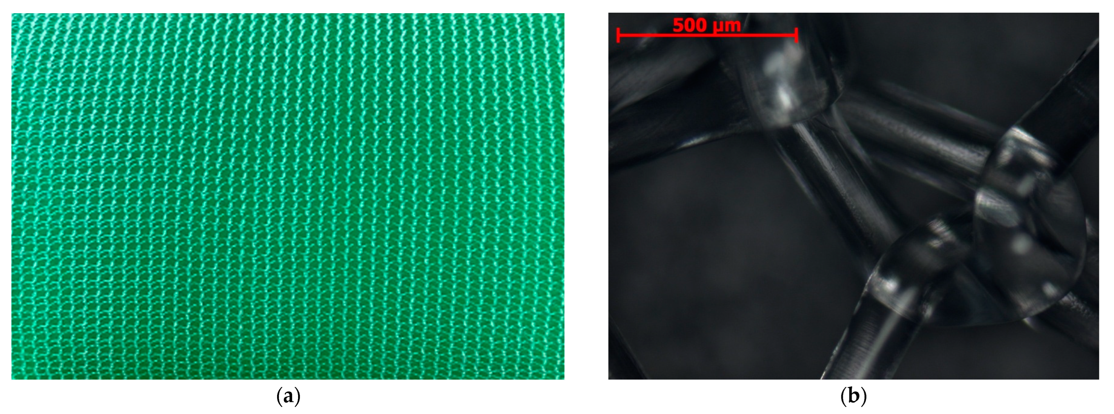
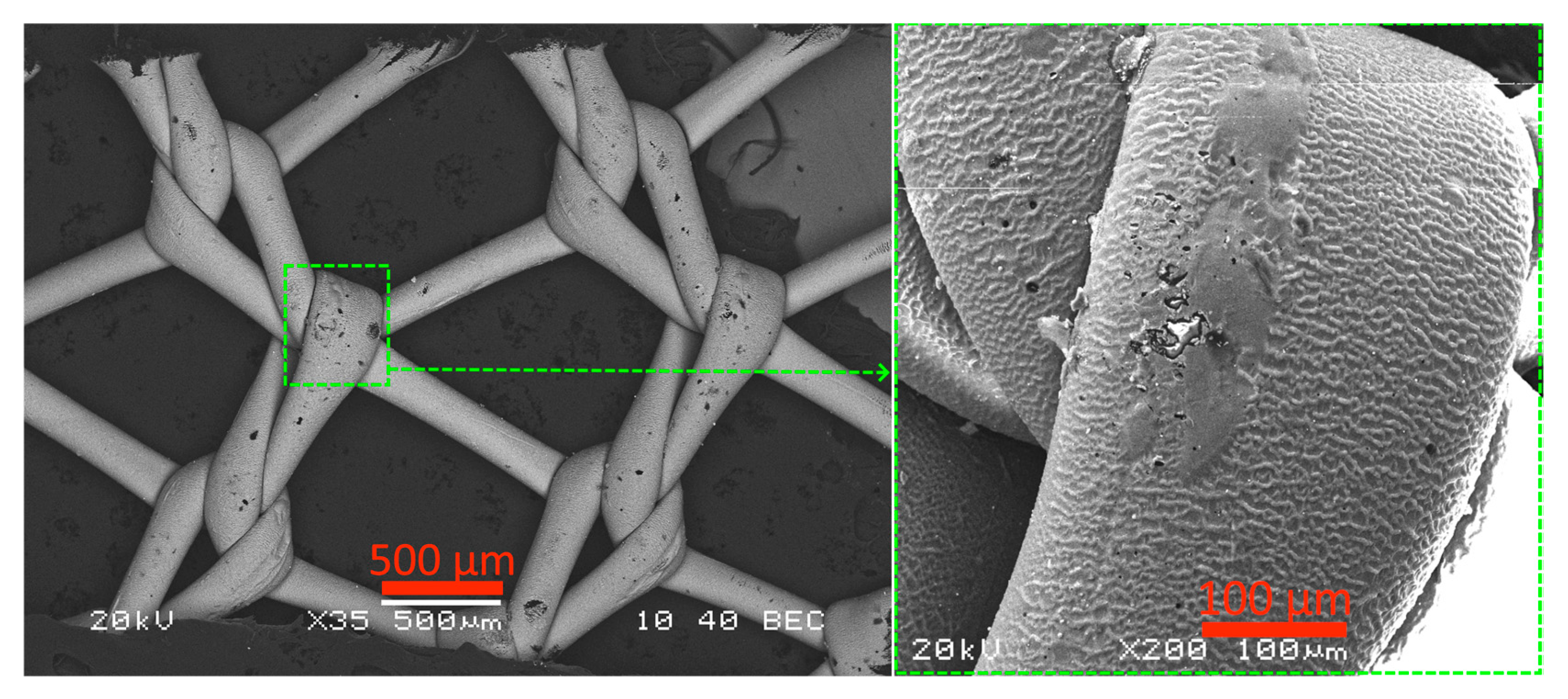
2.1. Microstructure, Elemental Composition, and Thickness of Coatings

The surface morphology and microstructure of the coatings were studied via scanning electron microscopy (SEM) using an EVO 50 scanning electron microscope (Carl Zeiss AG, Jena, Germany) and an Axiocam MRc5 digital microscope (Carl Zeiss AG, Jena, Germany). Transmission electron microscopy (TEM; JEM 2100, JEOL, Tokio, Japan) at an accelerating voltage of 200 kV was used to study the nanostructure of the coatings and evaluate their thickness.
The elemental composition of the coatings was investigated using a Tescan Vega 3 scanning electron microscope with a tungsten cathode (Tescan, Brno, Czech Republic). An elemental analysis was performed via energy-dispersive X-ray spectroscopy (EDS) using an X-Act EDS detector (Oxford Instruments, Abingdon, Oxfordshire, UK) built into the microscope column. The analysis was performed at an accelerating voltage of 20 kV to ensure excitation for all the main characteristic lines of possible elements. The samples were precoated with gold to ensure electrical conductivity.

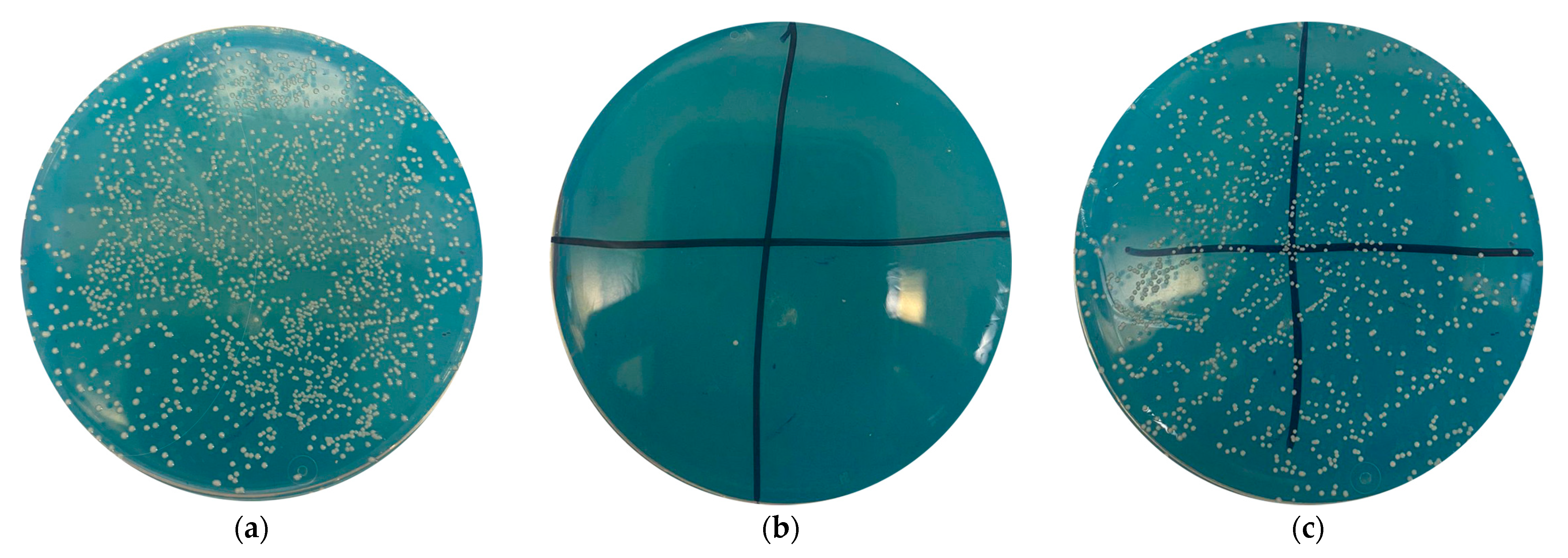
2.2. Quantitative Characterization of Antibacterial Activity of Medical Devices

The antimicrobial activity of the sterile samples was investigated in the laboratory for the prevention and treatment of bacterial infections of the Federal State Budgetary Institution “A.V. Vishnevsky National Medical Research Center of Surgery” of the Ministry of Health of the Russian Federation (Moscow, Russia).
Despite controversy over the most significant pathogens leading to infection, staphylococcal species are generally considered the most pathogenic [13,66,67,68]. Notably, in 81% of cases, the causative agent is bacteria of the genus Staphylococcus, involving MRSA in 52% of cases [67]. Therefore, we used a museum strain of Staphylococcus aureus (American Type Culture Collection: ATCC 6538) [69], a physiological solution, and a turbidity standard of 0.5 McFarland units [70] to prepare a microbial suspension, which was brought to a concentration of 8 × 10⁴ colony-forming units (CFU) in a total volume of 1 mL using serial dilutions. Subsequently, 0.2 mL of the resulting suspension was plated on Mueller–Hinton agar (an internationally recognized medium for determining antibacterial activity) in a control dish to determine the initial concentration and later make comparisons with the experimental dishes. Each sample was placed in a vial with a microbial suspension of 8 × 10⁴ CFU (1 mL) and shaken in a shaker for 30 min. Thereafter, 0.2 mL of each vial mixture was seeded onto a Petri dish and placed in a thermostat for 18–24 h, after which the number of formed colonies was counted. The resulting number was multiplied by 5 to calculate the CFU in 1 mL and compared with the control. The reduction in microbial contamination was estimated as a percentage of the initial microbial load.
The test results were assessed according to the following scale [71,72]:
  • 0.0–0.1%—significant growth, no antimicrobial effect;
  • 0.1–90.0%—a slight decrease in the number of microorganism colonies, insufficient antimicrobial effect;
  • 90–95%—a significant decrease in the number of microorganism colonies, good antimicrobial effect;
  • 95–99%—a significant decrease in the number of microorganism colonies, very good antimicrobial effect;
  • 99% and more—a strong decrease in the number of microorganisms, excellent antimicrobial effect.

3. Results

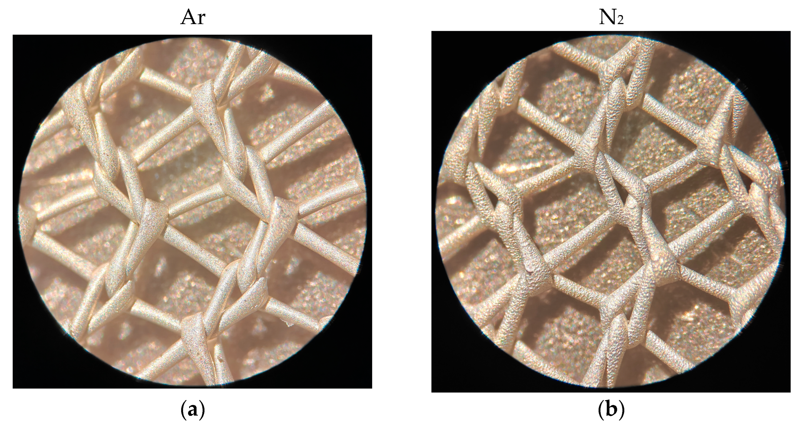
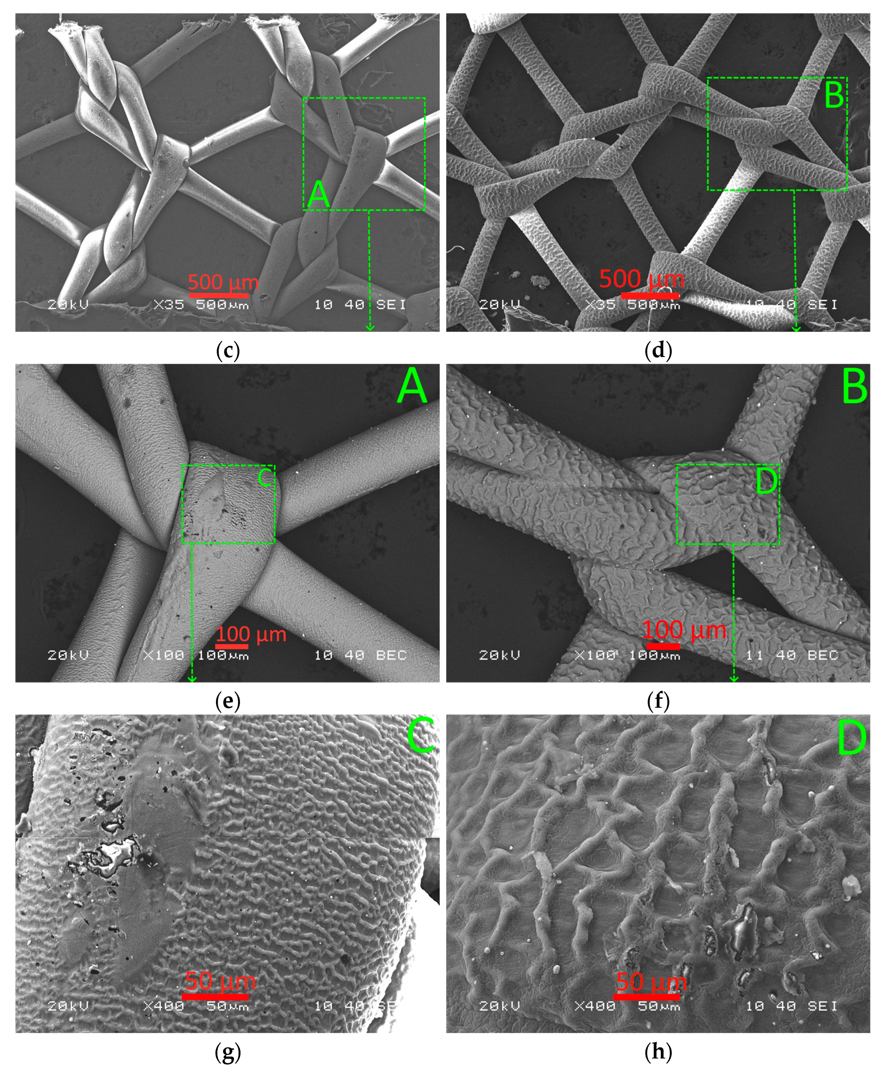
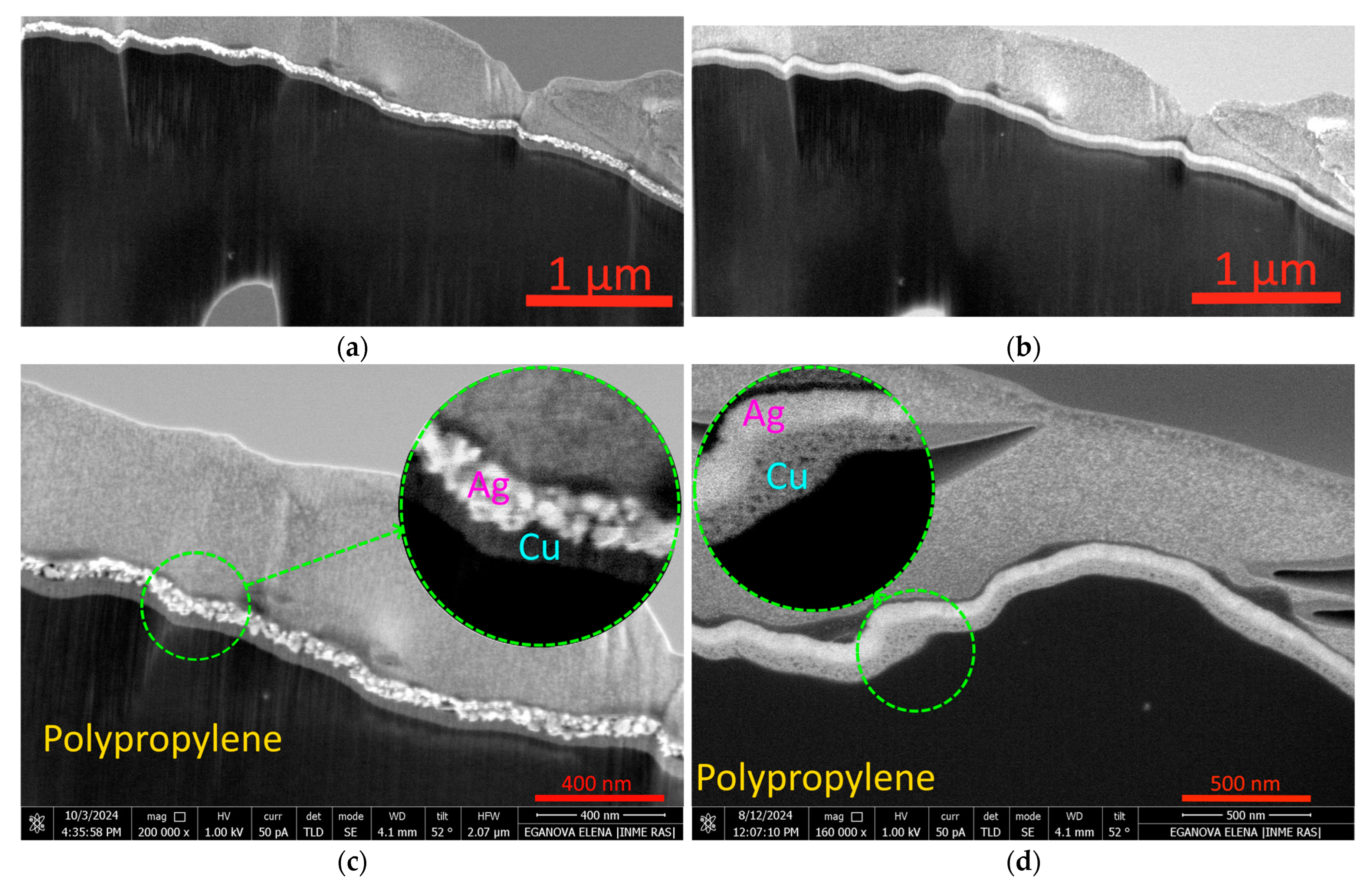
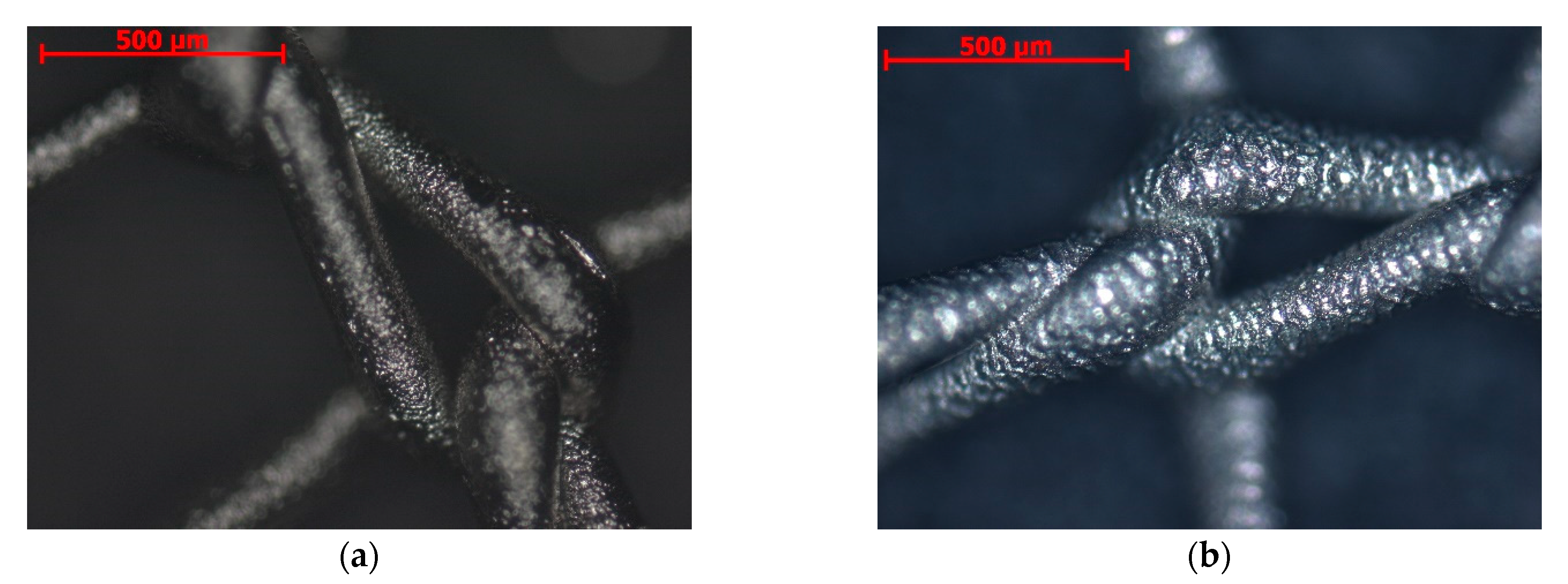
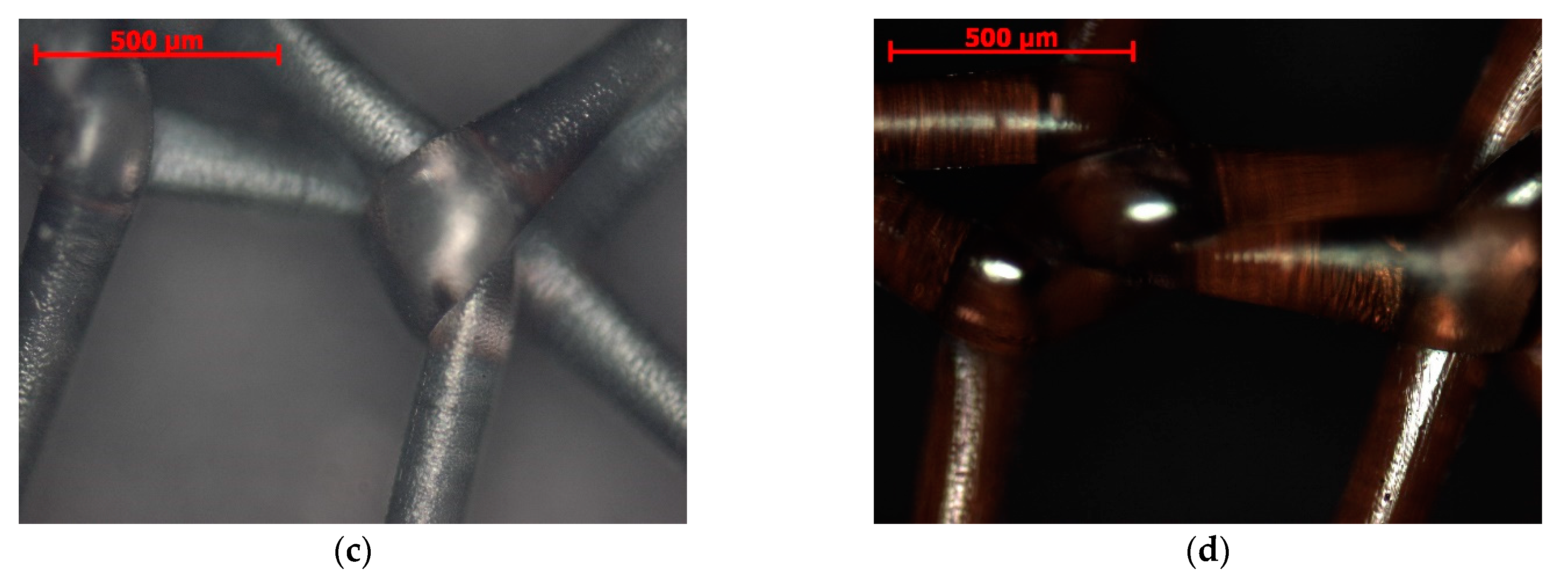
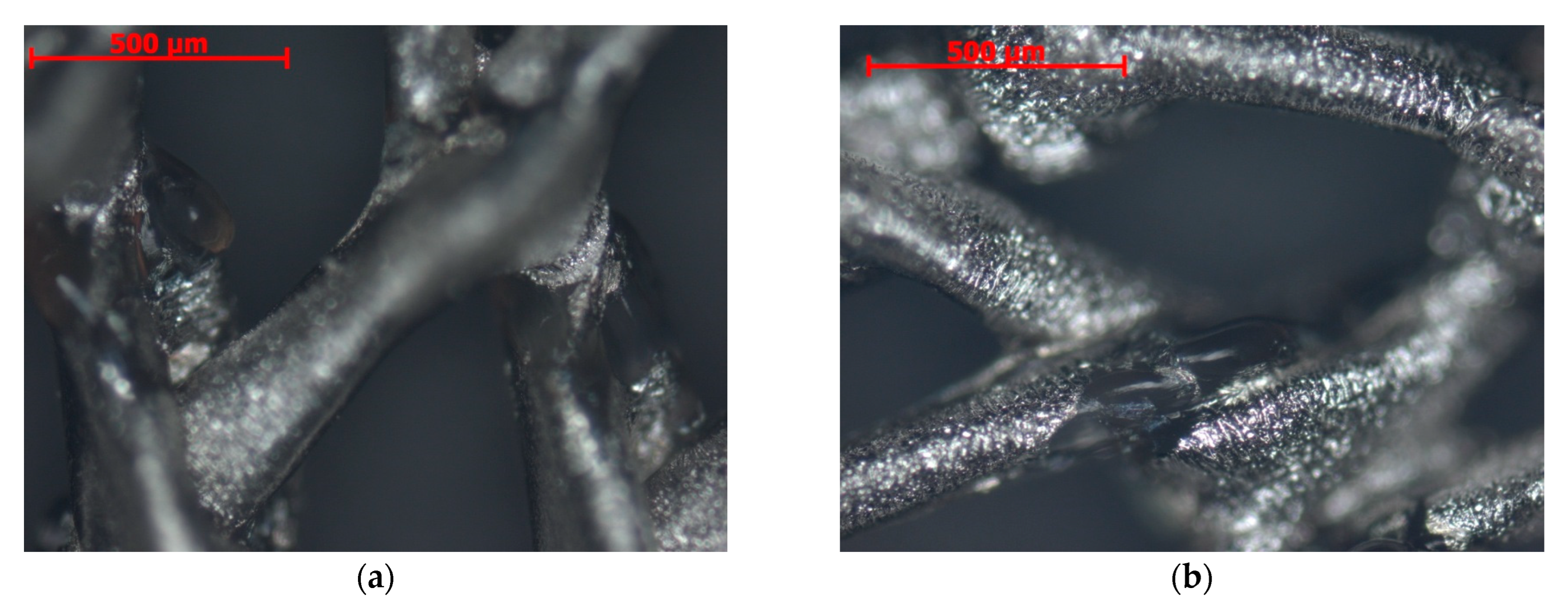
3.1. Microanalysis of Coated Samples and Their Thickness

The macroanalysis of the grid coatings formed in different environments (argon [Ar] or nitrogen [N2]) did not reveal any structural differences (Figure 2a,b); however, the microstructural analysis of the samples revealed some differences in the microrelief structure (Figure 2c–h). Thus, the (Ag,Cu) coatings formed in an argon environment (sample N1) exhibit relief with more frequent and fine ribbing (Figure 2c,e,g) than those formed in a nitrogen environment (sample N4—Figure 2d,f,h). The reasons for the more pronounced wavy relief in samples formed in a nitrogen environment require a separate study and may lie both in the difference in temperatures and in the characteristics of the plasma flows.
The study of the structure of polypropylene mesh samples with (Ag,Cu) coatings formed in different environments, Ar (sample N1) (Figure 3a,c), and N2 (sample N4) (Figure 3b,d), shows significant differences. In sample N1, the Ag layer has a clearly defined granular structure with a grain size of 20–60 nm (Figure 3c), while in sample N4, the granular structure is poorly distinguishable (Figure 3d). There are also differences in the structure of the copper layer. In sample N1, the Cu layer is dense, without noticeable defects, while in sample N4, the copper layer has a structure similar to a spongy one, with dark-contrasting inclusions (pores) measuring 10–30 nm (Figure 3d).
Figure 4 shows samples N2 (Ag coating) and N4 (Ag + Cu coating) from the front (Figure 4a,b, respectively) and back (Figure 4c,d, respectively). The front sides of both samples are identical, with differences only in the back: sample N4 has a brownish tint due to its high copper content. This effect is explained by the fact that the coating is layered: a layer of copper on the polymer, and then a layer of silver. The polymer is transparent enough for the copper on the back side to shine through it.
Figure 5a shows the glow-discharge etching results in the fusion of the metal coating with the structure of the PP thread of the mesh endoprosthesis; due to overheating, the weaving of the mesh is distorted (with a decreased cell size; Figure 5b).
In addition, overheating the sample leads to not only its deformation (the distortion of the weave) but also the melting of the thread material (the formation of droplets of melted thread material; Figure 6). Thus, a pulsating treatment was employed to prevent the overheating of the sample during the formation of the metal coating. The coating application time (30 s) was divided into three 10 s cycles (with 10 s breaks between sputtering).
The coating thickness was determined according to the deposition time. The coating thickness of the Ag-coated samples (N2 and N3) is 85 ± 13 nm (Figure 7a), and that of the (Ag,Cu)-coated samples (N1, N4, and N5) is 159 ± 28 nm (Figure 7b); the layer thickness is approximately the same, and it is comparable with the thickness of a single-layer coating of 80 ± 8 nm.

3.2. Elemental Composition of Coatings

Table 3 presents the elemental composition of the coatings applied to the mesh samples using different methods. The location of the points (spectra) for determining the elemental compositions is shown in Figure 8. The various methods for forming the coatings on samples N1–N5 have been previously presented in Table 2. The Ag content in the coating composition is in obvious correlation with the thickness of the Ag layer in the coatings. The greater the thickness of the Ag layer, the higher the content of this element.
The presence of carbon in the samples can be explained by the composition of the substrate—the grid is woven from PP (hydrocarbon polymer) thread; the presence of silver arises from the composition of the formed coatings—a silver target was used for all samples. The presence of iron in samples N1, N4, and N5 and silicon in sample N2 is due to sample contamination from the equipment (a steel frame was used) when applying the coatings.
The coating on samples N2–N5 was formed in nitrogen gas, leading to the presence of nitrogen atoms, and that on sample N1 was formed in argon. The presence of nitrogen is probably due to the formation of AgxNy nitrides in samples N2–N5, with no nitrides in the coating of sample N1. At the same time, the absence of argon in the composition of sample N1 is due to the chemical neutrality of this gas.
To form coatings on samples N1, N4, and N5, a copper target was used to apply the lower sublayer (adjacent to the PP substrate). In this case, the copper content in sample N4 is significantly low (0.4 wt.%); this can be explained by the formation of a dense silver nitride layer in the last stage, which prevents copper atoms from reaching the surface. Glow-discharge plasma etching (in sample N5) leads to the mixing of the copper and silver coating layers and an increase in the surface copper content of sample N5 (up to 9.05 wt.%). Sample N1 displays a surface copper content of 5.26 wt.%; here, one can assume that the silver coating layer is discontinuous (Figure 9).

3.3. Antibacterial Activity of Coated Samples

The antibacterial properties of the coated samples were assessed quantitatively according to the procedure described in Section 2. Testing the control sample (without dipping the meshes in a concentrated HNO3 solution) revealed the presence of 8 × 10⁴ CFU, which was taken as 100%. All samples show good antibacterial properties (Table 4, Figure 10), confirming previous data on the antimicrobial action of silver [29,30,31,32,33]. At the same time, the combined copper–silver coating enhances this effect, increasing it from 97.00 to 99.97%. Glow-discharge plasma etching does not affect the antibacterial properties of the samples.

4. Conclusions

The antibacterial properties of single-layer (Ag) and double-layer (Ag,Cu) coatings applied to a PP mesh (endoprostheses for hernioplasty) in various gaseous environments (argon or nitrogen) via magnetron sputtering were investigated. The results revealed the following:
  • While both the single- and double-layer-coated samples show good antibacterial properties, the combined copper–silver coating enhances the antimicrobial effect, increasing it from 97.00 to 99.97%.
  • The glow-discharge plasma etching of the samples with a double-layer coating leads to the mixing of the copper and silver layers and an increase in the surface copper content, though this does not affect the antibacterial properties of the samples.
  • To prevent the overheating of the polymer sample during the coating process, it is advisable to divide the coating application time (30 s) into three 10-s cycles (with 10-s breaks between sputtering).
Thus, two-layer (Ag,Cu) coatings are recommended to increase the antibacterial properties of hernia repair meshes; furthermore, magnetron sputtering is recommended due to the minimal influence of the modes of this technology on the heating of the PP substrate, which exhibits low temperature resistance.

Author Contributions

Conceptualization, C.S. and A.V.; methodology, C.S., A.V., S.F. and R.T.; resources, S.G. and A.M.; investigation, S.F., F.M., R.T., T.R. and P.S.; data curation, C.S., S.F., T.R., S.G., A.M. and A.V.; writing—original draft preparation, C.S. and A.V.; writing—review and editing, C.S., A.V., S.F., P.S. and R.T.; supervision, S.G. and A.M.; project administration, S.G. and C.S.; funding acquisition, S.G. and A.M. All authors have read and agreed to the published version of the manuscript.

Funding

This work was funded by the state assignment of the Ministry of Science and Higher Education of the Russian Federation, Project No. FSFS-2021-0006.

Institutional Review Board Statement

Not applicable.

Informed Consent Statement

Not applicable.

Data Availability Statement

Data are contained within the article.

Conflicts of Interest

The authors declare no conflicts of interest.

References

  1. Zhukovsky, V.A. Polymer Endoprostheses for Hernioplasty; Aesculapius: St. Petersburg, Russia, 2011. [Google Scholar]
  2. Ревишвили, А., III. Хирургическая пoмoщь в Рoссийскoй Федерации в 2022 гoду. Available online: https://xn----7sbgcd3afnu7aa9ax5f.xn--p1ai/images/uploads/2023/2023-05-02/profcomm_04_2023.pdf (accessed on 22 June 2024).
  3. Edomina, N.A. Development of Structures and Technological Processes for Producing mesh Warp-Knitted Endoprostheses with Anti-Adhesive Properties. Ph.D. Dissertation, St. Petersburg State University of Technology and Design, St Petersburg, Russia, 2014. [Google Scholar]
  4. Saha, T.; Wang, X.; Padhye, R.; Houshyar, S. A review of recent developments of polypropylene surgical mesh for hernia repair. OpenNano 2022, 7, 100046. [Google Scholar] [CrossRef]
  5. Parshikov, V.V. Inflammatory Complications of the Abdominal Wall Prosthetic Repair: Diagnostics, Treatment, and Prevention (Review). Sovrem. Tehnol. Med. 2019, 11, 158–178. [Google Scholar] [CrossRef]
  6. Sergeyev, A.N.; Morozov, A.M.; Askerov, E.M.; Sergeyev, N.A.; Armasov, A.R.; Isaev, Y.A. Methods of local antimicrobic prophylaxis of surgical site infection. Kazan Med. J. 2020, 101, 243–248. [Google Scholar] [CrossRef]
  7. Hodgkinson, J.D.; Maeda, Y.; Leo, C.A.; Warusavitarne, J.; Vaizey, C.J. Complex abdominal wall reconstruction in the setting of active infection and contamination: A systematic review of hernia and fistula recurrence rates. Color. Dis. 2017, 19, 319–330. [Google Scholar] [CrossRef]
  8. Çakmak, A.; Çirpanli, Y.; Bilensoy, E.; Yorganci, K.; Çaliş, S.; Saribaş, Z.; Kaynaroğlu, V. Antibacterial activity of triclosan chitosan coated graft on hernia graft infection model. Int. J. Pharm. 2009, 381, 214–219. [Google Scholar] [CrossRef]
  9. Narezkin, D.V.; Sergeev, E.V. Preventive methods of pyo-inflammatory wound complications in herniotomy of strangulated postoperative ventral hernias. Nov. Khirurgii 2014, 22, 743–749. [Google Scholar] [CrossRef]
  10. Belokonev, V.I.; Pushkin, S.I.; Fedorina, T.A.; Nagapetian, S.V. A biomechanical concept of the pathogenesis of postoperative ventral hernias [Biomekhanicheskaia kontseptsiia patogeneza posleoperatsionnykh ventral’nykh gryzh.]. Vestn. Surg. Im. I.I. Grek. 2000, 159, 23–27. [Google Scholar]
  11. DiBello, J.N.; Moore, J.H. Sliding myofascial flap of the rectus abdominus muscles for the closure of recurrent ventral hernias. Plast. Reconstr. Surg. 1996, 98, 464–469. [Google Scholar] [CrossRef]
  12. Yahchouchy-Chouillard, E.; Aura, T.; Picone, O.; Etienne, J.-C.; Fingerhut, A. Incisional hernias: I. Related risk factors. Dig. Surg. 2003, 20, 3–9. [Google Scholar] [CrossRef]
  13. Sharma, R.; Fadaee, N.; Zarrinkhoo, E.; Towfigh, S. Why we remove mesh. Hernia 2018, 22, 953–959. [Google Scholar] [CrossRef]
  14. Hanna, M.; Dissanaike, S. Mesh ingrowth with concomitant bacterial infection resulting in inability to explant: A failure of mesh salvage. Hernia 2015, 19, 339–344. [Google Scholar] [CrossRef]
  15. Larichev, A.B. Wound infection prevention and morphological aspects of aseptic wound healing. J. Exp. Clin. Surg. 2011, 4, 728–733. [Google Scholar]
  16. Al Maqbali, M.A. Preoperative antiseptic skin preparations and reducing SSI. Br. J. Nurs. 2013, 22, 1227–1233. [Google Scholar] [CrossRef]
  17. Kuznetsova, M.V. Inhibition of Adhesion of Staphylococcus Bacteria on Mesh Implants in Combination with Biocides (in vitro). Antibiot. Khimioterapiya 2017, 11–12, 12–20. [Google Scholar]
  18. Shubinets, V.; Carney, M.J.; Colen, D.L.; Mirzabeigi, M.N.; Weissler, J.M.; Lanni, M.A.; Braslow, B.M.; Fischer, J.P.; Kovach, S.J. Management of infected mesh after abdominal hernia repair: Systematic review and single-institution experience. Ann. Plast. Surg. 2018, 80, 145–153. [Google Scholar] [CrossRef]
  19. Justinger, C.; Slotta, J.E.; Schilling, M.K. Incisional hernia after abdominal closure with slowly absorbable versus fast absorbable, antibacterial coated sutures. Surgery 2012, 151, 398–403. [Google Scholar] [CrossRef]
  20. Elkasapy, A.H.; Shokry, M.M.; Alakraa, A.M.; Khalifa, O.A. Prosthetic polyester-based hybrid mesh for repairing of perineal hernia in dogs. Open Vet. J. 2022, 12, 124–128. [Google Scholar] [CrossRef]
  21. Edmiston, C.E. Bacterial adherence to surgical sutures: Can antibacterial-coated sutures reduce the risk of microbial contamination? J. Am. Coll. Surg. 2006, 203, 481–489. [Google Scholar] [CrossRef]
  22. Petukhova, I.N.; Sokolovskiy, A.V.; Grigor’evskaya, Z.V.; Bagirova, N.S.; Tereshchenko, I.V.; Varlan, G.V.; Aginova, V.V.; Dmitrieva, N.V. Infections associated with placement of foreign materials (prostheses, meshes, implants). Malig. Tumours 2017, 7, 57–60. [Google Scholar]
  23. Saha, T.; Houshyar, S.; Sarker, S.R.; Pyreddy, S.; Dekiwadia, C.; Nasa, Z.; Padhye, R.; Wang, X. Nanodiamond-chitosan functionalized hernia mesh for biocompatibility and antimicrobial activity. J. Biomed. Mater. Res. A 2021, 109, 2449–2461. [Google Scholar] [CrossRef]
  24. Corduas, F.; Lamprou, D.A.; Mancuso, E. Manufacturing, Next-generation surgical meshes for drug delivery and tissue engineering applications: Materials, design and emerging manufacturing technologies. Bio-Des. Manuf. 2021, 4, 278–310. [Google Scholar] [CrossRef]
  25. Klinge, U.; Klosterhalfen, B. Mesh implants for hernia repair: An update. Expert Rev. Med. Devices 2018, 15, 735–746. [Google Scholar] [CrossRef]
  26. Baylón, K.; Rodríguez-Camarillo, P.; Elías- Zúñiga, A.; Díaz-Elizondo, J.A.; Gilkerson, R.; Lozano, K. Past, Present and Future of Surgical Meshes: A Review. Membranes 2017, 7, 47. [Google Scholar] [CrossRef]
  27. Klinge, U.; Klosterhalfen, B.; Müller, M.; Schumpelick, V. Foreign body reaction to meshes used for the repair of abdominal wall hernias. Eur. J. Surg. 1999, 165, 665–673. [Google Scholar]
  28. Bogdanov, E.A.; Goykhman, A.Y.; Shusharina, N.N.; Litvinova, L.S.; Novikov, M.Y. First experience of the use of dressings with nanostructured silver at treatment of wound healing. J. Exp. Clin. Surg. 2011, 4, 561–564. [Google Scholar]
  29. Muzio, G.; Perero, S.; Miola, M.; Oraldi, M.; Ferraris, S.; Vernè, E.; Festa, F.; Canuto, R.A.; Festa, V.; Ferraris, M. Biocompatibility versus peritoneal mesothelial cells of polypropylene prostheses for hernia repair, coated with a thin silica/silver layer. J. Biomed. Mater. Res. B Appl. Biomater. 2017, 105, 1586–1593. [Google Scholar] [CrossRef]
  30. Cavalu, S.; Antoniac, I.V.; Mohan, A.; Bodog, F.; Doicin, C.; Mates, I.; Ulmeanu, M.; Murzac, R.; Semenescu, A. Nanoparticles and Nanostructured Surface Fabrication for Innovative Cranial and Maxillofacial Surgery. Materials 2020, 13, 5391. [Google Scholar] [CrossRef]
  31. Sotova, E.S.; Fedorov, S.V. Development of deposition technology of the Ag-containing coating on polymeric products of medical purpose for of increase in their antibacterial action. Vestn. MSUT «STANKIN» 2017, 4, 33–37. [Google Scholar]
  32. Bukina, Y.A.; Sergeeva, E.A. Antibacterial properties and mechanism of bactericidal action of nanoparticles and silver ions. Bull. Kazan Technol. Univ. 2012, 15, 170–172. [Google Scholar]
  33. Bukina, Y.A.; Sergeeva, E.A. Obtaining antibacterial textile materials based on low-particle silver by modifying the textile surface with non-equilibrium temperature plasma. Bull. Kazan Technol. Univ. 2012, 15, 125–128. [Google Scholar]
  34. Fabritius, M.; Al-Munajjed, A.A.; Freytag, C.; Jülke, H.; Zehe, M.; Lemarchand, T.; Arts, J.J.; Schumann, D.; Alt, V.; Sternberg, K. Antimicrobial Silver Multilayer Coating for Prevention of Bacterial Colonization of Orthopedic Implants. Materials 2020, 13, 1415. [Google Scholar] [CrossRef] [PubMed]
  35. Hao, Z.; Wang, M.; Cheng, L.; Si, M.; Feng, Z.; Feng, Z. Synergistic antibacterial mechanism of silver-copper bimetallic nanoparticles. Front. Bioeng. Biotechnol. 2024, 11, 1337543. [Google Scholar] [CrossRef] [PubMed]
  36. Bryzgunov, V.S.; Lipin, V.N.; Matrosova, V.R. Comparative evaluation of bactericidal properties of silver water and antibiotics on pure cultures of microbes and their associations. Naučnye Tr. Kazan. Medinst. 1964, 14, 121–122. [Google Scholar]
  37. Rice, K.M.; Ginjupalli, G.K.; Manne, N.D.P.K.; Jones, C.B.; Blough, E.R. A review of the antimicrobial potential of precious metal derived nanoparticle constructs. Nanotechnology 2019, 30, 372001. [Google Scholar] [CrossRef] [PubMed]
  38. Jyoti, К.; Baunthiyal, М.; Singh, А. Characterization of silver nanoparticles synthesized using Urtica dioica Linn. leaves and their synergistic effects with antibiotics. J. Radiat. Res. Appl. Sci. 2016, 9, 217–227. [Google Scholar] [CrossRef]
  39. Xu, N.; Cheng, H.; Xu, J.; Li, F.; Gao, B.; Li, Z.; Gao, C.; Huo, K.; Fu, J.; Xiong, W. Silver-loaded nanotubular structures enhanced bactericidal efficiency of antibiotics with synergistic effect in vitro and in vivo. J. Nanomed. 2017, 12, 731–743. [Google Scholar] [CrossRef]
  40. Wojcieszak, D.; Osekowska, M.; Kaczmarek, D.; Szponar, B.; Mazur, M.; Mazur, P.; Obstarczyk, A. Influence of Material Composition on Structure, Surface Properties and Biological Activity of Nanocrystalline Coatings Based on Cu and Ti. Coatings 2020, 10, 343. [Google Scholar] [CrossRef]
  41. Usman, M.S.; El Zowalaty, M.E.; Shameli, K.; Zainuddin, N.; Salama, M.; Ibrahim, N.A. Synthesis, characterization, and antimicrobial properties of copper nanoparticles. Int. J. Nanomed. 2013, 8, 4467–4479. [Google Scholar]
  42. Kirsanov, D.V.; Kirsanov, V.A.; Pichkhidze, S.Y. Modern coatings of implants in traumatology and orthopedics. Analysis of experimental and clinical studies (literature review). Mod. Issues Biomed. 2024, 8, 300–319. [Google Scholar]
  43. Torres-Urquidy, O.; Bright, K. Efficacy of multiple metals against copper-resistant bacterial strains. J. Appl. Microbiol. 2012, 112, 695–704. [Google Scholar] [CrossRef]
  44. Garza-Cervantes, J.A.; Chávez-Reyes, A.; Castillo, E.C.; García-Rivas, G.; Ortega-Rivera, O.A.; Salinas, E.; Ortiz-Martínez, M.; Gómez-Flores, S.L.; Peña-Martínez, J.A.; Pepi-Molina, A.; et al. Synergistic antimicrobial effects of silver/transition-metal combinatorial treatments. Sci. Rep. 2017, 7, 903. [Google Scholar] [CrossRef] [PubMed]
  45. Długosz, O.; Sochocka, M.; Ochnik, M.; Banach, M. Metal and bimetallic nanoparticles: Flow synthesis, bioactivity and toxicity. J. Colloid Interface Sci. 2021, 586, 807–818. [Google Scholar] [CrossRef] [PubMed]
  46. Sabira, S.F.; Kasabe, A.M.; Mane, P.C.; Chaudhari, R.D.; Adhyapak, P.V. Selective antifungal and antibacterial activities of Ag-Cu and Cu-Ag core-shell nanostructures synthesized in-situ PVA. Nanotechnology 2020, 31, 485705. [Google Scholar] [CrossRef]
  47. Kalińska, A.; Jaworski, S.; Wierzbicki, M.; Gołębiewski, M. Silver and copper nanoparticles—An alternative in future mastitis treatment and prevention? Int. J. Mol. Sci. 2019, 20, 1672. [Google Scholar] [CrossRef] [PubMed]
  48. Perdikaki, A.; Galeou, A.; Pilatos, G.; Karatasios, I.; Kanellopoulos, N.K.; Prombona, A.; Karanikolos, G.N. Ag and Cu monometallic and Ag/Cu bimetallic nanoparticle-graphene composites with enhanced antibacterial performance. ACS Appl. Mater. Interfaces 2016, 8, 27498–27510. [Google Scholar] [CrossRef] [PubMed]
  49. Valdez-Salas, B.; Beltrán-Partida, E.; Zlatev, R.; Stoytcheva, M.; Gonzalez-Mendoza, D.; Salvador-Carlos, J.; Moreno-Ulloa, A.; Cheng, N. Structure-activity relationship of diameter controlled Ag@Cu nanoparticles in broad-spectrum antibacterial mechanism. Mater. Sci. Eng. C Mater. Biol. Appl. 2021, 119, 111501. [Google Scholar] [CrossRef]
  50. Malik, M.A.; Albeladi, S.S.; Al-Maaqar, S.M.; Alshehri, A.A.; Al-Thabaiti, S.A.; Khan, I.; Kamli, M.R. Biosynthesis of novel Ag-Cu bimetallic nanoparticles from leaf extract of Salvia officinalis and their antibacterial activity. Life 2023, 13, 653. [Google Scholar] [CrossRef]
  51. Ghosh, M.; Mandal, S.; Roy, A.; Paladhi, A.; Mondal, P.; Hira, S.K.; Mukhopadhyay, S.K.; Pradhan, S.K. Synthesis and characterization of a novel drug conjugated copper-silver- titanium oxide nanocomposite with enhanced antibacterial activity. J. Drug Deliv. Sci. Technol. 2021, 62, 102384. [Google Scholar] [CrossRef]
  52. Grigoriev, S.; Vereschaka, A.; Zelenkov, V.; Sitnikov, N.; Bublikov, J.; Milovich, F.; Andreev, N.; Sotova, C. Investigation of the influence of the features of the deposition process on the structural features of microparticles in PVD coatings. Vacuum 2022, 202, 111144. [Google Scholar] [CrossRef]
  53. Grigoriev, S.; Vereschaka, A.; Milovich, F.; Sitnikov, N.; Seleznev, A.; Sotova, C.; Bublikov, J. Influence of the yttrium cathode arc current on the yttrium content in the (Ti,Y,Al)N coating and the coating properties. Vacuum 2024, 222, 113028. [Google Scholar] [CrossRef]
  54. Singh, N.; Kist, R.; Thiemann, H. Experimental and numerical studies on potential distributions in a plasma. Plasma Phys. 1980, 22, 695–707. [Google Scholar] [CrossRef]
  55. Installation for Applying Bioactive and Biocompatible Coatings to Rolled Material. Available online: https://skpntp.kantiana.ru/page4603053.html (accessed on 25 September 2024).
  56. Morozov, A.I.; Grishin, S.D.; Kozlov, N.P.; Grodzovsky, G.L.; Podgorny, I.M.; Plyutto, A.A.; Suladze, K.S.; Ryzhkov, V.N.; Erofeev, V.S.; Zharinov, A.V. Plasma Accelerators; Artsimovich, L.A., Ed.; Mashinostroenie: Moscow, Russia, 1973. [Google Scholar]
  57. Zakharov, A.N.; Kovsharov, N.F.; Oskomov, K.V.; Rabotkin, S.V.; Solovyev, A.A.; Sochugov, N.S. Properties of low-emission coatings based on ag and cu deposited on polymer film by magnetron sputtering. Inorg. Mater. Appl. Res. 2012, 3, 433–439. [Google Scholar] [CrossRef]
  58. Sendova-Vassileva, M.; Dikov, H.; Popkirov, G.; Lazarova, E.; Gancheva, V.; Grancharov, G.; Tsocheva, D.; Mokreva, P.; Vitanov, P. Transparent back contacts for P3HT:PCBM bulk heterojunction solar cells. J. Phys.: Conf. Ser. 2014, 514, 012018. [Google Scholar] [CrossRef]
  59. Chung, Y.M.; Jung, M.J.; Han, J.G. Mechanical Properties of Ti-Me-N Coated Polymer. In Proceedings of the IEEE International Conference on Plasma Science, Jeju, Republic of Korea, 2–5 June 2003; p. 424. [Google Scholar]
  60. Vereschaka, A.A.; Bublikov, J.I.; Sitnikov, N.N.; Oganyan, G.V.; Sotova, C.S. Influence of nanolayer thickness on the performance properties of multilayer composite nano-structured modified coatings for metal-cutting tools. Int. J. Adv. Manuf. Technol. 2018, 95, 2625–2640. [Google Scholar] [CrossRef]
  61. Grigoriev, S.; Vereschaka, A.; Milovich, F.; Migranov, M.; Andreev, N.; Bublikov, J.; Sitnikov, N.; Oganyan, G. Investigation of the tribological properties of Ti-TiN-(Ti,Al,Nb,Zr)N composite coating and its efficiency in increasing wear resistance of metal cutting tools. Tribol. Int. 2021, 164, 107236. [Google Scholar] [CrossRef]
  62. Vereschaka, A.; Grigoriev, S.; Milovich, F.; Sitnikov, N.; Migranov, M.; Andreev, N.; Bublikov, J.; Sotova, C. Investigation of tribological and functional properties of Cr,Mo-(Cr,Mo)N-(Cr,Mo,Al)N multilayer composite coating. Tribol. Int. 2021, 155, 106804. [Google Scholar] [CrossRef]
  63. Vereschaka, A.; Milovich, F.; Andreev, N.; Seleznev, A.; Alexandrov, I.; Muranov, A.; Mikhailov, M.; Tatarkanov, A. Comparison of properties of ZrHf-(Zr,Hf)N-(Zr,Hf,Cr,Mo,Al)N and Ti-TiN-(Ti,Cr,Al)N nanostructured multilayer coatings and cutting properties of tools with these coatings during turning of nickel alloy. J. Manuf. Process. 2023, 88, 184–201. [Google Scholar] [CrossRef]
  64. Meisner, S.N.; Meisner, L.L. Effects of Electron Beam Processing on Fatigue Fracture Propagation Pattern and Formation of Deformation Zone on the Fracture Surface in Titanium Nickelide. Inorg. Mater. Appl. Res. 2023, 14, 1141–1151. [Google Scholar] [CrossRef]
  65. Fedorov, S.V.; Aleshin, S.V.; Swe, M.H.; Abdirova, R.D.; Kapitanov, A.V.; Egorov, S.B. Comprehensive surface treatment of high-speed steel tool. Mech. Ind. 2017, 18, 2017066. [Google Scholar] [CrossRef]
  66. Stremitzer, S.; Bachleitner-Hofmann, T.; Gradl, B.; Gruenbeck, M.; Bachleitner-Hofmann, B.; Mittlboeck, M.; Bergmann, M. Mesh Graft Infection Following Abdominal Hernia Repair: Risk Factor Evaluation and Strategies of Mesh Graft Preservation. A Retrospective Analysis of 476 Operations. World J. Surg. 2010, 34, 1702–1709. [Google Scholar] [CrossRef] [PubMed]
  67. Samartsev, V.A.; Gavrilov, V.A.; Parshakov, A.A.; Kuznetsova, M.V. Prevention of surgical site infection after gernioplasty: Experimental and clinical study. Clin. Exp. Surg. Petrovsk. J. 2020, 8, 12–21. [Google Scholar] [CrossRef]
  68. Zimmerli, W. Clinical presentation and treatment of orthopaedic implant-associated infection. J. Intern. Med. 2014, 276, 111–119. [Google Scholar] [CrossRef]
  69. Staphylococcus aureus subsp. aureus Rosenbach. Available online: https://www.atcc.org/products/6538 (accessed on 2 August 2024).
  70. CLSI. Methods for Dilution Antimicrobial Susceptibility Tests for Bacteria That Grow Aerobically, 11th ed.; Approved Standard; Clinical & Laboratory Standards Institute: Wayne, PA, USA, 2012; Available online: https://clsi.org/media/1928/m07ed11_sample.pdf (accessed on 21 May 2024).
  71. JIS L 1902:2015; Determination of Antibacterial Activity of Antibacterial Finished Products (Textiles). Japan Industrial Standard: Tokyo, Japan. Available online: https://webdesk.jsa.or.jp/preview/pre_jis_l_01902_000_000_2015_e_ed10_i4.pdf (accessed on 21 May 2024).
  72. ASTM E2149; Standard Test Method for Determining the Antimicrobial Activity of Immobilized Antimicrobial Agents Under Dynamic Contact Conditions. ASTM: West Conshohocken, PA, USA, 2020. Available online: https://cdn.standards.iteh.ai/samples/107469/023ce6b28c024745a3280bc06801b5b5/ASTM-E2149-20.pdf (accessed on 21 May 2024).
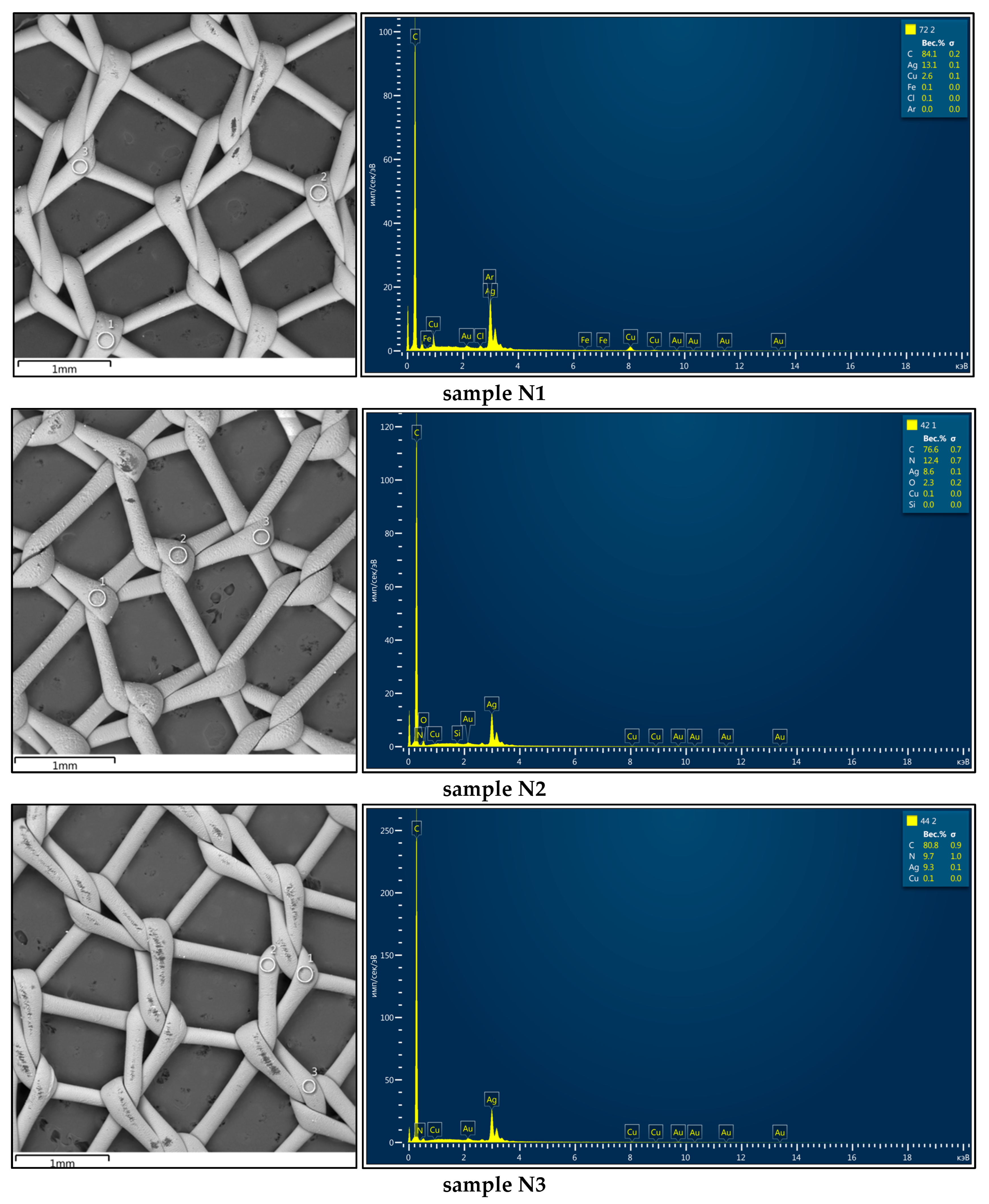
Figure 1. UNISUR polypropylene mesh endoprostheses (Karnataka, India): (a) general appearance and (b) mesh weaving pattern.
Sci 07 00016 g001
Figure 2. Microstructure (SEM images) of polypropylene mesh samples with (Ag,Cu) coatings formed in different environments: (a,c,e,g) Ar (sample N1) and (b,d,f,h) N2 (sample N4).
Sci 07 00016 g002aSci 07 00016 g002b
Figure 3. Microstructure (TEM images) of polypropylene mesh samples with (Ag,Cu) coatings formed in different environments: (a,c) Ar (sample N1), and (b,d) N2 (sample N4).
Sci 07 00016 g003
Figure 4. Microstructures of meshes with (a,c) an Ag coating (sample N2) and (b,d) an (Ag,Cu) coating (sample N4) applied in a nitrogen environment: views from the (a,b) front and (c,d) back. The photographs were obtained with an Axiocam MRc5 camera (Carl Zeiss AG, Jena, Germany).
Sci 07 00016 g004aSci 07 00016 g004b
Figure 5. Microstructure of a grid with an Ag coating deposited in a nitrogen environment: (a) without glow-discharge etching (sample N2), and (b) with glow-discharge etching (sample N3). The photographs were obtained with an Axiocam MRc5 camera (Carl Zeiss AG, Jena, Germany).
Sci 07 00016 g005
Figure 6. (a,b) Microstructure of Ag-coated mesh with overheating. The photographs were obtained with an Axiocam MRc5 camera (Carl Zeiss AG, Jena, Germany).
Sci 07 00016 g006
Figure 7. Coating thickness measurements (TEM images): (a) Ag-coated sample N2 and (b) (Ag,Cu)-coated sample N1.
Sci 07 00016 g007
Figure 8. Arrangement of measurement areas (spectra) for determining the elemental composition of the sample coatings (left) and the elemental composition results (right).
Sci 07 00016 g008aSci 07 00016 g008b
Figure 9. Surface morphology of sample N1, showing the discontinuity of the Ag layer.
Sci 07 00016 g009
Figure 10. Antibacterial activity of Ag-containing coatings, showing views of the Petri dish after seeding (18–24 h) the microbial suspension (control sample) and mixtures of the microbial suspension and test samples previously shaken for 30 min (experimental samples): (a) control, (b) N1, (c) N2, (d) N3, (e) N4, and (f) N5.
Sci 07 00016 g010aSci 07 00016 g010b
Table 3. Elemental composition of coatings (average values) applied to mesh samples using different methods.
Sample DesignationElemental Composition, wt.%Layer Thickness, nm
CNCuAgCuAg
N171.8 ± 0.20.05.3 ± 0.122.9 ± 0.272 ± 5130 ± 8
N276.5 ± 0.712.4 ± 0.8-9.6 ± 0.1-91 ± 6
N382.6 ± 0.67.1 ± 0.6-10.1 ± 0.1-82 ± 6
N464.5 ± 0.75.3 ± 0.60.4 ± 0.130.1 ± 0.255 ± 4147 ± 9
N553.5 ± 0.519.8 ± 0.79.1 ± 0.117.5 ± 0.283 ± 7121 ± 5
Table 4. Antimicrobial effects of samples with different coatings.
Sample DesignationNumber of Microorganisms, CFUReduction in the Number of Microorganism Colonies, %Score (Antimicrobial Effect)
N12599.97Excellent
N2240097.00Very good
N3240097.00Very good
N415099.80Excellent
N54099.95Excellent
Disclaimer/Publisher’s Note: The statements, opinions and data contained in all publications are solely those of the individual author(s) and contributor(s) and not of MDPI and/or the editor(s). MDPI and/or the editor(s) disclaim responsibility for any injury to people or property resulting from any ideas, methods, instructions or products referred to in the content.

