Proceeding Paper

Exploring Schiff Bases as Promising Alternatives to Traditional Drugs in the In Silico Treatment of Anti-Leishmaniasis as Trypanothione Reductase Inhibitors †

by
Diego R. Peixoto
1,
Carlos S. H. Shiraishi
2,3,4,*,
Rui M. V. Abreu
2,3,
Osmair V. Oliveira
5 and
José D. dos Santos
1
1
Laboratório de Química de Materiais e Modelagem Molecular (QMMOL), Campus Central: SEDE—CET, Universidade Estadual de Goiás, Anápolis 75132-903, Brazil
2
Centro de Investigação de Montanha (CIMO), Instituto Politécnico de Bragança, Campus de Santa Apolónia, 5300-253 Bragança, Portugal
3
Laboratório Associado para a Sustentabilidade e Tecnologia em Regiões de Montanha (SusTEC), Instituto Politécnico de Bragança, Campus de Santa Apolónia, 5300-253 Bragança, Portugal
4
Nutrition and Bromatology Group, Department of Analytical Chemistry and Food Science, Faculty of Science, Universidad de Vigo, E-32004 Ourense, Spain
5
Instituto Federal de Educação, Ciência e Tecnologia de São Paulo, Campus Catanduva, Catanduva 15808-305, Brazil
*
Author to whom correspondence should be addressed.
Presented at the 1st International Electronic Conference on Toxics, 20–22 March 2024; Available online: https://sciforum.net/event/IECTO2024.
Proceedings 2024, 102(1), 55; https://doi.org/10.3390/proceedings2024102055
Published: 25 November 2024

Abstract

:
Leishmaniasis, caused by the protozoan Leishmania spp. and transmitted by sandflies, affects 2 million people worldwide yearly and is recognized as a global problem by the WHO. Current treatments, including amphotericin B, Pentamidine, and Glucantime, show limited efficacy and serious side effects. Trypanothione reductase is a promising protein target for developing new promising drugs against Leishmaniasis. This study explores Schiff base compounds as potential alternatives to current treatments by inhibiting trypanothione reductase. Thirty-nine structures from the PubChem database were selected and analyzed using AutoDock Vina 1.1, an in silico molecular docking tool. Promising Schiff base candidates, indicated as compound 21 (3-Quinolinamine, N-(2-quinolinylmethylene)-, compound 24 (1,3-Bis[(E)-(2-Amino-4-Ethyl-5-Hydroxy-Phenyl)Methyleneamino]Urea, and compound 39 (Naphtaldehyde disulfide Schiff base), exhibited significant inhibitory binding affinity against trypanothione reductase, outperforming commercial inhibitors. Therefore, the present study proposes alternative Schiff base compounds for treating Leishmaniasis.

1. Introduction

Leishmaniasis, caused by an intracellular parasite transmitted by sandflies, is endemic in various regions, including Asia, Africa, the Americas, and the Mediterranean. Annually, 1.5 to 2 million new cases are reported, resulting in 70,000 deaths. Its manifestations range from cutaneous to visceral, depending on the species of Leishmania and the host’s immune response. Although several medications exist, such as trypanothione reductase inhibitors, as an important component in the interaction between the Leishmania parasite and the host, pentavalent antimonials are the most effective treatment option [1]; however, various adverse effects have been described. Antimoniates are especially effective in the treatment of cutaneous, mucocutaneous, and visceral leishmaniasis, and the most frequently reported adverse effects of antimonials include local irritation, anorexia, nausea, vomiting, myalgia, arthralgia, elevated liver enzymes, changes in urea and creatinine, and electrocardiographic changes such as T wave inversion, QT segment prolongation, ST segment depression, and sinus bradycardia, among others [2,3]. Other less effective compounds used to treat cutaneous leishmaniasis also present this problem, such as Pentamidine (musculoskeletal pain, anorexia, abdominal pain, nausea, vomiting, headache, asthenia, and fatigue), Amphotericin B (mild dyspnea and erythema) and Mitelfosine (kinetosis, vomiting and elevated aminotransferases) [4].
In this context, the exploration of Schiff bases emerges as a promising approach due to their therapeutic potential (antitumor, antimicrobial, analgesic, and anti-inflammatory activities) [5], as seen in the case of Leishmaniasis [6,7]. Schiff bases, classified as imines, are obtained by the condensation of aromatic amines (-NH2) with the carbonyl groups of aldehydes or ketones, capable of interacting with metal ions and other compounds for applicability in various technologies [8], as represented in Figure 1.
Conventional therapy for the treatment of Leishmaniasis with trypanothione reductase inhibition generates several adverse effects [9], and the use of Schiff bases is a potential alternative treatment [5]. Using in silico molecular docking methodologies to simulate how Schiff bases may interact with target proteins is a proven tool for analyzing potential candidate compounds. These computational approaches are valuable tools in drug development and may be used to screen promising molecules for leishmaniasis treatment, allowing for cost reduction, time efficiency, and large-scale screening of potential inhibitors.

2. Methods

2.1. Data Preparation for Virtual Screening

The structures of Schiff base compounds were obtained from the PubChem database (https://pubchem.ncbi.nlm.nih.gov/; accessed on 15 January 2024) using the search criteria “Schiff base compounds”, then selecting the first 39 compounds, downloaded in .sdf format (The data can be found in the supplementary materials), together with their respective commercial inhibitors (Pentamidine, Glucantime, Amphotericin B, Miltefosine and Paromomycin). These structures were initially prepared using the Biovia Discovery Studio 24.1 software and further processed with the AutoDock 4.2.6 program [10] to obtain the compounds in .pdbqt format needed for docking studies. Visualization was performed using the PyMOL 2.5.4 program (https://pymol.org/2/, accessed on 15 January 2024). The representation of the active site of the PDB structure 2JK6 Structure of Trypanothione Reductase from Leishmania infantum present in the Protein Data Bank (PDB), without mutations, with a resolution of 2.95 Å [11]. The file was prepared according to Leimann et al., 2023 [12], and AutoDock Vina 1.1 software (https://vina.scripps.edu/) [13] was used to perform molecular docking simulations and perform energy calculations of the interaction between the enzyme (Trypanothione Reductase) and the ligand, estimating the strength of the interaction between the two molecules [14]. Its input data include the enzyme and ligand in .pdbqt format and the file with simulation configurations. The active site coordinates for the 2JK6 structure were x = 29.416, y = 50.325, and z = −2.014, within a box of dimensions of x = 30 Å, y = 30 Å, and z = 30 Å.

2.2. Protein–Ligand Interaction Analysis

PLIP (Protein–Ligand Interaction Profiler) is an online service that analyzes interactions between enzymes and ligands in three-dimensional structures. Specifically, it is used to identify and visualize relevant non-covalent interactions that occur between enzymes and their associated ligands, automating the process of detecting different types of molecular interactions using files after molecular docking in .pdb format [14].

3. Results and Discussion

Virtual Screening

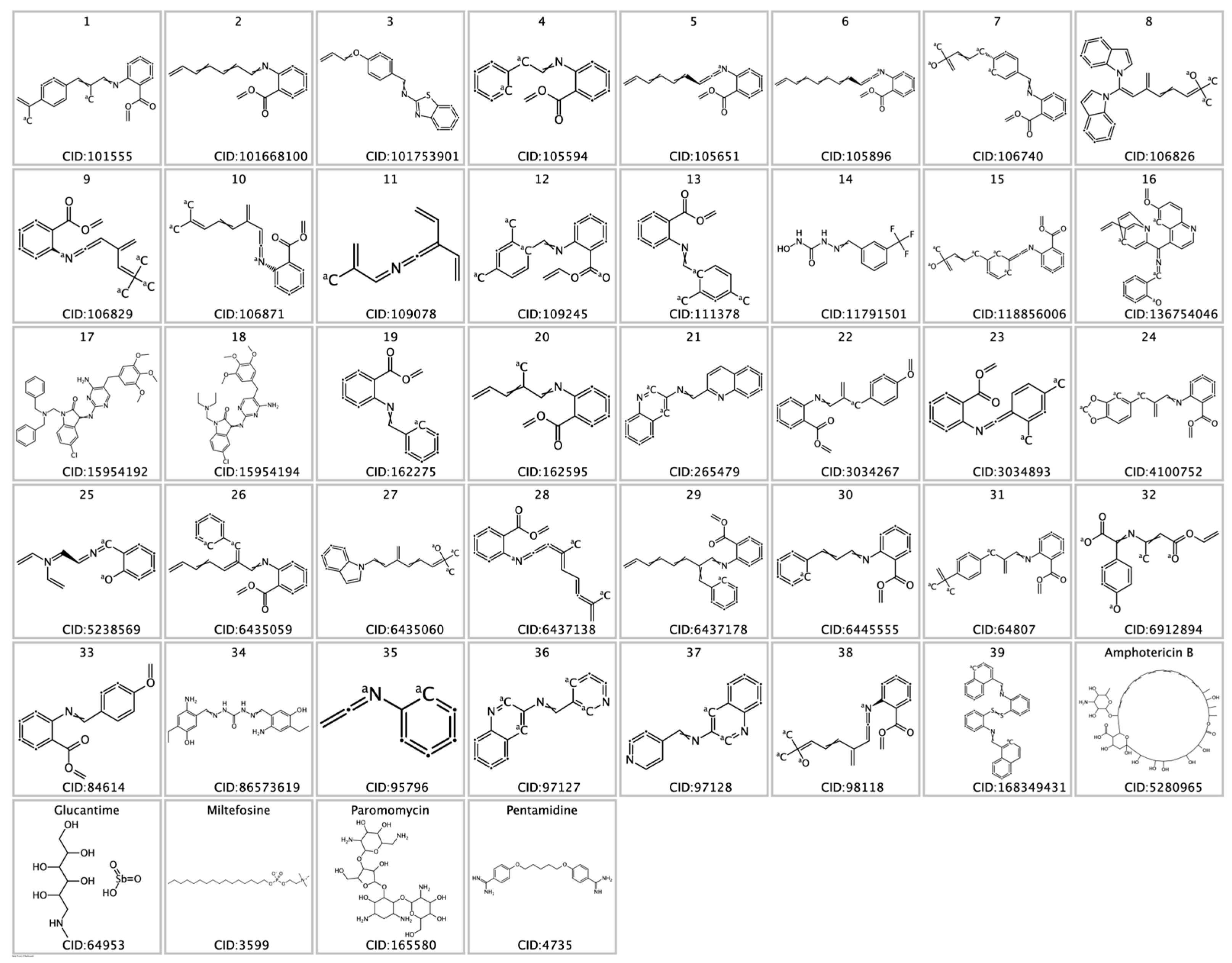
The Schiff base compound database used for virtual screening, shown in Figure 2, comprises a diverse collection of organic molecules characterized by the imine bond (-C=N-). Known for their versatility, these compounds are applied in drug synthesis, catalysis, and luminescent materials. This library provides a variety of structures for scientific research obtained from the PubChem database for exploring new drugs.
In the presence of the highlighted compounds, as shown in Table 1 and Figure 3, compound 21 (3-Quinolinamine, N-(2-quinolinylmethylene)-) presented the lowest predicted binding energy with a value of −10.5 kcal/mol. Analyzing the predicted interactions, it was possible to observe that residues Thr51, Lys60, Ile199, Phe203, and Ala338 present hydrophobic interaction with compound 21, while Ser14, Cys52, Ser178, Tyr198, Asp327, and Thr335 present predicted hydrogen bonds. These interactions suggest that compound 21 has a complex predicted binding nature with trypanothione reductase, with its non-polar regions avoiding interactions with water molecules and preferring to cluster with non-polar residues [15]. On the other hand, hydrogen bonds are the most significant predicted binding interaction as they are stronger than hydrophobic interaction, thus contributing more to the stability of the binding conformation between compound 21 and trypanothione reductase [16]. Therefore, the combination of these two types of interactions—hydrophobic and hydrogen—is probably responsible for the potent predicted binding between compound 21 and trypanothione reductase [17]. Like compound 21, compound 24 exhibits hydrophobic and hydrogen interactions, thus indicating that the predicted binding mode of compound 24 is similar to that of compound 21.
The presence of π–Perpendicular and π–Cation interactions in compound 39, the third compound with the lowest binding energy of −9.8 kcal/mol, adds complexity to its binding properties. The π–Perpendicular interactions refer to weak but highly specific bonds between pi and electron systems (usually present in aromatic rings) aligned perpendicular to each other. This orientation allows the formation of stacking interactions that can influence the stability of the molecular interaction between both molecules [18]. These interactions are often found in aromatic compounds, where aromatic rings overlap parallel or perpendicular. On the other hand, π–Cation interactions involve electrostatic attraction between a pi–electron system (such as an aromatic ring) and a cation (a positively charged ion) [19]. This interaction occurs when the cation is drawn towards the dense and delocalized electron cloud of the pi system, resulting in a weak but essential bond for the structure and stability of the binding between the compound and the protein target. In addition to hydrophobic and hydrogen interactions, π interactions may provide the compound with greater adaptability and specificity in binding to its target enzymes, making it a promising candidate for therapeutic or other specific purposes.
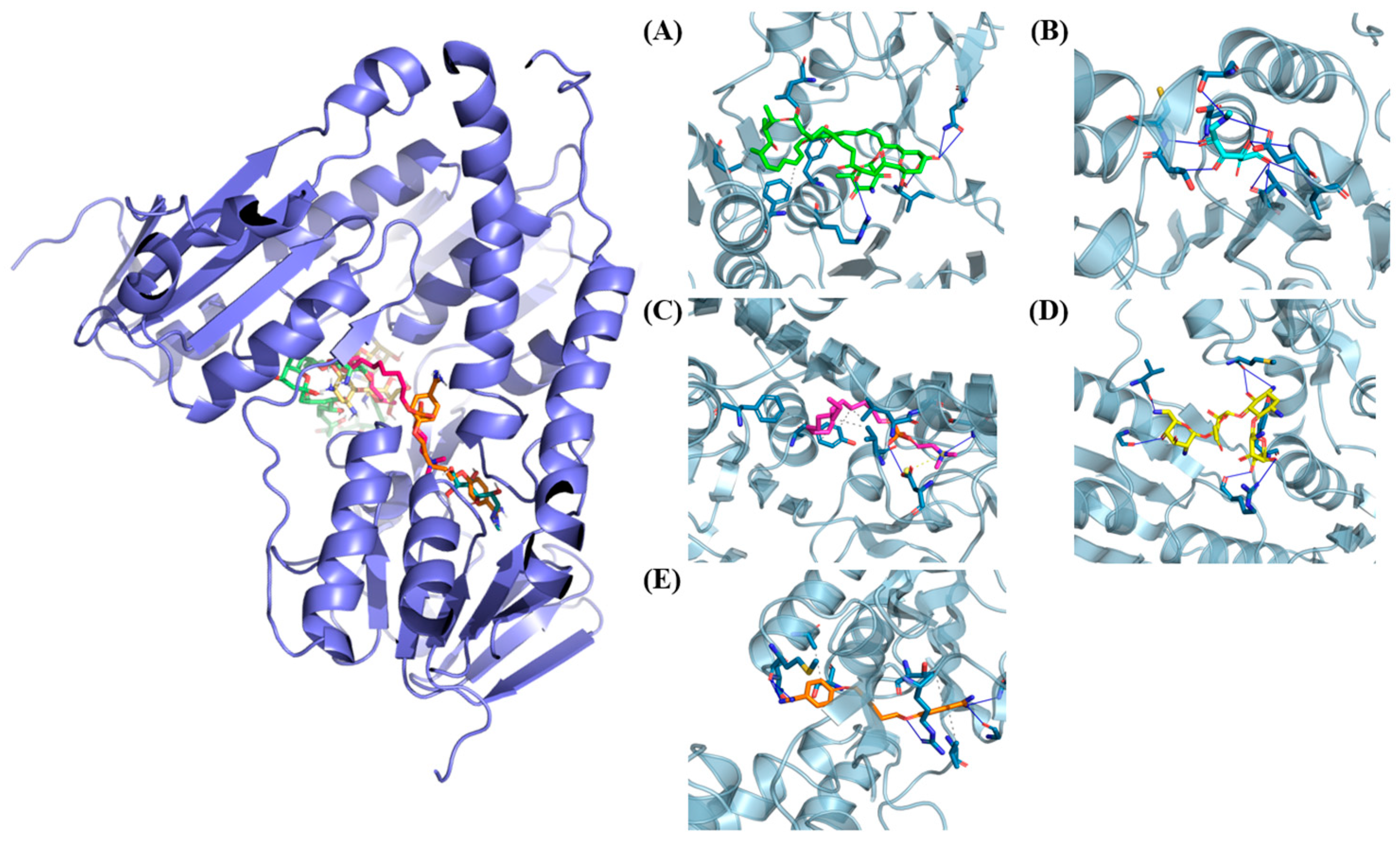
Analyzing Table 1, it is possible to identify residues shared between compound 21 and known commercial inhibitors (Figure 4) in the predicted binding conformation with trypanothione reductase. Among the common residues, those involved in hydrophobic and hydrogen interactions stand out. An example is the Thr51 residue, which demonstrates its importance by participating in hydrophobic interactions with compound 21 while also interacting with some of the analyzed commercial inhibitors such as Glucantime and Amphotericin B. Another residue, Lys60, is notable for participating in hydrophobic interactions with compound 21 and simultaneously engaging in a specific π–Cation interaction with Amphotericin B. Another residue, Ile199, is also predicted to perform hydrophobic interactions with compound 21 and Pentamidine, while Phe203 and Ala338 are involved in hydrophobic interactions with compound 21, Pentamidine, and Glucantime.
Compound 24 also interacts with key residues similar to commercial inhibitors, indicating similarities in their interaction conformations; a notable example is residue Thr51. The repeated Thr51 presence suggests that it is an essential residue for interactions with potential inhibitors against trypanothione reductase. Other shared residues, including Ile199, Phe203, and Ala338, also present in compound 24, Pentamidine, and Glucantime interactions with trypanothione reductase. These residues can be essential anchor points for interactions, providing a basis for the stable binding of these compounds. However, the nuances of individual interactions, the types of forces involved, and molecular conformations may differ, contributing to the unique profiles of each compound in terms of affinity, potency, and selectivity against trypanothione reductase.
It is noticeable that compound 39 also shares key residues with commercial inhibitors, suggesting the possibility of similarities in molecular interactions. A notable example is Thr51 residue, a common interaction point with compound 39 and some commercial inhibitors. Furthermore, Ile199 residue interacts with compound 39 and Pentamidine, which may be an essential anchor point contributing to the observed hydrophobic interactions.

4. Conclusions

This study highlights the complexity and urgency associated with Leishmaniasis as a significant challenge in public health. Current treatments are hampered by toxicity, high costs, and resistance, and this fact reinforces the critical need to seek practical and affordable therapeutic alternatives. Trypanothione reductase is a known promising protein target against Leishmaniasis. Schiff bases are a promising chemical template to discover new promising compounds that aim to improve the quality of life for patients and reduce the economic burdens associated with treatment. Thirty-nine Schiff bases were selected for virtual testing against trypanothione reductase. The results were obtained through molecular docking simulations using AutoDock Vina software. The compounds that present best predicted inhibition activity against trypanothione reductase were compound 21 (3-Quinolinamine, N-(2-quinolinylmethylene) with a predicted ΔG value of −10.5 kcal/mol, compound 24 (1,3-Bis[(E)-(2-Amino-4-Ethyl-5-Hydroxy-Phenyl)Methyleneamino]Urea) with a predicted ΔG value of −10.4 kcal/mol, and compound 39 (a Naphtaldehyde disulfide) Schiff base with a predicted ΔG value of −9.8 kcal/mol, suggesting possible efficacy against Leishmaniasis. However, experimental validation should be performed to demonstrate their effectiveness in subsequent analyses. Therefore, this research contributes substantially to developing new potential therapeutic strategies against Leishmaniasis, highlighting the possible contributions of Schiff Bases in treating this illness.

Supplementary Materials

Author Contributions

C.S.H.S., R.M.V.A., O.V.O. and J.D.d.S. were primarily responsible for the methodology, while C.S.H.S. and R.M.V.A. also contributed significantly to its development. C.S.H.S. and R.M.V.A. were responsible for the software aspect of the project, including its development and implementation. Additionally, C.S.H.S. and R.M.V.A. undertook the validation and formal analysis of the methodology. The investigation phase involved contributions from D.R.P., C.S.H.S. and R.M.V.A., with C.S.H.S. providing essential resources for the research. Data curation was performed by D.R.P. and C.S.H.S.; D.R.P. and C.S.H.S. collaborated on the original draft preparation; while R.M.V.A., O.V.O. and J.D.d.S. provided critical input during the writing, review, and editing process. Visualization tasks were handled by C.S.H.S. and R.M.V.A., O.V.O. and J.D.d.S. supervised the project, ensuring its progress and quality, and also took on project administration duties. All authors have read and agreed to the published version of the manuscript.

Funding

C.S.H.S. thanks the Fundação para a Ciência e Tecnologia (FCT), Portugal, for the Ph.D. Grant 2023.04950.BD.

Institutional Review Board Statement

Not applicable.

Informed Consent Statement

Not applicable.

Data Availability Statement

The original contributions presented in this study are included in the Supplementary Materials.

Conflicts of Interest

The authors declare no conflict of interest.

References

  1. Okwor, I.; Uzonna, J. Social and Economic Burden of Human Leishmaniasis. Am. J. Trop. Med. Hyg. 2016, 94, 489. [Google Scholar] [CrossRef] [PubMed]
  2. Arenas, R.; Torres-Guerrero, E.; Quintanilla-Cedillo, M.R.; Ruiz-Esmenjaud, J. Leishmaniasis: A Review. F1000Res 2017, 6, 750. [Google Scholar] [CrossRef]
  3. Rath, S.; Augusto Trivelin, L.; Imbrunito, T.R.; Tomazela, D.M.; De Jesús, M.N.; Calvo Marzal, P.; De Andrade, H.F.; Gustavo Tempone, A. Antimoniais Empregados No Tratamento Da Leishmaniose: Estado Da Arte. Quim. Nova 2003, 26, 550–555. [Google Scholar] [CrossRef]
  4. Oliveira, L.F.; Schubach, A.O.; Martins, M.M.; Passos, S.L.; Oliveira, R.V.; Marzochi, M.C.; Andrade, C.A. Systematic review of the adverse effects of cutaneous leishmaniasis treatment in the New World. Acta Tropica 2011, 118, 87–96. [Google Scholar] [CrossRef] [PubMed]
  5. Teran, R.; Guevara, R.; Mora, J.; Dobronski, L.; Barreiro-Costa, O.; Beske, T.; Pérez-Barrera, J.; Araya-Maturana, R.; Rojas-Silva, P.; Poveda, A.; et al. Characterization of Antimicrobial, Antioxidant, and Leishmanicidal Activities of Schiff Base Derivatives of 4-Aminoantipyrine. Molecules 2019, 24, 2696. [Google Scholar] [CrossRef] [PubMed]
  6. Gundampati, R.K.; Jagannadham, M. V Molecular Docking Based Inhibition of Trypanothione Reductase Activity by Taxifolin Novel Target for Antileishmanial Activity. J. Appl. Pharm. Sci. 2012, 2, 133–136. [Google Scholar] [CrossRef]
  7. Schubach, A.O.; Leon, L. Parte VIII-Tratamento e Diagnóstico Das Leishmanioses 23. Modelos de Estudo Para o Desenvolvimento de Drogas Anti-Leishmania; Editora Fiocruz: Rio de Janeiro, Brazil, 2014; pp. 413–428. [Google Scholar] [CrossRef]
  8. Khan, M.I.; Khan, A.; Hussain, I.; Khan, M.A.; Gul, S.; Iqbal, M.; Inayat-Ur-Rahman; Khuda, F. Spectral, XRD, SEM and Biological Properties of New Mononuclear Schiff Base Transition Metal Complexes. Inorg. Chem. Commun. 2013, 35, 104–109. [Google Scholar] [CrossRef]
  9. Sheikh, S.Y.; Hassan, F.; Shukla, D.; Bala, S.; Faruqui, T.; Akhter, Y.; Khan, A.R.; Nasibullah, M. A Review on Potential Therapeutic Targets for the Treatment of Leishmaniasis. Parasitol. Int. 2024, 100, 102863. [Google Scholar] [CrossRef] [PubMed]
  10. Morris, G.M.; Huey, R.; Olson, A.J. Using AutoDock for Ligand-Receptor Docking. Curr. Protoc. Bioinform. 2008, 24, 8.14.1–8.14.40. [Google Scholar] [CrossRef] [PubMed]
  11. Baiocco, P.; Colotti, G.; Franceschini, S.; Ilari, A. Molecular Basis of Antimony Treatment in Leishmaniasis. J. Med. Chem. 2009, 52, 2603–2612. [Google Scholar] [CrossRef] [PubMed]
  12. Leimann, F.V.; de Souza, L.B.; de Oliveira, B.P.M.; Rossi, B.F.; da Silva, P.S.; Shiraishi, C.S.H.; Kaplum, V.; Abreu, R.M.; Pereira, C.; Barros, L.; et al. Evaluation of Berberine Nanoparticles as a Strategy to Modulate Acetylcholinesterase Activity. Food Res. Int. 2023, 173, 113295. [Google Scholar] [CrossRef] [PubMed]
  13. Trott, O.; Olson, A.J. AutoDock Vina: Improving the Speed and Accuracy of Docking with a New Scoring Function, Efficient Optimization, and Multithreading. J. Comput. Chem. 2010, 31, 455–461. [Google Scholar] [CrossRef] [PubMed]
  14. Adasme, M.F.; Linnemann, K.L.; Bolz, S.N.; Kaiser, F.; Salentin, S.; Haupt, V.J.; Schroeder, M. PLIP 2021: Expanding the Scope of the Protein–Ligand Interaction Profiler to DNA and RNA. Nucleic Acids Res. 2021, 49, W530–W534. [Google Scholar] [CrossRef] [PubMed]
  15. Sun, Q. The Hydrophobic Effects: Our Current Understanding. Molecules 2022, 27, 7009. [Google Scholar] [CrossRef] [PubMed]
  16. Van der Lubbe, S.C.C.; Fonseca Guerra, C. The Nature of Hydrogen Bonds: A Delineation of the Role of Different Energy Components on Hydrogen Bond Strengths and Lengths. Chem. Asian J. 2019, 14, 2760–2769. [Google Scholar] [CrossRef] [PubMed]
  17. Salentin, S.; Schreiber, S.; Haupt, V.J.; Adasme, M.F.; Schroeder, M. PLIP: Fully Automated Protein-Ligand Interaction Profiler. Nucleic Acids Res. 2015, 43, W443–W447. [Google Scholar] [CrossRef] [PubMed]
  18. Headen, T.F.; Howard, C.A.; Skipper, N.T.; Wilkinson, M.A.; Bowron, D.T.; Soper, A.K. Structure of π-π Interactions in Aromatic Liquids. J. Am. Chem. Soc. 2010, 132, 5735–5742. [Google Scholar] [CrossRef] [PubMed]
  19. Mahadevi, A.S.; Sastry, G.N. Cation-π Interaction: Its Role and Relevance in Chemistry, Biology, and Material Science. Chem. Rev. 2013, 113, 2100–2138. [Google Scholar] [CrossRef] [PubMed]
Figure 1. General equation for the formation of the Schiff base compounds.
Proceedings 102 00055 g001
Figure 2. Compounds that make up the Schiff base library (139) and commercial inhibitors with their respective CID (PubChem Compound Identification, an access identifier with integers for identifying chemical structures in the database) codes.
Proceedings 102 00055 g002
Figure 3. In (A), trypanothione reductase (PDB: 2JK6) is depicted in cartoon format overlaid with the surface and the Schiff base inhibitors. These inhibitors are shown in stick format and are colored according to the following legend: (B) Compound 21 (3-Quinolinamine, N-(2-quinolinylmethylene)-), (C) Compound 24 (2-((3-(1,3-Benzodioxol-5-yl)-2-methylpropylidene)amino)benzoic amethyl ester), and (D) Compound 39 (Benzoic acid, 2-((2-(phenylmethylene)octylidene)amino)-, methyl ester).
Proceedings 102 00055 g003
Figure 4. In trypanothione reductase (PDB: 2JK6) is represented using the cartoon format overlaid with the surface. This representation allows the identification of the commercial inhibitors in the enzyme’s active site. These inhibitors are shown in stick format and are colored according to the following legend: (A) Amphotericin B (Green), (B) Glucantime (Pool Blue), (C) Miltefosine (Pink), (D) Paromomycin (Yellow), and (E) Pentamidine (Orange).
Proceedings 102 00055 g004
Table 1. Interaction of compounds with residues present in the structure of trypanothione reductase (PDB:2JK6).
CodeCID CodeAffinity (kcal/mol)Interaction TypeResidues
Promising Compounds
21265479−10.5HydrophobicThr51, Lys60, Ile199, Phe203, and Ala338
Hydrogen bondSer14, Cys52, Ser178, Tyr198, Asp327, and Thr335
2486573619−10.4HydrophobicThr51, Lys60, Ile199, Phe203, and Ala338
Hydrogen bondSer14, Cys52, Ser178, Tyr198, Asp327, and Thr335
39168349431−9.8HydrophobicThr51, Lys61, Tyr198, Ile199, Leu334, Ala365, Phe367, and Pro435
Hydrogen bondThr355
π-PerpendicularTyr198
π-CationLys60
Commercial inhibitors
APentamidine−8.8HydrophobicVal36, Thr160, and Ala338
HydrogenSer14, Thr51, Cys52, Ala159, Gly161, Asp327, Val328, and Thr335
BGlucantime−5.9HydrogenSer14, Thr51, Cys52, Ala159, Gly161, Asp327, Val328, and Thr335
CAmphotericin B−9HydrophobicTyr198, Phe230, Leu334, and Thr374
HydrogenArg228, Ile285, and Asn306
DMiltefosine−5.8HydrophobicTyr198, Phe230, Val332 and Leu334
HydrogenSer14, Asp327, and Thr335
Salt BridgesAsp327
EParomomycin−6.2HydrogenTyr198, Arg228, Met333, Val362, and Gly376
Disclaimer/Publisher’s Note: The statements, opinions and data contained in all publications are solely those of the individual author(s) and contributor(s) and not of MDPI and/or the editor(s). MDPI and/or the editor(s) disclaim responsibility for any injury to people or property resulting from any ideas, methods, instructions or products referred to in the content.