Nevus in the Oral Cavity †

1. Introduction
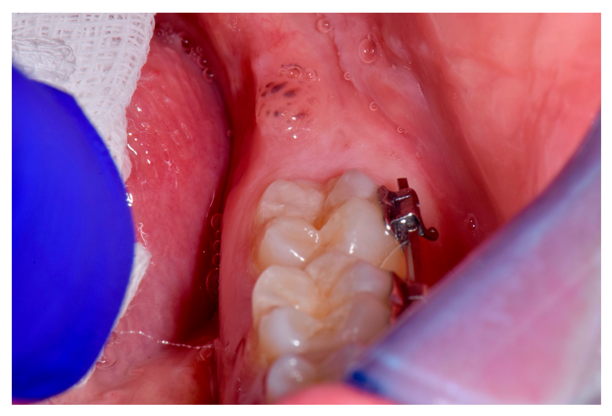
2. Case
3. Treatment
4. Conclusions
Conflicts of Interest
References
- Popa, C.; Stelea, C.; Popa, R.; Popescu, E. Oral and perioral endogenous pigmented lesions. Rev. Med. Chir. Soc. Med. Nat. Iasi 2008, 112, 1054–1060. [Google Scholar] [PubMed]
- Tavares, T.S.; Meirelles, D.P.; de Aguiar MC, F.; Caldeira, P.C. Pigmented lesions of the oral mucosa: A cross-sectional study of 458 histopathological specimens. Oral Dis. 2018, 24, 1484–1491. [Google Scholar] [CrossRef] [PubMed]
- Freitas, D.A.; Bonan, P.R.; Sousa, A.A.; Pereira, M.M.; Oliveira, S.M.; Jones, K.M. Intramucosal nevus in the oral cavity. J. Contemp. Dent. Pract. 2015, 16, 74–76. [Google Scholar] [CrossRef] [PubMed]

© 2019 by the authors. Licensee MDPI, Basel, Switzerland. This article is an open access article distributed under the terms and conditions of the Creative Commons Attribution (CC BY) license (http://creativecommons.org/licenses/by/4.0/).
Share and Cite
Sorrentino, D.; Decani, S.; Zenoni, C.; Sardella, A. Nevus in the Oral Cavity. Proceedings 2019, 35, 74. https://doi.org/10.3390/proceedings2019035074
Sorrentino D, Decani S, Zenoni C, Sardella A. Nevus in the Oral Cavity. Proceedings. 2019; 35(1):74. https://doi.org/10.3390/proceedings2019035074
Chicago/Turabian StyleSorrentino, Daniela, Sem Decani, Camilla Zenoni, and Andrea Sardella. 2019. "Nevus in the Oral Cavity" Proceedings 35, no. 1: 74. https://doi.org/10.3390/proceedings2019035074
APA StyleSorrentino, D., Decani, S., Zenoni, C., & Sardella, A. (2019). Nevus in the Oral Cavity. Proceedings, 35(1), 74. https://doi.org/10.3390/proceedings2019035074