Proceeding Paper

Modification of Ca2+-Crosslinked Sodium Alginate/Gelatin Films with Propolis for an Improved Antimicrobial Action †

by
Natália Cândido Homem
*,
Catarina Alexandra Fortuna dos Santos Miranda
,
Joana Isabel da Costa Antunes
,
Maria Teresa Sousa Pessoa de Amorim
and
Helena Prado Felgueiras
Centre for Textile Science and Technology (2C2T), Campus Azurém, University of Minho, 4800-058 Guimarães, Portugal
*
Author to whom correspondence should be addressed.
Presented at the First International Conference on “Green” Polymer Materials 2020, 5–25 November 2020; Available online: https://sciforum.net/conference/CGPM2020.
Proceedings 2021, 69(1), 4; https://doi.org/10.3390/CGPM2020-07180
Published: 3 November 2020

Abstract

:
Problems associated with microbial resistance to antibiotics are growing due to their overuse. In this scenario, plant extracts such as the propolis extract (PE) have been considered as potential alternatives to antibiotics in the treatment of infected wounds, due to its antimicrobial properties and ability to induce tissue regeneration. To improve the long-term effectiveness of PE in wound healing, polymeric films composed of biodegradable and biocompatible polymers are being engineered as delivery vehicles. Here, sodium alginate/gelatin (SA/GN) films containing PE were prepared via a simple, green process of solvent casting/phase inversion technique, followed by crosslinking with calcium chloride (CaCl2) solutions. The minimum inhibitory concentration (MIC) of PE was established as 0.338 mg/mL for Staphylococcus aureus and 1.353 mg/mL for Pseudomonas aeruginosa, the most prevalent bacteria in infected wounds. The PE was incorporated within the polymeric films before (blended with the polymeric solution) and after (immobilization via physisorption) their production. Flexible, highly hydrated SA/GN/PE films were obtained, and their antibacterial activity was assessed via agar diffusion and killing time kinetics examinations. Data confirmed the modified films effectiveness to fight bacterial infections caused by S. aureus and P. aeruginosa and their ability to be applied in the treatment of infected wounds.

1. Introduction

The problems associated with antimicrobial resistance against conventional antibiotics are growing due to overuse of these antimicrobial agents. Thus, several efforts have been made in order to minimize the pace of resistance by analyzing emergent microorganisms and resistance mechanisms, as well as newer potential antimicrobial agents [1] such as biomolecules, which can present inhibition behavior against the most common bacteria found in infected wounds. In this scenario, some plant extracts have been considered as potential alternatives to antibiotics or to be used in synergy with them, once they can present promising properties (e.g., antimicrobial, antifungal, antiviral, and regenerative properties) [2,3].
Propolis, also known as “bee glue”, is a natural resinous multifunctional substance collected and used by bees to preserve and protect their hives against bacterial and fungal attacks [4,5]. It is characterized as a complex mixture of chemical constituents such as resins, waxes, pollen, essential oils, and organic compounds, with a high polyphenol content [6]. The propolis extract (PE) has been used for centuries in folk medicine due to its great biological properties, ascribed to the presence of flavonoids, that include antimicrobial, antioxidant, and anti-inflammatory activities as well as the ability to induce tissue regeneration [7]. Concerning its antibacterial activity, two mechanisms can be considered. First, it acts directly on the microorganism, subsequently stimulating the immune system which results in the activation of natural defenses of the organism, consequently fighting infection [8]. Besides, several studies state that propolis has no toxicity and no side effects in animal models or humans, which enables it to be used in biomedical applications such as wound healing scaffolds [9].
Although PE can be considered as a potential alternative to antibiotics, in order to improve stability and long-term effectiveness of PE in wound healing treatments, polymeric films composed of biodegradable and biocompatible polymers can be engineered to be used as a 2 in 1 solution: the film would act as a protective barrier while a controlled release of the propolis extract would allow the wound to receive an antibacterial treatment without the need of antibiotics presence [10]. In this scenario, the polymers choice plays a key role in the final efficiency of the films, once they must be biocompatible and non-toxic, but still retain some characteristics such as good thermal and mechanical resistance [11]. Thus, natural-based polymers, such as sodium alginate and gelatin, have been explored in this aim.
Sodium alginate (SA) is a natural linear polysaccharide obtained from brown seaweeds, composed of 1,4-linked-β-d-mannuronic (M) and α-l-guluronic (G) monomers [12]. SA is water soluble, non-toxic, biodegradable, and biocompatible, as well as capable of holding large amounts of water, and thus it can be used on the production of hydrogels for biomedical applications. Additionally, SA films can be easily produced by the phase inversion technique, once that it interacts almost instantaneously with divalent cations (such as Ca2+, from CaCl2 aqueous solutions) in an irreversible manner, forming hydrogel-like films. However, SA films alone do not present interesting long-term applicability, as they are mechanically weak in wet conditions. Thus, other biopolymers such as gelatin are commonly used concomitantly with SA, as they can interact electrostatically and form a polyelectrolyte complex with enhanced performance [13]. Gelatin (GN) is a biodegradable protein derived from the controlled structural and chemical degradation of collagen, and contains many functional groups and cell binding sites in its structure, which increase its cell binging ability, making it desirable for tissue engineering applications [11].
In this context, the aim of this study was to evaluate the feasibility of the production of SA/GN films via solvent casting/phase inversion technique, and their modification with PE, in order to engineer hydrogel-like films to be used as delivery platforms for PE controlled release. The incorporation of PE was achieved by two different methods: blending of PE at the polymeric solutions before films casting and immersion of SA/GN films (immobilization via phisysorption) within PE solutions. The differences on surface chemistry and chemical composition of the produced films were evaluated, as well as their antibacterial activity against Pseudomonas aeruginosa and Staphyloccocus aureus, which are among the most common bacteria found in infected wounds.

2. Experiments

2.1. Materials

Hydroalcoholic propolis extract was purchased from Drasanvi (Vila Nova de Gaia, Portugal), and the alcohol content within the extract was left to evaporate before use. Gelatin (~300 Bloom, Type A, from porcine skin) and sodium alginate (alginic acid sodium salt, from brown algae, ≥2000 cP) applied on the films production were purchased from Sigma-Aldrich (Merck, Darmstadt, Germany). Calcium chloride (CaCl2 anhydrous, Chem-Lab, Zedelgem, Belgium) 2 wt% solution was employed as coagulation bath. Growth media trypticase soy broth (TSB), trypticase soy agar (TSA), nutrient broth (NB), and nutrient agar (NA) were purchased from VWR (the first for the Gram-positive bacteria and the second for the Gram-negative), while Mueller Hinton broth (MHB) was obtained from CondaLab. Gram-negative Pseudomonas aeruginosa (P. aeruginosa, ATCC 25853) and Gram-positive Staphylococcus aureus (S. aureus, ATCC 6538) bacteria were supplied from American Type Culture Collection (ATCC).

2.2. MICs

Minimum inhibitory concentrations (MICs) of PE for P. aeruginosa and S. aureus bacteria were assessed via broth microdilution method [14]. Initially, 100 μL of PE was added to the first column of a 96-well plate. Subsequently, serial dilutions (1:2) were performed by adding MHB in the next wells, reaching a final volume of 50 μL. Then, 50 μL of bacteria suspensions previously prepared in MHB (1 × 10⁷ CFUs/mL) was added to these wells. Additionally, free bacteria and MHB suspensions were used as control. Subsequently, the absorbance at 600 nm of the plate (zero time, t0h) was measured in an EZ READ 2000 Microplate Reader (Biochrom, Cambridge, UK). The plate was then left under incubation at 120 rpm and 37 °C for 24 h, and the absorbance after this period was also recorded (24 h time, t24h). MIC was established as the concentration of PE in which the growth of bacteria was not seen anymore, visually and by comparing the absorbance of samples at t0h and t24h. Finally, aliquots of the wells in which the MIC was observed, and also in the before and after wells (concentration higher and lower than MIC value), were diluted in PBS and cultured in NA and TSA at 37 °C for 24 h, to count the resulting colonies.

2.3. Polymeric Films Production

The polymeric films were produced via solvent casting/phase inversion technique. Initially, SA solutions (2% w/v) were prepared by dissolving SA in dH2O under stirring for 3 h at 50 °C. Meanwhile, GN solutions (~1% w/v) were also prepared, using dH2O as solvent, to be later mixed with SA solutions in a polymer ratio of 70:30 v/v (SA/GN films). This blend was left under low stirring for 1 h, until a homogeneous solution was obtained. For the SA/GN/PEb films (in which “b” means before casting, i.e., the PE was incorporated during blending) production, a PE solution at a concentration proportional to the P. aeruginosa MIC value (1.6% v/v) was prepared, by mixing PE with dH2O for 30 min and posteriorly adding this solution to the SA/GN blend previously prepared. In order to produce the SA/GN and SA/GN/PEb films, the respective solutions were then poured into glass Petri dishes and left at 4 °C for 24 h to remove air bubbles. After this period, the films were dried at RT for 6 days and subsequently dipped for 1 h in the coagulation bath (CaCl2 2 wt% solution). The prepared films were taken out from the Petri dishes, washed three times with dH2O (150 rpm, 5 min each washing cycle), and finally stored for further use. For SA/GN/PEa films (in which “a” means after casting, i.e., immobilization via physisorption) production, the SA/GN films were cut in circular (disk) samples of 6 mm diameter and then immersed in PE solutions for 96 h.

2.4. Films Characterization via ATR-FTIR

The differences in surface chemistry and chemical composition of the films were analyzed by attenuated total reflectance-Fourier transform infrared spectroscopy (ATR-FTIR). The equipment used was an IRAffinity-1S (Shimadzu, Kyoto, Japan), coupled with a HATR 10 accessory with a diamond crystal. The spectra were obtained in a wavenumber range of 400 to 4000 cm−1, and for each film a total of 200 scans were performed at a spectral resolution of 2 cm−1.

2.5. Water Absorption and Retention Capacity of SA/GN Films

The water absorption and retention capacity of the SA/GN films unloaded and loaded with PE were assessed [10]. Hydrated 6 mm diameter samples of each film were wiped with delicate task wipes to remove excess of surface water, weighed (noted as Ww) and measured to evaluate their thickness. The samples were dried at 45 °C for 72 h, and then weighed again (noted as Wd). The water absorption rate was calculated according to Equation (1) [15]:
W a t e r   c o n t e n t = W w W d W d × 100 %
Each sample was measured in triplicate.

2.6. Agar Disk Diffusion Tests

The films antibacterial activity was initially assessed visually against P. aeruginosa and S. aureus via the Kirby–Bauer method, as previously described [2]. Inoculums for both bacteria were prepared in TSB and incubated overnight at 37 °C and 120 rpm. The inoculums concentrations were adjusted to 1.0 × 10⁷ colony-forming units (CFUs)/mL. Aliquots of 100 µL of each bacteria solution were collected and spread along the respective agar media, casted within 90 mm diameter Petri dishes. After bacteria solution absorption, disk samples of all films produced (11 mm diameter) were placed in the bacteria-containing agar plates center and incubated for 24 h at 37 °C. After this period, zones of inhibition (ZoI) were observed and measured (mm) to verify the films antibacterial efficiency.

2.7. Killing Time Kinetics

To evaluate the efficiency of PE incorporated films throughout time, P. aeruginosa and S. aureus NB and TSB suspensions, respectively, adjusted to a concentration of 1 × 10⁵ CFUs/mL were prepared. Then, to these suspensions, circular samples of all films produced (6 mm diameter) were added. Unloaded films (SA/GN) were used as control. These bacteria-containing suspensions were incubated at 37 °C and 120 rpm, and at predetermined time points, 0, 1, 2, 6, and 24 h, aliquots were collected. These aliquots were then serially diluted in PBS, plated in NA/TSA, and incubated for more 24 h at 37 °C. The resulting colonies were counted and results were expressed in total number of bacteria colonies per time period or in log reduction relating the loaded samples with the unloaded. All measurements were performed in triplicate, and the results were processed at GraphPad Prism 7.0 software.

3. Results and Discussion

3.1. MICs

Prior to the production and loading of films, the MICs of PE against the Gram-positive S. aureus and the Gram-negative P. aeruginosa were established as 0.338 mg/mL and 1.353 mg/mL, respectively. These results are in agreement with other findings which state that that Gram-positive bacteria are sensitive to low propolis concentrations, while Gram-negative bacteria only can be inhibited with higher propolis doses, due to their structural differences [8]. It is also reported that the bioactivity of propolis is not directly related to the concentration of the biological active substances (e.g., phenolic acid esters and flavonoids), but to the synergistic activity between them [16]. The MICs assessment was essential in this study, once that the further loading of SA/GN with PE was performed at a concentration expected to be effective against both bacteria, based on the obtained MIC values.

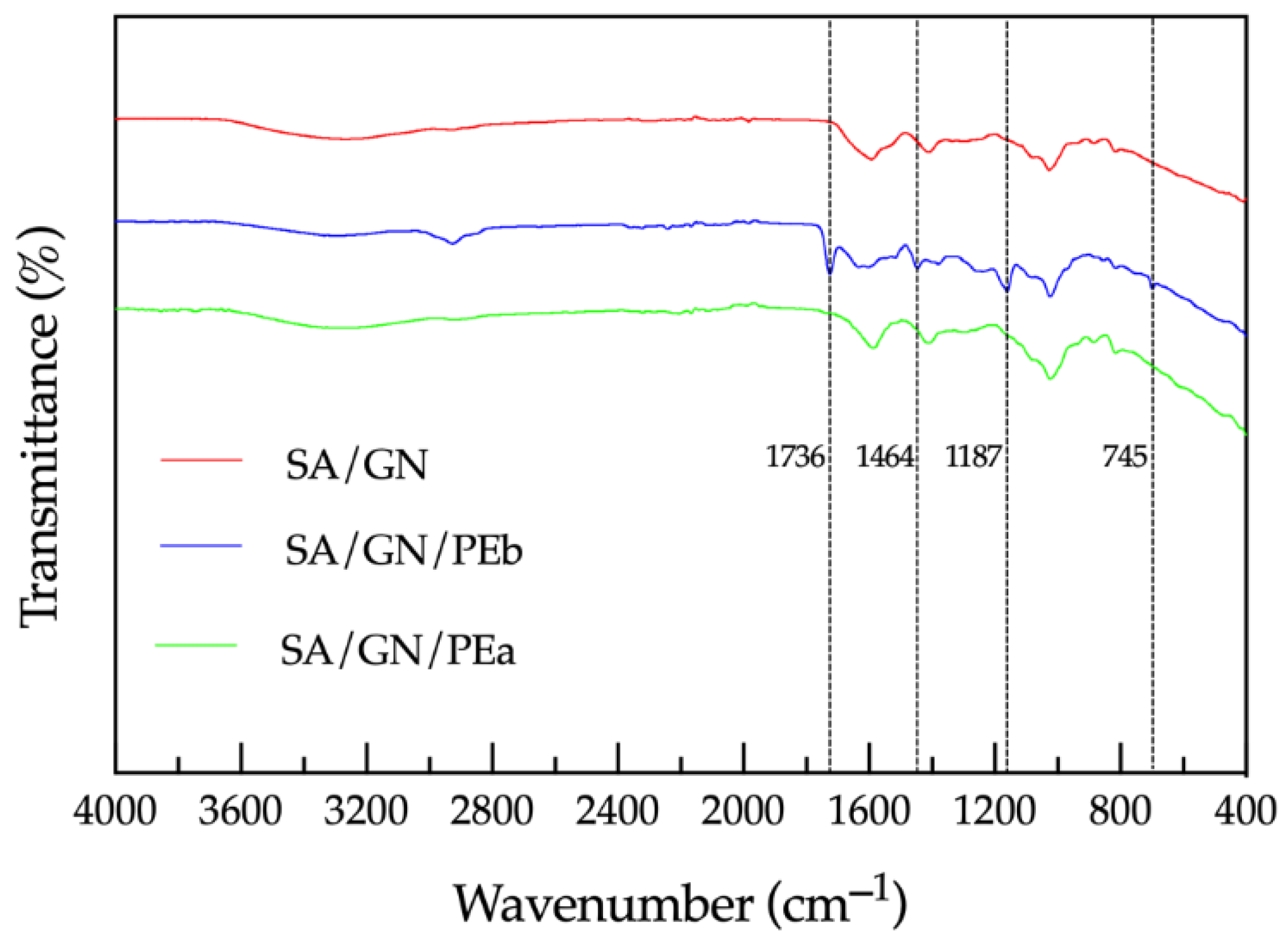
3.2. Films Characterization via ATR-FTIR

The SA/GN- and PE-loaded samples were analyzed via ATR-FTIR, and the spectra collected are presented at Figure 1. As can be observed, a very assorted amount of peaks and absorption bands can be identified. First, the broad band at 3330 cm−1 observed for all samples can be attributed to the O–H stretching vibration of the hydroxyl and/or phenolic groups. The presence of SA can be confirmed by the peaks at 1626 and 1445 cm−1, which can be attributed to asymmetric and symmetric stretching vibration of COO groups. Furthermore, GN associated peaks can be observed at 1152 and 1043 cm−1, being attributed to C–O stretching of carboxylic acid and C–N stretching of amines, respectively [15,17]. Besides, several peaks associated to PE (highlighted at Figure 1) can be observed. For instance, the peak at 1736 cm−1, associated to the carbonyl group (C=O) stretching vibrations of the ester bond, is not observed in SA/GN or SA/GN/PEa, while for the SA/GN/PEb it appears with a very pronounced intensity. This happens also with the peaks at 1464, 1187, and 1045 cm−1, which are attributed to the presence of flavonoids (C–H deformations and aromatic stretching), aromatic C–H deformation, and CH2 rocking of hydrocarbons originating from beeswax, respectively. Furthermore, the changing of a single peak into a doublet at 1645 and 1649 cm−1 (assigned to aromatic ring C=C stretching) indicates the presence of phenolic groups associated with PE [18,19]. These differences between SA/GN/PEa and SA/GN/PEb samples can indicate that the incorporation of PE during SA/GN films production was more effective in maintaining a proper amount of propolis available for antimicrobial action.

3.3. Water Absorption and Retention Properties of SA/GN Films

Water absorption of SA/GN and PE loaded films was evaluated and is presented at Table 1. As can be observed, the SA/GN and the SA/GN/PEa films presented higher water retention rates than the SA/GN/PEb sample. This can be explained by the fact that the SA/GN/PEb sample already possesses PE in its composition at the casting time, while the incorporation time of PE in the SA/GN/PEa film may not be enough to retain PE at the films after the washing period, and thus both samples SA/GN and SA/GN/PEa present similar water retention behavior. Furthermore, propolis is considered as a small molecule when compared to polymers, thus being easily located between huge polymer molecules, limiting the interactions between polymer chains and water molecules, affecting its water retention capacity [20].

3.4. Agar Disk Diffusion Tests

Unloaded and propolis-loaded films were examined for their ability to diffuse the antimicrobial agent within agar containing Gram-positive and Gram-negative microbial cells. Figure 2 reports the observations made. In most samples, bacteria were seen to grown around the film regardless of impregnation method or tested bacteria; thus, suggesting the inability of the propolis biomolecule to diffuse along the solid agar and kill bacteria. Still, upon closer observation, diffusion of propolis can be perceived, even if at a very small scale (slight discoloration), on the agar plates containing the SA/GN/PEb. This is of particular interest as it would be expected the films loaded after production to have the biomolecule more readily available to fight bacteria. Data suggest that more propolis was impregnated on the blended films and that it is able to diffuse from the sample to the solid exterior in a very small and slow manner.

3.5. Killing Time Kinetics

The antibacterial efficacy of propolis-loaded films was examined against the Gram-positive bacteria S. aureus and the Gram-negative bacteria P. aeruginosa for a period of 24 h (Figure 3).
Data reported the loaded films to have an active, quick effect against the bacteria, and that this effect was more pronounced against P. aeruginosa in the first moments of interaction. Indeed, in the first hour S. aureus was seen to grow when placed in contact with the SA/GN/PEa film. This suggests that, even though with time S. aureus becomes more susceptible to the action of propolis than P. aeruginosa, its effectiveness is only sensed later, namely, after the second hour of contact. Propolis acts by affecting the permeability of the cell membrane of the bacteria, disrupting their structure or decreasing the bacteria mobility. Moreover, as mentioned before, reports state that the antimicrobial activity of propolis is higher against Gram-positive than Gram-negative bacteria due to the species-specific structure of the cell outer membrane of the latter and its production of hydrolytic enzymes, which break down the active ingredients of propolis, inactivating them [8,9,16]. These reports are consistent with the observation from Figure 3 as well as the MIC assessments.
According to the results, the modification of the films by means of the blending method is considered the most effective to retain the biomolecule antimicrobial action. It is likely that most of the physisorbed propolis on the SA/GN/PEa films to be lost during washing processes, compromising the film capacity to fight microorganisms. Generally, data reported the successful action of the loaded films and confirmed the initial observations made from the film diffusion tests (Figure 2).

4. Conclusions

SA/GN films were successfully produced via solvent casting/phase inversion method. PE incorporation within SA/GN/PEb films was confirmed by FTIR. The SA/GN/PEa films showed higher water retention abilities and inferior antibacterial performance when compared to SA/GN/PEb films, thus leading to a conclusion that the incorporation of PE at blending solutions was more efficient. The SA/GN/PEb films were able to inhibit the growth of S. aureus and P. aeruginosa in a more pronounced and sustained manner than SA/GN films, demonstrating that PE incorporation was beneficial for antibacterial purposes. These results suggest that SA/GN/PE films could be used as alternatives to conventional treatments applied to infected wounds, for their improved bacterial inhibition. Further studies aiming at evaluating the loading capacity and release behavior of PE from SA/GN/PE films in physiological media are ongoing.

Author Contributions

Conceptualization, N.C.H., C.A.F.d.S.M., J.I.d.C.A., and H.P.F.; writing—original draft, N.C.H.; revision and editing, J.I.d.C.A., N.C.H., and H.P.F.; supervision, H.P.F.; funding acquisition, M.T.S.P.d.A. and H.P.F. All authors have read and agreed to the published version of the manuscript.

Acknowledgments

The authors acknowledge the Portuguese Foundation for Science and Technology (FCT), FEDER funds by means of Portugal 2020 Competitive Factors Operational Program (POCI), and the Portuguese Government (OE) for funding the project PEPTEX with reference PTDC/CTM-TEX/28074/2017 (POCI-01-0145-FEDER-028074). The authors also acknowledge project UID/CTM/00264/2020 of Centre for Textile Science and Technology (2C2T), funded by national funds through FCT/MCTES. C.S.M. acknowledges FCT for the PhD grant with reference 2020.08547.BD.

Conflicts of Interest

The authors declare no conflict of interest.

References

  1. Aslam, B.; Wang, W.; Arshad, M.I.; Khurshid, M.; Muzammil, S.; Rasool, M.H.; Nisar, M.A.; Alvi, R.F.; Aslam, M.A.; Qamar, M.U.; et al. Antibiotic resistance: A rundown of a global crisis. Infect. Drug Resist. 2018, 11, 1645–1658. [Google Scholar] [CrossRef] [PubMed]
  2. Felgueiras, H.P.; Homem, N.C.; Teixeira, M.A.; Ribeiro, A.R.M.; Antunes, J.C.; Amorim, M.T.S.P. Physical, Thermal, and Antibacterial Effects of Active Essential Oils with Potential for Biomedical Applications Loaded onto Cellulose Acetate/Polycaprolactone Wet-Spun Microfibers. Biomology 2020, 10, 1129. [Google Scholar] [CrossRef] [PubMed]
  3. Beluci, N.D.C.L.; Homem, N.C.; Amorim, M.T.S.P.; Bergamasco, R.; Vieira, A.M.S. Biopolymer extracted from Moringa oleifera Lam. in conjunction with graphene oxide to modify membrane surfaces. Environ. Technol. 2019, 1–12. [Google Scholar] [CrossRef] [PubMed]
  4. Oryan, A.; Alemzadeh, E.; Moshiri, A. Potential role of propolis in wound healing: Biological properties and therapeutic activities. Biomed. Pharmacother. 2018, 98, 469–483. [Google Scholar] [CrossRef] [PubMed]
  5. Bosio, K.; Avanzini, C.; D’Avolio, A.; Ozino, O.; Savoia, D. In vitro activity of propolis against Streptococcus pyogenes. Lett. Appl. Microbiol. 2000, 31, 174–177. [Google Scholar] [CrossRef] [PubMed]
  6. Juliano, C.; Pala, C.; Cossu, M. Preparation and characterisation of polymeric films containing propolis. J. Drug Deliv. Sci. Technol. 2007, 17, 177–181. [Google Scholar] [CrossRef]
  7. Rufatto, L.C.; Dos Santos, D.A.; Marinho, F.; Henriques, J.A.P.; Ely, M.R.; Moura, S. Red propolis: Chemical composition and pharmacological activity. Asian Pac. J. Trop. Biomed. 2017, 7, 591–598. [Google Scholar] [CrossRef]
  8. Przybyłek, I.; Karpiński, T.M. Antibacterial Properties of Propolis. Molecules 2019, 24, 2047. [Google Scholar] [CrossRef] [PubMed]
  9. Al-Ani, I.; Zimmermann, S.; Reichling, J.; Wink, M. Antimicrobial Activities of European Propolis Collected from Various Geographic Origins Alone and in Combination with Antibiotics. Medicines 2018, 5, 2. [Google Scholar] [CrossRef] [PubMed]
  10. Felgueiras, H.P.; Teixeira, M.A.; Tavares, T.D.; Homem, N.C.; Zille, A.; Amorim, M.T.P. Antimicrobial action and clotting time of thin, hydrated poly(vinyl alcohol)/cellulose acetate films functionalized with LL37 for prospective wound-healing applications. J. Appl. Polym. Sci. 2019, 137, 48626. [Google Scholar] [CrossRef]
  11. Miranda, C.S.; Ribeiro, A.R.M.; Homem, N.C.; Felgueiras, H.P. Spun Biotextiles in Tissue Engineering and Biomolecules Delivery Systems. Antibiotics 2020, 9, 174. [Google Scholar] [CrossRef] [PubMed]
  12. Baldevraj, R.M.; Jagadish, R. Incorporation of chemical antimicrobial agents into polymeric films for food packaging. In Multifunctional and Nanoreinforced Polymers for Food Packaging; Elsevier BV: Amsterdam, The Netherlands, 2011; pp. 368–420. [Google Scholar]
  13. Saravanan, M.; Rao, K.P. Pectin–gelatin and alginate–gelatin complex coacervation for controlled drug delivery: Influence of anionic polysaccharides and drugs being encapsulated on physicochemical properties of microcapsules. Carbohydr. Polym. 2010, 80, 808–816. [Google Scholar] [CrossRef]
  14. Wiegand, I.; Hilpert, K.; Hancock, R.E.W. Agar and broth dilution methods to determine the minimal inhibitory concentration (MIC) of antimicrobial substances. Nat. Protoc. 2008, 3, 163–175. [Google Scholar] [CrossRef] [PubMed]
  15. Wang, Q.-Q.; Liu, Y.; Zhang, C.-J.; Zhu, P. Alginate/gelatin blended hydrogel fibers cross-linked by Ca2+ and oxidized starch: Preparation and properties. Mater. Sci. Eng. C 2019, 99, 1469–1476. [Google Scholar] [CrossRef] [PubMed]
  16. Jug, M.; Zovko-Končić, M.; Kosalec, I. Modulation of antioxidant, chelating and antimicrobial activity of poplar chemo-type propolis by extraction procures. LWT Food Sci. Technol. 2014, 57, 530–537. [Google Scholar] [CrossRef]
  17. Devi, N.; Kakati, D.K. Smart porous microparticles based on gelatin/sodium alginate polyelectrolyte complex. J. Food Eng. 2013, 117, 193–204. [Google Scholar] [CrossRef]
  18. Do Nascimento, T.G.; da Silva, P.F.; Azevedo, L.F.; da Rocha, L.G.; de Moraes Porto, I.C.C.; e Moura, T.F.A.L.; Basílio-Júnior, I.D.; Grillo, L.A.M.; Dornelas, C.B.; da Silva Fonseca, E.J.; et al. Polymeric Nanoparticles of Brazilian Red Propolis Extract: Preparation, Characterization, Antioxidant and Leishmanicidal Activity. Nanoscale Res. Lett. 2016, 11, 1–16. [Google Scholar] [CrossRef] [PubMed]
  19. Svečnjak, L.; Marijanović, Z.; Okińczyc, P.; Kuś, P.M.; Jerković, I. Mediterranean Propolis from the Adriatic Sea Islands as a Source of Natural Antioxidants: Comprehensive Chemical Biodiversity Determined by GC-MS, FTIR-ATR, UHPLC-DAD-QqTOF-MS, DPPH and FRAP Assay. Antioxidants 2020, 9, 337. [Google Scholar] [CrossRef] [PubMed]
  20. Ahi, Z.B.; Renkler, N.Z.; Seker, M.G.; Tuzlakoglu, K. Biodegradable Polymer Films with a Natural Antibacterial Extract as Novel Periodontal Barrier Membranes. Int. J. Biomater. 2019, 2019, 1–7. [Google Scholar] [CrossRef] [PubMed]
Figure 1. ATR-FTIR spectra of the SA/GN films unloaded and loaded with PE.
Proceedings 69 00004 g001
Figure 2. Propolis diffusion examinations from the loaded SA/GN films against S. aureus and P. aeruginosa bacteria cultured on solid media (agar).
Proceedings 69 00004 g002
Figure 3. Time kill kinetics of the SA/GN, SA/GN/Pea, and SA/GN/PEb films, incubated from 1 h to 24 h, in contact with (a) S. aureus and (c) P. aeruginosa bacteria. Relative log reduction rates of the propolis-loaded films compared to the control samples (SA/GN) against the (b) S. aureus and (d) P. aeruginosa.
Proceedings 69 00004 g003
Table 1. Water retention and average thickness of SA/GN-loaded and unloaded films.
SampleWater Retention (%)Thickness (mm)
SA/GN2704.821.908
SA/GN/PEb1776.331.095
SA/GN/PEa2954.540.605
Publisher’s Note: MDPI stays neutral with regard to jurisdictional claims in published maps and institutional affiliations.

