Abstract
This study investigated the marginal adaptation of three recently introduced bioceramic root repair materials, EdgeBioCeramic RetroFill, Endocem MTA, and One-Fil PT, using VP-SEM analysis. Extracted single-rooted lower incisors were used to simulate retrograde fillings. The results showed no statistically significant differences in the marginal gap between the materials and the dentin walls. All three materials exhibited good dimensional stability, with gap sizes comparable to previously published research on similar materials. The mean GAP was 3.91 ± 2.56 for EdgeBioCeramic RetroFill, 4.32 ± 2.69 for Endocem MTA, and 4.50 ± 2.54 for One-Fil PT. This study employed VP-SEM, a valuable tool for analyzing bioceramic materials without altering their properties. The findings suggest the possibility of daily clinical use of these bioceramics by endodontists and general practitioners that could find applications in retrograde fillings and perforation repairs. However, further in vivo studies are needed to confirm long-term stability and assess the influence of sample preparation methods.
1. Introduction
Endodontic treatment was codified by Schilder in the late 1970s; since then, it has been clearly defined that the success of the treatment depends on shaping, cleaning and filling [1,2]. Therefore, in the last decades, shaping techniques have increased as well as the performance of the shaping instrument, with the development of a new heat-treated alloy and the rise in knowledge of the behavior of nickel titanium (Ni-Ti) files inside the root canal. In the same way, the cleaning systems have improved drastically [3,4]. Indeed, despite the clear advantages of NaOCl as a solvent for the organic tissues having been demonstrated, several activation techniques have been proposed to enhance the effectiveness of the irrigation and enhance the cleaning of the root canal [5,6]. On the other hand, despite sealers playing a critical role in root canal treatment success by filling the endodontic space and avoiding bacterial replication, not many innovations have been proposed since the 1970s. Indeed, despite several techniques having been proposed for the thermo-plasticization of the gutta-percha (GP), the main limitation of this technique is proper in the combination of GP and traditional sealers. The bioceramic sealers, introduced in the last ten years in the marketplace, create a tight seal between the filling material and the dentinal walls. This seal prevents bacterial leakage, promotes healing, and contributes to the longevity of the restoration. Bioceramic sealers have emerged as a promising alternative to traditional resin-based sealers due to their biocompatibility, excellent physical properties, and antibacterial properties [7,8].
These kinds of sealers, formulated with calcium silicate, have become a game-changer in endodontics due to their exceptional biological and physicochemical properties. These biocompatible and bioactive materials release calcium ions during setting, promoting the formation of hydroxyapatite (HA), a substance that mimics the natural tooth structure. The advantages highlighted by the literature include high biocompatibility, since they minimize tissue irritation, making them ideal for use near the delicate dental pulp; impeccable chemical stability, since they guarantee long-lasting performance within the biological environment; reduced shrinkage, since, they do not contract during setting, preventing unwanted tension within the tooth; post-setting expansion, since this unique characteristic promotes optimal adhesion to the canal walls, meticulously sealing even the most intricate anatomical variations; reduced inflammation, since in the case of accidental extrusion beyond the canal, they do not trigger inflammatory responses in the surrounding periapical tissues; promotion of bone regeneration, since the formation of hydroxyapatite fosters bone regrowth and aids in the repair of damaged tissues; elevated pH, since the creation of an alkaline environment hinders bacterial proliferation, contributing to the success of the endodontic treatment; hydrophilic nature, since their affinity for water facilitates adhesion to the dentin walls, ensuring a watertight seal; exceptional sealing ability, since they effectively prevent bacteria and microorganisms from entering the root canals, thereby preventing infections and future complications; and ease of use, since their malleable nature and workable consistency simplify their application and manipulation during endodontic procedures.
According to Camilleri, bioceramic sealers can be classified based on two factors [9,10]:
- Chemical Composition: Hydraulic aluminate cements or hydraulic calcium silicate-based cements. Moreover, calcium silicate-based cements are further differentiated into two distinct types: cements containing Portland cement and pure calcium silicate-based cements [11,12].
The term hydraulic is used to describe the setting reaction which needs to be performed in a wet environment [13].
- 2.
- Clinical Application: Intracoronal (within the crown), intraradicular (within the root canal), or extraradicular (beyond the root tip).
The intracoronal application consists of Vital Pulp Therapy (VPT): This is a treatment approach aimed at preserving the health of the dental pulp, the inner tissue of the tooth. VPT procedures are used for teeth with reversible pulpitis (inflammation) or minor injuries [14]. The types of VPT procedures covered are [15,16]:
- Indirect pulp capping: Protects exposed dentin without directly contacting the pulp tissue.
- Partial pulpotomy: Removes a small portion of the inflamed pulp tissue and places a protective material over the remaining healthy pulp.
- Revascularization: Attempts to revive a non-vital but not necrotic pulp tissue in immature teeth.
The intraradicular application consists of the use of cement inside the root canal. To be more precise, in the last revision of the literature, Camilleri highlights the advantages of these sealers during the sealing procedure: regarding the root canal filling, the comparison between hydraulic calcium silicate sealers with a different type of sealer (epoxy resin) in terms of post-operative pain (6 h to 7 days) after root canal treatment in mature permanent teeth showed that bioceramic sealers could lead to less post-operative pain after the root-filling procedure. Importantly, no serious side effects or significant differences in pain between the two types of sealers have been reported [9,10].
Regarding the apexification procedure, the comparison between a hydraulic cement (Mineral Trioxide Aggregate, MTA) and calcium hydroxide (CH) for apexification in immature permanent teeth with dead pulp tissue concluded that MTA performed better than CH, with failures only occurring in the CH group. The utilization of cutting-edge bioceramic sealers coincides with the advancement of obturation techniques. These innovative methods aim to maximize the three-dimensional sealing of the entire root canal system. Among these, techniques utilizing heated gutta-percha have demonstrated superior efficacy in terms of sealing ability and resolving periapical lesions when compared to traditional methods employing single cones or cold lateral condensation with older sealers. The advent of bioceramic sealers in endodontics marks a turning point, offering dental professionals a valuable tool for achieving optimal outcomes in root canal treatments. Their biocompatibility, bioactivity, and exceptional sealing properties contribute significantly to a more predictable and successful endodontic experience for both dentists and patients [9].
The extraradicular application of these materials in the extraradicular region of teeth comprises root-end surgery and perforation repair. According to the review by Camilleri, the comparison between bioceramic root repair material and SuperEBA and/or gutta-percha showed favorable outcomes at 12 months and no significant differences between groups using MTA and other materials were reported. Gutta-percha was found to be inferior to MTA for long-term (1–6 years) success [10].
Despite the improvement achieved in all phases of root canal treatment, the percentage of success is still at 90 percent. For this reason, sometimes there is a need for a surgical approach with retrograde treatment. Previously published research suggests that surgical endodontic treatment may offer a higher success rate compared to non-surgical retreatment, with studies reporting a 78% success rate for surgery versus 71% for retreatment after four years [17]. Another literature review indicates that periapical surgery obtains successful results in 84% of cases and that microsurgery techniques obtain better results than conventional techniques [18]. Regarding the materials used for the retrofilling technique, it has been observed that the most innovative materials (MTA, RRM, SuperEBA, or IRM) offer better results in terms of success than amalgam. Within these, MTA is the one that seems to have better results than the rest. Of course, not only the type of material used will be decisive in the success of periapical surgery since there are multiple factors that can influence the result; however, it can be determined that one of the influential factors is the material used, so using materials that increase the success rate according to scientific evidence will increase the probability of success in the treatment. Indeed, MTA obtains better results than IRM, while the comparison of the rest of the materials did not yield conclusive results. In the comparison of MTA and SuperEBA, the results favor MTA. As for RRM, it obtains better results than MTA. Additionally, between IRM and SuperEBA, the results favor SuperEBA [18].
These clinical studies point toward apical leakage as a major factor contributing to treatment failure in surgical endodontics. Several investigations have emphasized the importance of using filling materials that resist dimensional changes and maintain stability over time, alongside proper filling techniques, to minimize leakage. In the last years, MTA and bioceramic root repair material have been proposed as gold standard materials for retrograde filling. In the current study, three recently introduced materials have been studied, One-Fil PT, Endocem MTA, and EdgeBioCeramic RetroFill. Therefore, the aim of this manuscript is to investigate and evaluate using a Variable Pressure Scanning Electron Microscope (VP-SEM) as the interface between these RRMs and dentin with a particular focus on the formation and significance of the gap between the sealer and the canal wall.
2. Materials and Methods
2.1. Ethical Committee Approval
This research received approval from the ethics committee at Saveetha Dental College and Hospital (reference number: SRB/SDC/FACULTY/22/ENDO/049).
To determine the sample size, we employed G*Power software version 3.1 (Heinrich Heine University, Düsseldorf, Germany). We set the parameters based on prior research on the marginal gap and interfacial adaptation of different bioceramic sealers [19]. This included an alpha error of 0.05, a beta power of 0.90, and an effect size of 0.80. The software suggested a sample size of 60 teeth (20 per bioceramic sealer) to detect significant differences.
However, to account for potential variations arising during the compaction of the studied sealers, for which no existing data were available, we added an additional 6 samples per group. This resulted in a final sample size of 26 teeth per group, allocated based on the specific bioceramic sealer used.
2.2. Sample Preparation
This study used 78 single-rooted lower incisors (central or lateral) recently extracted. The sample preparations were performed according to a previously published study using VP-SEM analysis [20]. To maintain moisture within the dentinal tubules, each tooth underwent disinfection after extraction. This involved a 5-day soak in 5% sodium hypochlorite solution (OGNA, Padua, Italy) at body temperature (37 °C), followed by immediate storage in distilled water at room temperature (25 °C) for up to three months. Teeth exceeding this storage period were excluded. X-ray scans performed with Cone Beam Computed Tomography (CBCT) confirmed each tooth had a single, straight canal (curvature less than 10 degrees) with a similar width (slightly oval) across the crown, middle, and tip sections. Excluded teeth included those with previous root canal treatments, restorations extending beyond the enamel–cement junction (CEJ), posts, root resorption, or anatomical variations beyond the specified criteria.
The crowns were removed 1 mm above the CEJ using a water-cooled diamond disc, resulting in 12 mm root segments. After verifying an open canal using a #10 k-file, the working length was established by subtracting 0.5 mm from the entire root length, measured with the k-file reaching the tip under magnification. Following the manufacturer’s instructions, the canals were shaped using a HyFlex EDM/CM) with progressively larger instruments, using 15/03, 20/04, 25/-, and 30/04). Between each instrument change, the canals were irrigated for 20 s with 5% sodium hypochlorite solution. Final cleaning involved three cycles (each lasting 60 s) using 5% sodium hypochlorite, 17% EDTA solution to remove the smear layer, and again, 5% sodium hypochlorite. A device (EndoActivator) with a medium tip activated these solutions. The canals were then meticulously dried with sterile paper points before proceeding with the filling technique. After the orthograde treatment, the teeth were randomly divided into three groups. Therefore, each tooth was sectioned three millimeters from the apex and retro-prepared using a dedicated sonic tip (Komet, Verona, VR, Italy) for three millimeters, and filled with the bioceramic root repair material proper for the group, simulating a complete retrograde endodontic treatment.
2.3. SEM Observations
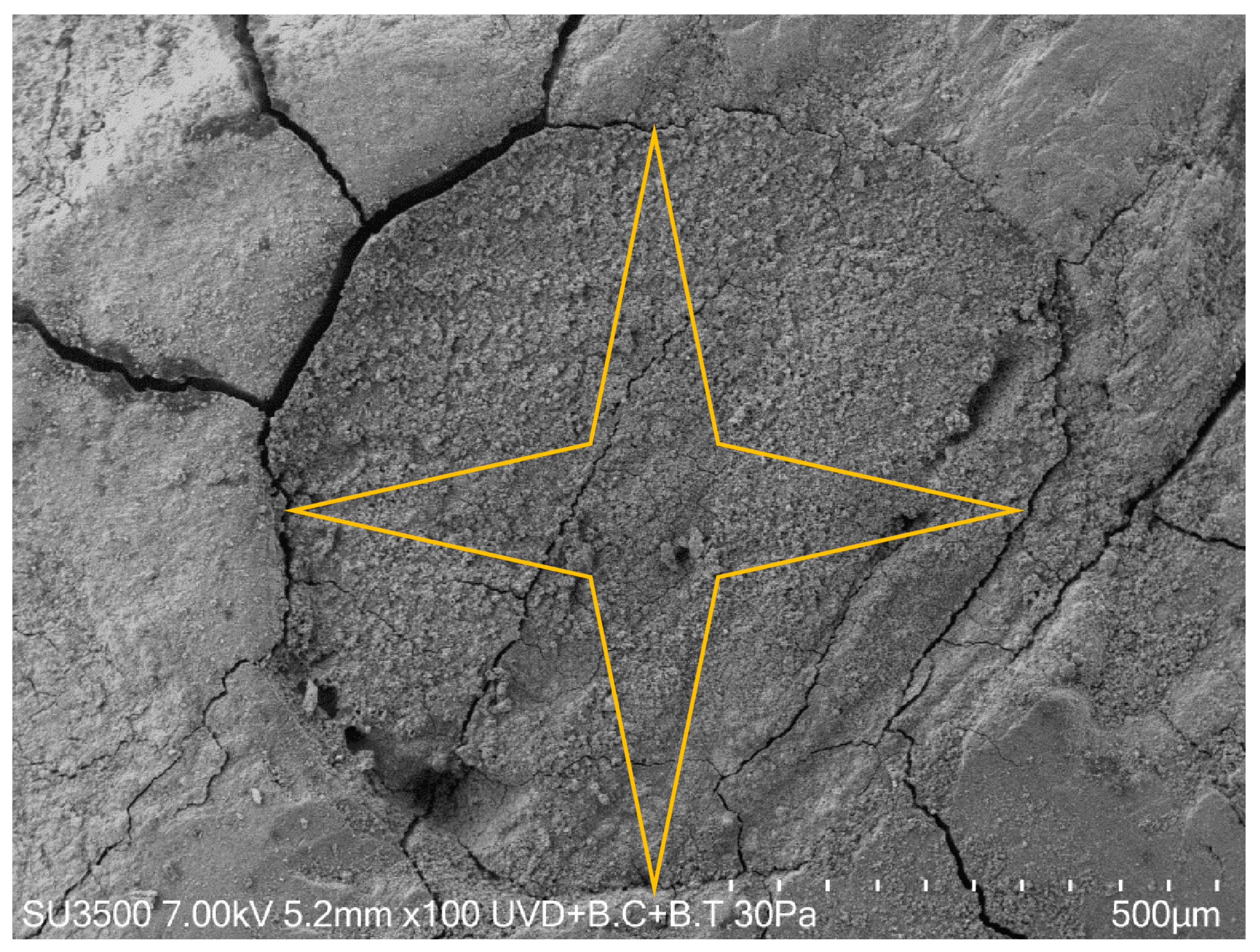
The samples were directly settled onto a carbon planchet stub without a conductive coating and observed with the variable pressure SEM Hitachi SU-3500 (Hitachi, Japan), setting the operating conditions at 30 Pa and 4.9–7.0 kV. Images were captured at several magnifications between 50× and 250×. Measurements were carried out on photos at 250× magnification. The marginal gap was measured using ImageJ software (Wayne Rasband; National Institute of Health, Bethesda, MD, USA). Following this, micrographs obtained from the SEM observation of each tooth section were digitally divided into 4 slices for the measurement of the gap in transverse sections. Precisely, the marginal gaps were evaluated using the freehand selection tool and the line selection tool of ImageJ software at the 4 points selected for the measurements, only after the parameters for measurements had been set correctly using the set scale and the scale bars of the pictures. For each section, the maximum value in terms of distance (µm) between the sealing materials and root canal wall was recorded, and the overall maximum gap value for each specimen was calculated by calculating the average of all the 4 slice values recorded (Figure 1). This procedure was repeated for each section of each tooth. The statistical analysis was then performed considering the overall maximum gap of each sectioned sample.
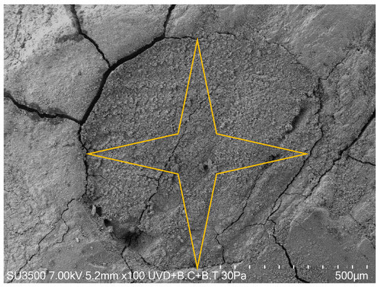
Figure 1.
SEM micrograph showing the measurement methodology at 50× and 100× magnification. Each transverse section was divided into 4 slices, and the major value in terms of the marginal gap of each slice was recorded using ImageJ software. The overall maximum gap value for each specimen resulted from calculating the average of all 4 slice values recorded.
2.4. Statistical Analysis
Statistical analysis was conducted using R software (version 4.2.0., R Foundation for Statistical Computing, Vienna, Austria). The normality distribution of continuous variables was evaluated both through visual and analytical methods with the Shapiro–Wilk test. Since the data did not follow a normal distribution, the Kruskal–Wallis test was used to compare the interfacial marginal adaptation among the three experimental groups. Post hoc Dunn’s test was conducted for pairwise comparison between the experimental groups at a 95% confidence level.
3. Results
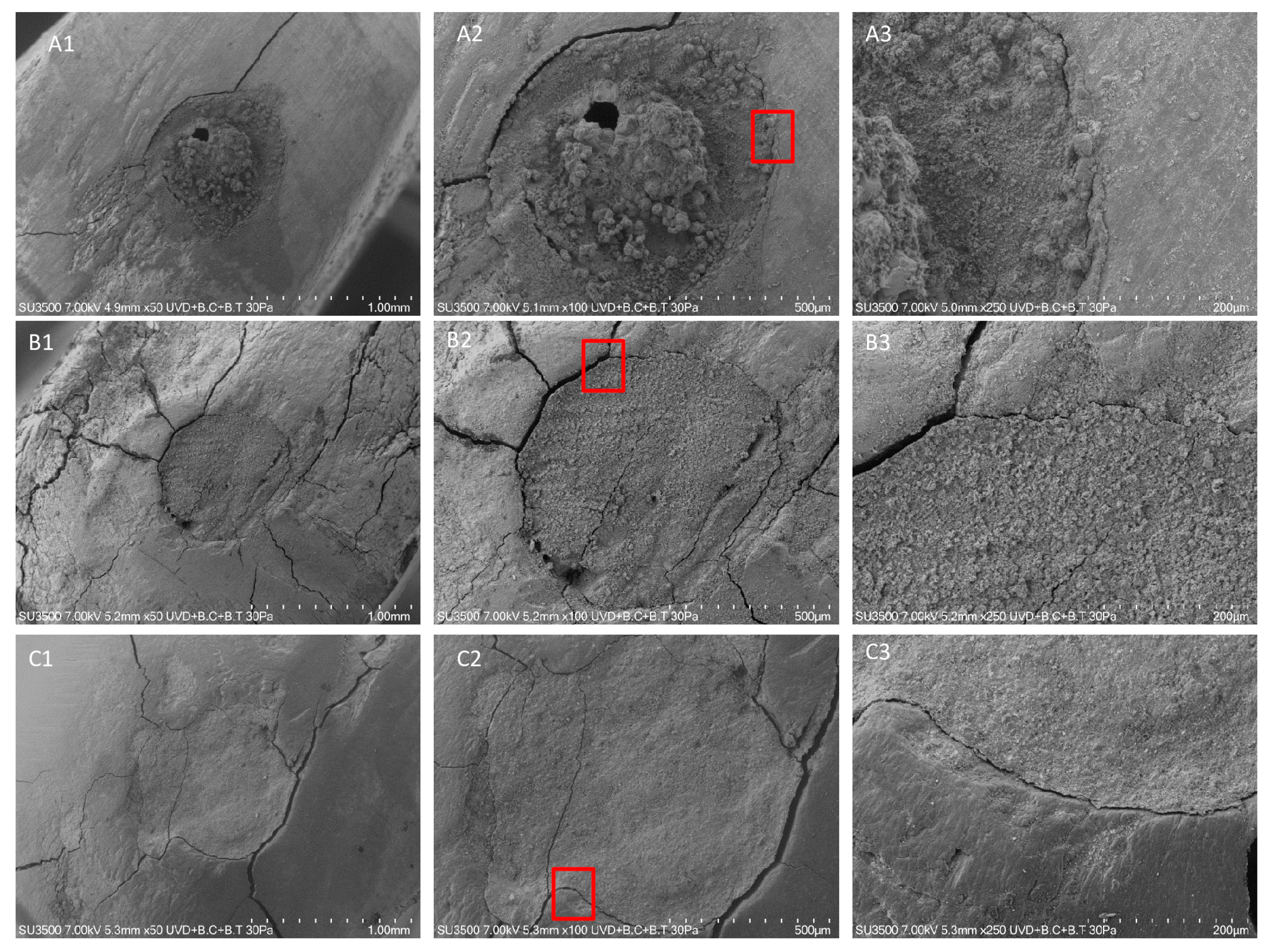
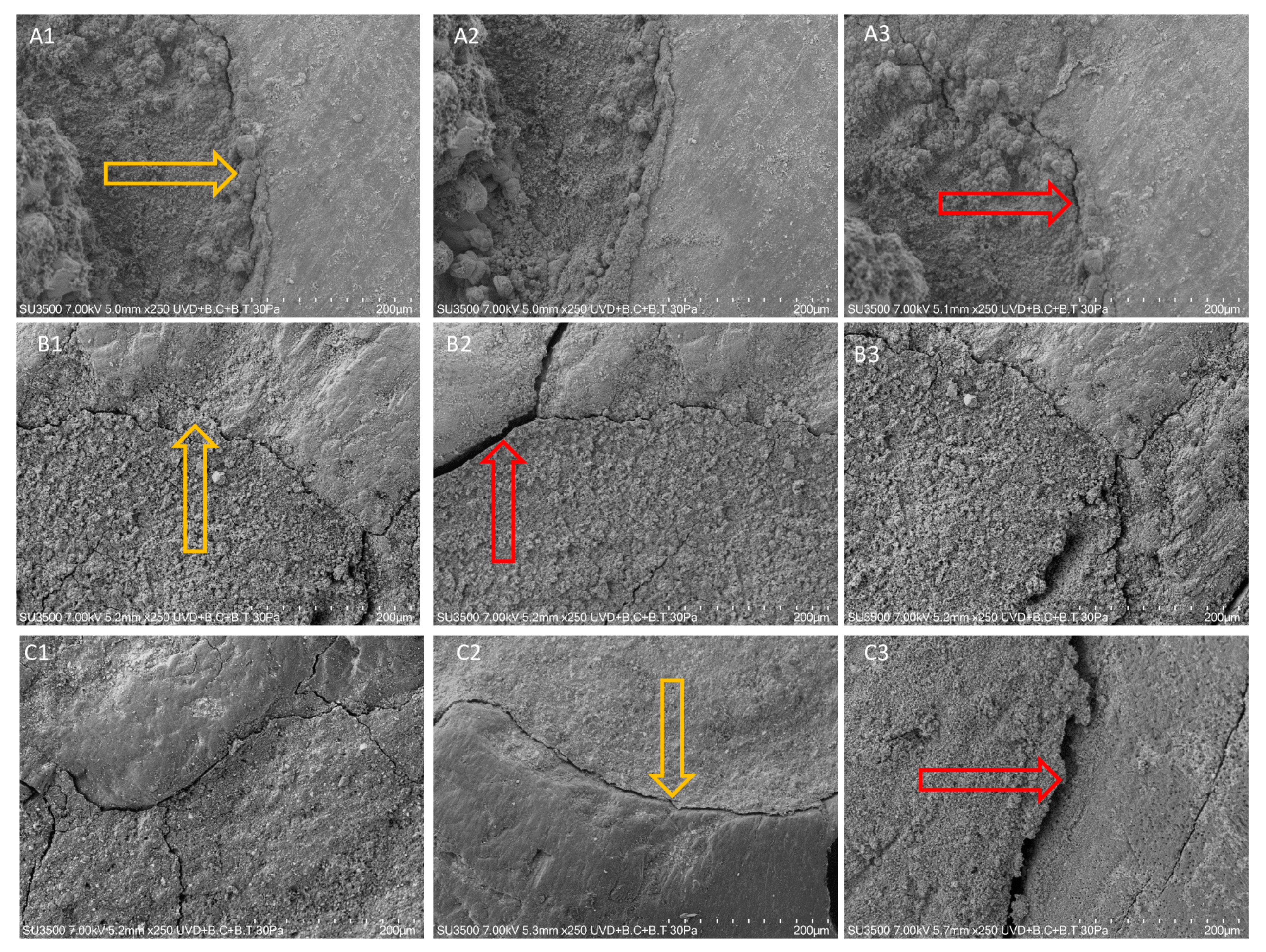
According to the collected data obtained from the VP-SEM observations and the performed statistical analyses, there was no statistically significant difference in terms of the marginal gap between the sealers and the dentinal walls in the three different bioceramic sealers used (p > 0.05). The results are schematically summarized in Table 1 and visually represented in Figure 1, Figure 2 and Figure 3. SEM micrographs show no gap between ERRM and dentin; Figure 1 shows the measurement methodology at 50× and 100× magnification. Figure 2 shows the three different root repair materials at different magnification. Figure 3 shows the three different root repair materials at 250× magnification. The yellow arrows indicate the minimum gap between the RRMs and the dentin wall while the red arrows indicate the maximum gap.

Table 1.
The mean (SD) of gap value in micrometers (µm) between the apical plug of the three RRMs and dentin wall of the root canal system at different times.
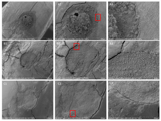
Figure 2.
SEM micrographs showing the three different root repair materials at different magnification. The red rectangle was designed to indicate the magnification site. (A) EdgeBioCeramic RetroFill, (B) Endocem MTA, and (C) One-Fil PT.

Figure 3.
SEM micrographs showing the three different root repair materials at 250× magnification. The yellow arrows indicate the minimum gap between the RRMs and the dentin wall while the red arrows indicate the maximum gap. (A) EdgeBioCeramic RetroFill, (B) Endocem MTA, and (C) One-Fil PT.
4. Discussion
Periapical surgery is a procedure indicated for the treatment of persistent apical periodontitis in an endodontically treated tooth when retreatment has not been successful or is not feasible [21]. The resection of the last 3 mm of the end of the root is carried out since the highest percentage of table accessory ducts are found in these 3 mm apical tissues [22]. After apical preparation, a filling material is used to seal the cavity at the end of the root. Prevention of microfiltration, biocompatibility, and material stability in apical tissues is very important. The filling material must adhere to the walls of the cavity and resist resorption and moisture ingress. A good-quality apical root canal filler is essential for the success of this surgical procedure [23].
The emergence of Mineral Trioxide Aggregate (MTA) in endodontics addressed a critical need for improved root repair materials. Ideal properties for such materials include effective sealing of pathways between the tooth’s internal anatomy and surrounding tissues, biocompatibility with human tissues, resistance to dissolution in oral fluids, and dimensional stability [24]. While MTA offers these advantages, research has identified limitations such as limited working time, slow setting, handling difficulties, and a lack of a suitable solvent for removal [25,26]. To overcome these shortcomings, novel bioceramic-based materials have been introduced to the endodontic market. In the current study, three recent bioceramics have been studied to evaluate their marginal gap with dentinal walls during the retrograde filling procedure: the Endocem MTA, the One-Fil PT, and the EdgeBioCeramic RetroFill [27].
The manufacturer highlights Endocem (EC) MTA’s advantages, which include its fast setting time (around 4 min) and user-friendly handling characteristics. Notably, it maintains a chemical composition similar to commercially available MTA, consisting primarily of calcium oxide, silicon dioxide, aluminum oxide, and radiopaque bismuth oxide, along with other metallic oxides [28,29,30]. Moreover, EC MTA consists of tiny particles of volcanic ash called pozzolan. This calcium silicate-based material interacts with calcium hydroxide, a product of the hydration process, to form additional cement-like substances. Research suggests that EC offers biocompatibility comparable to MTA, along with improved resistance to washout and minimal tooth staining.
One-Fil PT (OFPT) (Mediclus Co., Cheongju, Korea) is a recently released PPBC that is based on tricalcium silicate compound composition requiring the presence of water to set. The exact composition details of One-Fil PT, including any potential additives or modifiers, are, unfortunately, proprietary information and not publicly available [31].
According to the manufacturer, EdgeBioCeramic RetroFill stands out as a bioceramic material for repair procedures. Compared to traditional MTA, it boasts superior handling characteristics and promotes enhanced healing. A key advantage is its pre-mixed formulation, eliminating the need for manual mixing, unlike MTA. This translates to a more streamlined workflow. EdgeBioCeramic RetroFill offers a reduced working time of up to 30 min. The setting process initiates upon contact with moisture, with a minimum setting time of 2 h under normal conditions. The composition is tricalcium silicate, dicalcium silicate, zirconium oxide, tantalum, and calcium sulfate (anhydrous) [32].
While a material’s long-term clinical success is not solely dependent on marginal adaptation, this property serves as a valuable indicator of its sealing ability and resistance to leakage. In simpler terms, a well-adapted material effectively forms a tight seal, minimizing the potential for microleakage. To the best of our knowledge, no published study in the literature analyzed the marginal gap of these materials using VP-SEM. By the way, in the current literature, different studies have evaluated the marginal adaptation of EndoSequence Root Repair Material (ERRM) and MTA. According to Shokuhinejad et al., the mean value gap for the transversal section is 6.40 microns for MTA and 2.70 microns for ERRM in putty composition and 0.80 microns for ERRM in paste composition [33]. The results of the current manuscript are slightly higher, but this can be explained both by the use of real teeth samples instead of resin replicas and by the different composition of the material. Moreover, the minimum and maximum value are instead more similar to previously published studies. The results of the current study can be explained by the interaction of the alkaline compounds of the bioceramic materials that facilitate the penetration of the sealers [19]. Another published study on ERRM by Donfrancesco et al. shows similar results, with a mean value after 48 h of 4.32 microns, completely similar to the one observed in the current study [34]. Moreover, the previously mentioned study also used real tooth samples and the VP-SEM observation technique.
This study utilized a Variable Pressure Scanning Electron Microscope (VP-SEM) due to its unique ability to operate under variable pressure and humidity conditions. This capability is crucial because it prevents damage to MTA and bioceramic materials (like ERRM) when exposed to the electron beam. These specific operating parameters minimize the creation of artifacts that could arise from unsuitable observation conditions. Consequently, the measurements obtained in this study provide a more accurate representation of the materials under simulating clinical conditions [35]. Furthermore, the VP-SEM incorporates a Backscattered Electron (BSE) detector, which plays a vital role in differentiating between various material phases within the sample. This distinction helps to avoid inaccurate measurements by clearly defining the boundaries between the different materials. The BSE detector functions by counting the number of backscattered electrons, with the count directly proportional to the average atomic number of the sample. In simpler terms, brighter areas in a BSE image correspond to regions with a higher average atomic number, while darker areas indicate the opposite. This contrast allows researchers to effectively distinguish between different phases within the sample [36].
Since the marginal adaptation measurements reported and the use of VP-SEM, that does not modify the clinical conditions, we can state that Endocem MTA, One-Fil PT, and EdgeBioCeramic RetroFill have good stability over time. Indeed, the gap size of the material after the setting time is coherent with the current literature [33,34,37].
5. Conclusions
This manuscript suggests that all three bioceramic root repair materials studied may exhibit good dimensional stability. Therefore, it could broaden the clinical applications for both retrograde fillings in endodontic surgery and repairs of perforations that create a pathway between the root canal and surrounding periodontal tissue. Furthermore, the use of VP-SEM in this study implies that metal coating the samples might not significantly affect the long-term results for this type of analysis. However, to solidify these conclusions, further investigations are warranted. These future studies should aim to assess the dimensional stability of these materials over extended periods within a living organism (in vivo) and explore how sample preparation might influence the observed results.
Author Contributions
Conceptualization, O.D. and A.Z.; Methodology, O.D. and A.Z.; Validation, A.Z., L.T. and M.R.; Formal analysis, O.D.; Investigation, R.R.; Resources, L.T.; Data curation, R.R. and M.R.; Writing—original draft, M.S.; Writing—review & editing, O.D.; Visualization, R.R. and M.R.; Supervision, L.T.; Project administration, M.R.; Funding acquisition, R.R. All authors have read and agreed to the published version of the manuscript.
Funding
This research received no external funding.
Data Availability Statement
The original contributions presented in the study are included in the article, further inquiries can be directed to the corresponding author/s.
Conflicts of Interest
The authors declare no conflicts of interest.
References
- Schilder, H. Cleaning and Shaping the Root Canal. Dent. Clin. N. Am. 1974, 18, 269–296. [Google Scholar] [CrossRef] [PubMed]
- Schilder, H. Filling Root Canals in Three Dimensions. Dent. Clin. N. Am. 1967, 11, 723–744. [Google Scholar] [CrossRef]
- Alcalde, M.P.; Tanomaru-Filho, M.; Bramante, C.M.; Duarte, M.A.H.; Guerreiro-Tanomaru, J.M.; Camilo-Pinto, J.; Só, M.V.R.; Vivan, R.R. Cyclic and Torsional Fatigue Resistance of Reciprocating Single Files Manufactured by Different Nickel-Titanium Alloys. J. Endod. 2017, 43, 1186–1191. [Google Scholar] [CrossRef]
- Baek, S.-H.; Lee, C.-J.; Versluis, A.; Kim, B.-M.; Lee, W.; Kim, H.-C. Comparison of Torsional Stiffness of Nickel-Titanium Rotary Files with Different Geometric Characteristics. J. Endod. 2011, 37, 1283–1286. [Google Scholar] [CrossRef] [PubMed]
- Gomes, B.P.F.A.; Aveiro, E.; Kishen, A. Irrigants and Irrigation Activation Systems in Endodontics. Braz. Dent. J. 2023, 34, 1–33. [Google Scholar] [CrossRef] [PubMed]
- Dioguardi, M.; Gioia, G.D.; Illuzzi, G.; Laneve, E.; Cocco, A.; Troiano, G. Endodontic Irrigants: Different Methods to Improve Efficacy and Related Problems. Eur. J. Dent. 2018, 12, 459–466. [Google Scholar] [CrossRef]
- Pirani, C.; Camilleri, J. Effectiveness of Root Canal Filling Materials and Techniques for Treatment of Apical Periodontitis: A Systematic Review. Int. Endod. J. 2023, 56 (Suppl. 3), 436–454. [Google Scholar] [CrossRef] [PubMed]
- Aminsobhani, M.; Ghorbanzadeh, A.; Sharifian, M.R.; Namjou, S.; Kharazifard, M.J. Comparison of Obturation Quality in Modified Continuous Wave Compaction, Continuous Wave Compaction, Lateral Compaction and Warm Vertical Compaction Techniques. J. Dent. 2015, 12, 99–108. [Google Scholar]
- Camilleri, J.; Atmeh, A.; Li, X.; Meschi, N. Present Status and Future Directions: Hydraulic Materials for Endodontic Use. Int. Endod. J. 2022, 55, 710. [Google Scholar] [CrossRef]
- Camilleri, J. Classification of Hydraulic Cements Used in Dentistry. Front. Dent. Med. 2020, 1. [Google Scholar] [CrossRef]
- Aguilar, F.G.; Roberti Garcia, L.F.; Panzeri Pires-de-Souza, F.C. Biocompatibility of New Calcium Aluminate Cement (EndoBinder). J. Endod. 2012, 38, 367–371. [Google Scholar] [CrossRef] [PubMed]
- Oliveira, I.R.; Andrade, T.L.; Jacobovitz, M.; Pandolfelli, V.C. Bioactivity of Calcium Aluminate Endodontic Cement. J. Endod. 2013, 39, 774–778. [Google Scholar] [CrossRef] [PubMed]
- Castro-Raucci, L.M.S.; Teixeira, L.N.; Barbosa, A.F.S.; Fernandes, R.R.; Raucci-Neto, W.; Jacobovitz, M.; Oliveira, I.R.; de Oliveira, P.T. Calcium Chloride-Enriched Calcium Aluminate Cement Promotes in Vitro Osteogenesis. Int. Endod. J. 2018, 51, 674–683. [Google Scholar] [CrossRef] [PubMed]
- Li, X.; Pongprueksa, P.; Van Landuyt, K.; Chen, Z.; Pedano, M.; Van Meerbeek, B.; De Munck, J. Correlative Micro-Raman/EPMA Analysis of the Hydraulic Calcium Silicate Cement Interface with Dentin. Clin. Oral Investig. 2016, 20, 1663–1673. [Google Scholar] [CrossRef] [PubMed]
- Duncan, H.F.; Galler, K.M.; Tomson, P.L.; Simon, S.; El-Karim, I.; Kundzina, R.; Krastl, G.; Dammaschke, T.; Fransson, H.; Markvart, M.; et al. European Society of Endodontology Position Statement: Management of Deep Caries and the Exposed Pulp. Int. Endod. J. 2019, 52, 923–934. [Google Scholar] [CrossRef] [PubMed]
- AAE Position Statement on Vital Pulp Therapy. J. Endod. 2021, 47, 1340–1344. [CrossRef] [PubMed]
- Torabinejad, M.; White, S.N. Endodontic Treatment Options after Unsuccessful Initial Root Canal Treatment: Alternatives to Single-Tooth Implants. J. Am. Dent. Assoc. 2016, 147, 214–220. [Google Scholar] [CrossRef] [PubMed]
- Amador-Cabezalí, A.; Pardal-Peláez, B.; Quispe-López, N.; Lobato-Carreño, M.; Sanz-Sánchez, Á.; Montero, J. Influence of the Retrograde Filling Material on the Success of Periapical Surgery. Systematic Review and Meta-Analysis by Groups. Coatings 2022, 12, 1140. [Google Scholar] [CrossRef]
- Arikatla, S.K.; Chalasani, U.; Mandava, J.; Yelisela, R.K. Interfacial Adaptation and Penetration Depth of Bioceramic Endodontic Sealers. J. Conserv. Dent. 2018, 21, 373–377. [Google Scholar] [CrossRef]
- Zanza, A.; Reda, R.; Vannettelli, E.; Donfrancesco, O.; Relucenti, M.; Bhandi, S.; Patil, S.; Mehta, D.; Krithikadatta, J.; Testarelli, L. The Influence of Thermomechanical Compaction on the Marginal Adaptation of 4 Different Hydraulic Sealers: A Comparative Ex Vivo Study. J. Compos. Sci. 2023, 7, 10. [Google Scholar] [CrossRef]
- Kohli, M.R.; Berenji, H.; Setzer, F.C.; Lee, S.-M.; Karabucak, B. Outcome of Endodontic Surgery: A Meta-Analysis of the Literature-Part 3: Comparison of Endodontic Microsurgical Techniques with 2 Different Root-End Filling Materials. J. Endod. 2018, 44, 923–931. [Google Scholar] [CrossRef] [PubMed]
- Song, M.; Shin, S.-J.; Kim, E. Outcomes of Endodontic Micro-Resurgery: A Prospective Clinical Study. J. Endod. 2011, 37, 316–320. [Google Scholar] [CrossRef] [PubMed]
- Bodrumlu, E. Biocompatibility of Retrograde Root Filling Materials: A Review. Aust. Endod. J. 2008, 34, 30–35. [Google Scholar] [CrossRef] [PubMed]
- Johnson, B.R. Considerations in the Selection of a Root-End Filling Material. Oral Surg. Oral Med. Oral Pathol. Oral Radiol. Endod. 1999, 87, 398–404. [Google Scholar] [CrossRef] [PubMed]
- Charland, T.; Hartwell, G.R.; Hirschberg, C.; Patel, R. An Evaluation of Setting Time of Mineral Trioxide Aggregate and EndoSequence Root Repair Material in the Presence of Human Blood and Minimal Essential Media. J. Endod. 2013, 39, 1071–1072. [Google Scholar] [CrossRef] [PubMed]
- Parirokh, M.; Torabinejad, M. Mineral Trioxide Aggregate: A Comprehensive Literature Review--Part III: Clinical Applications, Drawbacks, and Mechanism of Action. J. Endod. 2010, 36, 400–413. [Google Scholar] [CrossRef] [PubMed]
- Cardinali, F.; Camilleri, J. A Critical Review of the Material Properties Guiding the Clinician’s Choice of Root Canal Sealers. Clin. Oral Investig. 2023, 27, 4147–4155. [Google Scholar] [CrossRef]
- Choi, Y.; Park, S.-J.; Lee, S.-H.; Hwang, Y.-C.; Yu, M.-K.; Min, K.-S. Biological Effects and Washout Resistance of a Newly Developed Fast-Setting Pozzolan Cement. J. Endod. 2013, 39, 467–472. [Google Scholar] [CrossRef]
- Kim, M.; Yang, W.; Kim, H.; Ko, H. Comparison of the Biological Properties of ProRoot MTA, OrthoMTA, and Endocem MTA Cements. J. Endod. 2014, 40, 1649–1653. [Google Scholar] [CrossRef]
- Kang, T.-Y.; Choi, J.-W.; Seo, K.-J.; Kim, K.-M.; Kwon, J.-S. Physical, Chemical, Mechanical, and Biological Properties of Four Different Commercial Root-End Filling Materials: A Comparative Study. Materials 2021, 14, 1693. [Google Scholar] [CrossRef]
- Song, M.; Lee, S.-M.; Bang, J.-Y.; Kim, R.H.; Kwak, S.W.; Kim, H.-C. Chemomechanical Properties and Biocompatibility of Various Premixed Putty-Type Bioactive Ceramic Cements. J. Endod. 2023, 49, 1713–1721. [Google Scholar] [CrossRef] [PubMed]
- EdgeBioCeramic RetroFillTM and Perforation Repair—Edge Endo LLC Online Store. Available online: https://store.edgeendo.com/edgebioceramic-retrofill-and-perforation-repair-p224.aspx (accessed on 31 May 2024).
- Shokouhinejad, N.; Nekoofar, M.H.; Ashoftehyazdi, K.; Zahraee, S.; Khoshkhounejad, M. Marginal Adaptation of New Bioceramic Materials and Mineral Trioxide Aggregate: A Scanning Electron Microscopy Study. Iran. Endod. J. 2014, 9, 144–148. [Google Scholar] [PubMed]
- Donfrancesco, O.; Seracchiani, M.; Morese, A.; Ferri, V.; Nottola, S.; Relucenti, M.; Gambarini, G.; Testarelli, L. Analysis of Stability in Time of Marginal Adaptation of Endosequence Root Repair Material on Biological Samples. Dent. Hypotheses 2020, 11, 11. [Google Scholar] [CrossRef]
- Donfrancesco, O.; Giudice, A.D.; Zanza, A.; Relucenti, M.; Petracchiola, S.; Gambarini, G.; Testarelli, L.; Seracchiani, M. Sem Evaluation of Endosequence Bc Sealer Hiflow in Different Environmental Conditions. J. Compos. Sci. 2021, 5, 99. [Google Scholar] [CrossRef]
- Relucenti, M.; Familiari, G.; Donfrancesco, O.; Taurino, M.; Li, X.; Chen, R.; Artini, M.; Papa, R.; Selan, L. Microscopy Methods for Biofilm Imaging: Focus on SEM and VP-SEM Pros and Cons. Biology 2021, 10, 51. [Google Scholar] [CrossRef]
- Tran, D.; He, J.; Glickman, G.N.; Woodmansey, K.F. Comparative Analysis of Calcium Silicate-Based Root Filling Materials Using an Open Apex Model. J. Endod. 2016, 42, 654–658. [Google Scholar] [CrossRef]
Disclaimer/Publisher’s Note: The statements, opinions and data contained in all publications are solely those of the individual author(s) and contributor(s) and not of MDPI and/or the editor(s). MDPI and/or the editor(s) disclaim responsibility for any injury to people or property resulting from any ideas, methods, instructions or products referred to in the content. |
© 2024 by the authors. Licensee MDPI, Basel, Switzerland. This article is an open access article distributed under the terms and conditions of the Creative Commons Attribution (CC BY) license (https://creativecommons.org/licenses/by/4.0/).