Review

Advancements in EBSD Techniques: A Comprehensive Review on Characterization of Composites and Metals, Sample Preparation, and Operational Parameters

1
Department of Mechanical and Industrial Engineering, Manipal Institute of Technology, Manipal Academy of Higher Education, Manipal 576104, Karnataka, India
2
Department of Mechanical Engineering, B.M.S. Institute of Technology and Management, Bangalore 560064, Karnataka, India
*
Author to whom correspondence should be addressed.
J. Compos. Sci. 2025, 9(3), 132; https://doi.org/10.3390/jcs9030132
Submission received: 23 December 2024 / Revised: 11 February 2025 / Accepted: 24 February 2025 / Published: 13 March 2025
(This article belongs to the Special Issue Metal Composites, Volume II)

Abstract

:
This comprehensive review focuses on the most recent advances in electron backscatter diffraction (EBSD) methods in the context of materials science and includes a thorough evaluation of the sample preparation procedures unique to EBSD as well as a complete examination of the important operational parameters inherent in EBSD setups. This review highlights the importance of customizing EBSD parameters for precise microstructural imaging and enhancing understanding of material behavior. While some studies have explored grain boundary characterization, stored energy, and crystallographic orientation using EBSD, there is a clear need for more comprehensive investigations to fully leverage its capabilities. Additionally, there is a significant gap in understanding the optimal choice of the reference plane in EBSD analysis, indicating the necessity for further research to improve EBSD analyses’ accuracy and efficacy. The review seeks to present a detailed and contemporary viewpoint on the many applications, sample preparation techniques, and optimal operational considerations that jointly increase the adaptability and efficacy of EBSD in materials science research by relying on the relevant literature.

1. Background Introduction

Microstructural analysis of modern-day materials has been very productive because of the usage of advanced microscopy techniques rather than traditional techniques. Electron backscatter diffraction (EBSD) is the ideal approach for understanding and studying the behavior of materials at the submicroscopic level [1]. EBSD is a scanning electron microscope (SEM) technology that captures the diffracted electron beam using a fluorescent screen, resulting in diffraction patterns [2]. When the beam covers the whole sample, the diffraction patterns acquired provide information on the lattice structure, grain boundaries, and crystal orientations [3].
Two-dimensional microscopes can only provide information about surface morphology, whereas recent advancements such as X-ray diffraction (XRD), energy dispersive X-ray spectroscopy (EDS), SEM, and EBSD provide critical information about materials at a deeper level, making it easier for a researcher to understand the behavior of materials and its relationship with physical and mechanical properties [4]. Seishi Kikuchi’s discovery, EBSD, can reveal remarkable information about materials with a working range of 10 nm to 1 cm [5]. The technique not only enables predictions of metallurgical behavior based on grain boundary characteristics, material energy, and crystallographic orientation but also integrates seamlessly into SEM, becoming a vital attachment for precise grain and precipitate orientation analysis (Figure 1a). The SEM’s electron beam interaction generates various signals on the specimen surface, including backscattered electrons with diffraction patterns (Figure 1b). These patterns, captured by a charge-coupled device (CCD) camera during a raster scan, yield an abundance of orientation data [6]. In materials science, where mechanical properties are intricately tied to microstructure and phase constituents, conventional optical or electron imaging techniques fall short [7]. EBSD’s unique capability to provide crystal orientation alongside morphological characteristics proves indispensable for accurate phase identification and differentiation [8].
In SEM, the incoming electron from the emitter reaches the surface of the specimen which is exposed underneath the electron beam. This electron undergoes a series of interactions with the material atoms. This interaction of the primary electrons results in the generation of different signals which could be X-ray, secondary electron, backscattered electron, etc. This is represented through the interaction volume [6]. All these signals can be detected in SEM using different detectors like an EDS which detects X-rays, a secondary electron (SE) detector for secondary electron imaging, and an EBSD detector for detecting diffraction signals generated from backscattered electrons [9,10]. These diffraction signals are generated from different crystallographic planes within the limited sample volume in the form of a pair of cones [11]. Depending on the orientation of crystallographic planes, the orientation of these cones will be different. So, within a point in the specimen where the primary electrons are incident just a few nanometers below it, the EBSD signals are generated, and these appear as cones [12]. There could be many pairs of such cones originating from a given point. So, if a plane is held in front of the specimen, these bunches of cones will intersect with the plane, giving rise to a set of bands, called Kikuchi bands [13]. For a given orientation of a grain, electron beam interaction with the specimen will be generated, as shown in Figure 1b. In SEM, the beam runs a raster scan over a selected area on the specimen surface. It scans line-by-line, so, at every point during a scan, the Kikuchi patterns corresponding to the orientation of the points are transferred onto the phosphor screen of the EBSD detector. Using a CCD camera, these patterns are captured instantaneously. Finally, when the scan is completed, all these saved patterns are transferred into an orientation value. This raw data collection of orientations yields an abundance of information [14].
EBSD is not confined to microstructure analysis alone; it excels in assessing plastic deformation on a smaller scale and detecting residual deformation post-sample processing. Despite alternative orientation techniques, such as electron channeling contrast imaging (ECCI) and selected area diffraction (SAD), EBSD stands out for its automation capability and ability to offer orientation data across large, scanned areas [15]. If a steel or any material is considered as a subject of study, their mechanical properties largely depend on the final microstructure and phase constituents. These morphological characteristics are usually analyzed using an optical microscope (OM) or SEM images; in these methods, phases are differentiated based on their morphological characteristics, but they fail to differentiate when there are similar morphologies for different phases [2]. EBSD analysis can overcome this difficulty by providing information regarding crystal orientation along with morphological characteristics, by which phase identification and differentiation can be performed with ease [16]. The significance of EBSD extends to its capacity to deliver detailed microstructural information within oxide layers without necessitating surface etching. For example, when dealing with two phases sharing the same crystal structure, such as magnetite and wustite within an oxide scale, accurate indexing becomes possible due to variations in lattice spacing [5,17]. Complemented by EDS, this method facilitates a thorough analysis of oxide scales. EBSD eliminates the need to etch the material’s surface before generating images of its microstructure. This technique proves instrumental in depicting texture relationships, crystallographic growth directions, and details on grain boundary misorientation. Notably, the capacity for precise phase identification stands out as a significant advantage of EBSD over conventional optical and electron imaging methods [18].
Sample preparation in the TEM procedure is time consuming and damaging, making it difficult to obtain information about grain size distribution and grain orientation effects. XRD (non-destructive) tests, on the other hand, cannot offer any information on local measurements below a 1 µm scale or less. Because it does not cause any harm to thin film materials when imaged in the SEM, EBSD can be considered a non-destructive test. EBSD has better spatial resolution than XRD, and EBSD also allows for a larger area of analysis than TEM [19]. In addition to its pivotal role in various industries, including metal processing, mining, aerospace, automobile, nuclear, microelectronics, ore refining, and academia, EBSD analysis has proven indispensable in examining a diverse range of crystalline materials. Metals, alloys, intermetallics, ceramics, thin films, nanostructures, semiconductors, superconductors, composites, bones, teeth, shells, organics, and polymers are among the materials actively subjected to EBSD scrutiny [20]. The versatility of EBSD extends to a spectrum of measurements, encompassing grain size, boundary characterization, texture (both general and local), recrystallization, deformation, structure analysis, phase identification, phase distribution, fracture analysis, corrosion analysis, orientation-related analysis, and strain analysis [21].
While EBSD analysis offers unparalleled insights, it is not without its limitations. The technique’s limited angular resolution, typically above 1°, poses a challenge, mitigated to some extent by the utilization of high-resolution electron backscatter diffraction (HR-EBSD), providing an increased field view with a resolution below 0.5° [22]. In the realm of in situ testing technologies, EBSD finds a place for capturing extensive crystallographic information at the micrometer scale, demanding comparatively less sample preparation. This method streamlines the examination of crucial factors such as grain boundaries, crystal orientation, and kernel average misorientation (KAM). The KAM’s most important use is that it can distinguish the crystal orientation between a certain center point and its neighbors. With these KAM findings, one may investigate the behavior of plastic deformation and dislocation density distribution, which aids in understanding the material’s mechanical characterization [7].
This study presents an overview of EBSD applications in the field of materials, sample preparation methods, and EBSD analysis operating parameters. This approach may be used with a variety of materials to provide essential information on grain size, orientations, misorientations, texture, and phase distributions. In certain ways, EBSD is a very strong technique for bridging the gap between microstructures and material properties.

2. Literature

Chen et al. [23] used EBSD analysis to study the severe plastic deformation of ultrafine-grained and nanomaterials. EBSD analysis was performed by varying the operating parameters and by using standard sample preparation methods. By using optimum parameter inputs, the authors were able to study the grains and sub-grains at the nanoscale level by using high-resolution SEM. Ion milling was proposed by the authors as a versatile and promising universal polishing procedure for EBSD preparation across a wide variety of materials. Their study revealed a critical association between step size and the maximum value of indexed points. The optimal step size, which is governed by magnification and board resolution/electronic step size, is critical for producing accurate and relevant findings. The authors’ use of EBSD was extremely efficient in gathering precise information on grains, sub-grains, texture, and grain boundary structure. Furthermore, the approach proved its adaptability in examining the mechanical properties of modern materials by analyzing the strain and stored energy within materials.
Hu et al. [24] used the EBSD approach and emphasized that improving tensile strength and consistent elongation in high-strength ultrafine-grained (UFG) Al alloys requires minimizing the grain boundary misorientation angle. They created a high-strength, ultrafine-grained Al alloy with a lamellar structure using cryomilling and consolidation (hot isostatic pressing), followed by a two-stage thermomechanical processing procedure that included high-temperature rotary swaging and room temperature high strain rate extrusion. The resultant lamellar structure was composed of multiple ultrafine grains linked together by grain boundaries with minor misorientation angles. Dislocations can glide over grain boundaries with low misorientation angles, resulting in their accumulation inside the lamellar band. As a result, this structural configuration provides an excellent combination of high uniform elongation and retained high strength. The major reason for achieving higher mechanical properties is because of the dislocation’s ability to transverse low angle grain boundaries (LAGBs) and gather within lamellar bands.
Lin et al. [25] performed an EBSD analysis on the behavior of Ni-based superalloy, which was subjected to hot deformation. Isothermal compression experiments with temperatures ranging from 920 to 1040 °C were used for this study. The results revealed that the LAGBs decreased with an increase in the deformation temperature, which is because of the decrease in dynamic recrystallization at low deformation temperatures. During hot deformation, both continuous dynamic recrystallization and discontinuous dynamic recrystallization were observed. Discontinuous dynamic recrystallization was observed to be the important mechanism of nucleation. So, using this EBSD analysis, authors were successful in analyzing the hot deformation behavior of Ni-based superalloy with the help of grain boundary information.
Sankaranarayanan et al. [26] studied the impact of heat treatment on the stress relaxation and recrystallization of the Mg nanocomposite using the EBSD technique, thereby improving the mechanical properties of the composite by altering the microstructure by varying the heat treatment parameters. From the results, it was found that the heat-treated samples exhibited more strain-free grains, confirming the effect of recrystallization and residual stress relaxation. The results highlighted the importance of heat treatment parameter optimization, changing which mechanical properties can be altered (improved) by reducing the residual stresses.
Morteza et al. [27] used EBSD to study the grain boundary modification and microstructural development of the Al 7075-T6 alloy, which was subjected to gas tungsten arc (GTA) welding followed by friction stir welding. The results revealed the formation of smaller grains in GTA welds after friction stir processing, whereas coarse grains were observed in GTA welds (no friction stir processing). After subjecting to friction stir processing, the tensile strength improved from 40 to 60%, and the dissolution of Al2Cu and MgZn2 was identified to be the major reason for the tensile strength improvement. The fracture occurred near the fusion zone in the original GTA weld; however, after friction stir processing, it migrated to the retarding side. The percentage of high-angle grain boundaries (HAGBs) increased from 78 to 91%, which was followed by the development of very fine equiaxed grains in the GTA weld joint. This behavior was ascribed to the dynamic recrystallization of the GTA weld joint during friction stir processing.
Lizheng et al. [28] fabricated composites by coating TiC on the Ti6Al4V alloy using laser cladding, and microstructure evolution was studied using EBSD. Using EBSD, average grain size, proportion of grain boundary angle, and texture intensity analysis was performed. From the results, it was concluded that the average grain size of α-Ti decreased when coated with TiC. Also, the LAGB of the composite coated with TiC was higher when compared to the uncoated composite. The texture strength and number of grains increased when coated with TiC.
Pirgazi et al. [29] studied the localized cracking of compacted graphitic iron (CGI—used in cylinder heads) caused by thermo-mechanical fatigue. The crack initiation, propagation, and growth of CGI were studied based on EBSD analysis. From the results, it was understood that the graphite particles are the major reason for localized cracking and that the density of the graphite particles on the fractured planes was almost twice in number when compared to other arbitrary planes of the structure. From their EBSD analysis, they proved that the crystal orientation does not have any effect on the crack propagation where graphite presence is dominating, although it might have some local effect far from graphite morphology.
Based on their 3D microstructural characterization (as shown in Figure 2), they concluded that the crystal plane parallel to the fracture plane has a random orientation and has no effect on localized cracks.
Molin et al. [30] initially studied the effect of welding on the average grain size of the base metal and weld seam using EBSD analysis. From the results, it was confirmed that the average grain size of the base metal was 20.6 µm, whereas the average grain size of the weld seam was 5.7 µm. It was interpreted that the welding operation had a heating effect on the material, which indirectly affected the refinement of the grain size of the material near the weld zones. Additionally, the authors aimed to study the short crack propagation effect of the weld seam sample. The major reason for studying the short crack propagation effect was that a short crack occupies almost 90% of fatigue life, whereas a long crack occupies a lesser portion. As the welding process provides an uneven surface, it was difficult to study the effect of a short crack on the weld seam sample. As the base metal sample consisted of similar phases, but with a larger grain size, the authors proceeded with the EBSD analysis of base metal samples. From the results, it was concluded that a transgranular fracture was dominant, but they have observed intergranular fractures at several places, which might be because of the plasticity difference between the phases present, that caused the mismatch and allowed the cracks to grow through the boundaries.
Sun et al. [31] performed EBSD-based analysis on the additively manufactured (AM) 316L stainless steel. As per the authors, the major reason for using EBSD analysis to study the AM material is because, by using an additive manufacturing technique, there is a high chance of the formation of heterogeneous grains and nonregular grain structures, which can be studied easily using EBSD analysis. The authors created 3D structures using ELAVO 3D to fill the gaps that 2D analysis was failing to capture during regular analysis. From the analysis results, they observed the formation of “tree-like” grains with different orientations; one appears to be “branch-like” and the other appears to be “trunk-like”. By using 3D characterization techniques, the capturing of the nucleation site was possible, which revealed that these two grains did not follow any specific pattern with their intragranular rotations, and the results of the crystallographic growth directions reconfirmed that the grains did not undergo any specific pattern during rotations, and the texture differences between them were distinguished clearly.
Koko et al. [22] studied the effect of reference pattern position (EBSP0) on the HR-EBSD analysis of cubic crystals with varying degrees of plastic deformation. The authors revealed that, if the reference pattern position is selected at a deformed point, it directly affects the EBSD analysis, providing undesired results. Reference patterns with higher geometrical necessary dislocation density cause more errors than selecting reference patterns with high lattice distortion. The authors were successful in establishing a relationship between the mean angular error and peak height, which can be used to find the optimal position of EBSP0 for improving HR-EBSD precision.
Jepson et al. [17] studied the effect of oxidation on 316 stainless steel at 1200 °C under different durations and cooling rates. Samples oxidized for 4 h were studied in detail using EBSD analysis. The results showed that there was a formation of three layers, each layer with different grains and different alloying content, like chromium or nickel. Using EBSD analysis, the authors were able to identify which layer experienced the internal oxidation effect and how the grains were formed (equiaxed or not).
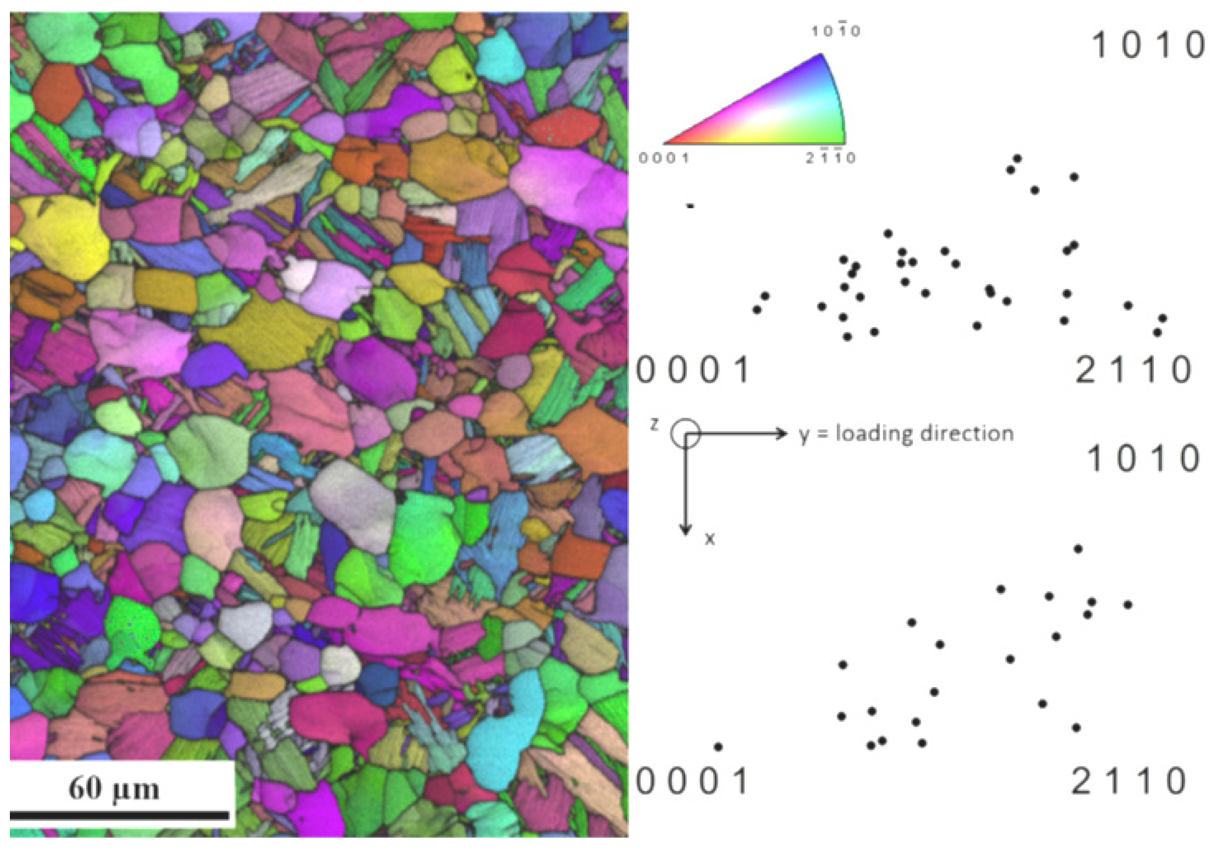
Hemery et al. [32] used EBSD to investigate the strain partitioning and deformation of the Ti-6Al-4V (bimodal) specimen under a tensile test. Figure 3 shows the IPF of crystallographic orientations of colonies and the nodules used. Lattice rotations and rotation axis were used to capture the active slip systems. Lattice rotations exhibited linear relationships with plastic strains, and these lattice rotation data were used for the direct quantification of the plastic strains. While in the case of strain partitioning, the operating slip mode plays a crucial role, the operating basal slip experienced a higher plastic strain than the prismatic slip mode.
EBSD analyses are also employed to study the phase transformation and recrystallization behavior of different materials. Su et al. [33] identified the phase transformation and recrystallization behavior of low-carbon steel during continuous cooling after the annealing process using an OM and EBSD setup. From the results, they concluded that, at different cooling rates, samples have undergone different phase transformations, which were captured and quantified successfully using EBSD analysis. EBSD analysis was successful to identify and quantify different phases like ferrite, pearlite, and bainite.
Pk et al. [34] studied the effect of SiC, dislocations, and misorientations on the mechanical properties of the aluminum metal matrix composite weld joints (as-weld and age-hardened). Samples were fine polished using an electropolishing technique and EBSD analysis was carried out using FEI Quanta-3D FEG (field emission gun) SEM, operated at 20 keV voltage. From the results, it was concluded that the non-indexed points were more in as-weld samples, which can be considered as dislocations, whereas in the case of the age-hardened samples, the number of dislocations was drastically reduced because of the precipitation effect, which was confirmed by IPF figures showing a smaller number of non-indexed points. Also, after age-hardening, the samples showed refinement in grain size, which was again confirmed by texture imaging from EBSD analysis. Both 8 and 12 wt.% SiC + age-hardened samples showed similar EBSD estimated misorientation values, but the mechanical properties of the 12 wt.% sample were higher, which says that the mechanical properties not only rely on the values of misorientations but also the distribution of them.
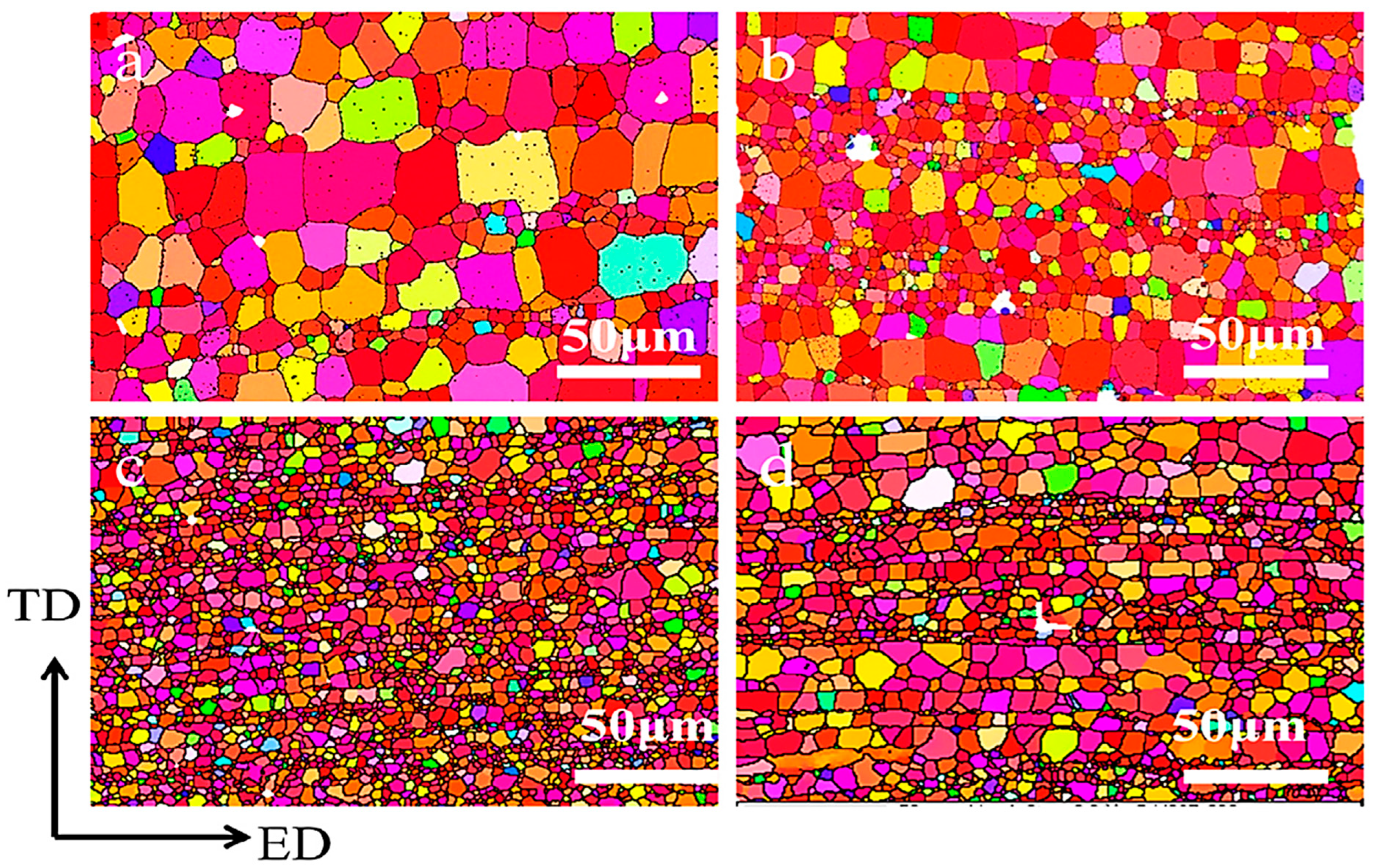
Yuan et al. [35] studied the effect of samarium (Sm) on Mg alloys, and the wt.% of Sm was varied from 1 to 4. Microstructural and phase analysis was performed using OM, SEM, EDAX, and XRD. EBSD analysis was also performed to observe the average grain size and texture intensity after the extrusion process. From Figure 4a–d, we can observe the recrystallization effect and drop in the average grain size until the 3 wt.% addition of Sm, after which the grain size increased. Also, the texture intensity of the alloys decreased from 12.5 to 7.8 multiples of a random distribution with the increase in the wt.% of Sm. From the mechanical testing results, it was concluded that the alloy with 3 wt.% of Sm outperformed other alloys, which can be justified by its lowest average grain size.
Wang et al. [36] studied the texture and microstructure evolution of friction stir welded aluminum alloys (AA5052-O and AA6061-T6) using the EBSD technique. After the friction stir welding, the area of study was categorized into four zones: (i) nugget zone (NZ), (ii) thermomechanically affected zone (TMAZ), (iii) heat-affected zone (HAZ), and (iv) base metals (BMs). From Figure 5a–d, we can see the orientation imaging microscopy images of the AA6061-T6 alloy, which indicate that the BM zone had high microstructural inhomogeneity, the NZ had equiaxed and fine grains, the HAZ showed similar behavior to BMs, whereas in the TMAZ, the grain structures were different from the NZ because of the strain deformation and heat, which restricted the recrystallization process. From the results of texture analysis, it was confirmed that the BMs transformed from {001}<100>C cube texture and {123}<634>S texture into {111}<112>A1 shear texture under the stirring shear force. Also, in the NZ and TMAZ, a large number of dislocations and precipitates were observed.
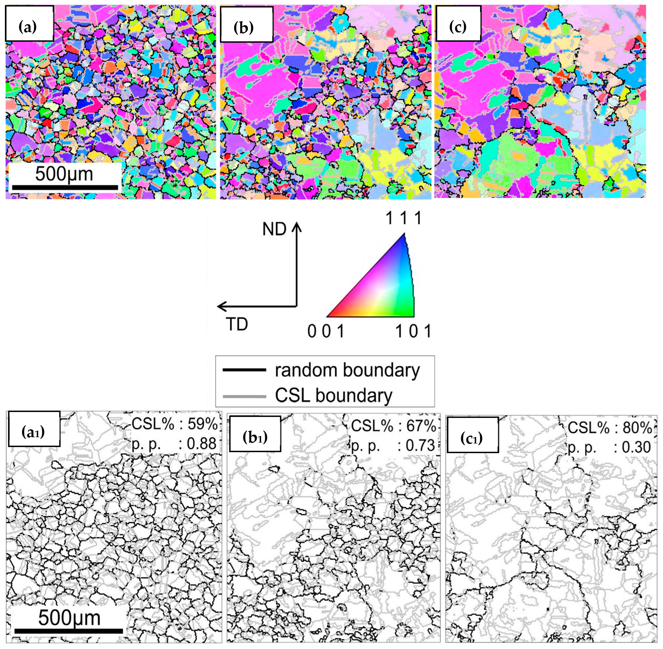
Shun et al. [37] studied the grain boundary character distribution (GBCD) evolution of 304 steel during the thermomechanical (TM) process using EBSD analysis. From the results, it was concluded that there was an increase in coincident site lattice (CSL) boundaries and a decrease in percolation probability (PP) when the sample was subjected to thermomechanical treatment. Figure 6 shows the IPF of the sample subjected to the TM process under different annealing durations, and the frequency of CSL% and PP is also shown. From Figure 6a–c and Figure 6a1–c1, we can see the growth of the clusters starting from the top left corner and bottom right corner and finally converging at the center. Clusters of grains were formed because of the strain-induced grain growth. Through EBSD analysis, the authors were able to identify the reason behind the disconnection of boundaries and establish a relation between the clusters of grains during the TM treatment.
Xie et al. [38] used EBSD analysis to determine the influence of the initial microstructure on the re-austenitization behavior of steels under various heating temperatures. The initial microstructures were studied, and it was observed that lath and granular bainite formation took place. Now, from the results of the EBSD analysis of two different samples with two different initial microstructures and temperatures, it was concluded that the martensite–austenite constituents have a great impact on the austenitizing behavior, which was also confirmed by the geometrically necessary dislocations (GNDs).
Dourandish et al. [39] studied the microstructural and phase changes in martensitic stainless steel during the forging process with the help of EBSD analysis. The results revealed different types of phases and compositions formed in the samples after the forging operation. High concentrations of alloying elements like Cr, Mo, and C within the eutectic carbide were identified, while the eutectic phase itself was found to be a combination of M23C6 and delta ferrite. Figure 7a,b show the orientation mapping of EBSD analysis, displaying the presence of the M23C6 and ferrite phases.
Weng et al. [40] studied the effect of the pre-aging treatment on the Cu-0.75Fe-0.35Ti (wt.%) alloy with the help of EBSD analysis. The samples subjected to cold rolling after the pre-aging treatment are termed “SARA”, whereas the samples subjected to cold rolling without pre-aging treatment are termed “SAR”. From the EBSD results, it was concluded that in both SAR and SARA samples, better results were achieved when samples were subjected to aging for 5 h. From the IPF images of SARA-aged for 5 h, a recrystallization effect was observed (Figure 8), whereas in the IPF images of SAR-aged for 5 h, recrystallization was not visible clearly. From this, it was understood that the pre-aging treatment promotes the recrystallization effect. Also in SARA samples, as the aging time increases, the density of low-angle boundaries (2–15°) decreases and high-angle boundaries (>15°) become more visible because of the recrystallization effect, as shown in Figure 9.
Sutcliffe et al. [41] studied the microstructural changes in a metallic specimen subjected to a polishing operation. EBSD analysis was used to study crystallographic orientation, twinning mechanisms, and grain boundaries. By capturing the high-quality Kikuchi patterns, the authors were able to identify the origin of the stress within the material.
Baghdadchi et al. [42] studied the phase formations and quantifications of the FDX 27 (UNS S82031) TRIP DSS samples after subjecting them to mechanical (MP) and electrolytic polishing (EP). Strain-induced martensite was observed in samples that were subjected to MP, whereas EP resulted in martensitic-free surfaces. Through EBSD analysis, the identification of martensite was difficult as both ferrite and martensite have almost similar lattice structures. Also, it was difficult to identify using OM. To overcome this trouble, the authors have developed a new procedure involving Beraha color etching, which helped to identify martensite separately using an optical microscope. The authors have developed a new progressive method for EBSD analysis as well, which has six steps that helped in the identification and quantification of martensite, ferrite, and austinite.
Roghani et al. [43] studied the microstructural changes in the AA1050 + CuO composite subjected to an annealing and accumulative roll bonding (ARB) process using FE-SEM with EBSD. From the results (Figure 10a–c), it was concluded that for the annealed sample, the grain distribution was equiaxed and the direction was majorly (111) and (001), whereas in the case of the samples subjected to the ARB process, the grains were elongated towards the rolling direction (111). Texture analysis confirmed the development of Brass {011} <211>, Cube {001} <100>, and S{123} <634>.
Jayashree et al. [44] investigated the effect of TIG welding and age hardening on the Al 6061 + SiC composites with the help of EBSD analysis. From the results, it was concluded that for the specimens subjected to TIG welding and aging, grain refinement was observed along with partial texturing (near- ND//<111> and ND//<110> orientations). Improvement in the mechanical properties of these specimens was correlated with the formation of intermetallic phases, grain refinement, and texturing of near ND//<110> grains.
Liu et al. [45] studied the tensile deformation behavior of Fe–32Ni samples under different temperatures ranging from 25 to 800 °C using EBSD analysis. From the EBSD results, it was understood that before performing the tensile test, crystal orientation was consistent with an average grain size of 10 µm, 93.1% of HAGBs were observed, and from the KAM maps, it was understood that the surface is almost free of defects. After deformation, it was noted that with an increase in the tensile strain, LAGBs increased and HAGBs decreased. LAGB transformation is predominantly associated with dislocation movement, which is caused by work hardening during plastic deformation, resulting in an ongoing supply of dislocations. Dense dislocations reorganize into a more ordered state to reduce system energy, generating new substructures that contribute to LAGBs. The average KAM value increased more pronouncedly at 800 °C, especially at grain boundaries where strain buildup caused crack initiation and expansion.
Ball et al. [46] studied dual-phase steel samples, using DCT and EBSD methods, providing a unique registration approach that effectively matched 2D DCT slices with EBSD grain maps. The EBSD scan revealed complex grain morphologies at austenitic–ferritic interfaces, with lath-like features ranging in size from 10 to 20 µm. Disagreements in phase fractions occurred, with EBSD suggesting 40% ferrite by area compared to DCT’s 20%, which was ascribed to variances in grain size influencing phase balance. Grain diameters determined by EBSD were much lower than DCT, indicating differences in diameter distributions between ferrite and austenite. Furthermore, EBSD revealed that ferrite grains were less spherical, in contrast to the DCT findings.
Conde et al. [47] performed EBSD research on microstructural changes during solubilization, tempering, and aging in the examination of PBF-L 18Ni steel exposed to various heat treatments. Misorientation histograms demonstrated greater low-angle grain boundaries in the solubilized state, which corresponded to bigger grain sizes and a lower HAGB frequency, whereas the pole figure suggested a decrease in the 100-texture aligned with the building direction. Because of the intrinsic properties of maraging steel, the solubilization treatment failed to eradicate the bimodal misorientation structure, resulting in enhanced LAGB compared to the tempered, aged, or as-built conditions. Furthermore, solubilization treatment increased band contrast, lowered grain orientation spread value, and increased particle size, indicating efforts to reduce microstructure energy.
Wang et al. [48] used EBSD and TEM to investigate the relationship between microstructure parameters (grain size and distribution) and the mechanical properties of TWIP steels. EBSD provided credible information on the samples’ plastic deformation (at varied strain rates). IPF maps revealed that materials subjected to a 0.4 strain produced ultrafine grains, increasing strength and the hardening rate. PFs revealed that there is no discernible change in the orientation distribution. For samples with little deformation, KAM values near grain boundaries are insignificant; however, as the strain rate rose, KAM values rapidly increased. The combined study of IPFs, KAM mappings, and PFs data revealed clear information about the microstructure and mechanical behavior of the samples.
HR-EBSD can provide information at a microscopic scale; if researchers opt for the evaluation of small strains using HR-EBSD, high-quality Kikuchi patterns can be achieved; however, the evaluation of large plastic strains was not thoroughly explored because they compromise the quality of the Kikuchi patterns [49], as shown in Figure 11.
Ronith et al. [50] used HR-EBSD to investigate the elastic strain behavior of a deforming crack tip in zirconium material. They compared the EBSD results to crystal plasticity-based finite element (CPFE) analysis. According to the results, HR-EBSD showed promising results in analyzing elastic stresses under substantial plastic deformation, which is beyond its usual limited application. HR-EBSD and CPEF data showed good agreement; the authors also concluded that HR-EBSD can be utilized to analyze samples subjected to greater applied forces, with or without a non-trivial strain floor.
Gardner et al. [51] conducted weighted burgers vector (WBV) and HR-EBSD analyses on common earth minerals and determined that both methods may provide precise data on dislocation structures in crystalline materials. HR-EBSD improves the precision of WBV, allowing it to offer more precise results.
Ronit et al. [49] compared traditional EBSD and HR-EBSD analyses of complicated geometrical formations. Based on the data, they concluded that HR-EBSD outperforms traditional EBSD, particularly when investigating localized dislocations. At complicated structures and high deformation levels, HR-EBSD was able to identify individual slip bands and provide a better description of dislocation types; also, HR-EBSD correlates better with TEM than traditional EBSD.
Karunanithi et al. [52] studied the mechanical behavior of the Ti-6Al-5V alloy which was prepared by mechanical alloying and the spark plasma sintering method. HR-EBSD, XRD, and TEM were used to correlate the mechanical behavior and microstructure. From the results, it was concluded that by increasing the milling time (120 h), structural refinement was observed, which was confirmed by HR-EBSD, which led to an improvement in the mechanical properties. From the results of the IPFs and PFs, it was concluded that the milled sample exhibited enhanced crystallographic orientation compared to the blended sample, and the milled sample exhibited a uniform distribution and had a ring structure in the texture. Also, for the 120 h sample, a decrement in LAGBs and slight increment in HAGBs were noticed. From KAM analysis, it was concluded that the 120 h sample had more dislocation density defects than the blended sample.
Gussev et al. [53] employed in situ SEM-EBSD to investigate the strain-induced phenomena in irradiated metallic materials. The results of SEM-EBSD revealed that the authors were able to discover and analyze a variety of findings, including lattice rotation, dislocation channeling, strain localization, and strain-induced phenomena. The authors identified a major research gap as the difficulty in determining or measuring local strain states, which can be addressed by fine tuning the approach.
Wei et al. [54] worked on measuring the pattern shifts in HR-EBSD with bigger lattice rotations. The measurement of displacement vectors during lattice rotations is a significant difficulty. A new approach was devised to evaluate pattern shifts, lattice rotations, and elastic strain recovery. This method effectively addressed the aforementioned difficulty by producing correct results that were validated by dynamic simulations. They found that the devised algorithm is suitable for accurate HR-EBSD readings.
Gallet et al. [55] performed a quantitative comparison of XRD, electron channeling contrast imaging (ECCI), TEM, EBSD, and HR-EBSD measurement techniques. The authors concluded that for low deformation measurements, TEM, ECCI, HR-EBSD, and EBSD were useful; however, XRD had uncertainties. For higher deformation, all other technologies were saturated except for HR-EBSD and EBSD, which were successful in capturing the dislocation density and revealing that they grow with strain. Finally, the investigators concluded that the combination of ECCI with EBSD gives efficient and complete dislocation population information at different densities.
Siska et al. [56] analyzed the stress and stain fields of individual {10–12} twins in the Mg alloy using 3D crystal plasticity and HR-EBSD. The authors concluded that HR-EBSD was useful in providing data regarding strain and stress fields, as well as the spatial activity of individual slip systems and shear stress distribution. These HR-EBSD data were successfully used for 3D crystal plasticity simulations.
Ernould et al. [57] employed HR-EBSD to investigate threading screw dislocations in a GaN layer. The authors employed a piezoelectric field dislocation mechanics model in conjunction with HR-EBSD to investigate the faults and their impact on properties. They concluded that the combination of the mechanics model with EBSD was useful, despite its spatial resolution limitation. HR-EBSD successfully detected in-plane elastic strains, shear strains, normal strains, and elastic fields. Finally, the authors suggested that by utilizing this strategy, one may work on crystal defects, disclinations, and disoriented domains.
Andrew et al. [58] explored a new analytical method that combined HR-EBSD with a physical reference pattern library. This study focused on Ti-6Al-4V material in both annealed and strained states. The results showed that this method (experimental reference + HR-EBSD) produced reliable results that are consistent with those obtained using high-energy synchrotron X-ray diffraction. They found a residual stress of 1 GPa and strain measurements as small as 0.001.
Optical distortions can alter the accuracy of measurements while using HR-EBSD; to solve this, Ernould et al. [59] developed a method that used digital image correlation (DIC) to address distortions in collected patterns. This was incorporated into the Gauss–Newton algorithm used by HR-EBSD. The authors took into account a variety of elements, including optical center positions, distortion coefficients, and angles, as well as the numerical cost of rectification, and validated its effectiveness in producing accurate results. The authors found that addressing these optical distortions would increase HR-EBSD’s accuracy and precision.
Ruggels et al. [60] employed HR-EBSD to investigate the dislocation structures of 316L austenitic stainless steel produced by additive manufacturing. HR-EBSD was used to measure changes in elastic stresses in deformed regions near grain boundaries and dislocation structures. The authors stated that HR-EBSD helped them discover flaws, dislocation line vectors, and Burgers vectors (BVs).
Ruggles et al. [61] performed HR-EBSD analysis on austenitic stainless steel and tantalum, and they correlated the results with TEM and ECCI. The authors eliminated some of the measured lattice distortions, which were problematic and enhanced the accuracy of the results; they were successful to identify dislocations and their BVs. This method was validated using the TEM and ECCI results.
Sperry et al. [62] analyzed the slip system activity in titanium alloy by comparing various methods like SEM-DIC, AFM, ECCI, and HR-EBSD. For analysis purposes, the authors deformed the titanium sample, and with the combination of the above methods, the authors were able to extract accurate information on orientation and strain gradients, the presence of GNDs, and out-of-plane displacements. The authors concluded that this multi-modal method gives a better understanding of strain gradient models and dislocation-based crystal plasticity.
Szilvia et al. [63] studied the GNDs in compressed Cu micropillars using 3D HR-EBSD. The evolution of GNDs under different strains was studied, and from the results, they concluded that Cu micropillars, when compressed, displayed less distortions compared to bigger materials because they displayed better hardness when compressed. Three-dimensional HR-EBSD allowed to visualize the tiny distortions in crystal lattices more clearly, so authors termed the usage of 3D HR-EBSD “effective” as they could study the material behavior at a small scale.
Andani et al. [64] worked on deformed Mg-4Al alloy and determined the effect of grain boundaries on the strength of slip systems using HR-EBSD. Micro Hall–Petch coefficients were calculated and linked to grain boundary features, and they made sure that the angle between the slip plane traces is marked as a critical factor, followed by the angle between slip directions. The authors concluded that this critical information would help altering the models for grain size effects in additional studies involving texture analysis and the Hall–Petch phenomenon.
Hansen et al. [65] researched about back stress and GNDs in tantalum using EBSD and the crystal plasticity finite element method (CPFEM). The growth of GNDs and their effect on back stress in large-grained tantalum were deeply studied. From the results, it was concluded that misaligned slip systems had a higher impact in increasing GND content. The clustering of GNDs was observed, which led to cumulative back stress. Through simulation (super dislocation model), it was concluded that 25% of the flow stress during deformation was mainly because of back stress. The authors highlighted the importance of back stress in plasticity models and the prominence of GNDs during a stress response.
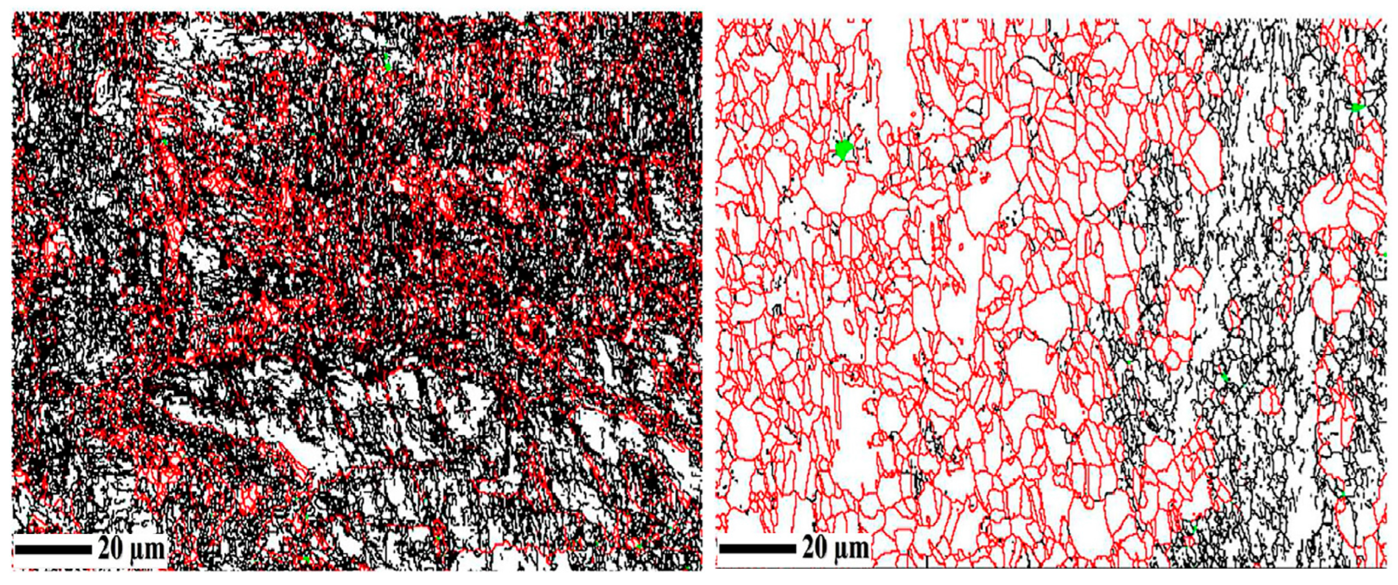
Gussev et al. [66] conducted in situ tensile tests and performed EBSD analysis on the neutron-irradiated 304L steel samples. Microstructural changes during deformation were captured and analyzed. The difference between irradiated samples and nonirradiated samples is that, in irradiated samples, during the initial stages of deformation, hot spots were observed with high local misorientations. Also, grain reference orientation deviation (GROD) increased rapidly compared to the nonirradiated sample, as shown in Figure 12.
Tanaka et al. [67] accurately determined the crystal orientation and pattern center by using a pattern matching approach with HR-EBSD. Even with the presence of image binning, noise, and optical distortion, the authors achieved results with high precision and accuracy with errors less than 10−5 of the pattern width and <0.01° for orientation. The authors listed some of the limitations like image sensor distortion and lens distortion, solving, which we can further improve, the precision of the output.
Tripathi et al. [68] varied the accelerating voltage and compared the spatial resolution values of Mg (light metal—low atomic number) and tungsten (heavy metal—higher atomic number) using EBSD. The authors varied the voltage from 5 to 30 keV. A straight high-angle grain boundary, parallel and perpendicular to the tilt axis, was chosen for study. The results concluded that in the Mg specimen, 240 nm lateral resolution was obtained for 5 keV, whereas 3500 nm was observed when the voltage was raised from 15 to 30 keV. For the tungsten specimen at 5 keV, a 60 nm resolution was observed.
Koko et al. [69] analyzed the in situ crack growth of silicon wafer at the micron scale using HR-EBSD. In this work, the authors developed a novel method to quantify the energy released from the crack during testing, without using any sort of simulation. The results were in line with the standard crack propagation values. The authors suggested that HR-EBSD is a promising method to study how cracks develop and behave under loading in smaller structures like coatings and inclusions.
Britton et al. [70] analyzed the relationship between lattice rotation, pattern shift measurement precision, and the accuracy of measuring elastic strain using HR-EBSD. Observations were made by analyzing diffraction patterns, the quantitative data of the modulation transfer function (MTF), and simulations. The authors concluded that exposure time was the major factor influencing sensitivity, and the strain resolution was increased using software including high bit-depth images. Measurement accuracy and numerical image shift precision were found to be linearly related. The authors provided useful insights into optimizing HR-EBSD so that accurate measurements can be obtained by varying exposure time and bit-depth considerations.
Tong et al. [71] studied the effect of pattern overlap on the accuracy of HR-EBSD measurements. The authors concluded that HR-EBSD measurements were accurate up to an 18 nm distance from the grain boundary. Within the proximity limit to the grain boundary, the pattern overlap affected the elastic strain measurements. The authors suggested that to obtain precise measurements, the identification of peak height and mean angular error is important. From statistical analysis, the authors concluded that the accuracy and precision of the HR-EBSD results are not greatly impacted when two grains overlap in the vicinity of a grain boundary.
Jun et al. [72] studied the deformation behavior of copper samples under uniaxial tension using HR-EBSD. From the results, they concluded that different orientations had different dislocations; <110> grains revealed dislocation cells, and <001> and <111> exhibited dislocation bands. As deformation increased, high GNDs tended to combine into hard grains with high Taylor factors, and strain differences between grains might get absorbed into these hard grains. The authors also suggest that further analysis needs to be performed on the correlation between deformation behavior, dislocation networks, and grain orientation. A similar study was performed by Guo et al. [73], where they subjected copper samples to uniaxial tension and studied the plastic deformation using HR-EBSD measurements. HR-EBSD data were also used to analyze GND density.
Guo et al. [74] subjected titanium to deformation under low strain conditions and studied the grain boundaries and slip bands. From the results, the authors observed blocked slip bands with and without stress concentration and slip transfer. From HR-EBSD measurements collected during slip transfer, the authors concluded that there is no significant stress intensity at the intersection of slip bands. They also found that blocked slip bands induced stress concentration and the measurements showed stress intensity for wrong slip plane alignments.
Andani et al. [75] investigated the Hall–Petch effect in Mg alloys, mainly concentrating on the micro-Hall–Petch coefficient values for the prismatic slip. Utilizing an innovative experimental method, they initiated prismatic slip bands in Mg-4Al at low-stress levels. HR-EBSD played a crucial role in measuring residual stress and calculating resolved shear stress ahead of blocked prismatic slip bands at different grain boundaries. The study revealed significant variation in prismatic values, which were nearly three times larger than those for the basal micro-Hall–Petch.
Kalácska et al. [76] studied the structural changes in copper single crystal after indentation using the HR-EBSD technique and focused ion beam (FIB) slicing. From the results, it was concluded that at some regions, antisymmetric stress levels were observed, and external deformation was majorly driven by the dislocations and movement of lattice defects. The authors suggested that the study related to external deformation on crystalline materials is also important.
Vermeij et al. [77] proposed a new method using both HR-EBSD and finite-strain high angular digital image correlation (IDIC). Dynamically simulated patterns were validated using this method and it showed that for medium and small strains, the rotation and strain errors were below 10−5, and for high strains, they were three times below 10−5, which was achieved even in the presence of 2% image noise.
Johannes et al. [78] studied the deformation evolution of tungsten micro-cantilever subjected to micromechanical testing using in situ HR-EBSD. FIB milling and nanoindentation were used for the study. GNDs and stresses were determined, and elastic strains and strain gradients were measured using EBSD. It was concluded that along the cantilever, the width stresses changed from tensile to compressive.
Vilalta et al. [79] worked on InAlN thin films and examined the lattice rotation, lattice strain, and dislocation densities using ECCI and HR-EBSD. From the results, it was concluded that using both ECCI and HR-EBSD, the precise finding of strain gradients was possible and successfully separated different dislocation densities. Compared to tilts that were perpendicular to the plane, the rotations within the plane were more significant, which in turn suggests that there was a higher density of mixed and edge dislocations.
Britton et al. [80] conducted two case studies to investigate the deformation behavior of copper and steel using HR-EBSD. The authors concluded that compared to Hough transform-based EBSD, HR-EBSD provides better angular resolution and precise measurements. This is possible because HR-EBSD uses cross-correlation-based image analysis.
Farhan et al. [81] studied (001)-oriented strontium titanate (STO) through Berkovich indentation using HR-EBSD and etch-pit analysis. The authors were able to identify various pop-in events and dislocation densities which are dependent on depth, and elastic strain fields in STO along <110> were noted. The authors concluded that by using both HR-EBSD and etch-pit analysis, it is possible to determine and understand dislocation dynamics and indentation effects.
Jackson et al. [82] analyzed how kinematically and dynamically simulated patterns change the accuracy and precision of HR-EBSD measurements. From the results, it was concluded that at low levels of distortion, dynamic patterns show greater precision and accuracy, whereas kinematic patterns perform well at high levels of tetragonality. Strain determination and low noise are some benefits of dynamic patterns.
Eskandari et al. [83] studied the effect of dynamic impact loading on Mn-steel using HR-EBSD. The authors concluded that high-strain deformation led to the formation of adiabatic shear bands (ASBs) and plastic deformation mechanisms. Different phases were identified and analyzed, deformation twins were identified adjacent to ASB, and strain-induced phases were noticed in neighboring grains.
Table 1 summarizes the findings of different EBSD investigations conducted by multiple authors using a variety of equipment. Table 1 shows that multiple authors employed EBSD to investigate several factors such as grain size, phases, deformation behaviors, and texture analysis, all of which are significant in understanding the mechanical properties of materials. Knowledge and comparing these results allow for the identification of trends or inconsistencies under different circumstances, resulting in a greater knowledge of the materials’ behaviors.
Accurate EBSD analysis is only achievable if the sample preparation is flawless. Smirnov et al. [84] studied the specimen preparation methods of metal matrix composites (MMCs) for EBSD analysis. The authors came up with six steps of specimen preparation, following which they achieved enhanced EBSD measurements. The authors concluded that after the final step of polishing, the height difference between the matrix and reinforcement particle should not exceed 2 µm for enhanced EBSD measurements near reinforcements. The sample preparation process involves a series of steps designed to unveil the microstructural details of the specimen. The detailed procedure is as follows: The initial step involves mounting the specimen using Bakelite or epoxy resin to attain proper stability for the next steps [46]. The second step is to subject the sample to a traditional polishing or grinding process which removes irregularities on the surface and provides a clear path for a finer polishing process. For grinding, grit papers of different grades can be used, and for polishing, Al2O3 or diamond paste can be used [11]. The third and final step involves a final polishing using either vibratory (employed to reduce further surface roughness) [85] or electrolytic polishing (removes surface debris and provides mirror finish) [42] or colloidal silica polishing (provides a smooth surface and fine polishing) [86]. Each material needs to be polished as per its reflective property and complexity; studies performed by various researchers on the sample preparation methods are presented in Table 2.
Table 2 shows that in the majority of the cited studies, the first step of polishing is mechanical polishing or grinding to achieve a smooth initial surface, and the final polishing is conducted with colloidal silica or diamond paste or an electrolyte solution to achieve a finer and desired surface finish. Many of the authors primarily employed the electropolishing approach to eliminate residual damage or contaminants. Depending on the required surface properties, polishing procedures must be selected. Initially, only mechanical polishing was utilized, but in recent years, more advanced processes like ion milling and electropolishing have been used to provide a finer and better surface quality with little material degradation. Also, various materials require different polishing processes. For example, electropolishing is appropriate for stainless steel and metallic specimens, but other materials, such as single crystal silicon, require particular treatment, such as immersing them in HF solution. Depending on the material hardness, equipment such as vibratory polishers or ion milling can be utilized.
Operating parameters play an important role in any analysis instrument. Different working parameters used by various authors are presented in Table 3. Over the years, there has been a trend of raising the voltage while decreasing the current to improve signal-to-noise ratios and minimize beam damage. As shown in Table 3, the important working parameters in EBSD are the voltage, probe current, tilt angle, step size, and working distance. Many researchers have played around with these parameters and tried to finalize the optimum working parameters for EBSD analysis. Voltage (keV) determines the energy and depth of the penetration of the electrons. The majority of the authors opted for the 20 keV values which provide a proper balance between the energy and depth. Similarly, the tilt angle is most important as it helps in capturing maximum signals by minimizing the shadows. Most of the researchers suggested that a tilt angle of 70° provides better resolution and clarity in observing Kikuchi bands. Researchers have used different probe current (nA) values for their respective studies, which provide a good relationship between the sample and signal strength. Similarly, the vacuum level was varied, which shows the adaptiveness of the EBSD system at different atmospheric conditions. So, there is a need to tailor this approach of setting vacuum and probe current values to set the optimal signal strength without reducing the structural integrity of the sample. Scan step size is another important parameter in EBSD analysis. A larger step size might be efficient, but it fails to provide detailed results, whereas a smaller step size can provide greater granular inspection views, which are helpful for material characterization. Depending on the resolution and clarity of the output, the working distance is altered, and it varies from material to material, based on the size and shape of the material. Researchers prefer to play around with these working distance values for better clarity and precision in their output, and this approach is similar in the case of magnification parameters as well.

3. Conclusions

This review paper highlights the significance of EBSD parameters, how they must be customized to obtain exact microstructural images, and how researchers could alter these parameters to gain a better knowledge of material behavior and EBSD operation.
A limited study was focused on grain boundary characterization, the stored energy of materials, and the crystallographic orientation of grains or textures utilizing the advanced capabilities of the EBSD technique. There exists a need for more elaborate investigations to unlock the full potential of EBSD in understanding these critical aspects of material microstructures.
Another significant gap concerns the limited exploration of the direction of the reference plane in EBSD analysis. The literature reveals that detailed investigations into the optimal choice of the reference plane are missing. A more complex understanding of this factor is crucial for achieving the best possible results in EBSD analyses, emphasizing the need for further research in this domain.
A considerable literature gap is observed in the realm of multi-phase material characterization using EBSD. The existing body of work provides only limited insights into the application of EBSD in the analysis of materials with multiple phases. Closing this gap requires dedicated research efforts to unveil the full potential of EBSD in elucidating the complex microstructures inherent in multi-phase materials. Addressing these gaps through future research endeavors will undoubtedly contribute to advancing our understanding of materials at the microscale and harnessing the full capabilities of EBSD for comprehensive characterization studies.
HR-EBSD is a versatile method for studying the microstructures of various materials; it can generate data that allow authors to correlate microstructure traits with mechanical properties. Under various loading conditions, HR-EBSD can be an effective tool for studying dislocations, strain localization, and stress distributions.
Combining HR-EBSD with other techniques like ECCI, TEM, and in situ tests improves understanding of the material reaction to deformation and allows authors to conduct in-depth research.
Some of the gaps identified as potential areas for future research include nanoscale deformation investigations, integration with simulation approaches, and machine learning to refine measurements and improve prediction skills. HR-EBSD analysis is lagging in the field of biomaterials, advanced ceramics, and composites.

Author Contributions

Conceptualization, S.D., S.K., S.S. and G.S.; methodology, M.S., N.K. and S.M.A.; software, N.K., G.A. and M.S.; validation, G.S., S.D., S.S. and M.S.; formal analysis, G.S., S.M.A. and G.A.; investigation, S.D., S.M.A., S.S. and M.S.; resources, S.D., S.K., S.S. and G.S.; data curation, S.M.A., G.A. and N.K.; writing—original draft preparation, S.D., S.K., G.S. and S.S.; writing—review and editing, G.A. and M.S.; visualization, G.A., N.K. and S.M.A.; supervision, G.S., S.K., S.S. and M.S.; project administration, S.D.; funding acquisition, S.S., N.K. and G.A. All authors have read and agreed to the published version of the manuscript.

Funding

This research received no external funding.

Data Availability Statement

No new data were created or analyzed in this study.

Conflicts of Interest

The authors declare no conflicts of interest.

References

  1. Wan, Y.; Jiang, W.; Song, M.; Huang, Y.; Li, J.; Sun, G.; Shi, Y.; Zhai, X.; Zhao, X.; Ren, L. Distribution and formation mechanism of residual stress in duplex stainless steel weld joint by neutron diffraction and electron backscatter diffraction. Mater. Des. 2019, 181, 108086. [Google Scholar] [CrossRef]
  2. Zhao, H.; Wynne, B.; Palmiere, E. A phase quantification method based on EBSD data for a continuously cooled microalloyed steel. Mater. Charact. 2017, 123, 339–348. [Google Scholar] [CrossRef]
  3. Bordín, S.P.F.; Ranalli, J.M.; Galván, V.; Castellano, G.; Limandri, S. Combined EBSD, SEM and EDS for detecting minor phases on austenitic stainless steels. Mater. Charact. 2022, 195, 112525. [Google Scholar] [CrossRef]
  4. Adams, B.L.; Kalidindi, S.R.; Fullwood, D.T. Electron backscatter diffraction microscopy and basic stereology. In Microstructure Sensitive Design for Performance Optimization; Elsevier: Amsterdam, The Netherlands, 2013; pp. 341–371. [Google Scholar] [CrossRef]
  5. Higginson, R.L.; Jepson, M.A.E.; West, G.D. Use of EBSD to characterise high temperature oxides formed on low alloy and stainless steels. Mater. Sci. Technol. 2006, 22, 1325–1332. [Google Scholar] [CrossRef]
  6. Ali, A.; Zhang, N.; Santos, R.M. Mineral Characterization Using Scanning Electron Microscopy (SEM): A Review of the Fundamentals, Advancements, and Research Directions. Appl. Sci. 2023, 13, 12600. [Google Scholar] [CrossRef]
  7. Tomaz, R.F.; Santos, D.B.; Camey, K.; Barbosa, R.; Andrade, M.S.; Escobar, D.P. Complex phase quantification methodology using electron backscatter diffraction (EBSD) on low manganese high temperature processed steel (HTP) microalloyed steel. J. Mater. Res. Technol. 2019, 8, 2423–2431. [Google Scholar] [CrossRef]
  8. Janda, A.; Ebenbauer, S.; Prestl, A.; Siller, I.; Clemens, H.; Spoerk-Erdely, P. In-situ high-temperature EBSD characterization during a solution heat treatment of hot-rolled Ti-6Al-4V. Mater. Charact. 2022, 192, 112207. [Google Scholar] [CrossRef]
  9. Gao, W.; Lu, J.; Zhou, J.; Liu, L.; Wang, J.; Zhang, Y.; Zhang, Z. Effect of grain size on deformation and fracture of Inconel718: An in-situ SEM-EBSD-DIC investigation. Mater. Sci. Eng. A 2022, 861, 144361. [Google Scholar] [CrossRef]
  10. Li, W.-S.; Gao, H.-Y.; Nakashima, H.; Hata, S.; Tian, W.-H. In-situ EBSD study of deformation behavior of retained austenite in a low-carbon quenching and partitioning steel via uniaxial tensile tests. Mater. Charact. 2016, 118, 431–437. [Google Scholar] [CrossRef]
  11. Baek, M.-S.; Kim, K.-S.; Park, T.-W.; Ham, J.; Lee, K.-A. Quantitative phase analysis of martensite-bainite steel using EBSD and its microstructure, tensile and high-cycle fatigue behaviors. Mater. Sci. Eng. A 2020, 785, 139375. [Google Scholar] [CrossRef]
  12. Karlsen, M.; Hjelen, J.; Grong, Ø.; Rørvik, G.; Chiron, R.; Schubert, U.; Nilsen, E. SEM/EBSD based in situ studies of deformation induced phase transformations in supermartensitic stainless steels. Mater. Sci. Technol. 2008, 24, 64–72. [Google Scholar] [CrossRef]
  13. Meaden, G.; Dingley, D.J.; Day, A.P. A review of EBSD: From rudimentary on line orientation measurements to high resolution elastic strain measurements over the past 30 years. IOP Conf. Ser. Mater. Sci. Eng. 2018, 375, 012003. [Google Scholar] [CrossRef]
  14. Wilkinson, A.; Britton, T.; Jiang, J.; Meaden, G.; Dingley, D. High Accuracy EBSD-A Review of Recent Applications, Innovations, and Remaining Challenges. Microsc. Microanal. 2011, 17, 402–403. [Google Scholar] [CrossRef]
  15. Biswas, K.; Sivakumar, S.; Gurao, N. Electron Microscopy in Science and Engineering. 2022. Available online: https://link.springer.com/bookseries/15345 (accessed on 3 January 2024).
  16. Fu, J. A general approach to determine texture patterns using pole figure. J. Mater. Res. Technol. 2021, 14, 1284–1291. [Google Scholar] [CrossRef]
  17. Jepson, M.; Higginson, R. The use of EBSD to study the microstructural development of oxide scales on 316 stainless steel. Mater. High Temp. 2005, 22, 195–200. [Google Scholar] [CrossRef]
  18. Feng, S.; Jin, Z.; Du, W.; Han, I.; Lui, A.; Zhou, X.; Shearing, P.R.; Grant, P.S.; Liotti, E. The mechanism of Fe-rich intermetallic compound formation and growth on inoculants revealed by electron backscattered diffraction and X-ray imaging. Mater. Des. 2023, 232, 112110. [Google Scholar] [CrossRef]
  19. Yamasaki, S.; Matsuo, H.; Morikawa, T.; Tanaka, M. Acquisition of microscopic and local stress-strain curves by combination of HR-EBSD and DIC methods. Scr. Mater. 2023, 235, 115603. [Google Scholar] [CrossRef]
  20. Nalepka, K.; Skoczeń, B.; Schmidt, R.; Ciepielowska, M.; Schmidt, E.; Chulist, R. Microstructure evolution in the context of fracture in austenitic steels under complex loads at cryogenic temperatures. Mater. Charact. 2023, 197, 112654. [Google Scholar] [CrossRef]
  21. Mathisen, M.B.; Eriksen, L.; Yu, Y.; Jensrud, O.; Hjelen, J. Characterization of microstructure and strain response in Ti–6Al–4V plasma welding deposited material by combined EBSD and in-situ tensile test. Trans. Nonferrous Met. Soc. China 2014, 24, 3929–3943. [Google Scholar] [CrossRef]
  22. Koko, A.; Tong, V.; Wilkinson, A.J.; Marrow, T.J. An iterative method for reference pattern selection in high-resolution electron backscatter diffraction (HR-EBSD). Ultramicroscopy 2023, 248, 113705. [Google Scholar] [CrossRef]
  23. Chen, Y.-J.; Hjelen, J.; Roven, H.J. Application of EBSD technique to ultrafine grained and nanostructured materials processed by severe plastic deformation: Sample preparation, parameters optimization and analysis. Trans. Nonferrous Met. Soc. China 2012, 22, 1801–1809. [Google Scholar] [CrossRef]
  24. Hu, T.; Ma, K.; Topping, T.; Saller, B.; Yousefiani, A.; Schoenung, J.; Lavernia, E. Improving the tensile ductility and uniform elongation of high-strength ultrafine-grained Al alloys by lowering the grain boundary misorientation angle. Scr. Mater. 2014, 78-79, 25–28. [Google Scholar] [CrossRef]
  25. Lin, Y.; Wu, X.-Y.; Chen, X.-M.; Chen, J.; Wen, D.-X.; Zhang, J.-L.; Li, L.-T. EBSD study of a hot deformed nickel-based superalloy. J. Alloys Compd. 2015, 640, 101–113. [Google Scholar] [CrossRef]
  26. Sankaranarayanan, S.; Sabat, R.; Jayalakshmi, S.; Suwas, S.; Gupta, M. Effect of nanoscale boron carbide particle addition on the microstructural evolution and mechanical response of pure magnesium. Mater. Des. 2014, 56, 428–436. [Google Scholar] [CrossRef]
  27. Shamanian, M.; Mostaan, H.; Safari, M.; Szpunar, J.A. Friction stir modification of GTA 7075-T6 Al alloy weld joints: EBSD study and microstructural evolutions. Arch. Civ. Mech. Eng. 2017, 17, 574–585. [Google Scholar] [CrossRef]
  28. Zhang, L.; Zhao, Z.; Bai, P.; Du, W. EBSD investigation on microstructure evolution of in-situ synthesized TiC/Ti6Al4V composite coating. Mater. Lett. 2021, 290, 129449. [Google Scholar] [CrossRef]
  29. Pirgazi, H.; Ghodrat, S.; Kestens, L.A. Three-dimensional EBSD characterization of thermo-mechanical fatigue crack morphology in compacted graphite iron. Mater. Charact. 2014, 90, 13–20. [Google Scholar] [CrossRef]
  30. Su, M.; Xu, L.; Peng, C.; Han, Y.; Zhao, L. Fatigue short crack growth, model and EBSD characterization of marine steel welding joint. Int. J. Fatigue 2022, 156, 106689. [Google Scholar] [CrossRef]
  31. Sun, Z.; Tsai, S.-P.; Konijnenberg, P.; Wang, J.-Y.; Zaefferer, S. A large-volume 3D EBSD study on additively manufactured 316L stainless steel. Scr. Mater. 2023, 238, 115723. [Google Scholar] [CrossRef]
  32. Hémery, S.; Villechaise, P. In situ EBSD investigation of deformation processes and strain partitioning in bi-modal Ti-6Al-4V using lattice rotations. Acta Mater. 2019, 171, 261–274. [Google Scholar] [CrossRef]
  33. Su, B.; Lin, H.-P.; Kuo, J.-C.; Pan, Y.-T. EBSD investigation on microstructure transformation in low carbon steel during continuous cooling. Can. Met. Q. 2014, 53, 352–361. [Google Scholar] [CrossRef]
  34. Pk, J.; MC, G.; Sharma, S.; Shetty, R.; Hiremath, P.; Shettar, M. The effect of SiC content in aluminum-based metal matrix composites on the microstructure and mechanical properties of welded joints. J. Mater. Res. Technol. 2021, 12, 2325–2339. [Google Scholar] [CrossRef]
  35. Yuan, M.; He, C.; Dong, Z.; Jiang, B.; Song, B.; Guo, N.; Liu, T.; Guo, S.; Pan, F. Effect of Sm addition on the microstructure and mechanical properties of Mg-xSm-0.4Zr alloys. J. Mater. Res. Technol. 2023, 23, 4814–4827. [Google Scholar] [CrossRef]
  36. Wang, B.; Lei, B.-B.; Zhu, J.-X.; Feng, Q.; Wang, L.; Wu, D. EBSD study on microstructure and texture of friction stir welded AA5052-O and AA6061-T6 dissimilar joint. Mater. Des. 2015, 87, 593–599. [Google Scholar] [CrossRef]
  37. Tokita, S.; Kokawa, H.; Sato, Y.S.; Fujii, H.T. In situ EBSD observation of grain boundary character distribution evolution during thermomechanical process used for grain boundary engineering of 304 austenitic stainless steel. Mater. Charact. 2017, 131, 31–38. [Google Scholar] [CrossRef]
  38. Xie, Z.; Han, P.; Liu, Z.; Wang, X.; Shang, C. Influence of initial microstructure on reaustenitization behavior in low alloy steel by in-situ high-temperature EBSD characterization. Mater. Lett. 2023, 350, 134876. [Google Scholar] [CrossRef]
  39. Dourandish, S.; Jahazi, M.; Ebrahimi, G.; Ebacher, L. Influence of eutectic phase precipitation on cracking susceptibility during forging of a martensitic stainless steel for turbine shaft applications. J. Mater. Res. Technol. 2021, 13, 260–270. [Google Scholar] [CrossRef]
  40. Weng, K.; Wang, Y.; Song, Y.; Fan, Y.; Zhao, H.; Hong, Z.; Song, K.; Dong, X.; Guo, C. Optimizing strength and electrical conductivity of Cu–Fe–Ti alloy by pre-aging treatment. J. Mater. Res. Technol. 2023, 26, 2009–2016. [Google Scholar] [CrossRef]
  41. Sutcliffe, J.; Petherbridge, J.; Cartwright, T.; Springell, R.; Scott, T.; Darnbrough, J. Preparation and analysis of strain-free uranium surfaces for electron and x-ray diffraction analysis. Mater. Charact. 2019, 158, 109968. [Google Scholar] [CrossRef]
  42. Baghdadchi, A.; Hosseini, V.A.; Karlsson, L. Identification and quantification of martensite in ferritic-austenitic stainless steels and welds. J. Mater. Res. Technol. 2021, 15, 3610–3621. [Google Scholar] [CrossRef]
  43. Roghani, H.; Borhani, E.; Jafarian, H.R. Effect of a trace amount addition of CuO on aluminum sheet processed by accumulative roll bonding with the common roots and rapid annealing. J. Mater. Res. Technol. 2021, 15, 4257–4271. [Google Scholar] [CrossRef]
  44. Jayashree, P.; Basu, R.; Sharma, S.S. An electron backscattered diffraction (EBSD) approach to study the role of microstructure on the mechanical behavior of welded joints in aluminum metal matrix composites. Mater. Today Proc. 2021, 38, 490–493. [Google Scholar] [CrossRef]
  45. Liu, L.; Huang, W.; Ruan, M.; Chen, Z. Effects of temperatures on microstructure evolution and deformation behavior of Fe–32Ni by in-situ EBSD. Mater. Sci. Eng. A 2023, 875, 145097. [Google Scholar] [CrossRef]
  46. Ball, J.A.; Oddershede, J.; Davis, C.; Slater, C.; Said, M.; Vashishtha, H.; Michalik, S.; Collins, D.M. Registration between DCT and EBSD datasets for multiphase microstructures. Mater. Charact. 2023, 204, 113228. [Google Scholar] [CrossRef]
  47. Conde, F.; Ribamar, G.; Escobar, J.; Jardini, A.; Oliveira, M.; Oliveira, J.; Avila, J. EBSD-data analysis of an additive manufactured maraging 300 steel submitted to different tempering and aging treatments. Mater. Charact. 2023, 203, 113064. [Google Scholar] [CrossRef]
  48. Cai, W.; Sun, C.; Zhang, H.; Wang, C.; Meng, L.; Fu, M. Delving into the intrinsic co-relation between microstructure and mechanical behaviour of fine-/ultrafine-grained TWIP steels via TEM and in-situ EBSD observation. Mater. Charact. 2024, 210, 113780. [Google Scholar] [CrossRef]
  49. Roy, R.; Shaik, A.; Topping, M.; Long, F.; Daymond, M.R. Investigation of fine-scale dislocation distributions at complex geometrical structures by using HR-EBSD and a comparison with conventional EBSD. Mater. Charact. 2024, 207, 113498. [Google Scholar] [CrossRef]
  50. Roy, R.; Topping, M.; Daymond, M.R. In-situ assessment of microscale crack tip fields in zirconium. Int. J. Mech. Sci. 2024, 264, 108812. [Google Scholar] [CrossRef]
  51. Gardner, J.; Wallis, D.; Hansen, L.N.; Wheeler, J. Weighted Burgers Vector analysis of orientation fields from high-angular resolution electron backscatter diffraction. Ultramicroscopy 2024, 257, 113893. [Google Scholar] [CrossRef]
  52. Karunanithi, R.; Prashanth, M.; Kamaraj, M.; Sivasankaran, S.; Kumaraswamidhas, L.; Alhomidan, A.A. Synthesis, characterization, and mechanical behavior of ultra-fine-grained Ti-6Al-5V alloy prepared by mechanical alloying and spark plasma sintering. Mater. Today Commun. 2024, 38, 108228. [Google Scholar] [CrossRef]
  53. Gussev, M.; McClintock, D.; Byun, T.; Lach, T. Recent progress in analysis of strain-induced phenomena in irradiated metallic materials and advanced alloys using SEM-EBSD in-situ tensile testing. Curr. Opin. Solid State Mater. Sci. 2024, 28, 101132. [Google Scholar] [CrossRef]
  54. Li, W.; Wang, Y.; Zhou, X.; Xu, J.; Zhang, R.; Zeng, Y.; Miao, H. Measurement of the pattern shifts for HR-EBSD with larger lattice rotations. Ultramicroscopy 2023, 247, 113697. [Google Scholar] [CrossRef] [PubMed]
  55. Gallet, J.; Perez, M.; Guillou, R.; Ernould, C.; Le Bourlot, C.; Langlois, C.; Beausir, B.; Bouzy, E.; Chaise, T.; Cazottes, S. Experimental measurement of dislocation density in metallic materials: A quantitative comparison between measurements techniques (XRD, R-ECCI, HR-EBSD, TEM). Mater. Charact. 2023, 199, 112842. [Google Scholar] [CrossRef]
  56. Siska, F.; Drozdenko, D.; Mathis, K.; Cizek, J.; Guo, T.; Barnett, M. Three-dimensional crystal plasticity and HR-EBSD analysis of the local stress-strain fields induced during twin propagation and thickening in magnesium alloys. J. Magnes. Alloy. 2023, 11, 657–670. [Google Scholar] [CrossRef]
  57. Ernould, C.; Taupin, V.; Beausir, B.; Fundenberger, J.; Maloufi, N.; Guyon, J.; Bouzy, E. Characterization of a nanopipe dislocation in GaN by means of HR-EBSD and field dislocation mechanics analysis. Mater. Charact. 2022, 194, 112351. [Google Scholar] [CrossRef]
  58. Deal, A.; Spinelli, I.; Chuang, A.; Gao, Y.; Broderick, T. Measuring residual stress in Ti-6Al-4V with HR-EBSD, using reference patterns from annealed material. Mater. Charact. 2021, 175, 111027. [Google Scholar] [CrossRef]
  59. Ernould, C.; Beausir, B.; Fundenberger, J.-J.; Taupin, V.; Bouzy, E. Integrated correction of optical distortions for global HR-EBSD techniques. Ultramicroscopy 2021, 221, 113158. [Google Scholar] [CrossRef] [PubMed]
  60. Ruggles, T.; Kacher, J.; Nowell, M.; Wright, S. HR-EBSD based Characterization of Dislocations in Additive Manufactured 316L Stainless Steel. Microsc. Microanal. 2021, 27, 2670–2672. [Google Scholar] [CrossRef]
  61. Ruggles, T.; Yoo, Y.; Dunlap, B.; Crimp, M.; Kacher, J. Correlating results from high resolution EBSD with TEM- and ECCI-based dislocation microscopy: Approaching single dislocation sensitivity via noise reduction. Ultramicroscopy 2020, 210, 112927. [Google Scholar] [CrossRef]
  62. Sperry, R.; Han, S.; Chen, Z.; Daly, S.H.; Crimp, M.A.; Fullwood, D.T. Comparison of EBSD, DIC, AFM, and ECCI for active slip system identification in deformed Ti-7Al. Mater. Charact. 2021, 173, 110941. [Google Scholar] [CrossRef]
  63. Kalácska, S.; Dankházi, Z.; Zilahi, G.; Maeder, X.; Michler, J.; Ispánovity, P.D.; Groma, I. Investigation of geometrically necessary dislocation structures in compressed Cu micropillars by 3-dimensional HR-EBSD. Mater. Sci. Eng. A 2020, 770, 138499. [Google Scholar] [CrossRef]
  64. Andani, M.T.; Lakshmanan, A.; Sundararaghavan, V.; Allison, J.; Misra, A. Quantitative study of the effect of grain boundary parameters on the slip system level Hall-Petch slope for basal slip system in Mg-4Al. Acta Mater. 2020, 200, 148–161. [Google Scholar] [CrossRef]
  65. Hansen, L.T.; Fullwood, D.T.; Homer, E.R.; Wagoner, R.H.; Lim, H.; Carroll, J.D.; Zhou, G.; Bong, H.J. An investigation of geometrically necessary dislocations and back stress in large grained tantalum via EBSD and CPFEM. Mater. Sci. Eng. A 2020, 772, 138704. [Google Scholar] [CrossRef]
  66. Gussev, M.; Leonard, K. In situ SEM-EBSD analysis of plastic deformation mechanisms in neutron-irradiated austenitic steel. J. Nucl. Mater. 2019, 517, 45–56. [Google Scholar] [CrossRef]
  67. Tanaka, T.; Wilkinson, A.J. Pattern matching analysis of electron backscatter diffraction patterns for pattern centre, crystal orientation and absolute elastic strain determination–accuracy and precision assessment. Ultramicroscopy 2019, 202, 87–99. [Google Scholar] [CrossRef] [PubMed]
  68. Tripathi, A.; Zaefferer, S. On the resolution of EBSD across atomic density and accelerating voltage with a particular focus on the light metal magnesium. Ultramicroscopy 2019, 207, 112828. [Google Scholar] [CrossRef]
  69. Koko, A.; Becker, T.H.; Elmukashfi, E.; Pugno, N.M.; Wilkinson, A.J.; Marrow, T.J. HR-EBSD analysis of in situ stable crack growth at the micron scale. J. Mech. Phys. Solids 2023, 172, 105173. [Google Scholar] [CrossRef]
  70. Britton, T.; Jiang, J.; Clough, R.; Tarleton, E.; Kirkland, A.; Wilkinson, A. Assessing the precision of strain measurements using electron backscatter diffraction–part 1: Detector assessment. Ultramicroscopy 2013, 135, 126–135. [Google Scholar] [CrossRef] [PubMed]
  71. Tong, V.; Jiang, J.; Wilkinson, A.J.; Ben Britton, T. The effect of pattern overlap on the accuracy of high resolution electron backscatter diffraction measurements. Ultramicroscopy 2015, 155, 62–73. [Google Scholar] [CrossRef]
  72. Jiang, J.; Ben Britton, T.; Wilkinson, A.J. The orientation and strain dependence of dislocation structure evolution in monotonically deformed polycrystalline copper. Int. J. Plast. 2015, 69, 102–117. [Google Scholar] [CrossRef]
  73. Guo, Y.; Collins, D.; Tarleton, E.; Hofmann, F.; Tischler, J.; Liu, W.; Xu, R.; Wilkinson, A.; Britton, T. Measurements of stress fields near a grain boundary: Exploring blocked arrays of dislocations in 3D. Acta Mater. 2015, 96, 229–236. [Google Scholar] [CrossRef]
  74. Guo, Y.; Britton, T.; Wilkinson, A. Slip band–grain boundary interactions in commercial-purity titanium. Acta Mater. 2014, 76, 1–12. [Google Scholar] [CrossRef]
  75. Andani, M.T.; Lakshmanan, A.; Sundararaghavan, V.; Allison, J.; Misra, A. Estimation of micro-Hall-Petch coefficients for prismatic slip system in Mg-4Al as a function of grain boundary parameters. Acta Mater. 2022, 226, 117613. [Google Scholar] [CrossRef]
  76. Kalácska, S.; Dankházi, Z.; Groma, I. Systematic study of structural changes in the vicinity of indentation marks with HR-EBSD. IOP Conf. Ser. Mater. Sci. Eng. 2018, 426, 012022. [Google Scholar] [CrossRef]
  77. Vermeij, T.; Hoefnagels, J. A consistent full-field integrated DIC framework for HR-EBSD. Ultramicroscopy 2018, 191, 44–50. [Google Scholar] [CrossRef] [PubMed]
  78. Ast, J.; Mohanty, G.; Guo, Y.; Michler, J.; Maeder, X. In situ micromechanical testing of tungsten micro-cantilevers using HR-EBSD for the assessment of deformation evolution. Mater. Des. 2017, 117, 265–266. [Google Scholar] [CrossRef]
  79. Vilalta-Clemente, A.; Naresh-Kumar, G.; Nouf-Allehiani, M.; Gamarra, P.; di Forte-Poisson, M.; Trager-Cowan, C.; Wilkinson, A. Cross-correlation based high resolution electron backscatter diffraction and electron channelling contrast imaging for strain mapping and dislocation distributions in InAlN thin films. Acta Mater. 2017, 125, 125–135. [Google Scholar] [CrossRef]
  80. Britton, T.B.; Hickey, J.L.R. Understanding deformation with high angular resolution electron backscatter diffraction (HR-EBSD). IOP Conf. Ser. Mater. Sci. Eng. 2018, 304, 012003. [Google Scholar] [CrossRef]
  81. Javaid, F.; Bruder, E.; Durst, K. Indentation size effect and dislocation structure evolution in (001) oriented SrTiO3 Berkovich indentations: HR-EBSD and etch-pit analysis. Acta Mater. 2017, 139, 1–10. [Google Scholar] [CrossRef]
  82. Jackson, B.E.; Christensen, J.J.; Singh, S.; De Graef, M.; Fullwood, D.T.; Homer, E.R.; Wagoner, R.H. Performance of Dynamically Simulated Reference Patterns for Cross-Correlation Electron Backscatter Diffraction. Microsc. Microanal. 2016, 22, 789–802. [Google Scholar] [CrossRef] [PubMed]
  83. Eskandari, M.; Mohtadi-Bonab, M.A.; Zarei-Hanzaki, A.; Odeshi, A.G.; Szpunar, J.A. High-Resolution EBSD Study of Adiabatic Shear Band and Neighboring Grains After Dynamic Impact Loading of Mn-Steel Used in Vehicle Structure. J. Mater. Eng. Perform. 2016, 25, 1611–1620. [Google Scholar] [CrossRef]
  84. Smirnov, A.S.; Belozerov, G.A.; Smirnova, E.O.; Konovalov, A.V.; Shveikin, V.P.; Muizemnek, O.Y. Specimen Preparation for Metal Matrix Composites with a High Volume Fraction of Reinforcing Particles for EBSD Analysis. J. Mater. Eng. Perform. 2016, 25, 2907–2913. [Google Scholar] [CrossRef]
  85. Dyakonov, G.; Zemtsova, E.; Mironov, S.; Semenova, I.; Valiev, R.; Semiatin, S. An EBSD investigation of ultrafine-grain titanium for biomedical applications. Mater. Sci. Eng. A 2015, 648, 305–310. [Google Scholar] [CrossRef]
  86. Abbasi, M.; Kim, D.-I.; Nelson, T.W.; Abbasi, M. EBSD and reconstruction of pre-transformation microstructures, examples and complexities in steels. Mater. Charact. 2014, 95, 219–231. [Google Scholar] [CrossRef]
  87. Serrano-Munoz, I.; Fernández, R.; Saliwan-Neumann, R.; González-Doncel, G.; Bruno, G. Dislocation structures after creep in an Al-3.85 %Mg alloy studied using EBSD-KAM technique. Mater. Lett. 2023, 337, 133978. [Google Scholar] [CrossRef]
  88. Mingard, K.; Roebuck, B.; Bennett, E.; Gee, M.; Nordenstrom, H.; Sweetman, G.; Chan, P. Comparison of EBSD and conventional methods of grain size measurement of hardmetals. Int. J. Refract. Met. Hard Mater. 2009, 27, 213–223. [Google Scholar] [CrossRef]
  89. Fullwood, D.; Vaudin, M.; Daniels, C.; Ruggles, T.; Wright, S.I. Validation of kinematically simulated pattern HR-EBSD for measuring absolute strains and lattice tetragonality. Mater. Charact. 2015, 107, 270–277. [Google Scholar] [CrossRef]
  90. Javaid, F.; Xu, Y.; Bruder, E.; Durst, K. Indentation size effect in tungsten: Quantification of geometrically necessary dislocations underneath the indentations using HR-EBSD. Mater. Charact. 2018, 142, 39–42. [Google Scholar] [CrossRef]
  91. Ernould, C.; Beausir, B.; Fundenberger, J.-J.; Taupin, V.; Bouzy, E. Global DIC approach guided by a cross-correlation based initial guess for HR-EBSD and on-axis HR-TKD. Acta Mater. 2020, 191, 131–148. [Google Scholar] [CrossRef]
  92. Ning, X.; Liang, Y.; Wang, Y.; Ye, F.; Lin, J. A quasi-in-situ EBSD study on the formation and development of η -fiber in 0.15 mm ultra-thin Fe-4.5 wt. % Si sheet. J. Alloys Compd. 2023, 969, 172372. [Google Scholar] [CrossRef]
  93. Seehaus, M.; Korte-Kerzel, S.; Sandlöbes-Haut, S. Influence of Si on the microstructure and C redistribution in martensitic steels. Mater. Des. 2023, 229, 111875. [Google Scholar] [CrossRef]
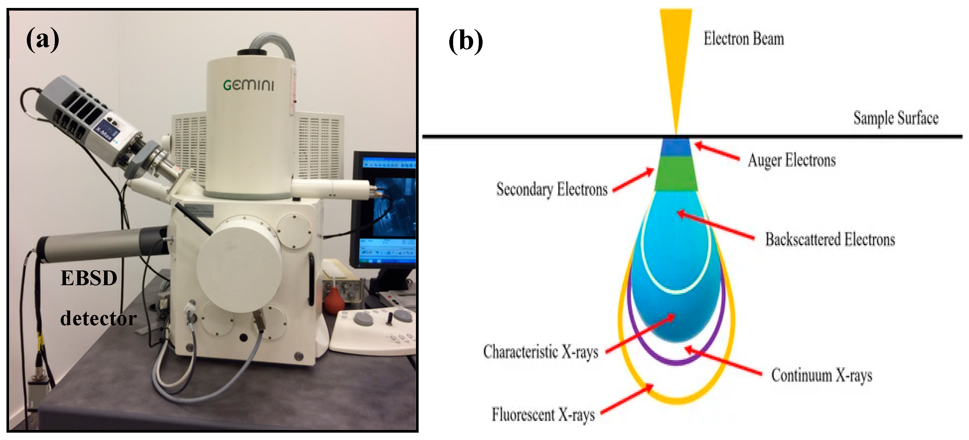
Figure 1. (a) EBSD detector, (b) electron beam interaction with the SEM specimen [6].
Jcs 09 00132 g001
Figure 2. Reconstructed 3D microstructure—crack is shown in black [29].
Jcs 09 00132 g002
Figure 3. IPF showing orientations and loading direction [32].
Jcs 09 00132 g003
Figure 4. (ad) IPF of samples (1–4 wt.% of Sm) subjected to the extrusion process [35].
Jcs 09 00132 g004
Figure 5. OIM images of AA6061-T6 alloy (a) BM, (b) HAZ, (c) TMAZ, (d) NZ [36].
Jcs 09 00132 g005
Figure 6. (ac) IPF and (a1c1) GBCD maps of 3% cold rolled specimens at different heating durations [37].
Jcs 09 00132 g006
Figure 7. Orientation mapping of EBSD analysis displaying (a) band contrast image, (b) phase map showing the presence of M23C6 (blue) and ferrite (red) phases [39].
Jcs 09 00132 g007
Figure 8. IPF map of SRA (left) and SARA (right) subjected to aging at 460 °C for 5 h [40].
Jcs 09 00132 g008
Figure 9. Grain boundary maps SARA subjected to aging at 460 °C for different periods of 2 h (left) and 5 h (right) [40].
Jcs 09 00132 g009
Figure 10. IPF mapping of (a) annealed aluminum, (b) aluminum sample subjected to ARB (6 cycles), (c) AA1050 + CuO composite subjected to ARB (6 cycles) [43].
Jcs 09 00132 g010
Figure 11. (a) Kikuchi pattern quality at loaded configuration. (b) Kikuchi pattern quality at undeformed state (taken from a different grain) [49].
Jcs 09 00132 g011
Figure 12. IPF, KAM, and GROD maps for (a) nonirradiated and (b) irradiated steel specimens at a comparable strain level [66].
Jcs 09 00132 g012
Table 1. EBSD setups in various studies: materials and outcomes comparison.
Sl. No.MaterialMethod of PreparationEBSD (Make and Model)Outcomes from EBSD AnalysisAuthor and Year of PublicationReference
1316 stainless steel-LEO VP 1530 FEG SEM fitted with TSL EBSDElemental presence and phase identification were identified using EBSD analysis, also the internal oxidation effect was identified.Jepson et al., 2005[17]
22-phase WC/Co 10 wt.% binder grades produced by
Sandvik Hard
Materials
EtchingZeiss Supra 40 FEGSEMGrain size was determined and compared results with conventional methods.Mingard et al., 2008[48]
3α-Ti, dual-phase steel, carbides in a superalloy, and Ti-6Al-4V alloyHot rolledJEOL JSM6500F Schottky emission ‘FE-SEM’Local deformation studies were carried out using EBSD.Wilkinson et al., 2010[49]
4Single crystal siliconDynamical simulations causing Bloch wave theory, quantitative
measurements of the detector Modulation Transfer Function (MTF)
JEOL JSM-6500F, patterns were captured using TSL-OIM DC v5.3 software and a Digiview II cameraStrain measurements were measured using EBSD and assessed the precision of the measurements using simulations.Britton et al., 2013[50]
5CGI with a pearlitic matrix-FEI Quanta 450 FEG-SEM. Analysis of EBSD results was performed using TSL software.Crystal orientation was identified using EBSD analysis and it was proved that unlike traditional conclusions, crystal orientation does not have any effect on the crack propagation where the density of graphite particles was higher.Pirgazi et al., 2014[29]
6Ti-6Al-4VAdditive layer manufacturingZeiss Ultra 55 LE FESEM fitted with a NORDIF UF−1000 ultra-fast EBSD detectorEBSD analysis was used to analyze information regarding phase identification, grain morphology, and crystal orientations. Also, with the usage of the new advanced EBSD setup, analyses of diffraction patterns across large areas were made easy.Borlaug et al., 2014[21]
7AA5052-O and AA6061-T6Friction stir weldingBruker e-Flash 1000 probe and data were processed using TSL OIM software (version 5.2)Microstructure, grain, texture, and misorientation studies were performed.Wang et al., 2015[36]
8Nickel superalloyHot compressions (920–1040 °C)JEOL-7001F1 FE-SEM with HKL Channel 5 SoftwareVariation in the fraction of low-angle grain boundaries during the compression process was noted and it was concluded that the discontinuous dynamic recrystallization was the major nucleation mechanism.Lin et al., 2015[25]
9Zircaloy-4 plate-Zeiss Auriga FEG-SEM
with Bruker eFlash EBSD camera
Elastic strains and lattice rotations were measured, and the effect of pattern overlap was analyzed using EBSD.Vivian et al., 2015 [51]
10OFHC polycrystal copper-JEOL JSM 6500-F SEM, TSL EBSD systemGNDs, orientation, and strain dependence were analyzed.Jun et al., 2015[52]
11SiGe-FE-SEM
(Hitachi model 4700, Japan), commercial EBSD setup
Absolute strains and lattice tetragonality were measured and kinematically simulated patterns were validated.David et al., 2015[53]
12Crystalline tungsten materials-High-resolution electron backscatter diffractionGNDs, elastic strain, and strain gradients, and stresses were determined.Johannes et al., 2016[54]
13Si/SiGe-FE-SEM (Hitachi model 4700; Japan), commercial EBSD setupComparison between dynamically and kinematically simulated patterns were analyzed.Brian et al., 2016[55]
14MMC (99.8% Al) as the matrix, SiC particles-TESCAN VEGA II XMU
scanning electron microscope with an OXFORD HKLNordlysF+ detachable device for EBSD
Specimen preparation for better measurements of EBSD was explored.Smirnov et al., 2016[56]
15Mn-SteelHot rolledHitachi SU6600
FE-SEM
equipped with a Nordlys Nano EBSD detector
Samples were subjected to dynamic impact and adiabatic shear bands were analyzed using EBSD.Eskandari et al., 2016[57]
16304 austenitic stainless steelThermomechanical treatmentHITACH SE-4300SE FE-SEM equipped with the orientation imaging microscopy (OIM) systemGrain boundary character distribution evolution during TM and annealing process were analyzed using EBSD.Shun et al., 2017[37]
17Al 7075-T6Rolling, gas tungsten arc welding, Friction stir weldingHitachi SEM equipped with EDS and EBSD was used, HKL CHANNEL5 software was used for visualization and processing of EBSD dataIPF maps were used to analyze the grain orientation and grain size, also low- and high-angle grain boundaries were determined. Kernel average misorientation maps were used to determine the average misorientation.Shamanian et al., 2017[27]
18Polycrystalline--Dynamically simulated patterns were validated using HR-EBSD and IDIC.T. Vermeij and J.P.M. Hoefnagels, 2018[58]
19TungstenEtchingTESCAN MIRA3 SEM, EDAX
TSL DigiView EBSD system, patterns were analyzed using Crosscourt V4.21 (CC4) from BLG Vantage Software
Inc. (Bristol, UK)
Quantification of GNDs under indentations was analyzed using EBSD.Farhan et al., 2018[59]
20CopperSolution heat treatedFEI Quanta 650 g FEG-SEM coupled with a Bruker eFlash HR EBSD detectorComparison between the measurements of conventional EBSD and HR-EBSD was made.Britton et al., 2018[60]
21Interstitial free (IF) steelvacuum-melted, hot rolled
and cold rolled, annealing
SEM: JEOL
7001F/7100F,
EBSD: EDAX DigiView camera
Elastic strain determination, crystal orientation, and pattern matching analysis was performed.Tomohito Tanaka and Angus J. Wilkinson, 2019[61]
22Pure magnesium and tungstenSolution heat treatedEBSD data were collected using Zeiss Crossbeam 1540 focused ion beam
system with a 464 × 464 pixel Hikari camera
Accelerating voltage was varied and spatial resolution results were studied.Abhishek Tripathi and Stefan Zaefferer, 2019[62]
23Crystalline materialAnnealedFEI Tecnai F30 TEM, TESCAN MIRA3
SEM equipped with an EDAX/TSL Hikari highspeed detector
Lattice distortions, GNDs were measured and compared results with TEM.Ruggles et al., 2019[63]
24Ti-6Al-4V-JEOL 6100 SEM equipped with an EBSD and EDAXLattice rotations concerning initial orientations were identified.Hemery et al., 2019[32]
25Metallic specimensPolishingZeiss EVO MA10 SEM fitted with an LaB6 source. Digi view 3 high-speed camera was used to capture data and EDAX OIM software was used for analysis purposes.Crystallographic orientation, twinning mechanisms, and grain boundary analysis were performed using EBSD, and the origin of stress within the metal specimen was identified.Sutcliffe et al., 2019[41]
26Nuclear-grade AISI 304L austenitic stainless steelCold-workingFEI VERSA 3D
SEM equipped with an Oxford Instruments Nordlys-2 EBSD system
Lattice rotations, phase instability, twinning, and misorientation evolution were analyzed.M.N. Gussev and K.J. Leonard, 2019[64]
27TantalumHardeningFEI Helios Nanolab 600 SEM using OIM DC 7.2 software
(EDAX-TSL)
GNDs and back stress were determined and correlated with mechanical properties.Hansen et al., 2019[65]
28Self-ion irradiated tungstenAnnealed, grindingZeiss Merlin field emission gun SEM, Atomic force microscopy (AFM), HR-EBSDDeformation behavior of irradiated tungsten material was analyzed using HR-EBSD.Suchandrima et al., 2020[66]
29Crystal copper micropillarsAnnealed,
hardening
Scanning transmission electron microscopy (STEM), FEI Quanta 3D, and Tescan Lyra3GND structures during deformation were analyzed using 3D HR-EBSD.Szilvia et al., 2020[67]
30Mg-4AExtrusionFEI Quanta 650 ESEM equipped with an integrated Oxford AZtec EDS and EBSD system, analysis was performed using CrossCourt4 (CC4) software package developed by BLG VantageEffect of grain boundary on the slip system was studied in detail.Andani et al., 2020[68]
31Al6061 + SiCStir castingFEI Quanta-3D FEG SEM equipped with an EBSD system from EDAX-TSLDislocations, misorientations, and grain boundary fractions were analyzed and correlated with effect on the mechanical properties of welded and age-hardened composites.Pk et al., 2021[34]
32Ti6Al4V + TiC compositeTiC coating using laser claddingHitachi S-3400N SEM equipped with an HKL-EBSD systemFrom EBSD analysis, the average grain size was measured, and it decreased in coated composites. Texture strength was also enhanced.Lizheng et al., 2021[28]
33FDX 27 (UNS S82031) TRIP DSSMechanical and electrolytic polishingZEISS Gemini SEM 450 equipped with a Symmetry S2 EBSD. Captured data were analyzed using AZtecCrystal 1.1 software.A six-step procedure was developed by authors for the EBSD analysis to identify the phases with similar lattice structures.Baghdadchi et al., 2021[42]
34AA1050 + CuOAccumulative roll bondingFE-SEM with EBSD was used to capture data and the data were analyzed using orientation imaging microscopy (version 7.3.1) analysis software and ImageJ software.Grain size analysis and texture analysis were performed using EBSD.Roghani et al., 2021[43]
35Al 6061 + SiCStir casting, TIG welding, and age hardeningFEI Quanta-3D FEG (field emission gun) SEM equipped with an EDAX-TSL EBSD- systemTexture analysis, misorientations, and grain size analysis were performed using EBSD.Jayashree et al., 2021[44]
36Crystalline material-HR-EBSD/HR-TKDOptical distortion in EBSD Lense was studied in detail.Ernould et al., 2021[69]
37Ti-7AlForged, annealed, quenchedTescan Mira III FEG-SEMSlip system activity and long-range rotation gradients were analyzed.Ryan et al., 2021[70]
38Ti-6Al-4VAnnealedHitachi SU70 FEG-SEM, HR-EBSDResidual stress and elastic strain were measured using reference patterns using EBSD.Andrew et al., 2021[71]
39GaN-FEG-SEM Jeol
F100
Elastic strains and rotation fields in defected area were assessed.Ernould et al., 2022[72]
40Mg-Al alloy-JEOL JSM-7001F and JSM-7900F FE SEM are both equipped with Oxford EBSD (Nordlys Nano and Symmetry) and EDS. Captured data were analyzed using Oxford Aztec 4.2 and TSL OIM 8.0 software to produce orientation maps, pole figures, and elemental profile.Orientation relationship between the grains and phase identification was performed using EBSD.Kang et al., 2022[73]
41Marine steelWeldingSEM (Sigma 300, Carl Zeiss) and EBSD device (Bruker, Ltd.). Images were post-processed by Esprit 2.1 software (Bruker, Ltd.).Phase identification, analysis of average grain size using IPF mapping, grain size reduced because of the heating effect during welding operation. Crack growth analysis using KAM and IPF mappings, short crack propagation was observed to be influential, and transgranular and intergranular fractures were observed. Molin et al., 2022[30]
42Single crystal silicon-Carl Zeiss
Merlin field emission gun scanning electron microscope (FEG-SEM)
Load fracture resistance of microstructural features was analyzed.Koko et al., 2022[74]
43316L stainless steelAdditive manufacturingSEM (ZEISS crossbeam XB 1540), EDAX Hikari camera, (ELAVO 3D) system was used for EBSD analysis. 2D and 3D EBSD analysis was performed using OIM Analysis 8.6.0101 and QUBE ver. 2.0.25.Nucleation sites, texture capturing, and grain orientations were identified and results were analyzed. Sun et al., 2023[31]
44Duplex stainless steel and silicon-Carl Zeiss Merlin field emission gun scanning electron microscope (FEG-SEM), Bruker eFlash CCD camera. Patterns were analyzed using MATLAB (XEBSD).Optimization of reference pattern position which improves the HR-EBSD precision.Koko et al., 2023[22]
45Mg alloy + Sm-JEOL JSM-7800 F SEM with EBSD setup and HKL-channel 5 software was used for analysis purposesAverage grain size and texture intensity were determined and correlated with the mechanical properties of the alloys prepared.Yuan et al., 2023[35]
46Low alloy steelHeat treatedTESCAN CLARA GMH (FE-SEM) equipped with Oxford Instruments SYMMETRY S2 EBSD detector. Captured data were analyzed using AZtecCrystal software.Austenite and martensitic phases were identified using EBSD analysis, also the dislocation densities were analyzed using GND.Xie et al., 2023[38]
47Outokumpo 2101 lean duplex
stainless steel
Hot rolledZeiss Supra 55VP SEM, TEM JEOL 2100 LaB6Dislocation densities were measured and compared the results with TEM and ECCI.Gallet et al., 2023[75]
48Cu-0.75Fe-0.35Ti (wt.%) alloySolutionizing, cold rolling, and agingHelios G4 CX (SEM) with an HKL-EBSD systemRecrystallization effect, formation of low- and high-angle grain boundaries, and density of GNDs were analyzed using EBSD and results are correlated with mechanical properties.Weng et al., 2023[40]
49Plagioclase
Olivine
-FEI
Quanta 650 FEG-SEM, NordlysNano
EBSD detector and AZtec software
Precision of HR-EBSD measurements can be improved by using WBV method.Joe et al., 2023[76]
50Magnesium alloy-FEI Quanta and Zeiss Auriga SEMComparison of shear stress and spatial activity of slips was made between HR-EBSD and simulations.Siska et al., 2023[77]
51Fe-4.5 wt.% Si sheetHot rolled, cold rolled, and annealedFE-SEM (Zeiss Gemini450) equipped with an EBSD system, collected data were processed using software HKL-Channel 5Texture analysis of cold rolled, hot rolled, and annealed specimens was performed, and IPF and KAM maps were also analyzed. Improved mechanical properties after being subjected to different treatments were justified by relating them to changes in orientation, grain size, and texture of samples.Ning et al., 2023[78]
52Martensitic steelsCasting and rollingDual-Beam-FIB Helios Nanolab 600i (FEI), Hikari detector (EDAX), and analysis was performed using MTEX 5.7.0 toolbox based on MatlabThe Greninger–Troiano orientation connection between austenite and martensite was discovered using EBSD measurements in both FeNiC and FeNiCSi alloys.Seehaus et al., 2023[79]
53Austenitic stainless steel-JEOL 7000 FEG-SEM equipped with a Nordlys EBSD detector, indexing was performed using software AztecComparison between DCT and EBSD methods was made and concluded that for multi-phase characterization, the DCT method is the best.Ball et al., 2023[46]
54Maraging 300 steel powderAdditive manufacturing, tempering, and agingSEM FEI Quanta 650 equipped
with a Schottky FEG coupled with a high-speed EBSD system, processed using MTEX software
Grain boundary orientation was majorly studied and results are related to the performance of the sample.Conde et al., 2023[47]
55Zr foil-A NewTech MT1000 in situ tensile rig mounted in an FEI Nova Nano
SEM, Bruker eFlash EBSD detector
Elastic strains at microscopic scale were investigated with the help of HR-EBSD.Roy et al., 2023[80]
56TWIP steelAir-filled vacuum furnace (melting), hot rolled, cold rolled, annealedField emission electron microscopy (FEI)
Quanta 650 FEG SEM), equipped with an EBSD probe (EDAX-TSL)
Density of GNDs was analyzed and correlated with mechanical properties.Wang et al., 2024[81]
Table 2. Polishing methods employed for EBSD analysis on diverse materials.
Author & YearMaterialPolishing MethodReference
Jepson et al., 2005316 stainless steel
  • Specimens were subjected to grinding followed by polishing with diamond paste (6 to 1 μ)
  • Final polishing was made with colloidal silica for 25 min
[17]
Mingard et al., 20082-phase WC/Co 10 wt.% binder grades produced by Sandvik Hard Materials
  • Thermosetting resin was used to mount all the samples and standard metallographic techniques were utilized to get a surface finish
[48]
Wilkinson et al., 2010α-Ti, dual-phase steel, carbides in a superalloy, and Ti-6Al-4V alloy
  • α-Ti: 2500 grit paper was used to grind the samples, then repeatedly polished using colloidal silica and hydrogen peroxide mixture in a ratio of 5:1
  • Dual-phase steel: Mechanical grinding and polishing was performed. Again, electropolished with the help of Struers A8 electrolyte.
  • Ti-6Al-4V alloy: colloidal silica was used to make polishing for the grinded failed samples.
[49]
Britton et al., 2013 Single crystal silicon
  • Soaked in 40% HF and cleaned with acetone to remove contamination
[50]
Borlaug et al., 2014Ti-6Al-4V
  • Alcohol–silica suspension (0.4 μm), and the addition of H2O2 (hydrogen peroxide) for mirror finish
  • Polishing time—10 min, RPM—150
  • Ultrasonic cleaning—10 min
  • Plasma cleaning—5 min
  • Ion milling and sputtering for 5 min
[21]
Pirgazi et al., 2014CGI with a pearlitic matrix
  • Mechanical polishing was performed using (oxide polishing suspension), also silica was used as abrasive particles (0.04 μm)
  • Final polishing was made using an automatic polishing machine
[29]
Lin et al., 2015Nickel superalloy
  • Samples were initially ground to 0.07 to 0.08 mm thickness
  • Followed by twinjet electropolishing using ethanol and perchloric acid (9:1)
[25]
Vivian et al., 2015 Zircaloy-4 plate
  • Zircaloy-4 plate was cut and 4000 grit paper was used to grind. Colloidal silica and hydrogen peroxide solution in the ratio of 5:1 was used to polish for 4.5 h.
  • Also, methanol and perchloric acid in the ratio of 9:1 were used for electropolishing cooled with liquid nitrogen to obtain good quality EBSD patterns.
[51]
Jun et al., 2015OFHC polycrystal copper
  • 85% phosphoric acid was used to immerse the specimens to do electropolishing, which acts as an anode and copper sheet as a cathode.
[52]
Johannes et al., 2016Crystalline tungsten materials
  • Edge of a tungsten single crystal specimen was mechanically and electrochemically polished and micro-cantilevers were fabricated on it using FIB milling.
[54]
Smirnov et al., 2016MMC (99.8% Al) as the matrix, SiC particles
  • Diamond suspension was utilized to conduct mechanical polishing. Then, polished again using colloidal oxide suspension.
  • Electrolyte consisting of 10% HClO4 and 90% CH3COOH was used to achieve electrochemical polishing of SiC50/Al
[56]
Eskandari et al., 2016Mn-Steel
  • Polish was conducted mechanically using SiC papers
  • Further, residual strain on the surface was eliminated by electropolishing
[57]
Shun et al., 2017304 austenitic stainless steel
  • Specimens were electropolished with ethanol and perchloric acid (10%) at −0.15 °C and 25 V
[37]
Shamanian et al., 2017Al 7075-T6
  • Initial polishing was performed using grit papers and diamond polishing paste
  • Final polishing was performed using VibroMet 2 Vibratory polisher (Buehler) with colloidal silica slurry (50 nm) for about 6 h
[27]
Farhan et al., 2018Tungsten
  • Electropolishing was conducted for 20–30 s after grinding the specimens
[59]
Britton et al., 2018Copper
  • Initially polished with 4000 SiC, final polishing was performed using Gatan PECSII argon ion polisher
[60]
Hemery et al., 2019Ti-6Al-4V
  • Initially, specimens were subjected to grinding with 4000 SiC-grade paper
  • Pre-polishing was performed using a diamond paste of 9 μ
  • Final polishing was made using colloidal silica (0.04 μm) and H2O2 (10%)
[32]
Sutcliffe et al., 2019Metallic specimens
  • SiC P360 to P4000-grade papers were used for the first stage of polishing
  • Finally, samples were ultrasonically cleaned and subjected to electropolishing
[41]
M.N. Gussev and K.J. Leonard, 2019Nuclear-grade AISI 304L austenitic stainless steel
  • The flat sides of all specimens were subjected to polishing with diamond lapping paper (3 mm), again electropolished at 30 V DC for 10 s with the help of Struers A2 electropolishing solution
[64]
Hansen et al., 2019Tantalum
  • After fine polishing, final samples thickness was maintained well below 1 mm
[65]
Tomohito Tanaka and Angus J. Wilkinson, 2019Interstitial free (IF) steel
  • The material was cut and colloidal silica was used for polishing the material
[61]
Abhishek Tripathi and Stefan Zaefferer, 2019Pure magnesium and tungsten
  • Diamond suspension was utilized for polishing
  • Ethanol and ortho-phosphoric acid in the volume ratio of 5:3 were used for electropolishing for 30 min
[62]
Ruggles et al., 2019Crystalline material
  • The prepared disk was jet polished using twin-jet electropolisher with the help of 90% of ethanol solution and 10% of perchloric
[63]
Suchandrima et al., 2020Self-ion irradiated tungsten
  • Colloidal silica and diamond paste were used to conduct polishing. And electropolishing was utilized in a 1% NaOH aqueous solution electrolyte.
[66]
Szilvia et al., 2020Crystal copper micropillars
  • A small area was subjected to polishing by a beam with FIB settings, which are similar to the pillar slicing parameters
[67]
Andani et al., 2020Mg-4Al
  • Samples were mechanically polished and final polishing was performed using 0.05 μm polycrystalline diamond solution
[68]
Baghdadchi et al., 2021FDX 27 (UNS S82031) TRIP DSS
  • Initially, samples were subjected to a grinding operation
  • One set of samples was subjected to mechanical polishing with diamond (3 and 9 µm) and alumina (0.05 µm) suspensions
  • The second set of samples was subjected to electrolytic polishing with 450 mLH2SO4, 300 g distilled water, 150 g citric acid, and 600 mL H3PO4
[42]
Roghani et al., 2021AA1050 + CuO
  • Samples were initially polished with sandpapers, followed by electropolishing with CH3OH (70%) and HNO3 (30%) for 20 s at −30 °C and 15 V
[43]
Pk et al., 2021Al6061 + SiC
  • Samples were electropolished with methanol and perchloric acid (20%) at −10 °C and 15 V
[34]
Dourandish et al., 2021Martensitic stainless steel
  • Specimens were polished with SiC (600–1200-grade paper)
  • Final polishing was performed using diamond paste (1 μm) and etched with Villela solution for 25 s
[39]
Ryan et al., 2021Ti-7Al
  • 600, 800, and 1200 grit paper of SiC was used to polish the simples. Diamond paste with colloidal silica mixture and 30% of hydrogen peroxide in the ratio of 4:1 was used.
  • Again, specimens were electropolished for 2.5 min using 300 mL methanol, 30 mL perchloric acid, and 200 mL butanol
[70]
Andrew et al., 2021Ti-6Al-4V
  • All specimens were hot-mounted in conductive resin and specimens were subjected to vibratory polish of 0.05 μm
[71]
Koko et al., 2022Single crystal silicon
  • Its pre-polished surface was parallel to (001). The loaded edges of the sample were manually abraded, which are parallel to (110).
[74]
Molin et al., 2022Marine steel
  • Electropolishing was performed using a 10% perchloric acid alcohol solution
  • Polishing time 10–15 s
  • Voltage 20–30 keV
[30]
Sun et al., 2023316L stainless steel
  • QATM Saphir X-Change was used to polish the specimens for EBSD analysis
[31]
Koko et al., 2023Duplex stainless steel and silicon
  • For polishing of specimens, SiC grit papers were used (240 to 4000 grit)
  • Followed by polishing with diamond paste (9 to 1 μ) and colloidal silica
  • Polishing time—120 min
  • RPM—50, Load—5 N
  • Followed by ultrasonic cleaning using ethanol for 20 min
[22]
Itziar et al., 2023Al-3.8 wt.% Mg alloy
  • Initial polishing was performed using grit papers and diamond slurry (3 µm)
  • 20 nm colloidal silica was used for final polishing.
  • Struers LectroPol-5 machine was used for surface finishing
[87]
Seehaus et al., 2023Martensitic Steels
  • Samples were polished initially with diamond suspension
  • Finally, electropolishing was performed using A2 (Struers) electrolyte
[79]
Ball et al., 2023Austenitic stainless steel
  • Samples were polished using colloidal silica (0.04 µm) and then electropolished with ethanol and perchloric acid (80:20) at 20 °C, 15 V for 20 s.
[46]
Conde et al., 2023Maraging 300 steel powder
  • Samples were polished with grit papers (80–1500), followed by diamond paste polishing with 3 and 1 µm.
[47]
Roy et al., 2023Zr foil
  • 2400 grit paper was used to grind the samples, then samples were subjected to electropolishing with 5% perchloric acid in methanol at −20 °C
[80]
Gallet et al., 2023Stainless steel
  • 1 μm diamond solution was used to perform polishing after grinding the samples mechanically.
  • Electropolishing was conducted using A2 electrolyte with Lectropol 5 device to avoid strain hardening of the surface.
[75]
Siska et al., 2023Magnesium alloy
  • Initial grinding with grit papers up to 4000, followed by diamond paste polishing (3 and 1 µm). Final surface treatment was performed using Ar ion-milling Leica EM RES102 system.
[77]
Wang et al., 2024TWIP steel
  • Defects were reduced by grinding specimens and grinded until surface became smooth and flat
  • 10% perchloric acid–ethanol (HClO4-C2H5OH) electrolyte solution was used for electropolishing in an ice water bath. It was carried out for 40–60 s at a voltage of 20 V to remove the damaged surface.
[81]
Table 3. Variations in parameters for effective operation and analysis in EBSD studies.
Author, Year, and ReferenceParameters Used for EBSD Analysis
Voltage (keV)Specimen Tilt Angle/(°)Probe Current (nA)Vacuum (Pa)EBSD Scan Step Size (μm)Working Distance (mm)Aperture (μm)MagnificationReference
Jepson et al., and 20052070---Varied-100[17]
Mingard et al., and 20081570--0.050.05–0.5605000, 10,000, and 20,000[88]
Britton et al., and 2013207010-10-f 0.95 lens-[70]
Borlaug et al., and 2014207037~2 × 10−40.2–0.522–24300100–1000[21]
Pirgazi et al., and 20142070--3--100[29]
Lin et al., and 20152070--1.5---[25]
Vivian et al., and 2015204010-10, 8, 6, 4, 2, 1, 0.5, 0.2, 0.1, and 0.05 [71]
Jun et al., and 201520-17-0.5---[72]
David et al., and 201520702-1---[89]
Brian et al., and 201620702-1---[82]
Smirnov et al., and 201630---300 nm9--[84]
Eskandari et al., and 201620---35 nm---[83]
Shun et al., and 20172570--5--100[37]
Shamanian et al., and 201720---0.01515--[27]
T. Vermeij and J.P.M. Hoefnagels, and 20182070------[77]
Farhan et al., and 2018200--100 nm8--[90]
Britton et al., and 2018-70 and 1010-0.316.4 and 15.7--[80]
M.N. Gussev and K.J. Leonard, and 201920708-0.5--650[66]
Hansen et al., and 2019----1-5---[65]
Hemery et al., and 201925705-0.4 hexagonal---[32]
Sutcliffe et al., and 2019Varied (1 to 7)70--Varied (0.1 to 1)-20-[41]
Tomohito Tanaka and Angus J. Wilkinson, and 20192070--0.2---[67]
Abhishek Tripathi and Stefan Zaefferer, and 201930, 15, 10, and 590--50 nm11-3500 at voltages of 10, 15, and 30 kV, and 15,000 at 5 kV[68]
Ruggles et al., and 2019207013-0.412--[61]
Suchandrima et al., and 202020-1510−5 mbar169 nm---[66]
Ernould et al., and 202020-7.1-220 nm15.26 and 16.29--[91]
Jayashree et al., and 20212070--0.5--100[44]
Pk et al., and 20212070--0.5--100[34]
Dourandish et al., and 20211070---3-100[39]
Baghdadchi et al., and 20212070--0.48 and 0.6310--[42]
Ernould et al., and 20212070--1 nm---[59]
Ryan et al., and 20213070148 μA-520--[62]
Andrew et al., and 2021-70--0.515--[58]
Ernould et al., and 2022157010-40 nm1850-[57]
Koko et al., and 2022207010-0.2518--[69]
Sun et al., and 20232070------[31]
Koko et al., and 2023207010-0.07518--[22]
Yuan et al., and 20232070--0.6--100[35]
Ning et al., and 202320-101 × 10−3----[92]
Seehaus et al., and 202315-1.4-0.25---[93]
Ball et al., and 202320-13-0.0125---[46]
Conde et al., and 20232070--0.8, 0.12, 0.025---[47]
Roy et al., and 2023301024-250 nm---[50]
Joe et al., and 202330701050plagioclase dataset-0.2 µm and olivine dataset-1.25 μm---[51]
Gallet et al., and 2023Polishing-1.5, EBSD -15, and BSE-20----7EBSD-60
BSE-20
[55]
Wang et al., and 2024-70---0.05--[48]
Roy et al., and 202430-24-250 nm---[49]
Disclaimer/Publisher’s Note: The statements, opinions and data contained in all publications are solely those of the individual author(s) and contributor(s) and not of MDPI and/or the editor(s). MDPI and/or the editor(s) disclaim responsibility for any injury to people or property resulting from any ideas, methods, instructions or products referred to in the content.