Article

Effect of Selenium–Arabinogalactan Nanocomposite on Environmental Bacteria

by
Elena I. Strekalovskaya
1,2,
Alla I. Perfileva
3,
Olga F. Vyatchina
2,
Devard I. Stom
4,5,6,
Aleksander V. Romashchenko
7,
Anna I. Kasatova
8,
Tatyana V. Kon’kova
9,
Boris G. Sukhov
9 and
Konstantin V. Krutovsky
10,11,12,13,*
1
Laboratory of Environmental Biotechnology, A.E. Favorsky Irkutsk Institute of Chemistry, Siberian Branch of the Russian Academy of Sciences, 664033 Irkutsk, Russia
2
Department of Microbiology, Irkutsk State University, 664003 Irkutsk, Russia
3
Laboratory of Plant-Microbe Interactions, Siberian Institute of Plant Physiology and Biochemistry, Siberian Branch of the Russian Academy of Sciences, 664033 Irkutsk, Russia
4
Research Institute of Biology, Irkutsk State University, 664003 Irkutsk, Russia
5
Baikal Museum, Siberian Branch of the Russian Academy of Sciences, 664520 Listvyanka, Russia
6
Department of Engineering Communications and Life Support Systems, Irkutsk National Research Technical University, 664074 Irkutsk, Russia
7
Laboratory of Animal Genetics, Institute of Citology and Genetics, Siberian Branch of the Russian Academy of Sciences, 630090 Novosibirsk, Russia
8
Laboratory of Radiation Biophysics and Biomedical Technologies, Budker Institute of Nuclear Physics, Siberian Branch of the Russian Academy of Sciences, 630090 Novosibirsk, Russia
9
Laboratory of Nanoparticles, V.V. Voevodsky Institute of Chemical Kinetics and Combustion, Siberian Branch of the Russian Academy of Sciences, 630090 Novosibirsk, Russia
10
Department of Forest Genetics and Forest Tree Breeding, Georg-August University of Göttingen, Büsgenweg 2, 37077 Göttingen, Germany
11
Laboratory of Population Genetics, N.I. Vavilov Institute of General Genetics, Russian Academy of Sciences, 119333 Moscow, Russia
12
Genome Research and Education Center, Laboratory of Forest Genomics, Department of Genomics and Bioinformatics, Institute of Fundamental Biology and Biotechnology, Siberian Federal University, 660036 Krasnoyarsk, Russia
13
Scientific and Methodological Center, G.F. Morozov Voronezh State University of Forestry and Technologies, 394036 Voronezh, Russia
*
Author to whom correspondence should be addressed.
J. Compos. Sci. 2025, 9(5), 210; https://doi.org/10.3390/jcs9050210
Submission received: 7 March 2025 / Revised: 16 April 2025 / Accepted: 23 April 2025 / Published: 26 April 2025

Abstract

:
It has been previously shown that a selenium (Se) nanocomposite (NC) based on the natural polysaccharide arabinogalactan (AG) produced from Siberian larch wood (Larix sibirica Ledeb.), containing 0.000625% of Se, has antibacterial properties against phytopathogens, such as Clavibacter sepedonicus, Pectobacterium carotovorum, and Phytophthora cactorum. The same concentration of Se/AG NC stimulated the growth and development of potato plants in vitro, as well as the formation of their roots, while Se did not accumulate in potato tissues after plant treatment. However, to realize the full potential of Se/AG NC in agriculture for fighting phytopathogens with the aim of developing commercial nanopreparations, additional toxicological studies are needed to fully address their effects. In this study, to assess the environmental risk of using Se/AG NCs, it was applied to a number of bacteria isolated from soil (Escherichia coli, Bacillus cereus, and B. megaterium), water (Micrococcus luteus, B. subtilis, and Sarcina flava), and activated sludge and wastewater of treatment facilities (Serratia marcescens, M. luteus, B. cereus, and Pseudomonas aeruginosa). When studying the antibacterial activity of Se/AG NC against 11 test cultures of bacteria using the agar diffusion method, it was shown that Se/AG NC had a toxic effect only at high concentrations in the range from 40 mg/mL Se/AG NC (1.68 mg/mL Se) to 0.625 mg/mL Se/AG NC (0.026 mg/mL Se) on two types of bacteria M. luteus isolated from the waters of Lake Baikal and B. cereus obtained from activated sludge of treatment facilities. The maximum diameter of the growth inhibition zone of the test cultures after exposure to different concentrations of Se/AG NC was noted for M. luteus (water) and E. coli (soil) at 40 mg/mL − 26.3 and 20.3 mm, respectively. Thus, the negative impact of Se/AG NC on bacteria from different ecological niches was registered only at high concentrations, similar to the predicted concentrations of Se/AG NC in wastewater, which demonstrates the environmental safety of Se/AG NC for use in agriculture.

1. Introduction

Selenium (Se) is a trace element found everywhere in the environment and is fundamental to human health. It is a component of many important enzymes in the body, such as glutathione peroxidase, thioredoxin reductase, and iodothyronine deiodinase, and plays a vital role in maintaining normal metabolism [1,2,3]. For humans and animals, plant foods are considered the main source of Se [3]. Se is a crucial micronutrient for maintaining plant physiological processes. It stimulates plant growth, improves crop quality, increases antioxidant capacity, and has a beneficial effect on plant resistance to various environmental stresses, such as cold, drought, ultraviolet radiation, soil salinity, and high temperatures [4,5]. Thus, Se-containing substances are actively used as fertilizers to regulate the growth of agricultural crops and produce Se-rich products. A promising alternative to traditional inorganic Se forms is Se nanoparticles (Se NPs), which have higher bioavailability and lower toxicity than selenate and selenite [6,7]. Various studies have reported that Se NPs act as biostimulants, antioxidants, and fungicides [3,8,9,10]. However, when using nanoforms of Se, the question arises: what dosage is optimal for plants and at the same time toxic to phytopathogens? In view of the dual effect of Se NPs on plants, as well as on environmental bacteria, in which numerous Se-containing enzymes have been found [2], it is important to determine acceptable and safe concentrations. It has been suggested that Se NPs may exert a hormesis effect, i.e., a biphasic response to the dose of nanoparticles (NPs), characterized by a beneficial effect at a low dose and an inhibitory or toxic effect at a high dose [11]. The safe limit of Se concentrations in Se NPs is relatively narrow [12], while their toxicity can be quite variable and depends not only on the concentration but also on the chemical nature of the NPs. At the same time, individual concentrations of Se NPs depend on a specific natural and artificial ecosystem environment (soil, water, plants, wastewater, etc.).
Nanotechnology is being actively introduced into various aspects of human economic activity. One of the areas of research in this area is the use of nanomaterials for biological and agricultural applications. Accordingly, we investigated the biological effects of chemically synthesized nanocomposites (NCs), which are NPs of metals or non-metals tightly packed into matrices of natural origin [13]. In particular, such elemental Se nanobiocomposites have shown a valuable set of properties such as low toxicity in vivo, antiradical activity [14] with a hepatoprotective effect [15], which could be studied and visualized using luminescence of elemental Se quantum dots [16]. In the development of complex plant health improvement, we synthesized and studied the biological activity of a number of different NCs based on Se [8,9], silver (Ag) [17], manganese (Mn) [18], and copper (Cu) [19] NPs packed in natural polymer matrices, such as carrageenan, arabinogalactan (AG), starch, and humic substances. It was found that these agents at final concentrations of 0.000625% for Se [8], Ag [17], and Cu [19] NPs and 0.00625% for Mn NPs [18] reduced the viability of the phytopathogenic bacteria Clavibacter sepedonicus, which causes ring rot of potatoes, and Pectobacterium carotovorum subsp. carotovorum, which affects a wide range of crops from vegetable crops to trees [10,20]. According to our data, NCs based on Se, Ag, Cu, and Mn NPs exerted bactericidal, bacteriostatic, and antibiofilm effects on the studied phytopathogenic bacteria. NCs based on Se NPs attach to the cell wall of C. sepedonicus [21], causing disruption of the cell transmembrane potential), leading to bacterial death. NCs based on Mn NPs are capable of causing deformation of bacterial cells and ruptures of their cell wall [18]. In addition, Se/AG NCs were effective against phytopathogenic fungi of the genus Phytophthora; they had a pronounced fungicidal effect on the fungus [22].
At the same time, we have shown that in the applied concentrations, which have an antimicrobial effect, NCs stimulated the growth and development of potatoes in vitro, both healthy and infected with C. sepedonicus [9,17,18,19], and reduced the intensity of colonization of potato plants by the pathogen. The effect of NCs is manifested both in the biometric characteristics of plants in vitro and the biochemical status of plants, including the pigment content, number of reactive oxygen species (ROSs), activity of antioxidant enzymes, and intensity of lipid peroxidation (LP). The positive effect of Se NC nanopriming on the germination of seeds of some cultivated plants was observed for soybeans, peas, and potatoes [23]. It has been demonstrated that Se/AG NCs are capable of exerting a nanopriming effect on soybean seeds infected with P. carotovorum, which is expressed as an increase in seed germination and improvement of some biometric characteristics of seedlings, as well as the antioxidant status of plants, reducing the number of ROSs and LP products in the tissues of soybean seedlings [20]. In the conditions of the field experiment, the absence of a negative effect of Se/AG NCs on potato productivity was demonstrated; moreover, stimulation of biometric indicators of potato productivity was observed after pre-planting treatment of tubers with Se/AG NCs in some years [10].
To study the ecological effect of Se/AG NC application, we investigated the accumulation of Se, which is in NCs in the form of NPs, in plant tissues after their treatment with Se/AG NCs at an effective concentration (0.000625%–0.00625% NPs of the element in the final solution) and the effect of Se/AG NCs on the viability of soil, mainly rhizosphere bacteria. It was shown that after the treatment of potato plants in vitro with Se/AG NCs, Se did not accumulate in their tissues [24]. A similar observation was obtained when potatoes were exposed to Mn/AG NCs in vitro [18] and Cu/AG NCs [19]. Preliminary experiments showed that Se/AG NCs did not inhibit the viability of soil bacteria Acinetobacter guillouiae, Rhodococcus erythropolis, and Pseudomonas oryzihabitans [22]. However, the effect of Ag/AG NCs on the growth and development of R. erythropolis and P. oryzihabitans was ambiguous; in some cases, NCs even stimulated bacterial growth.
Given the exponential growth of production and use of NPs in various sectors of the national economy, their entry into water and waste treatment facilities with subsequent accumulation and further transfer into natural ecosystems with wastewater and its sediment leads to the need to study the effect of NPs in different concentrations on various living organisms. Among the most important of them are environmental bacteria, which are also excellent indicators; due to their small size, they have a large relative contact surface with NPs and are able to react to their influence faster than more highly organized organisms.
Although predicted environmental concentrations of various NPs range from 0.088 ng/L to 10.16 mg/L in surface water, from 0.0164 to 400 μg/L in wastewater, and from 0.0093 to 2000 mg/kg in wastewater treatment plant sludge [25,26,27,28,29,30,31,32,33,34,35], their exact content in wastewater is quite difficult to determine. It is possible that the actual concentrations of NPs will be significantly higher than expected.
Therefore, concentrations of Se NPs in Se/AG NCs in the range of 0.04–2.21 mg/mL, which had the maximum biological effect in previous experiments, were selected in this study. The main aim of this study was to evaluate the antibacterial potential of relatively high concentrations of Se/AG NCs against bacteria isolated from natural (soil, water) and artificial (wastewater, activated sludge) ecosystem environments for a preliminary assessment of the environmental safety of the NC under study.

2. Materials and Methods

2.1. Synthesis of Se/AG NCs

The Se/AG NC was obtained by oxidation of an organophosphorus compound, diselenophosphinate [36], and Se NPs are stabilized by a natural polymer, the polysaccharide AG, isolated from the wood of Siberian larch (Larix sibirica Ledeb.). Polysaccharides are widely used as common matrix polymers for coating NPs; they stabilize and uniformly distribute Se NPs, making them biocompatible and less toxic [8,37]. Detailed synthesis and characterization of Se/AG NCs are described in [9]. Aqueous solutions of Se/AG NCs were used in our study in the following concentrations, taking into account the content of only Se NPs: 40 mg/mL (2.21 mg/mL Se NPs), 20 mg/mL (1.104 mg/mL), 10 mg/mL (0.55 mg/mL), 5 mg/mL (0.28 mg/mL), 2.5 mg/mL (0.14 mg/mL), 1.25 mg/mL (0.07 mg/mL), 0.625 mg/mL (0.04 mg/mL).

2.2. Atomic Emission Spectroscopy of Se/AG NCs Using Inductively Coupled Plasma Emission

Inductively Coupled Plasma Emission (ICPE) spectrometer ICPE-9820 (Shimadzu, Kyoto, Japan) was calibrated in the range of 0.1–10 μg/mL using a standard solution of a weighed portion of high-purity elemental Se in nitric acid with a dilution series. A weighed portion of the studied Se/AG NC was also dissolved in concentrated nitric acid until a transparent homogeneous solution was obtained, and the Se content was determined from the calibration curve.

2.3. Fourier-Transform Infrared Spectroscopy (FTIR) of Se/AG NCs

The Se/AG NC spectrum was recorded using a Bruker Vertex 80 spectrometer (Bruker Corporation, Billerica, MA, USA) in KBr pellets, in the spectral range from 400 to 4000 cm−1.

2.4. Optical Absorption of Se/AG NCs

The spectrum of 0.01 % aqueous suspensions of Se/AG NCs in 10 mm quartz cells were recorded using a UV-1900 UV-Vis Spectrophotometer (Shimadzu, Kyoto, Japan).

2.5. Dynamic Light Scattering of Se/AG NCs

Hydrodynamic diameter of Se/AG NCs in colloidal solutions was determined by the dynamic light scattering at 90°, and zeta potential (ζ) by electrophoresis in a U-shaped cuvette according to the protocols of the manufacturer Zetasizer Nano ZS (Malvern, UK).

2.6. Microbial Strains and Cultivation Conditions

The antibacterial activity of Se/AG NCs was studied against 11 test cultures isolated from soil (Escherichia coli, Bacillus cereus, and B. megaterium), Lake Baikal water (Micrococcus luteus, B. subtilis, and Sarcina flava), and activated sludge and wastewater (Serratia marcescens, M. luteus, B. cereus, and P. aeruginosa—two strains) of the treatment facilities of a petrochemical company (Baikal region, Russian Federation). Bacterial cultures were morphologically and biochemically identified using standard laboratory procedures [38]. All bacteria in the experiment were grown on Mueller–Hinton medium (State Scientific Center of Applied Microbiology and Biotechnology, Obolensk, Russia) at 37 °C for 24 h.

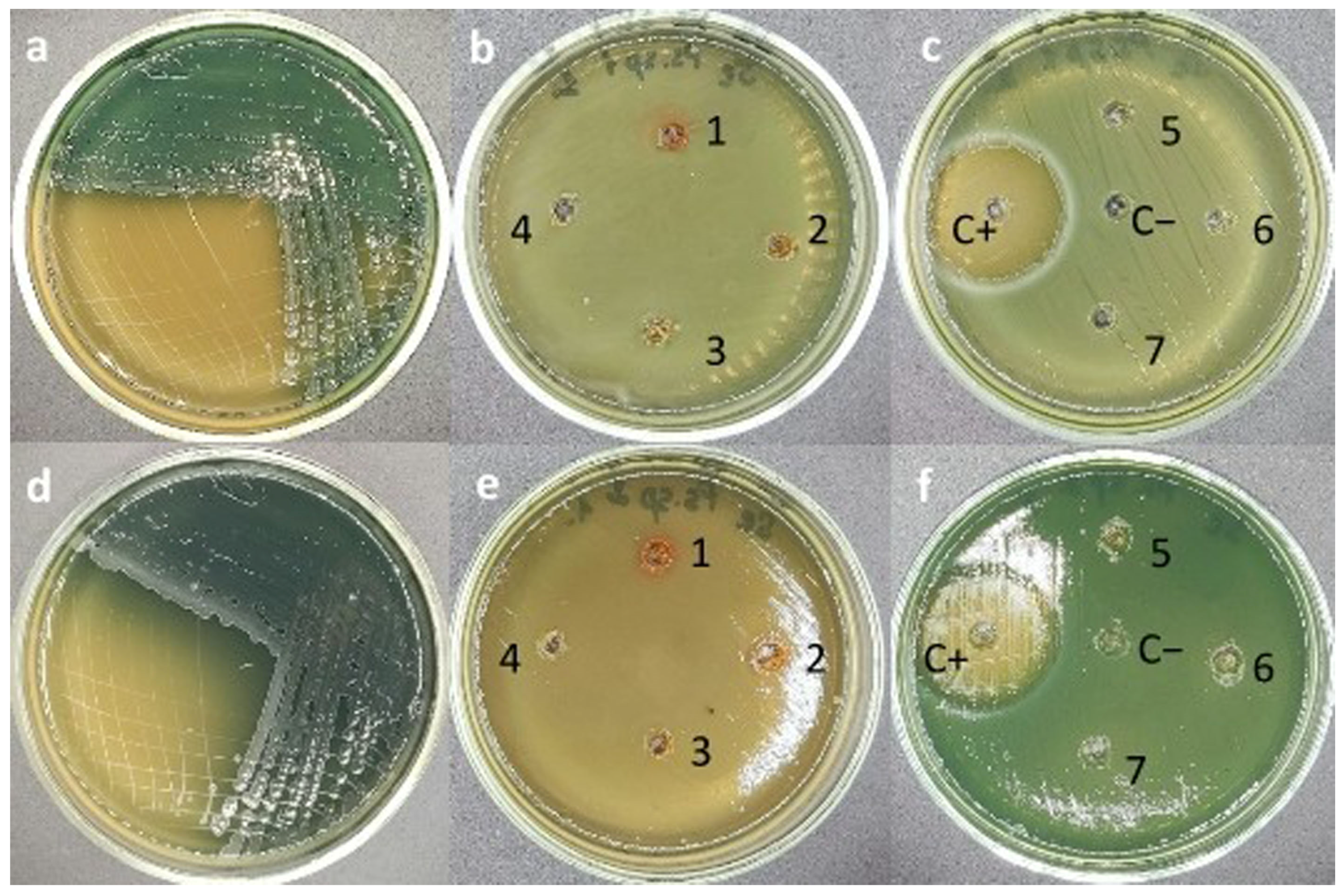
2.7. Evaluation of Antimicrobial Activity of Se/AG NCs by Agar Diffusion Method

The agar well diffusion method was performed to qualitatively screen the susceptibility of bacteria to selected concentrations of Se/AG NCs. The antimicrobial effect of Se/AG NCs was assessed on a solid Mueller–Hinton nutrient medium (State Research Center for Applied Microbiology and Biotechnology, Obolensk, Russia), onto which an 18–24 h bacterial suspension of the studied cultures was pre-inoculated using a swab by suspending the initial cultures in a physiological solution (0.86% NaCl) to approximately 108 CFU/mL using 0.5 McFarland turbidity standard [39]. After that, wells of 6 mm diameter were made in the agar using a sterile punch, into which different dilutions of Se/AG NCs were added. The antibacterial drug ceftriaxone (10 mg/mL, PAO Sintez, Kurgan, Russia) was used as a positive control (C+). Sterile distilled water was used as a negative control (C−). All wells were equidistant from each other to exclude diffusion of NPs in the agar. After 24 h of incubation in a thermostat at 37 °C, zones of growth inhibition of bacteria were determined around the wells with different concentrations of Se/AG NCs.
The results were assessed based on the diameter of the inhibition zones around the “well” (“holes”), including the diameter of the “well” itself. The zones were measured using a caliper. In some cases, the inhibition zones were oval in shape; in such cases, the largest and smallest diameters of the zone were measured, and the average value was calculated, which was taken as the indicator. The absence of inhibition zones of bacteria around the “well” indicated that the test culture was not sensitive to a given dilution of Se/AG NCs. The larger the inhibition zone of the test culture, the higher its sensitivity to Se/AG NCs [40,41].

2.8. Statistical Analysis

All experiments were carried out in triplicate, the obtained results were statistically processed with the calculation of mean values and standard deviations using the online program Calculator.net (https://www.calculator.net, accessed on 7 March 2025).

3. Results

3.1. Characteristic Se/AG NCs

As shown by a series of three measurements using the ICPE method, the average Se content in NCs was 5.52%. In the FTIR spectrum of Se/AG NCs, five intense infrared absorption regions were observed: a broad band at 3000–3800 cm−1, as well as at 2700–3000 cm−1, 1645–1651 cm−1, 1370–1452 cm−1, and 1000–1200 cm−1 (Figure 1).
In the optical absorption spectrum of Se/AG NCs, a broad descending absorption with a noticeable maximum in the region of 256 nm was observed in the entire range of 200–800 nm (Figure 2).
According to dynamic light scattering data, the aqueous colloidal solution of Se/AG NCs contains NPs with an average hydrodynamic size of about 140 nm (Figure 3a), which are negatively charged with an average surface charge value of −15.5 ± 5.92 mV (Figure 3b).

3.2. Effects of Se/AG NCs on Environmental Bacteria

A qualitative agar well diffusion test was performed to screen Se/AG NCs for its antimicrobial properties against Gram-positive (M. luteus, B. cereus, B. subtilis, B. megaterium, and S. flava) and Gram-negative (E. coli, S. marcescens, and P. aeruginosa) bacteria using different aqueous dilutions of Se/AG NCs (0.625–40 mg/mL). The obtained data are presented in Table 1.
The inhibition zones around the wells with positive control ranged from 19 to 59.6 mm. These zones were larger than those formed around the wells containing Se/AG NCs, confirming the higher growth inhibition effect of the commercial antibiotic against both Gram-positive and Gram-negative species of the studied bacteria. Despite high concentrations, Se/AG NCs in this study was completely ineffective against Gram-negative bacteria S. marcescens and P. aeruginosa producing two types of pigment, as well as against Gram-positive bacteria B. subtilis. At all concentrations studied, Se/AG NCs had a toxic effect only on two Gram-positive bacteria, M. luteus and B. cereus, isolated from Lake Baikal water and activated sludge of treatment facilities, respectively. The inhibition zone for M. luteus varied from 9.3 mm at a concentration of 0.04 mg/mL to 26.3 mm at 2.21 mg/mL Se NPs in the NC. For B. cereus, the minimum inhibition zone was 8.3 mm at a concentration of 0.04 mg/mL. With an increase in the concentration of Se NPs in the NC to 2.21 mg/mL, the maximum diameter of growth inhibition was 14.3 mm. The bacterium M. luteus isolated from activated sludge demonstrated a decrease in the diameter of growth inhibition zones (from 7 to 14.3 mm) compared to the similar isolate isolated from Lake Baikal water and lost sensitivity at a Se NP concentration in the NC equal to 0.04 mg/mL. With the exception of M. luteus isolated from water, the maximum diameter of the growth inhibition zone after exposure to ultra-high concentrations of Se/AG NCs was observed in E. coli isolated from soil—20.3 mm.
Figure 4 shows the effect of Se/AG NCs on bacteria, demonstrating a strong antibacterial effect.
Despite the lack of antibacterial effect of Se/AG NCs on isolates of P. aeruginosa (producing the fluorescent siderophore pigment pyoverdin, which colors the culture medium in green) and P. aeruginosa (producing the greenish-blue phenazine pigment pyocyanin, which colors the culture medium in blue-green) isolated from wastewater treatment plants, suppression of both pigments of the isolates was observed after exposure to Se/AG NCs. Pigmentation in the P. aeruginosa isolate producing pyoverdin was suppressed at all tested concentrations of Se/AG NCs, while pyocyanin production in the other P. aeruginosa strain was abolished at high concentrations (from 5 to 40 mg/mL) of Se/AG NCs, and the strains did not lose their ability to grow (see Figure 5). However, no effect of Se/AG NCs on the production of lemon-yellow pigment in M. luteus isolates was observed.
Overall, our study data showed the bactericidal effect of high doses of Se/AG NCs only against two types of bacteria—M. luteus and B. cereus.

4. Discussion

The broad band observed in the FTIR spectrum of Se/AG NCs in the region of 3000–3800 cm−1, as well as the band at 1645–1651 cm−1, are related to vibrations of hydrated hydroxyl groups of the polysaccharide. The band at 2700–3000 cm−1 belongs to the stretching vibrations of C–H bonds of carbohydrate units of macromolecules; the same bonds are responsible for the absorption of deformation vibrations in the region of 1370–1452 cm−1. The group of bands at 1000–1200 cm−1 can be attributed to vibrations of C–O–C ether groups of polysaccharide macromolecules.
Obviously, the contribution to the broad optical absorption of Se/AG NCs in the ultraviolet and visible spectra can be made by both the superimposed exciton and plasmon absorption bands of nanosized semiconductor Se, and the absorption bands of the AG polysaccharide. In particular, the observed pronounced absorption band in the 256 nm spectrum can most likely be attributed to the superposition of the optical absorption of exciton excitations of polydisperse Se quantum dots [42] and the aldehyde groups of the open terminal carbohydrate units of AG macromolecules, which are in dynamic equilibrium with the cyclic hemiacetal carbohydrate units [43].
Comparing the obtained dynamic light scattering data on the average size of the studied NC particles (Se NPs packed in AG macromolecules), equal to 140 nm, with the average sizes of Se NPs of 40–60 nm for this NC obtained previously using transmission electron microscopy (TEM) [9], it can be assumed that one diffusion-mobile Se/AG NC particle contains from one to several Se NPs. The observed negative charge of NC particles −15.50 mV may manifest itself in some charge discrimination of their delivery to the same negative surface of microbial cells. This may lead to differentiation of cytotoxicity in relation to microbial cells, for example, according to the principle of substrate preference of the AG shell of Se NPs for different microorganisms, which requires further detailed studies.
Concentrations of Se NPs used in agriculture vary within different limits. Thus, Se NPs has proven itself as a stimulator of plant growth and development (tobacco, eggplant, potato, tomato, spinach, etc.) in concentrations from 0.5 mg/L to >500 mg/L, and of biofortification (spinach, onion, rice, wheat)—up to 22 mg/L [44,45,46,47,48,49,50,51,52]. In pest control as nematicides and insecticides against Meloidogyne incognita, Anopheles stephensi, Culex quinquefasciatus, and Aedes aegypti and as an antimicrobial agent against Alternaria solani, Sclerospora graminicola, Macrophomina phaseolina, Sclerotinia sclerotiorum, Diaporthe longicolla, B. subtilis, Staphylococcus aureus, and E. coli, Se NPs had a positive effect at doses of 0.1 mg/mL–500 mg/L and 0.1 mg/L–1000 mg/L, respectively [47,52,53,54,55,56,57,58,59,60]. According to our data, it has been previously shown that the Se/AG NC has antibacterial properties against phytopathogens C. sepedonicus, P. carotovorum, and Phytophthora cactorum, while the active concentration of Se/AG NCs did not exceed 50 µg/mL (3 µg/mL Se) [9,20,22]. At the same time, this concentration did not affect the representatives of soil microbiome A. guillouiae, R. erythropolis, and P. oryzihabitans [22], which is confirmed by the data of our experiment. Thus, the data obtained by us in the present study indicate the relative ecological safety of Se/AG NCs, which we obtained by chemical synthesis.
The agar diffusion test was conducted as a qualitative test to observe and predict the antibacterial effect of Se/AG NCs. Interestingly, according to the results obtained in this work, the inhibitory effect of Se/AG NCs did not depend on the classification of bacteria as Gram-positive or Gram-negative forms, which can suggest that the difference in the structure of the bacterial wall does not affect the antibacterial activity of Se NPs. The nonspecific action of Se NPs associated with the structure of the bacterial cell wall has been known previously. It was shown on Gram-positive (S. aureus, B. cereus, and B. subtilis) and Gram-negative bacteria (P. aeruginosa, E. coli, and Vibrio parahemolyticus) that interaction of Se NPs with bacterial cells results in the release of proteins and polysaccharides from the cell. It was found that protein and polysaccharide leaks were due to changes in membrane permeability and destruction of cell walls. And changes in the intensity of ROSs indicated that oxidative damage can play a significant role in antibacterial processes [61].
There are several explanations in the literature for the mechanisms by which Se NPs influence bacterial cells. One of them suggests that Se NPs can attach to bacterial cells through charge attraction, which in turn alters the bacterial membrane and changes the permeability of the microbial membrane. In response, genes that cause oxidative stress through increased ROS production are overexpressed in bacterial cells [62].
Dang-Bao et al. [63] believe that the antibacterial mechanism of Se NPs against the Gram-positive bacterium S. aureus involves the direct adhesion of Se NPs to the bacterial surface and the effect on the structural integrity of the membrane. The attachment of Se NPs to the membrane dramatically changes the permeability of the membrane. Se NPs penetrate into the bacterial cell. In the presence of oxygen and proton, Se NPs disintegrate into ions, which facilitates infusion. Once inside the cell, they interact with its internal components, causing them to be damaged to the point that they can no longer perform important cellular functions of the cell [63]. Thus, Se NPs interact with DNA and lipids. Se NPs disrupt protein synthesis by binding to the ribosome and denaturing it, causing the translation process to cease. Se NPs cause denaturation and reading shift of the DNA molecule during transcription, as well as to disruption of cell division. The interaction makes bacteria incapable of cell division and reproduction, which ultimately leads to death. Se NPs can also generate ROSs and free radicals, causing irreversible oxidative damage to bacteria. Se NPs affect critical signaling pathways that are essential for the life cycle of bacteria. The destruction of essential cellular components, such as proteins, DNA, and RNA, caused by oxidative stress leads to changes in membrane permeability and increased leakage of biological components from the cell [63].
According to [64], the main mechanism of the destructive effect of Se NPs on bacterial cells is associated with the generation of ROSs under the influence of NPs. In particular, the antimicrobial effect of various Se compounds is associated with the formation of free radicals [65]. With regard to Se, it is known that Se NPs kill bacteria by activating ROSs, which destroy their cell membrane and lead to the death of bacterial cells. ROS production in response to treatment with biogenic Se NPs was analyzed in P. aeruginosa, S. aureus, and Burkholderia cenocepacia strains [66]. All tested bacterial strains showed an increase in ROSs generated after treatment with biogenic Se NPs compared to untreated controls. The number of ROSs observed in the experiments was of the same order of magnitude as the number of ROSs generated by some antibiotics [66]. In the bacterial strains of S. aureus and E. coli in the groups treated with Se NPs, the number of ROSs increased with the increase in Se NPs, suggesting that the ROS generation in bacteria was stimulated due to the presence of Se NPs, which was more likely to induce oxidative stress to kill bacteria [67].
Thus, it is suggested that Se NPs can alter microbial morphology, reduce microbial membrane permeability, and lead to oxidative stress through the generation of ROSs. Se NPs initiate their activity by attaching to the outer surface of the microbial cell, causing membrane rupture, pit formation, and disruption of ion transport activity. Then, all intracellular forms such as DNA, plasmids, and various bacterial organelles become targets of Se NPs in the microbial cell [68]. Cellular toxicity now occurs as a result of oxidative stress created by the accumulation of ROSs. Finally, in the acidic environment, ionic species (Se+ ions) are formed, causing cellular toxicity and genotoxicity by interacting with negatively charged cell organelles.
In addition, using TEM of microbial cell sections, in our previous work, we documented the antiphytopathogenic cytotoxicity of Se/AG NCs, accompanied by the attachment of NPs to the bacterial cell surface followed by direct penetration of Se NPs into microbial cells [21]. This may indicate a very likely, as previously proposed [69], initial active trophic uptake by microbial cells of polysaccharide AG macromolecules with encapsulated Se NPs through the penetration of NPs into bacterial cells by simple diffusion, as well as through the mechanisms of induced transport of compounds into the cell from the external environment [70]. Enzymatic degradation of these macromolecules and bioutilization of released monosaccharides can lead to the release of NPs from the polysaccharide matrix and their penetration into the cell. Observation of Se NPs inside microbial cells suggests direct cytotoxicity of Se NPs penetrating into cells, bypassing the stage of release of mobile ionic forms of Se with their subsequent toxic effect on cells. However, a combined mechanism of action on microbial cells of both Se ions released from Se NPs and Se NPs penetrating into cells is not excluded. Therefore, a detailed identification of the mechanism of protective antiphytopathogenic action of the present Se/AG NCs requires further detailed studies.
Despite the fact that most microorganisms have not encountered such NPs during evolution, they have nevertheless developed various mechanisms of adaptation to adverse conditions and toxicity, especially microorganisms in the wastewater and activated sludge from treatment facilities. Although wastewater is considered one of the main sources of NPs distribution in the natural environment, the adaptation of microorganisms is not limited to treatment facilities [71]. One of the mechanisms of adaptation of Gram-negative bacteria can be considered porins (transmembrane channel-forming proteins) of the outer cell membrane, the diameter of which varies from 1 to 6 nm. Many studies showed that the size of NPs is one of the main factors of their antimicrobial activity. The sizes of Se NPs in our study were 40–60 nm, which probably hinders their penetration into most Gram-negative bacterial cells. The only exception was E. coli isolated from the soil environment, which showed sensitivity to Se/AG NCs at its high concentrations (5–40 mg/mL). This is probably due to the presence of at least two types of porins (OmpC and OmpF) in E. coli, with a pore diameter exceeding the size of most ions released from NPs, which allows them to penetrate into the periplasmic space of the bacterial cell. It is believed that this occurs through active copper transporters CTR1 [72]. In addition, the mechanism of adaptation of Gram-negative bacteria to NPs is manifested in the overexpression by bacterial cells of extracellular substances, such as exopolysaccharides, which are secreted by bacteria in the form of free mucus, capsules, and sheaths, forming an extracellular matrix that prevents direct contact of NPs with bacteria and promotes agglomeration and deactivation of NPs [73,74]. Gram-positive bacteria do not have this ability, as a result of which they are more vulnerable due to the simple organization of their cell wall.
Since NPs from both household and industrial products will enter the environment, such as wastewater, it is important to study their effect on bacteria, the main component of activated sludge. The most sensitive microorganism from activated sludge to Se/AG NCs in all studied concentrations (0.625–40 mg/mL) was the B. cereus strain. Representatives of the genus Bacillus in activated sludge play an important role in flocculation, oxidation of aliphatic hydrocarbons [71]. The Se/AG NC was effective at concentrations ranging from 1.25 to 40 mg/mL on a bacteria M. luteus capable of degrading a range of aromatic, aliphatic, and xenobiotic compounds. In fact, bacteria and NPs may develop symbiotic or similar synergistic relationships under certain environmental conditions. Bacteria can also potentially be influential catalysts. They can change the oxidation state of many elements, facilitate the removal of certain molecules containing NPs. All of these are critical for the development of microbial processes that can concentrate, remove, and recover various pollutants including NPs from industrial wastewater [75]. Bacteria can also catalyze reactions that can lead to the aggregation of NPs or the conjugation of molecules that facilitate their removal. Thus, on the one hand, NPs can have an adverse effect on bacteria, and on the other hand, they interact with bacteria, facilitating autoaggregation and, ultimately, the removal of NPs from the wastewater stream. Similar types of mechanisms are also possible in sewage sludge, which is a repository of diverse microbial systems and can lead to numerous mechanisms—complexation, aggregation, etc. [76].
For bacteria, an increase in the concentration of toxic substances is a factor that causes a combined stress response [77]. The bactericidal effect of NPs depends on their concentration in the preparation; many researchers have proven that the higher the concentration of NPs, the deeper the damage to cellular structures and the more pronounced the antibacterial effect [67].
Therefore, bacteria have developed complex homeostatic mechanisms using multiple strategies to neutralize the toxic effects of NPs that are also described above. In some cases, bacterial adaptive mechanisms can cause changes in pigment production. In our study, Se NPs were shown to successfully suppress pigment synthesis in Pseudomonas spp. Microbial pigments such as pyoverdin and pyocyanin in Pseudomonas species are pathogenicity factors because they exhibit anti-inflammatory and cytotoxic properties [78,79]. These pigments are involved in bacterial quorum sensing, virulence, and iron uptake by bacteria [80]. Thus, the pigments themselves become logical targets for therapeutic interventions, including those using NPs. Pseudomonas spp. are capable of synthesizing a whole complex of oxidatively active pigments of the phenazine series, the composition (qualitative and quantitative) of which depends on the cultivation conditions, components of the medium, individual characteristics of bacterial strains and sources of isolation. Phenazines are characterized by a unique mechanism of antibacterial action and high activity, including against phytopathogenic fungi and bacteria, as well as the ability to improve the penetration of mineral salts into plants [81,82]. The obtained results are confirmed by another study on the effect of NPs on bacterial pigment formation [79,83,84] and also indicate the potential of using Se/AG NCs in relation to human and animal pathogens belonging to the genus Pseudomonas, which includes P. aeruginosa.
Thus, the assessment of environmental risks of the impact of nanomaterials on biological objects, including ecologically important microbial communities, is necessary to predict potential toxic or non-toxic impacts by selecting or changing the characteristics of nanomaterials (size, shape, concentration, etc.) for their successful use in natural and artificial ecosystems. Certainly, the effect of Se/AG NCs on environmental bacteria needs additional studies, such as determining the minimum inhibitory concentration of Se/AG NCs, identification of morphological changes occurring in bacteria under the influence of Se/AG NCs using scanning electron microscopy, measuring ROSs in bacterial cells, high concentrations of which can lead to oxidative stress.

5. Conclusions

Se/AG NCs, which are negatively charged composite Se/AG particles that are diffusion-mobile in aqueous media, showed a weak, insignificant antimicrobial effect against both Gram-negative and Gram-positive bacteria even in supercritical concentrations, which allows us to classify it as an environmentally friendly NC, which will undoubtedly open up new areas of its application in the future. In all critically high concentrations studied (0.625–40 mg/mL), Se/AG NCs had a negative effect only on two strains—B. cereus from activated sludge and M. luteus from water. The susceptibility of strains from the highest to the lowest sensitivity to Se/AG NCs was in the following order: M. luteus (water) < B. cereus (activated sludge) < S. flava (water) < M. luteus (activated sludge) < E. coli (soil) < B. megaterium (soil) < B. cereus (soil). The detected low toxicity of Se/AG NPs towards bacteria from natural and artificial environments is probably due to the AG matrix used, which can prevent active release of Se ions.

Author Contributions

Conceptualization, E.I.S., A.I.P., B.G.S. and K.V.K.; methodology, E.I.S., A.I.P., O.F.V., B.G.S., A.I.K., A.V.R. and T.V.K.; software, E.I.S., A.I.P., B.G.S. and K.V.K.; validation, E.I.S., A.I.P., O.F.V., D.I.S., B.G.S. and K.V.K.; formal analysis, E.I.S. and A.I.P.; investigation, E.I.S., A.I.P., B.G.S. and K.V.K.; resources, E.I.S., A.I.P. and B.G.S.; data curation, E.I.S., A.I.P., B.G.S. and K.V.K.; writing—original draft preparation, E.I.S., A.I.P., O.F.V., B.G.S., A.I.K., A.V.R. and T.V.K.; writing—review and editing, E.I.S., A.I.P., O.F.V., D.I.S., B.G.S., A.I.K., A.V.R., T.V.K. and K.V.K.; visualization, E.I.S., B.G.S., A.I.K., A.V.R. and T.V.K.; supervision, K.V.K.; project administration, E.I.S.; funding acquisition, E.I.S. All authors have read and agreed to the published version of the manuscript.

Funding

This research was carried out with the support of the state program 124022100043f5 funded by the Ministry of Science and Higher Education of the Russian Federation within the framework of the basic project “Development of biotechnological solutions to environmental problems of industrial production”. It was also funded by the Russian Science Foundation, grant No. 25-24-20046, “Evaluation of the effect of chalcogen- and metal-containing nanocomposites on the expression level of pathogen-dependent (PR) genes of cultivated plants of the Siberian region when infected with the phytopathogenic bacterium Pectobacterium carotovorum”. The dynamic light scattering was carried out in the specific pathogen free (SPF) vivarium of the Institute of Cytology and Genetics of the Siberian Branch of the Russian Academy of Sciences and supported by basic research project FWNR-2025-0019.

Data Availability Statement

Data are contained within this article and available from the authors upon request.

Acknowledgments

We are grateful to G. O. Zhdanova (Laboratory of Aquatic Toxicology, Research Institute of Biology, Irkutsk State University, Russia) and N. E. Bukovskaya (Department of Microbiology, Irkutsk State University, Russia) for providing the bacteria strains. We also express our gratitude to the Baikal Analytical Center for Collective Use of the Siberian Branch of the Russian Academy of Sciences and the Center for Collective Use of Analytical Equipment of Irkutsk State University for the opportunity to use their equipment and facilities.

Conflicts of Interest

The authors declare no conflicts of interest.

Abbreviations

The following abbreviations are used in this paper:
AGarabinogalactan
CFUcolony forming unit
DNAdeoxyribonucleic acid
ICPEinductively coupled plasma emission
LPlipid peroxidation
NCnanocomposite
NPnanoparticle
RNAribonucleic acid
ROSreactive oxygen species
Se/AG NCNanocomposite (NC) based on selenium (Se) and arabinogalactan (AG)

References

  1. Genchi, G.; Lauria, G.; Catalano, A.; Sinicropi, M.S.; Carocci, A. Biological activity of selenium and its impact on human health. Int. J. Mol. Sci. 2023, 24, 2633. [Google Scholar] [CrossRef] [PubMed]
  2. Islam, M.R.; Akash, S.; Jony, M.H.; Alam, M.N.; Nowrin, F.T.; Rahman, M.M.; Thiruvengadam, M. Exploring the potential function of trace elements in human health: A therapeutic perspective. Mol. Cell. Biochem. 2023, 478, 2141–2171. [Google Scholar] [CrossRef] [PubMed]
  3. Song, J.; Yu, S.; Yang, R.; Xiao, J.; Liu, J. Opportunities for the use of selenium nanoparticles in agriculture. NanoImpact 2023, 31, 100478. [Google Scholar] [CrossRef]
  4. Feng, R.; Wei, C.; Tu, S. The roles of selenium in protecting plants against abiotic stresses. Environ. Exp. Bot. 2013, 87, 58–68. [Google Scholar] [CrossRef]
  5. Bano, I.; Skalickova, S.; Sajjad, H.; Skladanka, J.; Horky, P. Uses of selenium nanoparticles in the plant production. Agronomy 2021, 11, 2229. [Google Scholar] [CrossRef]
  6. Ferro, C.; Florindo, H.F.; Santos, H.A. Selenium nanoparticles for biomedical applications: From development and characterization to therapeutics. Adv. Healthc. Mater. 2021, 10, 2100598. [Google Scholar] [CrossRef] [PubMed]
  7. Yang, R.; Li, Q.; Zhou, W.; Yu, S.; Liu, J. Speciation analysis of selenium nanoparticles and inorganic selenium species by dual-cloud point extraction and ICP-MS determination. Anal. Chem. 2022, 94, 16328–16336. [Google Scholar] [CrossRef]
  8. Perfileva, A.I.; Nozhkina, O.A.; Graskova, I.A.; Sidorov, A.V.; Lesnichaya, M.V.; Aleksandrova, G.P.; Sukhov, B.G. Synthesis of selenium and silver nanobiocomposites and their influence on phytopathogenic bacterium Clavibacter michiganensis subsp. sepedonicus. Russ. Chem. Bull. 2018, 67, 157–163. [Google Scholar] [CrossRef]
  9. Perfileva, A.I.; Nozhkina, O.A.; Ganenko, T.V.; Graskova, I.A.; Sukhov, B.G.; Artem’ev, A.V.; Krutovsky, K.V. Selenium nanocomposites in natural matrices as potato recovery agent. Int. J. Mol. Sci. 2021, 22, 4576. [Google Scholar] [CrossRef]
  10. Perfileva, A.I.; Kharasova, A.R.; Nozhkina, O.A.; Sidorov, A.V.; Graskova, I.A.; Krutovsky, K.V. Effect of nanopriming with selenium nanocomposites on potato productivity in a field experiment, soybean germination and viability of Pectobacterium carotovorum. Horticulturae 2023, 9, 458. [Google Scholar] [CrossRef]
  11. Zhang, L.; Zeng, H.; Cheng, W.H. Beneficial and paradoxical roles of selenium at nutritional levels of intake in healthspan and longevity. Free Radic. Biol. Med. 2018, 127, 3–13. [Google Scholar] [CrossRef] [PubMed]
  12. Kapur, M.; Soni, K.; Kohli, K. Green synthesis of selenium nanoparticles from broccoli, characterization, application and toxicity. Adv. Tech. Biol. Med. 2017, 5, 1–7. [Google Scholar] [CrossRef]
  13. Trofimov, B.A.; Sukhov, B.G.; Aleksandrova, G.P.; Medvedeva, S.A.; Grishchenko, L.A.; Mal’kina, A.G.; Feoktistova, L.P.; Sapozhnikov, A.N.; Dubrovina, V.I.; Martynovich, E.F.; et al. Arabinogalactan-based nanocomposites with magnetic, optical, catalytic and biologically active properties. Dokl. Akad. Nauk. 2003, 393, 634–635. [Google Scholar] [CrossRef]
  14. Kolesnikova, L.I.; Karpova, E.A.; Vlasov, B.Y.; Sukhov, B.G.; Trofimov, B.A. Lipid peroxidation–antioxidant defense system during toxic liver damage and its correction with a composite substance containing selenium and arabinogalactan. Bull. Exp. Biol. Med. 2015, 159, 225–228. [Google Scholar] [CrossRef] [PubMed]
  15. Lesnichaya, M.V.; Karpova, E.A.; Sukhov, B.G. Effect of high dose of selenium nanoparticles on antioxidant system and biochemical profile of rats in correction of carbon tetrachloride-induced toxic damage of liver. Coll. Surf. B Biointerface 2021, 197, 111381. [Google Scholar] [CrossRef]
  16. Lesnichaya, M.V.; Shendrik, R.Y.; Sukhov, B.G. Relation between excitation dependent luminescence and particle size distributions for the selenium nanoparticles in κ-carrageenan shell. J. Lumin. 2019, 211, 305–313. [Google Scholar] [CrossRef]
  17. Graskova, I.A.; Perfilieva, A.I.; Nozhkina, O.A.; Sukhov, B.G.; Aleksandrova, G.P.; Trofimov, B.A. Silver-containing nanocomposites of humic substances, agents for healing of potatoes from the ring rot. Dokl. Biol. Sci. 2018, 483, 239–242. [Google Scholar] [CrossRef]
  18. Khutsishvili, S.S.; Perfileva, A.I.; Nozhkina, O.A.; Karepova, M.S.; Ganenko, T.V.; Sukhov, B.G.; Trofimov, B.A.; Krutovsky, K.V. Novel nanobiocomposites based on natural polysaccharides as universal trophic low-dose micronutrients. Int. J. Mol. Sci. 2021, 22, 12006. [Google Scholar] [CrossRef]
  19. Khutsishvili, S.S.; Perfileva, A.I.; Kon’kova, T.V.; Lobanova, N.A.; Sadykov, E.K.; Sukhov, B.G. Copper-containing bionanocomposites based on natural raw arabinogalactan as effective vegetation stimulators and agents against phytopathogens. Polymers 2024, 16, 716. [Google Scholar] [CrossRef]
  20. Perfileva, A.I.; Krutovsky, K.V. Effect of manganese- and selenium-containing nanocomposites on soybean resistance to Pectobacterium carotovorum and microbial landscape of soybean seedlings. Plant Growth Regul. 2024, 104, 389–402. [Google Scholar] [CrossRef]
  21. Perfileva, A.I.; Moty’leva, S.M.; Klimenkov, I.V.; Arsent’ev, K.Y.; Graskova, I.A.; Sukhov, B.G.; Trofimov, B.A. Development of Antimicrobial Nano-Selenium Biocomposite for Protecting Potatoes from Bacterial Phytopathogens. Nanotechnol. Russ. 2017, 12, 553–558. [Google Scholar] [CrossRef]
  22. Perfileva, A.I.; Tsivileva, O.M.; Nozhkina, O.A.; Karepova, M.S.; Graskova, I.A.; Ganenko, T.V.; Sukhov, B.G.; Krutovsky, K.V. Effect of natural polysaccharide matrix-based selenium nanocomposites on Phytophthora cactorum and rhizospheric microorganisms. Nanomaterials 2021, 11, 2274. [Google Scholar] [CrossRef] [PubMed]
  23. Nurminsky, V.N.; Perfileva, A.I.; Kapustina, I.S.; Graskova, I.A.; Sukhov, B.G.; Trofimov, B.A. Growth-stimulating activity of natural polymer-based nanocomposites of selenium during the germination of cultivated plant seeds. Dokl. Biochem. Biophys. 2020, 495, 296–299. [Google Scholar] [CrossRef] [PubMed]
  24. Graskova, I.A.; Perfileva, A.I.; Nozhkina, O.A.; Dyakova, A.V.; Nurminsky, V.N.; Klimenkov, I.V.; Sudakov, N.P.; Borodina, T.M.; Aleksandrova, G.P.; Lesnichaya, M.V.; et al. The effect of nanoscale selenium on the causative agent of rot pathogen and potato in vitro. Khimiya Rastit. Syr’ya 2019, 3, 345–354, (In Russian with English Abstract). [Google Scholar] [CrossRef]
  25. Blaser, S.A.; Scheringer, M.; MacLeod, M.; Hungerbühler, K. Estimation of cumulative aquatic exposure and risk due to silver: Contribution of nano-functionalized plastics and textiles. Sci. Total Environ. 2008, 390, 396–409. [Google Scholar] [CrossRef]
  26. Mueller, N.C.; Nowack, B. Exposure modeling of engineered nanoparticles in the environment. Environ. Sci. Technol. 2008, 42, 4447–4453. [Google Scholar] [CrossRef]
  27. Gottschalk, F.; Sonderer, T.; Scholz, R.W.; Nowack, B. Modeled environmental concentrations of engineered nanomaterials (TiO2, ZnO, Ag, CNT, fullerenes) for different regions. Environ. Sci. Technol. 2009, 43, 9216–9222. [Google Scholar] [CrossRef]
  28. Kiser, M.A.; Westerhoff, P.; Benn, T.; Wang, Y.; Perez-Rivera, J.; Hristovski, K. Titanium nanomaterial removal and release from wastewater treatment plants. Environ. Sci. Technol. 2009, 43, 6757–6763. [Google Scholar] [CrossRef]
  29. Arvidsson, R.; Molander, S.; Sandén, B.A.; Hassellöv, M. Challenges in exposure modeling of nanoparticles in aquatic environments. Hum. Ecol. Risk Assess. 2011, 17, 245–262. [Google Scholar] [CrossRef]
  30. Westerhoff, P.; Song, G.; Hristovski, K.; Kiser, M.A. Occurrence and removal of titanium at full scale wastewater treatment plants: Implications for TiO2 nanomaterials. J. Environ. Monit. 2011, 13, 1195–1203. [Google Scholar] [CrossRef]
  31. Praetorius, A.; Scheringer, M.; Hungerbühler, K. Development of environmental fate models for engineered nanoparticles—A case study of TiO2 nanoparticles in the rhine river. Environ. Sci. Technol. 2012, 46, 6705–6713. [Google Scholar] [CrossRef] [PubMed]
  32. Maurer-Jones, M.A.; Gunsolus, I.L.; Murphy, C.J.; Haynes, C.L. Toxicity of engineered nanoparticles in the environment. Anal. Chem. 2013, 85, 3036–3049. [Google Scholar] [CrossRef]
  33. Choi, S.; Johnston, M.; Wang, G.S.; Huang, C.P. A seasonal observation on the distribution of engineered nanoparticles in municipal wastewater treatment systems exemplified by TiO2 and ZnO. Sci. Total Environ. 2018, 625, 1321–1329. [Google Scholar] [CrossRef] [PubMed]
  34. Syafiuddin, A.; Salmiati, S.; Hadibarata, T.; Kueh, A.B.; Salim, M.R.; Zaini, M.A. Silver nanoparticles in the water environment in Malaysia: Inspection, characterization, removal, modeling, and future perspective. Sci. Rep. 2018, 8, 986. [Google Scholar] [CrossRef]
  35. Bathi, J.R.; Moazeni, F.; Upadhyayula, V.K.; Chowdhury, I.; Palchoudhury, S.; Potts, G.E.; Gadhamshetty, V. Behavior of engineered nanoparticles in aquatic environmental samples: Current status and challenges. Sci. Total Environ. 2021, 793, 148560. [Google Scholar] [CrossRef] [PubMed]
  36. Lesnichaya, M.V.; Malysheva, S.F.; Belogorlova, N.A.; Graskova, I.A.; Gazizova, A.V.; Perfilyeva, A.I.; Nozhkina, O.A.; Sukhov, B.G. Synthesis and antimicrobial activity of arabi-nogalactan-stabilized selenium nanoparticles from sodium bis(2-phenylethyl)diselenophosphinate. Russ. Chem. Bull. 2019, 68, 2245–2251. [Google Scholar] [CrossRef]
  37. Chen, W.; Li, Y.; Yang, S.; Yue, L.; Jiang, Q.; Xia, W. Synthesis and antioxidant properties of chitosan and carboxymethyl chitosan-stabilized selenium nanoparticles. Carbohydr. Polym. 2015, 132, 574–581. [Google Scholar] [CrossRef]
  38. Gerhardt, P.; Murray, R.G.E.; Costilow, R.N.; Nester, E.W.; Wood, W.A.; Krieg, N.R.; Phillips, G.B. (Eds.) Manual of Methods for General Bacteriology, 1st ed.; American Society for Microbiology: Washington, DC, USA, 1981; Volume 1, 524p. [Google Scholar]
  39. Gabrielyan, L.; Badalyan, H.; Gevorgyan, V.; Trchounian, A. Comparable antibacterial effects and action mechanisms of silver and iron oxide nanoparticles on Escherichia coli and Salmonella typhimurium. Sci. Rep. 2020, 10, 13145. [Google Scholar] [CrossRef]
  40. Islama, M.M.; Masumb, S.M.; Mahbuba, K.R.; Haquea, M.Z. Antibacterial activity of crab-chitosan against Staphylococcus aureus and Escherichia coli. J. Adv. Sci. Res. 2011, 2, 63–66. Available online: https://www.sciensage.info/index.php/JASR/article/view/61 (accessed on 30 August 2024).
  41. Aliasghari, A.; Khorasgani, M.R.; Vaezifar, S.; Rahimi, F.; Younesi, H.; Khoroushi, M. Evaluation of antibacterial efficiency of chitosan and chitosan nanoparticles on cariogenic streptococci: An in vitro study. Iran. J. Microbiol. 2016, 8, 93–100. Available online: https://pmc.ncbi.nlm.nih.gov/articles/PMC4906725 (accessed on 16 February 2025). [PubMed] [PubMed Central]
  42. Singh, S.C.; Mishra, S.K.; Srivastava, R.K.; Gopal, R. Optical properties of selenium quantum dots produced with laser irradiation of water suspended Se nanoparticles. J. Phys. Chem. C 2010, 114, 17374–17384. [Google Scholar] [CrossRef]
  43. Tunik, T.V.; Nemchenko, U.M.; Ganenko, T.V.; Yurinova, G.V.; Dzhioev, Y.P.; Sukhov, B.G.; Zlobin, V.I.; Trofimov, B.A. Synthesis and spectral characterization of new biodegradable arabinogalactan derivatives for diagnosis and therapy. Bull. Russ. Acad. Sci. Phys. 2019, 83, 343–349. [Google Scholar] [CrossRef]
  44. Domokos-Szabolcsy, E.; Márton, L.; Sztrik, A.; Babka, B.; Prokisch, J.; Fari, M. Accumulation of red elemental selenium nanoparticles and their biological effects in Nicotinia tabacum. Plant Growth Regul. 2012, 68, 525–531. [Google Scholar] [CrossRef]
  45. Gudkov, S.V.; Shafeev, G.A.; Glinushkin, A.P.; Shkirin, A.V.; Barmina, E.V.; Rakov, I.I.; Kalinitchenko, V.P. Production and use of selenium nanoparticles as fertilizers. ACS Omega 2020, 5, 17767–17774. [Google Scholar] [CrossRef]
  46. El-Batal, A.I.; Sidkey, N.M.; Ismail, A.A.; Arafa, R.A.; Fathy, R.M. Impact of silver and selenium nanoparticles synthesized by gamma irradiation and their physiological response on early blight disease of potato. J. Chem. Pharm. Res. 2016, 8, 934–951. [Google Scholar]
  47. Quiterio-Gutiérrez, T.; Cadenas-Pliego, G.; Hernández-Fuentes, A.D.; Sandoval-Rangel, A.; Benavides-Mendoza, A.; Cabrera-de la Fuente, M.; Juárez-Maldonado, A. The application of selenium and copper nanoparticles modifies the biochemical responses of tomato plants under stress by Alternaria solani. Int. J. Mol. Sci. 2019, 20, 1950. [Google Scholar] [CrossRef]
  48. Golubkina, N.A.; Folmanis, G.E.; Tananaev, I.G. Comparative evaluation of selenium accumulation by allium species after foliar application of selenium nanoparticles, sodium selenite and sodium selenite. Dokl. Biol. Sci. 2012, 444, 176–179. [Google Scholar] [CrossRef]
  49. Golubkina, N.A.; Folmanis, G.E.; Tananaev, I.G.; Krivenkov, L.V.; Kosheleva, O.V.; Soldatenko, A.V. Comparative Evaluation of Spinach Biofortification with Selenium Nanoparticles and Ionic Forms of the Element. Nanotechnol. Russ. 2017, 12, 569–576. [Google Scholar] [CrossRef]
  50. Wang, K.; Wang, Y.; Li, K.; Wan, Y.; Wang, Q.; Zhuang, Z.; Guo, Y.; Li, H. Uptake, translocation and biotransformation of selenium nanoparticles in rice seedlings (Oryza sativa L.). J. Nanobiotechnol. 2020, 18, 103. [Google Scholar] [CrossRef]
  51. Hu, T.; Li, H.; Li, J.; Zhao, G.; Wu, W.; Liu, L.; Wang, Q.; Guo, Y. Absorption and bio-transformation of selenium nanoparticles by wheat seedlings (Triticum aestivum L.). Front. Plant Sci. 2018, 9, 597. [Google Scholar] [CrossRef]
  52. Garza-García, J.J.; Hernández-Díaz, J.A.; Zamudio-Ojeda, A.; León-Morales, J.M.; Guerrero-Guzmán, A.; Sánchez-Chiprés, D.R.; García-Morales, S. The role of selenium nanoparticles in agriculture and food technology. Biol. Trace Elem. Res. 2022, 200, 2528–2548. [Google Scholar] [CrossRef] [PubMed]
  53. Udalova, Z.V.; Folmanis, G.E.; Khasanov, F.K.; Zinovieva, S.V. Selenium nanoparticles–an inducer of tomato resistance to the root-knot nematode Meloidogyne incognita (Kofoid et White, 1919) Chitwood 1949. Dokl. Biochem. Biophys. 2018, 482, 264–267. [Google Scholar] [CrossRef]
  54. Sowndarya, P.; Ramkumar, G.; Shivakumar, M.S. Green synthesis of selenium nanoparticles conjugated Clausena dentata plant leaf extract and their insecticidal potential against mosquito vectors. Artif. Cells Nanomed. Biotechnol. 2017, 45, 1490–1495. [Google Scholar] [CrossRef] [PubMed]
  55. Krishnan, M.; Ranganathan, K.; Maadhu, P.; Thangavelu, P.; Kundan, S.; Arjunan, N. Leaf extract of Dillenia indica as a source of selenium nanoparticles with larvicidal and antimicrobial potential toward vector mosquitoes and pathogenic microbes. Coatings 2020, 10, 626. [Google Scholar] [CrossRef]
  56. Salem, S.S.; Fouda, M.M.G.; Fouda, A.; Awad, M.A.; Al-Olayan, E.M.; Allam, A.A.; Shaheen, T.I. Antibacterial, cytotoxicity and larvicidal activity of green synthesized selenium nanoparticles using Penicillium corylophilum. J. Clust. Sci. 2021, 32, 351–361. [Google Scholar] [CrossRef]
  57. Nandini, B.; Hariprasad, P.; Prakash, H.S.; Shetty, H.S.; Geetha, N. Trichogenic-selenium nanoparticles enhance disease suppressive ability of Trichoderma against downy mildew disease caused by Sclerospora graminicola in pearl millet. Sci. Rep. 2017, 7, 612. [Google Scholar] [CrossRef]
  58. Vrandečić, K.; Ćosić, J.; Ilić, J.; Ravnjak, B.; Selmani, A.; Galić, E.; Vinković, T. Antifungal activities of silver and selenium nanoparticles stabilized with different surface coating agents. Pest Manag. Sci. 2020, 76, 2021–2029. [Google Scholar] [CrossRef] [PubMed]
  59. Huang, X.; Chen, X.; Chen, Q.; Yu, Q.; Sun, D.; Liu, J. Investigation of functional selenium nanoparticles as potent antimicrobial agents against superbugs. Acta Biomater. 2016, 30, 397–407. [Google Scholar] [CrossRef]
  60. Alagesan, V.; Venugopal, S. Green synthesis of selenium nanoparticle using leaves extract of Withania somnifera and its biological applications and photocatalytic activities. Bionanoscience 2019, 9, 105–116. [Google Scholar] [CrossRef]
  61. Zhang, H.; Li, Z.; Dai, C.; Wang, P.; Fan, S.; Yu, B.; Qu, Y. Antibacterial properties and mechanism of selenium nanoparticles synthesized by Providencia sp. DCX. Environ. Res. 2021, 194, 110630. [Google Scholar] [CrossRef]
  62. Seixas, A.F.; Quendera, A.P.; Sousa, J.P.; Silva, A.F.Q.; Arraiano, C.M.; Andrade, J.M. Bacterial Response to Oxidative Stress and RNA Oxidation. Front. Genet. 2022, 12, 821535. [Google Scholar] [CrossRef] [PubMed]
  63. Dang-Bao, T.; Ho, T.G.; Do, B.L.; Phung Anh, N.; Phan, T.D.T.; Tran, T.B.Y.; Duong, N.L.; Hong Phuong, P.; Nguyen, T. Green orange peel-mediated bioinspired synthesis of nanoselenium and its antibacterial activity against methicillin-resistant Staphylococcus aureus. ACS Omega 2022, 7, 36037–36046. [Google Scholar] [CrossRef] [PubMed]
  64. Huang, T.; Holden, J.A.; Heath, D.E.; O’Brien-Simpson, N.M.; O’Connor, A.J. Engineering highly effective antimicrobial selenium nanoparticles through control of particle size. Nanoscale 2019, 11, 14937–14951. [Google Scholar] [CrossRef] [PubMed]
  65. Tran, P.L.; Hammond, A.A.; Mosley, T.; Cortez, J.; Gray, T.; Colmer-Hamood, J.A.; Shashtri, M.; Spallholz, J.E.; Hamood, A.N.; Reid, T.W. Organo selenium coating on cellulose inhibits the formation of biofilms by Pseudomonas aeruginosa and Staphylococcus aureus. Appl. Environ. Microbiol. 2009, 75, 3586–3592. [Google Scholar] [CrossRef]
  66. Cremonini, E.; Boaretti, M.; Vandecandelaere, I.; Zonaro, E.; Coenye, T.; Lleo, M.M.; Lampis, S.; Vallini, G. Biogenic selenium nanoparticles synthesized by Stenotrophomonas maltophilia SeITE02 loose antibacterial and antibiofilm efficacy as a result of the progressive alteration of their organic coating layer. Microb. Biotechnol. 2018, 11, 1037–1047. [Google Scholar] [CrossRef]
  67. Mao, L.; Wang, L.; Zhang, M.; Ullah, M.W.; Liu, L.; Zhao, W.; Li, Y.; Ahmed, A.A.Q.; Cheng, H.; Shi, Z.; et al. In situ synthesized selenium nanoparticles-decorated bacterial cellulose/gelatin hydrogel with enhanced antibacterial, antioxidant, and anti-inflammatory capabilities for facilitating skin wound healing. Adv. Healthc. Mater. 2021, 10, e2100402. [Google Scholar] [CrossRef]
  68. Nastulyavichus, A.A.; Kudryashov, S.I.; Smirnov, N.A.; Khmel’nitskii, R.A.; Rudenko, A.A.; Mel’nik, N.N.; Kirilenko, D.A.; Brunkov, P.; Ionin, A.A. Laser formation of colloidal sulfur-and carbon-doped silicon nanoparticles. Opt. Spectrosc. 2020, 128, 897–901. [Google Scholar] [CrossRef]
  69. Papkina, A.V.; Perfileva, A.I.; Zhivet’yev, M.A.; Borovskii, G.B.; Graskova, I.A.; Klimenkov, I.V.; Lesnichaya, M.V.; Sukhov, B.G.; Trofimov, B.A. Complex effects of selenium-arabinogalactan nanocomposite on both phytopathogen Clavibacter michiganensis subsp. sepedonicus and potato plants. Nanotechnol. Russ. 2015, 10, 484–491. [Google Scholar] [CrossRef]
  70. Nallathamby, P.D.; Lee, K.J.; Desai, T.; Xu, X.H. Study of the multidrug membrane transporter of single living Pseudomonas aeruginosa cells using size-dependent plasmonic nanoparticle optical probes. Biochemistry 2010, 49, 5942–5953. [Google Scholar] [CrossRef]
  71. Strekalovskaya, E.I.; Sipkina, E.I. Nanostructured materials as hazardous wastewater micropullutants: Sources, behavior, and impact on functional bacterial community of activated sludge. Proc. Univ. Appl. Chem. Biotechnol. 2024, 14, 339–351, (In Russian with English Abstract). [Google Scholar] [CrossRef]
  72. Khina, A.G.; Krutyakov, Y.A. Similarities and differences in the mechanism of antibacterial action of silver ions and nanoparticles. Appl. Biochem. Microbiol. 2021, 57, 683–693. [Google Scholar] [CrossRef]
  73. Niño-Martínez, N.; Salas Orozco, M.F.; Martínez-Castañón, G.A.; Torres Méndez, F.; Ruiz, F. Molecular mechanisms of bacterial resistance to metal and metal oxide nanoparticles. Int. J. Mol. Sci. 2019, 20, 2808. [Google Scholar] [CrossRef] [PubMed]
  74. Pagnucco, G.; Overfield, D.; Chamlee, Y.; Shuler, C.; Kassem, A.; Opara, S.; Najaf, H.; Abbas, L.; Coutinho, O.; Fortuna, A.; et al. Metal tolerance and biosorption capacities of bacterial strains isolated from an urban water-shed. Front. Microbiol. 2023, 14, 1278886. [Google Scholar] [CrossRef]
  75. Jeong, H.; Byeon, E.; Kim, D.H.; Maszczyk, P.; Lee, J.S. Heavy metals and metalloid in aquatic invertebrates: A review of single/mixed forms, combination with other pollutants, and environmental factors. Mar. Pollut. Bull. 2023, 191, 114959. [Google Scholar] [CrossRef] [PubMed]
  76. Brar, S.K.; Verma, M.; Tyagi, R.D.; Surampalli, R.Y. Engineered nanoparticles in wastewater and wastewater sludge–evidence and impacts. Waste Manag. 2010, 30, 504–520. [Google Scholar] [CrossRef]
  77. Aljerf, L.; AlMasri, N. A gateway to metal resistance: Bacterial response to heavy metal toxicity in the biological environment. Ann. Adv. Chem. 2018, 2, 32–44. [Google Scholar] [CrossRef]
  78. Liu, G.Y.; Nizet, V. Color me bad: Microbial pigments as virulence factors. Trends Microbiol. 2009, 17, 406–413. [Google Scholar] [CrossRef]
  79. Beleneva, I.A.; Kharchenko, U.V. Ecological and biological aspects of effect of nanoparticles and toxic forms of metals on marine and opportunistic bacteria. Russ. J. Mar. Biol. 2024, 50, 135–145. [Google Scholar] [CrossRef]
  80. Dietrich, L.E.; Price-Whelan, A.; Petersen, A.; Whiteley, M.; Newman, D.K. The phenazine pyocyanin is a terminal signalling factor in the quorum sensing network of Pseudomonas aeruginosa. Mol. Microbiol. 2006, 61, 1308–1321. [Google Scholar] [CrossRef]
  81. Nansathit, A.; Phaosiri, C.; Pongdontri, P.; Chanthai, S.; Ruangviriyachai, C. Synthesis, isolation of phenazine derivatives and their antimicrobial activities. Walailak J. Sci. Technol. 2011, 6, 79–91. Available online: https://wjst.wu.ac.th/index.php/wjst/article/view/74 (accessed on 19 September 2024).
  82. Aziz, L.M.; Hamza, S.J.; Abdul-Rahman, I.A. Isolation and characterization of phenazine produced from mutant Pseudomonas aeruginosa. Al-Anbar J. Vet. Sci. 2012, 5, 42–53. [Google Scholar]
  83. Gómez-Gómez, B.; Arregui, L.; Serrano, S.; Santos, A.; Pérez-Corona, T.; Madrid, Y. Unravelling mechanisms of bacterial quorum sensing disruption by metal-based nanoparticles. Sci. Total Environ. 2019, 696, 133869. [Google Scholar] [CrossRef] [PubMed]
  84. Ali, S.G.; Ansari, M.A.; Alzohairy, M.A.; Alomary, M.N.; Jalal, M.; AlYahya, S.; Khan, H.M. Effect of biosynthesized ZnO nanoparticles on multi-drug resistant Pseudomonas aeruginosa. Antibiotics 2020, 9, 260. [Google Scholar] [CrossRef] [PubMed]
Figure 1. FTIR spectrum of the Se/AG NC.
Jcs 09 00210 g001
Figure 2. Optical absorption spectrum of the Se/AG NC.
Jcs 09 00210 g002
Figure 3. Distribution of hydrodynamic sizes (a) and surface potentials (b) of Se/AG NC particles in a colloidal solution.
Jcs 09 00210 g003
Figure 4. Examples of the effect of Se/AG NCs on some of the studied strains (a,b)—Escherichia coli strain (soil); (c,d)—Bacillus cereus strain (soil); (e,f)—Micrococcus luteus strain (activated sludge); 1, 2, 3, 4, 5, 6, and 7—designate wells with Se/AG NC concentrations of 40, 20, 10, 5, 2.5, 1.25, and 0.625 mg/mL, respectively; C−—negative control (sterile distilled water); C+—positive control (ceftriaxone, 10 mg/mL).
Jcs 09 00210 g004
Figure 5. Effect of Se/AG NCs on pigment formation by P. aeruginosa strains producing a fluorescent siderophore pigment pyoverdin (ac) and a greenish-blue phenazine pigment pyocyanin (df); 1, 2, 3, 4, 5, 6, and 7—wells with Se/AG NCs in concentrations of 40, 20, 10, 5, 2.5, 1.25, and 0.625 mg/mL, respectively; C−—negative control (sterile distilled water); C+—positive control (ceftriaxone, 10 mg/mL).
Jcs 09 00210 g005
Table 1. Mean diameter (± s.e., mm) of growth inhibition zones of test cultures after exposure to different concentrations of Se/AG NCs and in the positive control (ceftriaxone, 10 mg/mL).
Bacteria/SourceDilutions of Se/AG NCs, mg/mLCeftriaxone, mg/mL
40201052.51.250.62510
Gram-negative bacteria
Escherichia coli/soil20.3 ± 0.519.0 ± 0.817.3 ± 0.513.3 ± 0.500042.3 ± 2.1
Serratia marcescens/activated sludge000000041.6 ± 0.9
Pseudomonas aeruginosa
producing
pyoverdin/wastewater
000000027.6 ± 0.5
P. aeruginosa producing pyocyanin/wastewater000000028.6 ± 0.5
Gram-positive bacteria
Micrococcus luteus/water26.3 ± 0.522.6 ± 0.918.0 ± 4.315.0 ± 0.811.6 ± 0.510.6 ± 0.59.3 ± 0.557.0 ± 1.4
M. luteus/activated sludge14.3 ± 0.513.3 ± 1.213.3 ± 1.213.3 ± 1.28.3 ± 1.37.0 ± 0.0059.6 ± 0.5
Bacillus cereus/activated sludge16.3 ± 2.615.0 ± 3.713.3 ± 2.113.3 ± 1.711.0 ± 1.411.0 ± 1.48.3 ± 0.540.6 ± 0.5
B. cereus/soil11.3 ± 0.59.0 ± 0.80000027.6 ± 0.5
B. megaterium/soil11.6 ± 0.911.6 ± 0.98.6 ± 0.57.6 ± 0.500019.0 ± 0.8
B. subtilis/water000000045.3 ± 0.5
Sarcina flava/water16.0 ± 0.815.0 ± 1.613.0 ± 3.013.0 ± 0.812.3 ± 0.511.3 ± 1.0059.6 ± 0.5
Disclaimer/Publisher’s Note: The statements, opinions and data contained in all publications are solely those of the individual author(s) and contributor(s) and not of MDPI and/or the editor(s). MDPI and/or the editor(s) disclaim responsibility for any injury to people or property resulting from any ideas, methods, instructions or products referred to in the content.

