Article

Foam Cells Analysis from Retrieved Stroke Clot for the Identification of Atherothrombotic Etiology

by
Fabrizio Giammello
1,2,3,†,
Antonio Ciacciarelli
1,†,
Domenico Cosenza
4,†,
Santi Galletta
5,†,
Valeria Barresi
6,
Paolino La Spina
1,
Maria Carolina Fazio
1,
Jolanda De Caro
4,
Masina Cotroneo
1,
Cristina Dell’Aera
1,
Francesco Grillo
1,
Serena Ammendola
6,
Agostino Tessitore
7,
Sergio Lucio Vinci
7,
Rosa Fortunata Musolino
1,
Carmela Casella
1,‡ and
Antonio Toscano
1,*,‡
1
Stroke Unit, Department of Clinical and Experimental Medicine, University of Messina, 98124 Messina, Italy
2
Translational Molecular Medicine and Surgery, Department of Biomedical, Dental Science and Morphological and Functional Images, University of Messina, 98124 Messina, Italy
3
Neurology and Stroke Unit, Neuchâtel Hospital Network, 2000 Neuchâtel, Switzerland
4
Neurology Division, IRCCS Centro Neurolesi Bonino Pulejo, 98124 Messina, Italy
5
Service of Neurology and Neurorehabilitation, Neuchatel Hospital Network, 2000 Neuchâtel, Switzerland
6
Department of Diagnostics and Public Health, University of Verona, 37124 Verona, Italy
7
Neuroradiology Unit, Department of Biomedical Sciences and of Morphologic and Functional Images, University of Messina, 98124 Messina, Italy
*
Author to whom correspondence should be addressed.
These authors contributed equally to this work and should be considered co-first authors.
These authors contributed equally to this work and should be considered co-senior authors.
Clin. Transl. Neurosci. 2024, 8(2), 17; https://doi.org/10.3390/ctn8020017
Submission received: 5 December 2023 / Revised: 23 March 2024 / Accepted: 2 April 2024 / Published: 5 April 2024

Abstract

:
Background: In atherothrombotic acute ischemic stroke (AIS), when the atheroma breaks down, the clot can incorporate foam cells (FCs). The identification of the correct etiology is paramount for secondary stroke prevention. This study aims to evaluate the presence of the FC in the arterial clot, and to determine whether patients with FCs and patients without FCs (NFCs) had different cerebrovascular risk factors, haemato-chemical parameters, and atherosclerotic disease incidence, in order to predict the etiological diagnosis. Methods: We collected 100 clots retrieved by mechanical thrombectomy from 495 consecutive AIS patients with large vessel occlusion. An expert pathologist evaluated the FC presence by histological examination stained with hematoxylin and eosin. Results: We observed FCs in 29/100 (29%) of retrieved clots and divided the patients into two groups, with/without FCs. The two groups had similar clinical and laboratory features, with a discrepancy between the FC presence in the clot and the clinical etiological diagnosis, even if not statistically significant. Conclusions: Our study showed the presence of FCs in approximately one-third of the retrieved clots, but the identification of the clot that presumably comes from the atheromatous plaque rupture tended to disagree with the clinical diagnosis. Future studies may reveal their potential to disclose clot origin or specific patient characteristics, guiding treatment options.

1. Introduction

Ischemic stroke (IS) may be due to several etiological causes. Identifying and treating these causes are a fundamental step for a proper medical therapy. The most used criteria for etiologic classification of IS are those developed for the Trial of Org 10,172 in Acute Stroke Treatment (TOAST) that classify the stroke etiology in five subtypes: large-artery atherosclerosis, cardio-embolism, small-vessel occlusion, stroke of other determined etiology, and stroke of undetermined etiology [1]. The TOAST classification is based on clinical parameters and imaging data and the degree of reliability are divided into “probable” and “possible”.
The estimation of the incidence of stroke etiology is 15–48% for large-artery atherosclerosis, 21–37% for cardioembolic origin, 10–34% for small-vessel disease, and about 38% remain with unknown etiology [2]. The most frequent causes of IS are thrombosis, embolism, and cerebral hypoperfusion [3]. While lacunar infarct could be identified at the bedside examination, the clinical definition of embolic and thrombotic stroke do not rely on a gold standard to identify the cause in every case. Arboix et al. observed that some clinical features observed at stroke onset might be predictive of cerebral infarction subtype. In fact, they proposed atrial dysrhythmia and sudden onset to maximal deficit as significant predictors of cardioembolic etiology, whereas hypertension, chronic obstructive pulmonary disease, diabetes, dyslipidemia and older age would be independently related to atherothrombotic mechanism [4]. In embolic stroke, the migration of emboli can occur from the heart [5], especially during potential-embolus-forming arrhythmias (such as atrial fibrillation), or from artery due to the rupture of atherosclerotic lesions [6].
Thromboembolism is one of the most dangerous complications of atherosclerosis [7]. The atherosclerotic disease can arise from multiples factors that lead to the arterial wall modification. A chronic injury as hypertension, hyperlipidemia, blood flow disturbances or oxidative stress cause the exposition and the release of proteoglycans, chemokines and adhesion molecules. These molecules are linked to circulating low-density lipoproteins (LDLs), neutrophils and monocytes. Once penetrated in the sub-endothelium, LDLs get oxidated (LDL-ox). LDL-ox determine the activation of the adhesion molecules and the release of chemokines by the endothelium, which recruit monocytes [8]. After penetrating in the endothelium, monocytes are activated in macrophages, engulf the ox-LDL, through scavenger receptors [9,10], and become foam cells (FCs) [11]. FCs play a key role in all phases of atherosclerosis [10] and their presence in the vascular intima is a hallmark of atherosclerosis [9,10,12]. Previous studies have shown the causative role of genetic factors in macrophage metabolism, as they could be significant modifiers that explain the discrepancy of macrophage components [13,14].
Before the era of mechanical thrombectomy (MT), thrombus examination was only possible postmortem and in very few patients. An autoptic study [15] analyzed the thrombi etiology obtained from arteries of patients who died within 30 days after a stroke: they found the presence of cholesterol aggregates and FCs that were located in the thrombi from atheromatous plaques, both in situ and in embolic clots. In clinical practice, to identify the correct etiology of IS is paramount to perform the best therapy for secondary prevention. However, at present, no markers have been established to distinguish the embolic source. Patients with IS of embolic source undergo long diagnostic evaluations to determine underlying risk factors and potential pathogenesis, in order to provide the best possible therapy and minimize the risk of recurrence. However, the pathogenesis remains unclear in up to 40% of patients [16].
The introduction of stent retrievers and thromboaspiration techniques allows for the retrieval of clots from IS patients, and therefore for an in vivo histological analysis of embolic thrombus material to be performed [16]. Histological clot analysis, as an additional test, might be promising to improve diagnostic accuracy and simplify diagnostic cascades. Traditional teaching on thrombus categorization was based on the dominant composition (red, white, and mixed; platelet-rich, fibrin-rich, and erythrocyte-rich), and preventive treatment also relied on the simplistic concept that a thrombus of the arterial origin should be platelet-rich, whereas a cardioembolic thrombus would be erythrocyte/fibrin-rich [17]. Mounting studies have investigated the histopathological composition of cerebral clots from stroke patients [16,18,19,20,21,22,23,24,25,26], resulting in unclear findings. Nevertheless, histological thrombus features vary significantly according to the underlying cause and may help to differentiate between cardioembolic and noncardioembolic stroke [27]. As a matter of a fact, in the last few years, histological studies have shown that the thrombus composition is very heterogeneous between patients, but the distribution pattern of each thrombus component often differs between patients with cardiac thrombi and those with arterial thrombi, and the efficacy of endovascular thrombectomy is different according to the thrombus composition [17,18,21,23]. Therefore, research on histological features of thrombus may be helpful for determining the strategy of stroke prevention and also for improving reperfusion therapy, including the development of new thrombolytic agents [17].
Hence, the purpose of this study is to assess the presence of FCs in the retrieved clot obtained by MT and to compare histological findings with clinical features of acute IS (AIS) patients.

2. Materials and Methods
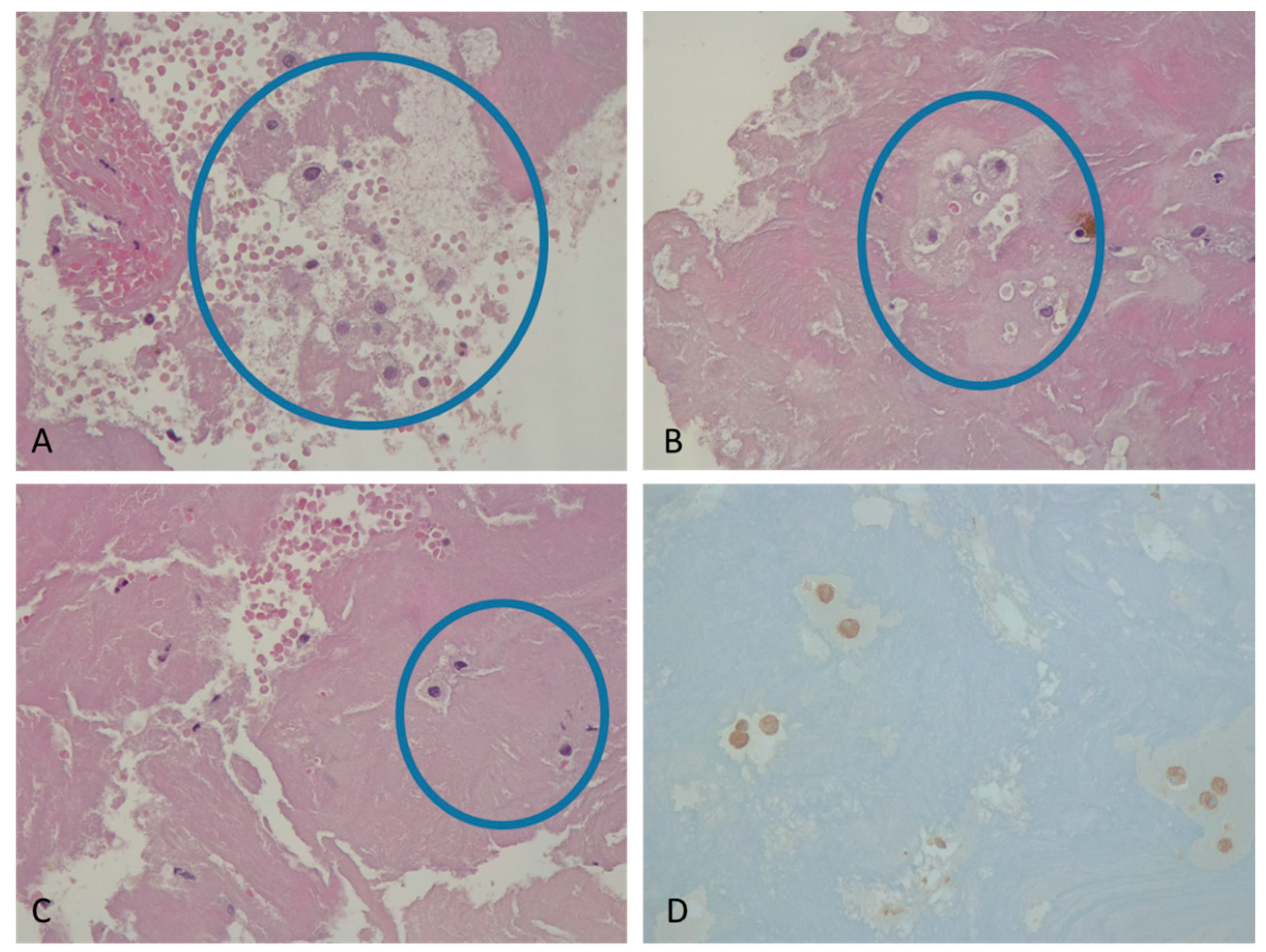
We prospectively included a cohort of 495 consecutive AIS patients with large vessel occlusion (LVO) admitted to our comprehensive stroke center (CSC) who had undergone endovascular treatment (EVT) from 2017 to 2019. Inclusion criteria were as follows: AIS patients ≥ 18 years with an occlusion of the proximal segments of middle, anterior and posterior cerebral arteries, the intracranial part of the internal carotid artery, or the basilar artery; MT with successful thrombus retrieval, regardless of reperfusion status; and informed consent of the patients or their legal representatives during the hospital treatment. Only patients with complete diagnostic and histological workup were included for further analysis, and therefore, we could collect one hundred clots retrieved by mean of MT from patients with AIS. According to the present state-of-the-art [28,29] methods, endovascular approaches included manual thromboaspiration thrombectomy, MT using stent retriever, or a combination of these. The decision concerning EVT was based on the neuroradiological addresses and the current guidelines [28,30], and it was performed with approved MT devices, using stent retrievers, large-bore aspiration catheters, or a combination of both. IV rT-PA was administered to eligible patients, according to Guidelines for Thrombolytic Therapy [29,31]. Collected data included medical history, neurological and physical examination, brain imaging and blood tests.
Each patient underwent a comprehensive clinical assessment to evaluate the presence of carotid atheromatous pathology: ultrasound images, computed tomography angiography (CTA), or magnetic resonance angiography (MRA).
The assumed etiology of AIS was determined according to the TOAST (Trial of Org 10,172 in Acute Stroke Treatment) criteria [1]: (1) cardioembolism; (2) large-artery atherosclerosis; (3) small-vessel occlusion; (4) other determined etiology; or (5) undetermined etiology. Mechanical thrombectomy was performed and thrombus material, taken during the retrieval procedure, was immediately fixed in a solution of 10% provided formalin. After fixation, samples were embedded in paraffin, and thereafter slices of samples were obtained and stained with hematoxylin and eosin (H&E) for histological assessment. An expert pathologist assessed the presence of FCs in each specimen, blinded to the clinical data (Figure 1). An additional pathologist re-reviewed the slides independently. The concordance between the two pathologists was almost perfect (K value: 0.82). In discordant cases, consensus was reached using a double-headed microscope. The presence of FCs was confirmed using immunohistochemistry against CD68 (clone KP1, dilution 1:100; Agilent Technologies, Santa Clara, CA, USA) and an automated immunostainer.
For the statistic description of continuous variables with normal distribution, we used mean and standard deviation (SD). For continuous variables with non-normal distribution, we used median, minimal and maximal values (Min Max), and first and third quartile (I-IIIQ) description. Qualitative data are reported as percentages (%) and frequencies (N). Interferential analyses of groups and correlation analysis were performed by appropriate non-parametric tests: Pearson’s chi-squared test (χ2) or Mann–Whitney U test. Multivariate analyses were performed by multivariate logistic regression tests. We considered as statistically significant p values lower than 0.05. The power calculation was performed with G*power [32], showing that we needed a sample size of n = 68 to detect a correlation between the atherosclerotic etiology of AIS following the TOAST classification and the presence of FCs in the retrieved clot obtained by MT, as well as to compare histological findings with a 2-sided 5% significance level and a power of 80%.

3. Results

We analyzed 100 samples of clots taken from patients affected by ischemic stroke; 29/100 samples had FCs. Consequently, we divided the patients into two groups: the group with FCs in the clot and the group without FCs (NFCs). The characteristics of the two groups (Table 1 and Table 2) were similar: diabetes (25% vs. 25.8%), arterial hypertension (75% vs. 78.8%), hypercholesterolemia (42.9% vs. 42.4%), smoking (17.9% vs. 13.8%), tumors (3.6% vs. 18.5%), potential-embolus-forming arrhythmias (48% vs. 39.3%), ejection fraction (EF) < 40% (9.1% vs. 10%), LDLs >100 mg/dL (56.5% vs. 41.4%), and high-density lipoprotein (HDL) below range (83.3% vs. 86.7%). We did not find significant statistic differences between the two groups in blood parameters: triglycerides, C-reactive protein (CRP), red cells, white cells, platelets, and international normalized ratio (INR). In our sample, the in-hospital mortality was too low to draw any conclusion from difference rate between the two populations, although most cases occurred in NFC patients (six out of seven in-hospital deaths).
We found an ipsilateral atheromatous lesion in the supra-aortic extra-cranial arteries in 18/23 patients of the FC group and in 38/56 of the NFC group. According to the TOAST classification, the etiological diagnosis in the FC group were athero-thrombotic in six (20.7%) patients, cardio embolic in 13 (44.8%) patients, and undetermined in 10 (34.5%) patients.
In the NFC group, the etiological diagnosis was atherothrombotic in 11 (15.5%) patients, cardio-embolic in 28 (39.4%) patients, undetermined in 31 (43.7%), and one (1.4%) patient with other determined etiology.
The chi-squared statistic resulted in 3.537 and a p-value 0.318. Using the Fisher’s exact test, the odds ratio was 1.491 and the p-value resulted in 0.324. For both tests, the p-values are greater than the significance level of 0.05; therefore, we found no significant difference in the distribution of TOAST categories between FCs and NFCs.
Data about in-hospital mortality and a detailed comparison between our findings and TOAST causes are reported as Supplementary Table S1.

4. Discussion

In our study, we histologically evaluated the presence of FCs in 100 clots retrieved from patients with AIS by mechanical thrombectomy. Since the EVT became a novel therapeutic option for IS, researchers focused on the histological analysis of the retrieved clot from the occluded artery [16,17]. This allowed us to characterize the composition of the clot with the purpose of understanding the pathophysiology, determining the etiology, and predicting the outcome of the IS. There are no clear associations between histological patterns or markers of retrieved clots and etiology of stroke. Previous studies focused mainly on the characterization of the clot composition classifying those clots rich in fibrin, red blood cells, white blood cells, platelets and cellular markers [33]. Other studies, using immunohistochemistry, determined the presence of cells of inflammation and immunity [20,24]. Few studies analyzed the presence of FCs in the arterial clots. Sadoshima et al. [15] observed the presence of FCs taken in patients who died for stroke, while Hashimoto et al. [34] described the presence of “atheromatous gruels” formed by FCs or foamy lipophages, cholesterol clefts and fibrous cap, in vivo retrieved clots. FCs are the principal constituents of the core in atheromatous plaques and their presence in the clot suggests a fissure of the plaque in the formation of the thrombus. Through optical microscopy, we observed the presence of FCs in 29/100 (29%) of analyzed clots. FCs exhibit impaired immune functions and prolonged inflammation, contributing to the progression of stroke pathology in the chain of chronic inflammation after stroke [35]. CRP has been suggested to promote platelet activation and FC generation through macrophages differentiation; it has been associated with large-artery atherotrombotic events [36]. However, according to the TOAST classification, only four of these patients were diagnosed as atherothrombotic stroke (13.8%) and 13 patients had an ipsilateral atheromatous lesion in the supra-aortic extra-cranial arteries. In our opinion, the discrepancy between the finding of an atheromatous lesion in the supra-aortic arteries and the presence of the FCs in the clot should not surprise us. Atheromatous lesions may break down completely, especially when IVT is performed in bridging procedures, leading to a negative observation in the neuroimaging. Furthermore, the aortic arch is an underestimated localization of atherosclerosis. Actually, 12/29 (41.4%) were classified as undetermined according to TOAST classification: five out of these twelve patients with FCs did not complete investigations, while one patient was classified as cryptogenic stroke after standard evaluation. The remaining six patients were considered with stroke of undetermined cause, due to the presence of no probable, but one or more possible, causes identified (stroke “of possibly determined origin”) [37]. Finally, it may not be excluded that small plaques, even in the presence of atrial fibrillation, may be the cause of the IS. This could explain the high rate of FCs found in patients who presumably had cardioembolic stroke (10/29, 34.5%). Thus, the TOAST classification is to be considered useful for identifying the stroke etiology, but the probabilistic nature of this tool should be taken into account. In this setting, we found that the histological observation of the FCs in the retrieved clot is a feasible method that can influence the physician toward a more reliable etiology identification and ultimately choose the proper therapy.

5. Limitations

There are intrinsic limitations of the clot analysis, often leading to inconsistent and conflicting results. The extraction of the clot during the endovascular procedure has numerous bias. For instance, cardio-embolic clots seem to be more fragile and difficult to retain, especially after IVT [38]. Moreover, it cannot be said that the orientation of the clot in the vessel, once retrieved, led to misinterpretation of the data obtained. The main pitfall of this study was the lack of systemic investigation of slices along the thrombus or volumetric assessment of the presence of FCs. In addition, we could not evaluate systematically the presence of macrophage markers with a preliminary blotting before assessing the analysis for lipids. A further limitation in this study was the lack of complete imaging assessment of atherosclerosis disease in all-arterial network that supplies the ischemic brain area (in particular the aortic trunk and intracranial vessels). Finally, the COVID-19 pandemic affected recruitment (suspension in 2020 and impossible restart, due to the redeployment of the clinical research workforce to support the pandemic clinical services), and therefore, we have not been able to increase the sample size.

6. Conclusions

In secondary stroke prevention, one of the main challenges is to identify the correct etiology of the clot formation in order to start as soon as possible the best medical therapy. Histological thrombus composition in LVO may represent an interesting research question, as it is an important feature that may help to specify stroke pathogenesis, differentiating between cardioembolic and atherothrombotic stroke causes. Further clinical and radiological factors predicting stroke etiology might support histological findings, i.e., considering the systematic assessment of hyperdense vessel signs in native computed tomography and/or susceptibility vessel signs in susceptibility-weighted imaging on brain magnetic resonance imaging [39]. Our study showed the presence of FCs in approximately one-third of the retrieved clots, but the identification of the clot that presumably comes from the atheromatous plaque rupture tended to disagree with the clinical diagnosis, even if the difference between FC and NFC groups did not reach the statistical significance. Future studies may reveal their potential to disclose clot origin or specific patient characteristics, guiding treatment options.

Supplementary Materials

The following supporting information can be downloaded at: https://www.mdpi.com/article/10.3390/ctn8020017/s1, Table S1: In-hospital mortality and detailed comparison between anatomopathological findings and presumed stroke etiology by TOAST criteria.

Author Contributions

F.G. (Fabrizio Giammello): Study concept or design; drafting/revision of the manuscript for content, including medical writing for content; analysis or interpretation of data. A.C.: Study concept or design; drafting/revision of the manuscript for content, including medical writing for content. D.C.: Study concept or design; major role in the acquisition of data. S.G.: Study concept or design; drafting/revision of the manuscript for content, including medical writing for content; analysis or interpretation of data. V.B.: Drafting/revision of the manuscript for content, including medical writing for content; major role in the acquisition of data; analysis or interpretation of data. P.L.S.: Drafting/revision of the manuscript for content, including medical writing for content. M.C.F.: Drafting/revision of the manuscript for content, including medical writing for content. J.D.C.: Major role in the acquisition of data. M.C.: Analysis or interpretation of data. C.D.: Analysis or interpretation of data. F.G. (Francesco Grillo): Analysis or interpretation of data. S.A.: Analysis or interpretation of data. A.T. (Agostino Tessitore): Major role in the acquisition of data. S.L.V.: Major role in the acquisition of data. R.F.M.: Drafting/revision of the manuscript for content, including medical writing for content. C.C.: Major role in the acquisition of data; study concept or design. A.T. (Antonio Toscano): Drafting/revision of the manuscript for content, including medical writing for content; analysis or interpretation of data. All authors have read and agreed to the published version of the manuscript.

Funding

This research did not receive any specific grant from funding agencies in the public, commercial, or not-for-profit sectors.

Institutional Review Board Statement

The paper does not report on primary research. Ethical review and approval were waived for this study because all data analyzed were collected as part of routine diagnosis and treatment.

Informed Consent Statement

Not applicable.

Data Availability Statement

Additional unpublished data from the study are not available.

Conflicts of Interest

The authors declare that they have no conflicts of interest related to this work.

References

  1. Adams, H.P.; Bendixen, B.H.; Kappelle, L.J.; Biller, J.; Love, B.B.; Gordon, D.L.; Marsh, E.E. Classification of subtype of acute ischemic stroke. Definitions for use in a multicenter clinical trial. TOAST. Trial of Org 10172 in Acute Stroke Treatment. Stroke 1993, 24, 35–41. [Google Scholar] [CrossRef] [PubMed]
  2. Palomeras Soler, E.; Casado Ruiz, V. Epidemiology and Risk Factors of Cerebral Ischemia and Ischemic Heart Diseases: Similarities and Differences. Curr. Cardiol. Rev. 2010, 6, 138–149. [Google Scholar] [CrossRef] [PubMed]
  3. Deb, P.; Sharma, S.; Hassan, K.M. Pathophysiologic mechanisms of acute ischemic stroke: An overview with emphasis on therapeutic significance beyond thrombolysis. Pathophysiology 2010, 17, 197–218. [Google Scholar] [CrossRef] [PubMed]
  4. Arboix, A.; Oliveres, M.; Massons, J.; Pujades, R.; García-Eroles, L. Early differentiation of cardioembolic from atherothrombotic cerebral infarction: A multivariate analysis. Eur. J. Neurol. 1999, 6, 677–683. [Google Scholar] [CrossRef] [PubMed]
  5. Savelieva, I.; Bajpai, A.; Camm, A.J. Stroke in atrial fibrillation: Update on pathophysiology, new antithrombotic therapies, and evolution of procedures and devices. Ann. Med. 2007, 39, 371–391. [Google Scholar] [CrossRef] [PubMed]
  6. Viles-Gonzalez, J.F.; Fuster, V.; Badimon, J.J. Atherothrombosis: A widespread disease with unpredictable and life-threatening consequences. Eur. Heart J. 2004, 25, 1197–1207. [Google Scholar] [CrossRef] [PubMed]
  7. Libby, P.; Ridker, P.M.; Hansson, G.K. Progress and challenges in translating the biology of atherosclerosis. Nature 2011, 473, 317–325. [Google Scholar] [CrossRef] [PubMed]
  8. Weber, C.; Noels, H. Atherosclerosis: Current pathogenesis and therapeutic options. Nat. Med. 2011, 17, 1410–1422. [Google Scholar] [CrossRef] [PubMed]
  9. Sargolzaei, J.; Chamani, E.; Kazemi, T.; Fallah, S.; Soori, H. The role of adiponectin and adipolin as anti-inflammatory adipokines in the formation of macrophage foam cells and their association with cardiovascular diseases. Clin. Biochem. 2018, 54, 1–10. [Google Scholar] [CrossRef] [PubMed]
  10. Chistiakov, D.A.; Melnichenko, A.A.; Myasoedova, V.A.; Grechko, A.V.; Orekhov, A.N. Mechanisms of foam cell formation in atherosclerosis. J. Mol. Med. 2017, 95, 1153–1165. [Google Scholar] [CrossRef] [PubMed]
  11. Cochain, C.; Zernecke, A. Macrophages in vascular inflammation and atherosclerosis. Pflug. Arch. Eur. J. Physiol. 2017, 469, 485–499. [Google Scholar] [CrossRef] [PubMed]
  12. Yu, X.H.; Fu, Y.C.; Zhang, D.W.; Yin, K.; Tang, C.K. Foam cells in atherosclerosis. Clin. Chim. Acta 2013, 424, 245–252. [Google Scholar] [CrossRef] [PubMed]
  13. Hai, Q.; Ritchey, B.; Robinet, P.; Alzayed, A.M.; Brubaker, G.; Zhang, J.; Smith, J.D. Quantitative Trait Locus Mapping of Macrophage Cholesterol Metabolism and CRISPR/Cas9 Editing Implicate an ACAT1 Truncation as a Causal Modifier Variant. Arter. Thromb. Vasc. Biol. 2018, 38, 83–91. [Google Scholar] [CrossRef] [PubMed]
  14. Ritchey, B.; Hai, Q.; Han, J.; Barnard, J.; Smith, J.D. Genetic variant in 3′ untranslated region of the mouse pycard gene regulates inflammasome activity. Elife 2021, 10, e68203. [Google Scholar] [CrossRef] [PubMed]
  15. Sadoshima, S.; Fukushima, T.; Tanaka, K. Cerebral artery thrombosis and intramural hemorrhage. Stroke 1979, 10, 411–414. [Google Scholar] [CrossRef] [PubMed]
  16. Sporns, P.B.; Hanning, U.; Schwindt, W.; Velasco, A.; Minnerup, J.; Zoubi, T.; Heindel, W.; Jeibmann, A.; Niederstadt, T.U. Ischemic Stroke: What Does the Histological Composition Tell Us about the Origin of the Thrombus? Stroke 2017, 48, 2206–2210. [Google Scholar] [CrossRef] [PubMed]
  17. Heo, J.H.; Nam, H.S.; Kim, Y.D.; Choi, J.K.; Kim, B.M.; Kim, D.J.; Kwon, I. Pathophysiologic and Therapeutic Perspectives Based on Thrombus Histology in Stroke. J. Stroke 2020, 22, 64–75. [Google Scholar] [CrossRef] [PubMed]
  18. Marder, V.J.; Chute, D.J.; Starkman, S.; Abolian, A.M.; Kidwell, C.; Liebeskind, D.; Ovbiagele, B.; Vinuela, F.; Duckwiler, G.; Jahan, R.; et al. Analysis of thrombi retrieved from cerebral arteries of patients with acute ischemic stroke. Stroke 2006, 37, 2086–2093. [Google Scholar] [CrossRef]
  19. Singh, P.; Kaur, R.; Kaur, A. Clot composition and treatment approach to acute ischemic stroke: The road so far. Ann. Indian Acad. Neurol. 2013, 16, 494–497. [Google Scholar] [CrossRef] [PubMed]
  20. Schuhmann, M.K.; Gunreben, I.; Kleinschnitz, C.; Kraft, P. Immunohistochemical Analysis of Cerebral Thrombi Retrieved by Mechanical Thrombectomy from Patients with Acute Ischemic Stroke. Int. J. Mol. Sci. 2016, 17, 298. [Google Scholar] [CrossRef]
  21. Niesten, J.M.; van der Schaaf, I.C.; van Dam, L.; Vink, A.; Vos, J.A.; Schonewille, W.J.; de Bruin, P.C.; Mali, W.P.T.M.; Velthuis, B.K. Histopathologic composition of cerebral thrombi of acute stroke patients is correlated with stroke subtype and thrombus attenuation. PLoS ONE 2014, 9, e88882. [Google Scholar] [CrossRef]
  22. Boeckh-Behrens, T.; Kleine, J.F.; Zimmer, C.; Neff, F.; Scheipl, F.; Pelisek, J.; Schirmer, L.; Nguyen, K.; Karatas, D.; Poppert, H. Thrombus histology suggests cardioembolic cause in cryptogenic stroke. Stroke 2016, 47, 1864–1871. [Google Scholar] [CrossRef] [PubMed]
  23. Kim, S.; Yoon, W.; Kim, T.; Kim, H.; Heo, T.; Park, M. Histologic analysis of retrieved clots in acute ischemic stroke: Correlation with stroke etiology and gradient-echo MRI. Am. J. Neuroradiol. 2015, 36, 1756–1762. [Google Scholar] [CrossRef] [PubMed]
  24. Dargazanli, C.; Rigau, V.; Eker, O.; Riquelme Bareiro, C.; Machi, P.; Gascou, G.; Arquizan, C.; Ayrignac, X.; Mourand, I.; Corlobe, A.; et al. High CD3+ cells in intracranial thrombi represent a biomarker of atherothrombotic stroke. PLoS ONE 2016, 11, e0154945. [Google Scholar] [CrossRef] [PubMed]
  25. Krajíčková, D.; Krajina, A.; Šteiner, I.; Vyšata, O.; Herzig, R.; Lojík, M.; Chovanec, V.; Raupach, J.; Renc, O.; Waishaupt, J.; et al. Fibrin clot architecture in acute ischemic stroke treated with mechanical thrombectomy with stent-retrievers―Cohort study―. Circ. J. 2018, 82, 866–873. [Google Scholar] [CrossRef] [PubMed]
  26. Brinjikji, W.; Duffy, S.; Burrows, A.; Hacke, W.; Liebeskind, D.; Majoie, C.B.L.M.; Dippel, D.W.J.; Siddiqui, A.H.; Khatri, P.; Baxter, B.; et al. Correlation of imaging and histopathology of thrombi in acute ischemic stroke with etiology and outcome: A systematic review. J. Neurointerv. Surg. 2017, 9, 529–534. [Google Scholar] [CrossRef] [PubMed]
  27. Sporns, P.B.; Hanning, U.; Schwindt, W.; Velasco, A.; Buerke, B.; Cnyrim, C.; Minnerup, J.; Heindel, W.; Jeibmann, A.; Niederstadt, T. Ischemic Stroke: Histological Thrombus Composition and Pre-Interventional CT Attenuation Are Associated with Intervention Time and Rate of Secondary Embolism. Cerebrovasc. Dis. 2017, 44, 344–350. [Google Scholar] [CrossRef] [PubMed]
  28. Turc, G.; Bhogal, P.; Fischer, U.; Khatri, P.; Lobotesis, K.; Mazighi, M.; Schellinger, P.D.; Toni, D.; De Vries, J.; White, P.; et al. European Stroke Organisation (ESO)—European Society for Minimally Invasive Neurological Therapy (ESMINT) Guidelines on Mechanical Thrombectomy in Acute Ischemic Stroke. J. Neurointervent. Surg. 2023, 15, e8. [Google Scholar] [CrossRef] [PubMed]
  29. Powers, W.J.; Rabinstein, A.A.; Ackerson, T.; Adeoye, O.M.; Bambakidis, N.C.; Becker, K.; Biller, J.; Brown, M.; Demaerschalk, B.M.; Hoh, B. Guidelines for the early management of patients with acute ischemic stroke: 2019 update to the 2018 guidelines for the early management of acute ischemic stroke a guideline for healthcare professionals from the American Heart Association/American Stroke A. Stroke 2019, 50, E344–E418. [Google Scholar] [CrossRef]
  30. Wahlgren, N.; Moreira, T.; Michel, P.; Steiner, T.; Jansen, O.; Cognard, C.; Mattle, H.P.; van Zwam, W.; Holmin, S.; Tatlisumak, T.; et al. Mechanical thrombectomy in acute ischemic stroke: Consensus statement by ESO-Karolinska Stroke Update 2014/2015, supported by ESO, ESMINT, ESNR and EAN. Int. J. Stroke 2016, 11, 134–147. [Google Scholar] [CrossRef] [PubMed]
  31. Ahmed, N.; Audebert, H.; Turc, G.; Cordonnier, C.; Christensen, H.; Sacco, S.; Sandset, E.C.; Ntaios, G.; Charidimou, A.; Toni, D.; et al. Consensus statements and recommendations from the ESO-Karolinska Stroke Update Conference, Stockholm 11–13 November 2018. Eur. Stroke J. 2019, 4, 307–317. [Google Scholar] [CrossRef] [PubMed]
  32. Faul, F.; Erdfelder, E.; Lang, A.-G.; Buchner, A. G*Power 3: A flexible statistical power analysis program for the social, behavioral, and biomedical sciences. Behav. Res. Methods 2007, 39, 175–191. [Google Scholar] [CrossRef] [PubMed]
  33. Di Meglio, L.; Desilles, J.-P.; Ollivier, V.; Nomenjanahary, M.S.; Di Meglio, S.; Deschildre, C.; Loyau, S.; Olivot, J.-M.; Blanc, R.; Piotin, M.; et al. Acute ischemic stroke thrombi have an outer shell that impairs fibrinolysis. Neurology 2019, 93, e1686–e1698. [Google Scholar] [CrossRef]
  34. Hashimoto, T.; Hayakawa, M.; Funatsu, N.; Yamagami, H.; Satow, T.; Takahashi, J.C.; Nagatsuka, K.; Ishibashi-Ueda, H.; Kira, J.-I.; Toyoda, K.; et al. Histopathologic Analysis of Retrieved Thrombi Associated with Successful Reperfusion after Acute Stroke Thrombectomy. Stroke 2016, 47, 3035–3037. [Google Scholar] [CrossRef] [PubMed]
  35. Zbesko, J.C.; Stokes, J.; Becktel, D.A.; Doyle, K.P. Targeting foam cell formation to improve recovery from ischemic stroke. Neurobiol. Dis. 2023, 181, 106130. [Google Scholar] [CrossRef] [PubMed]
  36. Llombart, V.; Garcia-Berrocoso, T.; Bustamante, A.; Fernandez-Cadenas, I.; Montaner, J. Cardioembolic Stroke Diagnosis Using Blood Biomarkers. Curr. Cardiol. Rev. 2013, 9, 340. [Google Scholar] [CrossRef] [PubMed]
  37. Saver, J.L. Cryptogenic Stroke. N. Engl. J. Med. 2016, 374, 2065–2074. [Google Scholar] [CrossRef] [PubMed]
  38. Duffy, S.; McCarthy, R.; Farrell, M.; Thomas, S.; Brennan, P.; Power, S.; O’hare, A.; Morris, L.; Rainsford, E.; MacCarthy, E.; et al. Per-Pass Analysis of Thrombus Composition in Patients With Acute Ischemic Stroke Undergoing Mechanical Thrombectomy. Stroke 2019, 50, 1156–1163. [Google Scholar] [CrossRef] [PubMed]
  39. Beyeler, M.; Grunder, L.; Göcmen, J.; Steinauer, F.; Belachew, N.F.; Kielkopf, M.; Clénin, L.; Mueller, M.; Silimon, N.; Kurmann, C.; et al. Absence of susceptibility vessel sign and hyperdense vessel sign in patients with cancer-related stroke. Front. Neurol. 2023, 14, 1148152. [Google Scholar] [CrossRef] [PubMed]
Figure 1. Microscopic view of a retrieved clot showed FCs in a hematoxylin–eosin-stained section. Presence of FCs in hematoxylin-and-eosin-stained histological slides of different clots (blue circles, AC). FCs were positive for CD68 immunohistochemistry (D).
Ctn 08 00017 g001
Table 1. Characteristics of patients.
NFC (n = 71)FC (n = 29)
Min IQMedianIIIQ MaxMin IQMedianIIIQMax
Triglycerides
mg/dL
406989111274427483106162
CRP
mg/dL
0.10.30.92.3523.70.10.20.419.7
Red cells
×106/mm3
2.474.094.554.946.153.094.234.514.635.44
White cells
×104/mm3
4.57.48.610.924.94.68.29.811.999.9
Platelets
×105/mm3
0.571.862.202.703.90.482.052.382.503.56
INR0.961.021.081.163.190.921.071.11.192.6
NFCs, Non-foam cells; FCs, foam cells; IQ, first interquartile; IIIQ, third interquartile; CRP, C-reactive protein; platelet; INR, international normalized ratio.
Table 2. Characteristics of patients.
NFC (n = 71) FC (n = 29)
%n%np Value
Diabetes25.817/66257/280.96
Hypertension78.852/667521/280.77
Hypercholesterolemia42.428/6642.912/280.98
Smoking13.89/6517.95/280.76
Tumors18.512/653.61/280.26
Potential-embolus-forming arrhythmia39.324/614812/250.54
EF < 40%106/609.12/220.46
Ipsilateral plaque in US study73.138/5658.318/230.47
LDL > 100 mg/dL41.424/5856.513/230.29
HDL below range86.752/6083.320/240.81
NFCs, Non-foam cells; FCs, foam cells; potential-embolus-forming arrhythmia: atrial flutter, atrial fibrillation; EF, ejection fraction; ipsilateral plaque at ultrasound (US) study: atheromasic plaque on the same side of cerebral ischemia; LDL, low-density lipoprotein; HDL, high-density lipoprotein.
Disclaimer/Publisher’s Note: The statements, opinions and data contained in all publications are solely those of the individual author(s) and contributor(s) and not of MDPI and/or the editor(s). MDPI and/or the editor(s) disclaim responsibility for any injury to people or property resulting from any ideas, methods, instructions or products referred to in the content.

