Review

Central and Peripheral Immunity Responses in Parkinson’s Disease: An Overview and Update

Lllian S. Wells Department of Neurosurgery, University of Florida, Gainesville, FL 32610, USA
*
Author to whom correspondence should be addressed.
Neuroglia 2025, 6(2), 17; https://doi.org/10.3390/neuroglia6020017
Submission received: 2 March 2025 / Revised: 27 March 2025 / Accepted: 31 March 2025 / Published: 4 April 2025

Abstract

:
Parkinson’s disease (PD) is a progressive neurodegenerative disorder characterized by motor and non-motor symptoms, with increasing evidence supporting the role of immune dysregulation in its pathophysiology. Neuroinflammation, mediated by microglial activation, pro-inflammatory cytokine production, and blood–brain barrier dysfunction, plays a crucial role in dopaminergic neuronal degeneration. Furthermore, peripheral immune changes, including T cell infiltration, gut microbiota dysbiosis, and systemic inflammation, contribute to disease progression. The bidirectional interaction between the central and peripheral immune systems suggests that immune-based interventions may hold therapeutic potential. While dopaminergic treatments remain the standard of care, immunomodulatory therapies, monoclonal antibodies targeting α-synuclein, and deep brain stimulation (DBS) have demonstrated immunological effects, though clinical efficacy remains uncertain. Advances in immune phenotyping offer new avenues for personalized treatment approaches, optimizing therapeutic responses by stratifying patients based on inflammatory biomarkers. This review highlights the complexities of immune involvement in PD and discusses emerging strategies targeting immune pathways to develop disease-modifying treatments.

1. Introduction

Parkinson’s disease (PD) is the second most common neurodegenerative disease, a chronic movement disorder that is clinically characterized by motor and non-motor symptoms [1,2]. Pathophysiologically, PD is associated with the preferential degeneration of dopaminergic neurons in the substantia nigra (SN) and misfolded α-synuclein aggregates resulting in Lewy bodies [1,2,3]. The subsequent dopamine deficiency in the basal ganglia causes a movement disorder with classical parkinsonian motor symptoms including bradykinesia, resting tremor, rigidity, and postural instability [1,2]. Although the etiology of PD remains unelucidated, research suggests multifactorial factors such as aging, genetics, and environmental exposures are at play [1,2,3]. Recently, the literature has emphasized the involvement of the immune system and neuroinflammation in the onset and progression of PD.
Neuroinflammation is a fundamental CNS immune response elicited by a plethora of inflammatory factors such as infection, tissue injury, and aging [3,4,5]. While acute neuroinflammation serves a neuroprotective role by safeguarding the CNS and facilitating tissue repair, chronic neuroinflammation has previously been associated with neurodegenerative disease and has become a prominent feature of PD pathophysiology [4,5,6]. In PD, chronic neuroinflammation induces sustained pro-inflammatory cytokine production, glial activation, and leukocyte infiltration, ultimately leading to neuronal damage and death [3,5,6,7]. Elevated systemic levels of pro-inflammatory cytokines, such as IL-1β, IL-2, IL-4, IL-6, TNFα, and IFN-γ, reflect the underlying neuroinflammatory processes and are associated with dopaminergic neuron degeneration in the SN [4,8,9,10]. An imbalance between elevated pro-inflammatory cytokines and reduced anti-inflammatory cytokines correlates with disease severity and worsening motor symptoms [7,9,11]. Moreover, the overexpression of these cytokines secreted by reactive microglia has implicated the involvement of microglia activation in the development and progression of PD [6]. Microglia, the resident macrophages in the brain, along with astrocytes mediate the neuroinflammatory response after being activated by toxic α-synuclein aggregates, [4,5,6,7]. Conversely, neuroinflammation has also been associated with increasing α-synuclein misfolding and aggregation [3,6]. Reactive microglia function as antigen-presenting cells (APCs) and secrete inflammatory mediators including TNFα, IL-6, and reactive oxygen species (ROS), which in turn exacerbate neurotoxic effects in dopaminergic neurons [3,6,12]. The crosstalk between microglia and astrocytes has been shown to amplify immune responses and induce apoptosis in dopaminergic neurons [3,5,13].
Beyond the central immune response, PD-associated neuroinflammation has been linked to abnormalities in the peripheral adaptive immune system [14] (summarized in Figure 1). The prodromal stage of PD is associated with peripheral inflammation, with non-motor symptoms reflecting α-synuclein pathology in the gut [5,6]. Misfolded α-synuclein has been linked to innate and adaptive immune response through interactions between microglia and T lymphocytes [3,5,6,9]. Studies on human PD pathology and animal models have revealed increased peripheral T lymphocytes and an abundance of APCs, suggesting communication between the peripheral nervous system (PNS) and the CNS [3,9]. Additionally, humoral immune changes in PD include B cells and plasma cells contributing to the production of α-synuclein antibodies [10,12,15,16]. Emerging evidence also highlights the role of gut microbiota in modulating peripheral immune responses, potentially influencing PD pathogenesis through alterations in microbial composition and metabolite production [3,5,6,7]. This pro-inflammatory state also compromises the blood–brain barrier (BBB), increasing its permeability and promoting alterations in the enteric nervous system via gut dysbiosis and a leaky gut mechanism [3,5,6,7]. The propagation of α-synuclein aggregates from the periphery to the CNS is enhanced by the leaky gut allowing leukocyte infiltration into the blood and subsequent circulation to the brain through altered BBB vascular permeability [3,5,6,7]. Further research on peripheral immune dysregulation holds promising therapeutic value for the development of PD inflammatory biomarkers and earlier detection of PD onset.
Although current PD therapies predominantly rely on symptomatic treatment, the development of innovative pharmacotherapeutics have shone a promising light on the future of disease-modifying treatments [17]. While dopamine replacement remains the gold standard for symptom relief, studies suggest that dopaminergic drugs may also exert immunomodulatory effects [18]. Evidence indicates that dopaminergic signaling influences CNS immune cell functions, including chemotaxis, phagocytosis, antigen presentation, and cytokine secretion [19,20,21]. Additionally, deep brain stimulation (DBS) has been investigated as a potential disease-modifying therapy [22]. DBS has been shown to reverse PD-associated immune changes, including alterations in circulating cytokine levels, and promote a shift from pro-inflammatory helper T cells to anti-inflammatory regulatory T cells [22,23,24]. Clinical trials are also exploring immunotherapies such as vaccines and monoclonal antibodies designed to target α-synuclein aggregates with high specificity [5,12,25]. However, further research is necessary to elucidate the underlying immunological mechanisms and optimize these novel therapeutic approaches [25]. In this review, we will explore the complexities of peripheral immune responses and dysregulation in PD while examining emerging the therapeutic strategies aimed at targeting these immune pathways.

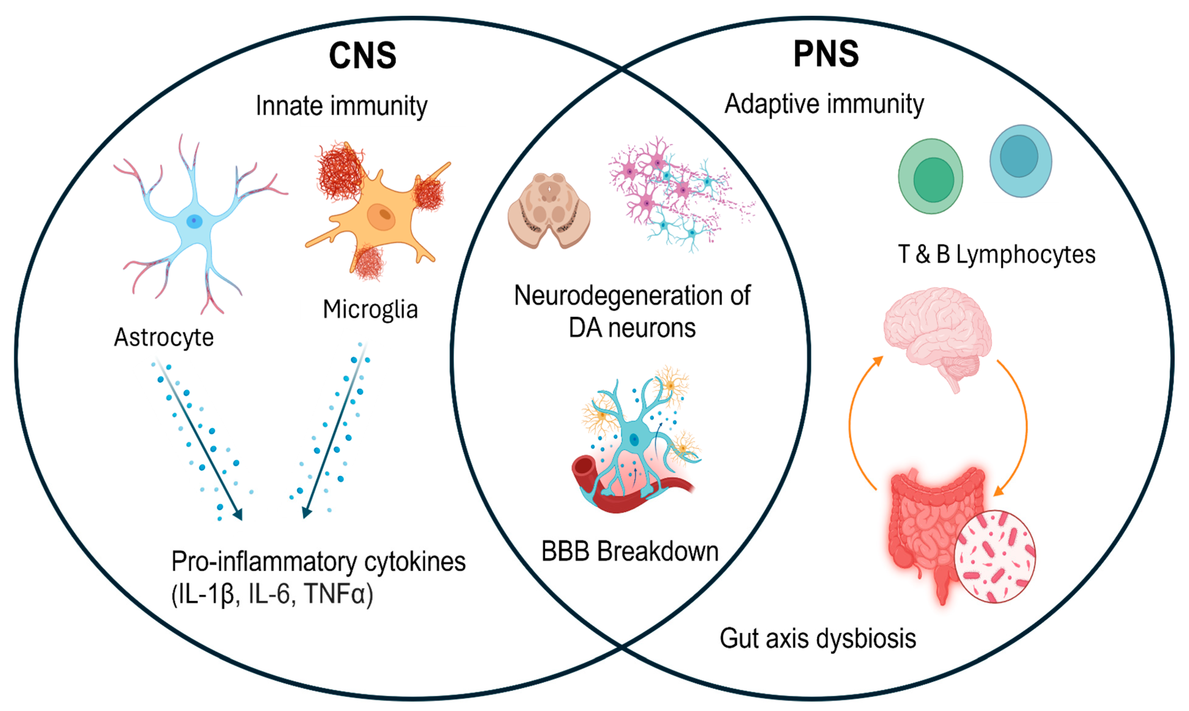
2. Bidirectional Central–Peripheral Immune Interplay

Peripheral immune activation has been implicated in the activation and maintenance of central neuroinflammation in PD pathogenesis. While components of the central–peripheral immune interplay remain to be elucidated, the current literature emphasizes a two-fold approach consisting of microglial activation and T cell infiltration within the CNS [6,7]. Microglia are the most abundant immune cells in the CNS and have been associated with neurodegenerative disease progression through α-synuclein mediated pathways [26,27]. The presence of peripheral immune components and autoantigens, such as α-synuclein, is capable of infiltrating the brain parenchyma due to compromised tight junctions, BBB breakdown, and “leaky gut” [7]. Within the CNS, activated microglia function as APCs and elicit CD4 T cell proliferation via MHC class II antigen presentation [28,29]. Interestingly, a study analyzing postmortem PD brains confirmed these findings of excessive microglial activation and discovered dopaminergic neurons in the SN overexpressed MHC-II cell surface receptors, namely HLA-DR [30]. Microglial activation combined with an abnormal expression of HLA molecules elicit central neuroinflammation, which is further propagated by CD4 and CD8 T lymphocyte infiltration through increased BBB permeability [3,30,31]. Imaging reports have supported theories on BBB breakdown in PD patients, demonstrating increased permeability and angiogenesis in the basal ganglia and SN [5,32,33]. The degenerative loss of tight junctions directly compromises the brain’s status as an immune-privileged site, as evidenced by an increase in macrophages, microglia, dendritic cells, and T cell infiltrates [5]. Notably, circulating lymphocytes in the periphery are significantly decreased in PD [22]. This shift could arise from multifactorial causes including a decrease in regulatory T cell expression and increased T cell infiltrates in the CNS [24,34]. The bidirectional immune interplay causes reactive microglia, among other primed immune cells, to perpetuate inflammatory cascades via the release of pro-inflammatory cytokines. The literature supports this theory of dysregulated cytokines influencing chronic neuroinflammation, as it evidently shows elevated levels of TNF-α, IL-1β, IL-6, and IFN-γ following microglia activation [3,35,36]. Innate immunity continuously aids in the proliferation of these signals through the formation of inflammasomes and the activation of macrophages into pro-inflammatory M1 states [5,6]. The adaptive immune system is also at play by inducing T cell differentiation into T helper 1 (Th1) and T helper 17 (Th17), further amplifying the secretion of IFN-γ, TNF-α, and IL-2 [5]. Taken together, the simultaneous dysregulation of pro-inflammatory cytokine production and reduction in anti-inflammatory modulators, such as T regulatory cells and M2 macrophages, explains one of the pathological hallmarks of PD.
While PD is characterized by CNS dopamine depletion, residual dopamine has been found to activate microglial cells and enhance the release of pro-inflammatory cytokines [19,37]. The role of dopamine and dopamine transporter (DAT) dysregulation has been explored as an additional link between the central and peripheral immune responses [37,38]. DAT is the major regulator of dopaminergic expression in the CNS and sets the tone for peripheral immune cell regulation, a major immunological function of dopamine [37,38]. Of note, DAT directly interacts with α-synuclein and consequently experiences a significant impairment of DAT uptake in both the peripheral and central dopaminergic neurons [39,40]. This decrease in DAT dopamine uptake has been clinically correlated to an earlier onset and increased the severity of dyskinetic symptoms [41]. The quantification of DAT uptake capacity serves as the gold standard for visualizing dopaminergic degeneration and death in PD patients, providing valuable insight to the disease’s progression and severity [38,42]. Similarly, immune phenotyping of lymphocyte infiltrates, HLA antigen presentation molecules, and dysregulated cytokines may elucidate disease mechanisms and guide early PD diagnosis and immunotherapeutics.
Studies investigating this bidirectional interplay provide the foundational basis for targeting peripheral immune pathways through a combination of therapeutics including anti-inflammatory drugs and immunosuppressants [13,43]. The application of immunosuppressants in the setting of PD could inhibit the release of aforementioned pro-inflammatory cytokines, prevent immune cell extravasation, and modulate MHC antigen presentation to T lymphocytes, with the common goal of mitigating central neuroinflammation [13,43,44]. Moreover, understanding the complex differentiation of T cells throughout PD pathogenesis may pave the way for immunomodulatory therapies that enhance regulatory T cell activity and subsequently restore prior imbalances in T lymphocytes and immune tolerance [13,45].

3. Peripheral Immune Dysregulation and Its Impact on PD Pathogenesis

Peripheral immune activation primes microglia in the CNS, promoting a pro-inflammatory response that amplifies neuroinflammation. In response to infection, microglia release pro-inflammatory cytokines, heightening inflammation in the CNS [46,47]. Systemic inflammation further sustains this response by recruiting immune cells that activate microglia, prolonging neuroinflammation [46]. A study conducted by Xie et al. demonstrated that peripheral immune activation drives neuroinflammation through microglia and monocytes. Lipopolysaccharide (LPS) administration in Sprague–Dawley rats elevated pro-inflammatory cytokines, including TNF-α and IL-1β [47]. Subsequent depletion of monocytes or microglia resulted in a significant reduction in the mRNA expression of both TNF- α and IL-1β compared to control mice, underscoring their role in neuroinflammation [47]. Similarly, Gaviglio et al. showed that IL-12 and IL-18 co-expression during LPS-induced sterile inflammation increased TNF-α, IL-1β, and IFN-γ secretion, leading to microglial activation via MHC-II upregulation [46]. IL-12 and IL-18 expression also increased the recruitment of pro-inflammatory leukocytes and monocytes, altering the immune cell composition of the brain and further increasing microglia activation.
Breakdown of the BBB allows infiltration of peripheral immune cells and inflammatory mediators into the CNS, perpetuating neuroinflammation and dopaminergic neuronal loss [48]. The BBB preserves CNS homeostasis but is compromised by systemic inflammation [48]. Mechanisms such as NADPH oxidase-driven ROS production, NOD-like receptor protein 3 (NLRP3) inflammasome activation, and gasdermin D signaling disrupt tight junctions, increasing permeability [49,50,51] (Figure 2). Al-Bachari et al. observed heightened BBB disruption in PD patients using contrast-enhanced MRI, particularly in posterior white matter regions [52]. This disruption enables the infiltration of T cells, which leads to enhanced microglial activation and hence neuroinflammation [53]. Additionally, CD4+ and CD8+ T cells have been found in elevated levels in postmortem brain tissue in mice, further confirming the role of peripheral immune cells in disease progression [34].
Dysbiosis of the gut microbiota and increased intestinal permeability (“leaky gut”) also contribute to systemic inflammation, which feeds into central neuroinflammatory processes. Gut microbiota dysbiosis, characterized by an elevated transport of toxic metabolites into the blood stream and increased gastric lining permeability, plays a major role in promoting systemic inflammation and driving neuroinflammation [54]. The gut microbiota plays an important role in regulating the transport of beneficial substances to the gut while preventing toxic metabolites from entering the bloodstream [54,55]. When dysregulation occurs, harmful substances such as LPS and other bacterial products enter systemic circulation, triggering cytokine production, which crosses the BBB, intensifying neuroinflammation [56,57]. Additionally, dysbiosis of the gut microbiota can alter tryptophan metabolism by activating the kynurenine pathway, leading to the production of quinolinic acid, a metabolite associated with increased neuroinflammation [58]. Elevated levels of quinolinic acid have been implicated in neurodegenerative diseases such as PD [55]. Its presence in cerebrospinal fluid is associated with increased pro-inflammatory cytokines, contributing to neuroinflammation and neurodegenerative disease progression [58].
Clinical evidence highlights systemic inflammation’s role in PD, such as correlations between neutrophil-to-lymphocyte ratio (NLR) and striatal DAT levels, linking peripheral immune changes to neurodegeneration. The NLR serves as a biomarker of systemic inflammation and has been linked to PD progression. Moghaddam et al. found that elevated NLR correlated with reduced DAT availability in the striatum, as assessed by DAT-SPECT imaging [59]. Their findings showed that, compared to healthy age- and sex-matched controls, patients with PD had significantly higher NLRs, which correlated negatively with specific binding ratios in the bilateral putamen and ipsilateral caudate [59]. They also analyzed subgroups of PD patients and found that a higher NLR was also associated with greater motor severity in tremor-dominant PD [59]. Another study conducted by Kim et al. found similar results, showing that a higher NLR was associated with a greater decline in motor function, as measured by part 3 of the Movement Disorder Society Unified Parkinson’s Disease Rating Scale (MDS-UPDRS) [60]. Additionally, DAT imaging revealed that patients with PD who had higher NLR at baseline exhibited a greater decrease in DAT activity in the caudate region [60]. Similarly, Muñoz-Delgado et al. and Zhang et al. confirmed that higher baseline NLR predicted greater DAT loss over time and worsening motor function [60,61,62]. These findings consistently support a link between systemic inflammation, dopaminergic decline, and disease severity.

4. Chronic Inflammation, Immune Senescence, and Central–Peripheral Immune Communication

Aging-related immune changes, termed “inflammaging”, contribute to a chronic pro-inflammatory state that exacerbates neurodegeneration in PD. “Inflammaging” is a natural phenomenon in which an imbalance between pro- and anti-inflammatory molecules results in persistent immune responses through both adaptive and innate mechanisms [63,64]. Sustained inflammation often leads to altered neuronal cell profiles which can lead to significant degeneration in patients with PD [7,65]. Rauschenberger et al. demonstrated that transgenic mice carrying the A30P/A53T double-mutated α-synuclein gene—a murine model for PD—exhibited significant dopaminergic neuron loss at 16–17 months compared to both younger transgenic mice and age-matched controls [66]. Additionally, the older transgenic mice showed an increased infiltration of CD8+ T cells, astrocytes, and CD11b-positive immune cells in the SN, a finding that was not observed in the control mice [66]. These results suggest a correlation between an upregulated immune response and dopaminergic neuron degeneration.
In another study, Zhou et al. identified a link between NLRP3 inflammasome activation and microglial neuroinflammation, which in turn contributes to dopaminergic degeneration [67]. They observed that serum from PD patients aged 63–78 exhibited higher levels of caspase-1 and IL-1β—both markers of NLRP3 inflammasome activity—compared to healthy controls. Moreover, the study demonstrates that overexpression of the α-synuclein gene, combined with increased oxidative stress and age-related mitochondrial dysfunction, leads to the upregulation of the NLRP3 inflammasome. To investigate this mechanism, the researchers isolated microglia from transgenic A53T mice expressing the mutated α-synuclein gene and from caspase-1 knockout mice. After 24 h of culture, microglia from the α-synuclein transgenic mice showed a tenfold increase in IL-1β production compared to those from the caspase-1 knockout mice. Furthermore, when the conditioned media from these cultures were applied to dopaminergic neurons, the media from the α-synuclein group had a significantly reduced number of dopaminergic neurons compared to that from the caspase-1 knockout group [67]. These findings suggest that NLRP3 activation can lead to increased dopaminergic neuron degeneration, highlighting the direct contribution of inflammasome activation to PD progression.
Immune senescence is characterized by altered T-cell profiles, such as reduced CD57 expression, which sustain inflammation and worsen PD progression. Another immunological consequence of aging is cell senescence, which is the reduced expression and regulation of immune cells. This natural process occurs as individuals age, and markers such as CD57 can indicate the stage of differentiation and aging in immune cells such as T cells [68]. A study conducted by Williams-Gray et al. demonstrated that PD patients aged 55 to 88 exhibited significantly reduced levels of CD57 in CD8+ peripheral blood mononuclear cells compared to age-matched controls (p = 0.005), suggesting prolonged inflammatory responses [69].
Experimental models (e.g., SAMP8 mice) confirm the role of immune aging in PD pathogenesis. Immune cell senescence associated with aging contributes to the progression of PD by disrupting physiological mechanisms like proteostasis, leading to the accumulation of abnormal proteins such as α-synuclein [70,71]. Additionally, senescent immune cells can upregulate cytokines such as IL-6, IL-1α, and IL-8, which can ultimately result in persistent inflammation [72]. SAMP8 mice, a senescence-accelerated model, exhibited greater α-synuclein aggregation and dopaminergic loss following α-synuclein preformed fibril injection compared to SAMR1 controls [71]. Moreover, transcriptomic analysis revealed upregulated neuroinflammatory genes (CCL21, IRF7, HCAR2), reinforcing the connection between immune aging and PD progression, making a case for sustained inflammation in PD [71].

5. Dopaminergic and Immunomodulatory Drug Therapies

Inflammation and immune responses have potential interactions at every stage of PD treatment. The American Association of Neurologists recommends initiating therapy with levodopa or dopamine agonists in patients diagnosed with PD who have motor symptoms inhibiting their daily living [73]. Limited research exists exploring the interplay of levodopa or dopamine agonists with the immune system in PD. In a 6-hydroxydopamine lesion mouse model of PD, supplementation of levodopa significantly decreased nigrostriatal dopaminergic neuron loss in IFN-γ knockout mice and wild-type mice at a similar rate [74]. This is the only study of its kind, but it provides initial evidence that the preservative effects of levodopa work on a pathway unrelated to inflammation. However, increased inflammation may exacerbate levodopa’s side effects, such as levodopa-induced dyskinesia [75]. The dopaminergic neuron loss characteristic of PD leads to dysregulation of glial dopamine receptors, sensitizing them to inflammatory impacts of ⍺-synuclein via exogenous dopamine supplemented by levodopa treatment [75,76].
Although immunomodulatory drugs have not yet demonstrated clinical efficacy in PD, numerous ongoing trials continue to assess their potential [77]. Since the etiology of PD is complex and not well understood, most clinical trials of immunomodulatory treatments are phase I or II trials, relying on inflammatory biomarkers or motor symptom improvements in animal models. The highest-powered studies in this category were conducted by the National Institute for Neurological Disorders and Stroke Exploratory Trials in Parkinson’s Disease, which found that pioglitazone, minocycline, creatine, coenzyme Q10, and immunophilin ligand GPI-1485 had no significant impact on MDS-UPDRS decline over a 3–6-year time period [78,79,80,81] (Figure 3).

6. Immune-Based Therapies in Clinical Trials

Like other experimental PD treatments, immune-based therapies often demonstrate biomarker improvements, such as reduced alpha-synuclein deposition, however they have failed to translate into meaningful clinical benefits [82] (Figure 3). Prasinezumab, a monoclonal antibody that binds aggregated ⍺-synuclein at the C-terminal, ameliorated neuronal damage, neuroinflammation, and motor symptoms in an animal model of PD [83]. Yet, in a phase II trial, intravenous prasinezumab failed to show differences in MDS-UPDRS decline or DAT SPECT imaging after five years, with a high incidence of infusion reactions [84]. Similar results were seen with another monoclonal antibody, cinpanemab, which selectively binds aggregated ⍺-synuclein [85]. After 52 weeks, early-PD patients treated with intravenous cinpanemab had no significant difference in DAT SPECT imaging or MDS-UPDRS decline compared to controls [86]. These results are especially frustrating given the promising results in animal models, underscoring the need for deeper investigation into PD pathogenesis before advancing immune-based therapies in clinical trials [87].

7. DBS and Immunomodulatory Effects

DBS is a key therapeutic option for patients with PD motor symptoms refractory to dopaminergic medications and significant dyskinesias [88]. Neurosurgeons target the subthalamic nucleus or globus pallidus internus with electrical stimulation delivered through implantable electrodes [88]. Despite being invented over three decades ago, the mechanism of DBS’s improvement in PD symptoms is not well understood [89]. Some research suggests DBS inhibits local neurons, while other evidence suggests local excitation and regulation [90,91]. Given the role of neuroinflammation in PD, recent studies have investigated the immunomodulatory effects of DBS [92].
Undoubtedly, the insertion of a foreign object into the tightly regulated nervous system generates an immune response. DBS lead insertion triggers an immune response within the CNS, activating microglia, astrocytes, and macrophages along the lead tract, releasing inflammatory mediators such as IL-1, IL-6, and TNF-⍺ [92]. Post-mortem studies show that a capsule of gliosis forms around the DBS lead shortly after surgery, which does not change in size for the rest of the patient’s life and may cause increased electrical impedance [93].
Despite mixed results in animal studies, there is little evidence in humans regarding DBS being neuroprotective [94,95,96,97] (Figure 3). However, DBS’s effects extend beyond electrical stimulation, with multiple studies confirming reductions in pro-inflammatory markers in PD patients with DBS [22,23,98]. Serum pro-hepcidin, the precursor to acute phase reactant hepcidin, was elevated in PD patients treated with DBS and dopaminergic medication versus PD patients treated with dopaminergic medication alone—an effect not seen with other implants like pacemakers [98]. Additionally, serum TNF-⍺ and CCL5, two pro-inflammatory markers, decreased after DBS, aligning with levels in age- and sex-matched non-PD controls [23]. A positive correlation between CCL5 and Hoehn–Yahr stage suggests that systemic inflammation contributes to motor dysfunction, reinforcing the hypothesis that DBS alleviates symptoms in part by modulating inflammation [23]. Additionally, a recent analysis of the immune cohort of the Parkinson’s Progression Marker Initiative alongside a cohort recruited from the University Hospital of Würzburg describes a significantly lower ratio of pro-inflammatory Th17 to anti-inflammatory Treg cells in PD patients with DBS versus patients without DBS [22]. These findings support inflammation’s role in PD motor symptoms and suggest DBS may exert anti-inflammatory benefits.

8. Integrating Immune Phenotyping in PD Management

Neuroinflammation influences CNS aging and plays a key role in PD pathophysiology [99]. Many inflammatory biomarkers have been connected to PD [100]. These markers reflect the underlying immune cells contributing to neuroinflammatory damage in the striatum and systemically [6]. Microglia induce neuroinflammation as the resident immune cells in the CNS, secreting inflammatory markers such as IL-6, IL-1, NOS, and TNF-⍺ [101]. Astrocytes also play a role as the mediators of neuroinflammation in the activated state, secreting IL-1, IL-5, IL-6, TNF-⍺, and TGF-β. Individual genetic and age-related immune responses necessitate personalized therapies for future PD treatments [102]. Current anti-inflammatory drug trials are non-specific, enrolling PD patients across broad age and symptom ranges, limiting efficacy insights [103]. As the role of neuroinflammation in PD pathogenesis becomes clearer and biomarker stratification improves, clinical trials can achieve greater precision, potentially validating treatments previously deemed ineffective [104].
Early PD patients have elevated IL-1 and IL-6 levels compared to healthy controls [105,106]. The population of PD patients also has a higher frequency of specific alleles encoding polymorphic regions of IL-1β and IL-6 [107]. Serum IL-1 levels are already used as a marker autoimmune for diseases like rheumatoid arthritis, with IL-1 blockers employed as treatment [108]. A future direction of PD research will be further exploration of the PD phenotypes connected to increased IL-1 and IL-6, as well as the treatment of PD patients who have elevated IL-1 with properly dosed IL-1 antagonists like anakinra or IL-6 antagonists such as sarilumab [108,109,110].
A recent study determined that in addition to innate inflammatory markers such as IL-1 and IL-6, plasma IL-2 is also higher in patients with PD versus healthy controls [111]. IL-2 influences T-lymphocyte differentiation, a key process in balancing the neuroinflammatory milieu [112,113]. Recombinant IL-2 is currently used to treat renal cell carcinoma and metastatic melanoma by stimulating non-selective T-cell stimulation [114]. However, contemporary research is engineering IL-2 molecules by chemically modifying IL-2 agonists to cause selective proliferation of anti-inflammatory lymphocytes while attenuating pro-inflammatory lymphocytes, a potentially promising therapy for a disease of lymphocyte-induced neuroinflammation as seen in PD [115]. Another potential application of immunophenotyping profile analysis of PD patients could be utilizing it to stratify PD patients. This may potentially guide optimal therapy combinations. By characterizing immune responses, clinicians can predict treatment responsiveness to anti-inflammatory agents, DBS, or combination therapies, paving the way for precision medicine in PD (Figure 3). Research on this front is currently lacking.

9. Future Directions in Advancing Combination Therapies

The complex pathogenesis of PD necessitates complex treatment, targeting alpha-synuclein aggregate formation, neuroinflammation, neuronal degeneration, and disruption of pathological motor signaling. Currently, the only viable PD combination therapies are levodopa with DBS, which alleviates symptoms but does not halt or reverse PD progression [116]. Future therapies targeting alpha-synuclein aggregate formation have the potential to work as adjunct therapies that could stop, slow, or even reverse PD synergistically with the standard of care. However, current evidence does not support using anti-alpha synuclein pharmaceuticals [117]. The same is true for non-specific anti-inflammatory or immunomodulating drugs [77]. One reason these drugs may not work is their inability to penetrate the BBB. Advances in BBB transport targeting such as polymeric drug delivery systems or nanoparticle packaging could allow previously inefficacious therapeutics to cross the BBB and have a greater effect in PD patients [118].
Stem cell therapies for neuronal regeneration, a previous approach (which failed to show benefit), is experiencing a resurgence in newer PD clinical trials [119]. After a 10-year moratorium on stem cell therapy for PD because of undecipherable data, stem cell technologies have vastly improved [120]. Recent phase I trials have proven the safety and anecdotal evidence of significantly improved MDS-UPDRS scores and radiological evidence of increased dopamine transmission [121,122]. Interestingly, one trial implanted human neural stem cells intranasally to be delivered via olfactory neural pathways in a minimally invasive approach [121]. Despite these promising developments, stem cell transplants address motor symptoms and neuronal degeneration but do not, to our knowledge yet, modify the underlying PD pathology of neuroinflammation and α-synuclein aggregation. Despite significant advancements in understanding PD pathophysiology, no therapy has yet halted disease progression. The current standard of PD treatment, including levodopa and DBS in conjunction with a healthier lifestyle, including a healthy diet and exercise, provide symptomatic relief but do not address the underlying mechanisms of neuroinflammation and α-synuclein aggregation (Figure 4). Continued research is essential to understanding PD pathophysiology and progression, particularly regarding inflammatory profile changes in the CNS and periphery, even with existing symptom-targeting treatments. Refining targeted modalities through a combined approach can enable earlier disease assessment, optimize management strategies, and improve patient outcomes.

10. Conclusions

Despite substantial progress in understanding PD pathophysiology, no current treatment halts disease progression. Neuroinflammation, immune dysregulation, and α-synuclein aggregation remain central to disease mechanisms, necessitating a multifaceted therapeutic approach. While traditional therapies such as levodopa and DBS provide symptomatic relief, emerging research on immune-based interventions, including monoclonal antibodies, anti-inflammatory drugs, and precision immunomodulation, presents promising avenues for disease modification. The integration of immune phenotyping into PD management may facilitate earlier diagnosis and optimized treatment strategies, guiding personalized therapeutic combinations. Continued research into central–peripheral immune interactions and targeted immunotherapies, as well as methods of delivering therapeutics across the BBB, are essential to advancing PD treatment and improving patient outcomes.

Author Contributions

Conceptualization: A.D.; methodology: A.D., G.E., H.H.; Literature review: G.E., H.H., M.G. and A.D.; writing—original draft preparation and figures, G.E., H.H., M.G.; writing—review and editing, H.H. and A.D.; supervision, A.D. All authors have read and agreed to the published version of the manuscript.

Funding

This research received no external funding.

Conflicts of Interest

The Authors declare no conflict of interest.

References

  1. Chia, S.J.; Tan, E.K.; Chao, Y.X. Historical Perspective: Models of Parkinson’s Disease. Int. J. Mol. Sci. 2020, 21, 2464. [Google Scholar] [CrossRef] [PubMed]
  2. De Virgilio, A.; Greco, A.; Fabbrini, G.; Inghilleri, M.; Rizzo, M.I.; Gallo, A.; Conte, M.; Rosato, C.; Appiani, M.C.; de Vincentiis, M. Parkinson’s disease: Autoimmunity and neuroinflammation. Autoimmun. Rev. 2016, 15, 1005–1011. [Google Scholar] [CrossRef] [PubMed]
  3. Marogianni, C.; Sokratous, M.; Dardiotis, E.; Hadjigeorgiou, G.M.; Bogdanos, D.; Xiromerisiou, G. Neurodegeneration and Inflammation-An Interesting Interplay in Parkinson’s Disease. Int. J. Mol. Sci. 2020, 21, 8421. [Google Scholar] [CrossRef]
  4. Isik, S.; Yeman Kiyak, B.; Akbayir, R.; Seyhali, R.; Arpaci, T. Microglia Mediated Neuroinflammation in Parkinson’s Disease. Cells 2023, 12, 1012. [Google Scholar] [CrossRef] [PubMed]
  5. Araújo, B.; Caridade-Silva, R.; Soares-Guedes, C.; Martins-Macedo, J.; Gomes, E.D.; Monteiro, S.; Teixeira, F.G. Neuroinflammation and Parkinson’s Disease-From Neurodegeneration to Therapeutic Opportunities. Cells 2022, 11, 2908. [Google Scholar] [CrossRef]
  6. Zhu, B.; Yin, D.; Zhao, H.; Zhang, L. The immunology of Parkinson’s disease. Semin. Immunopathol. 2022, 44, 659–672. [Google Scholar] [CrossRef]
  7. Bartl, M.; Xylaki, M.; Bähr, M.; Weber, S.; Trenkwalder, C.; Mollenhauer, B. Evidence for immune system alterations in peripheral biological fluids in Parkinson’s disease. Neurobiol. Dis. 2022, 170, 105744. [Google Scholar] [CrossRef]
  8. Karpenko, M.N.; Vasilishina, A.A.; Gromova, E.A.; Muruzheva, Z.M.; Miliukhina, I.V.; Bernadotte, A. Interleukin-1β, interleukin-1 receptor antagonist, interleukin-6, interleukin-10, and tumor necrosis factor-α levels in CSF and serum in relation to the clinical diversity of Parkinson’s disease. Cell. Immunol. 2018, 327, 77–82. [Google Scholar] [CrossRef]
  9. Heidari, A.; Yazdanpanah, N.; Rezaei, N. The role of Toll-like receptors and neuroinflammation in Parkinson’s disease. J. Neuroinflamm. 2022, 19, 135. [Google Scholar] [CrossRef]
  10. Liu, T.W.; Chen, C.M.; Chang, K.H. Biomarker of Neuroinflammation in Parkinson’s Disease. Int. J. Mol. Sci. 2022, 23, 4148. [Google Scholar] [CrossRef]
  11. Mogi, M.; Harada, M.; Narabayashi, H.; Inagaki, H.; Minami, M.; Nagatsu, T. Interleukin (IL)-1 beta, IL-2, IL-4, IL-6 and transforming growth factor-alpha levels are elevated in ventricular cerebrospinal fluid in juvenile parkinsonism and Parkinson’s disease. Neurosci. Lett. 1996, 211, 13–16. [Google Scholar] [CrossRef] [PubMed]
  12. Harms, A.S.; Ferreira, S.A.; Romero-Ramos, M. Periphery and brain, innate and adaptive immunity in Parkinson’s disease. Acta Neuropathol. 2021, 141, 527–545. [Google Scholar] [CrossRef]
  13. Tan, E.K.; Chao, Y.X.; West, A.; Chan, L.L.; Poewe, W.; Jankovic, J. Parkinson disease and the immune system—Associations, mechanisms and therapeutics. Nat. Rev. Neurol. 2020, 16, 303–318. [Google Scholar] [CrossRef]
  14. Xiao, Y.; Wei, Q.; Ou, R.; Yang, T.; Jiang, Q.; Hou, Y.; Zhang, L.; Liu, K.; Wang, S.; Lin, J.; et al. Association between peripheral adaptive immune markers and disease progression in Parkinson’s disease. J. Neurol. 2023, 270, 4444–4450. [Google Scholar] [CrossRef]
  15. Yanamandra, K.; Gruden, M.A.; Casaite, V.; Meskys, R.; Forsgren, L.; Morozova-Roche, L.A. α-synuclein reactive antibodies as diagnostic biomarkers in blood sera of Parkinson’s disease patients. PLoS ONE 2011, 6, e18513. [Google Scholar] [CrossRef] [PubMed]
  16. Papachroni, K.K.; Ninkina, N.; Papapanagiotou, A.; Hadjigeorgiou, G.M.; Xiromerisiou, G.; Papadimitriou, A.; Kalofoutis, A.; Buchman, V.L. Autoantibodies to alpha-synuclein in inherited Parkinson’s disease. J. Neurochem. 2007, 101, 749–756. [Google Scholar] [CrossRef] [PubMed]
  17. McFarthing, K.; Buff, S.; Rafaloff, G.; Pitzer, K.; Fiske, B.; Navangul, A.; Beissert, K.; Pilcicka, A.; Fuest, R.; Wyse, R.K.; et al. Parkinson’s Disease Drug Therapies in the Clinical Trial Pipeline: 2024 Update. J. Parkinsons Dis. 2024, 14, 899–912. [Google Scholar] [CrossRef]
  18. Channer, B.; Matt, S.M.; Nickoloff-Bybel, E.A.; Pappa, V.; Agarwal, Y.; Wickman, J.; Gaskill, P.J. Dopamine, Immunity, and Disease. Pharmacol. Rev. 2023, 75, 62–158. [Google Scholar] [CrossRef]
  19. Matt, S.M.; Gaskill, P.J. Where Is Dopamine and how do Immune Cells See it?: Dopamine-Mediated Immune Cell Function in Health and Disease. J. Neuroimmune Pharmacol. 2020, 15, 114–164. [Google Scholar] [CrossRef]
  20. Levite, M. Dopamine and T cells: Dopamine receptors and potent effects on T cells, dopamine production in T cells, and abnormalities in the dopaminergic system in T cells in autoimmune, neurological and psychiatric diseases. Acta Physiol. 2016, 216, 42–89. [Google Scholar] [CrossRef]
  21. Sarkar, C.; Basu, B.; Chakroborty, D.; Dasgupta, P.S.; Basu, S. The immunoregulatory role of dopamine: An update. Brain Behav. Immun. 2010, 24, 525–528. [Google Scholar] [CrossRef]
  22. McFleder, R.L.; Musacchio, T.; Keller, J.; Knorr, S.; Petschner, T.; Chen, J.; Muthuraman, M.; Badr, M.; Harder-Rauschenberger, L.; Kremer, F.; et al. Deep brain stimulation halts Parkinson’s disease-related immune dysregulation in the brain and peripheral blood. Brain Behav. Immun. 2025, 123, 851–862. [Google Scholar] [CrossRef] [PubMed]
  23. Chang, L.; Dong, W.W.; Luo, B.; Qiu, C.; Lu, Y.; Lin, X.J.; Zhang, W.B. Deep brain stimulation improves central nervous system inflammation in Parkinson’s disease: Evidence and perspectives. CNS Neurosci. Ther. 2023, 29, 2177–2185. [Google Scholar] [CrossRef] [PubMed]
  24. Chen, Y.; Qi, B.; Xu, W.; Ma, B.; Li, L.; Chen, Q.; Qian, Q.; Liu, X.; Qu, H. Clinical correlation of peripheral CD4+-cell sub-sets, their imbalance and Parkinson’s disease. Mol. Med. Rep. 2015, 12, 6105–6111. [Google Scholar] [CrossRef]
  25. Saleh, M.; Markovic, M.; Olson, K.E.; Gendelman, H.E.; Mosley, R.L. Therapeutic Strategies for Immune Transformation in Parkinson’s Disease. J. Parkinsons Dis. 2022, 12, S201–S222. [Google Scholar] [CrossRef]
  26. Bachiller, S.; Jiménez-Ferrer, I.; Paulus, A.; Yang, Y.; Swanberg, M.; Deierborg, T.; Boza-Serrano, A. Microglia in Neurological Diseases: A Road Map to Brain-Disease Dependent-Inflammatory Response. Front. Cell. Neurosci. 2018, 12, 488. [Google Scholar] [CrossRef]
  27. Li, Q.; Barres, B.A. Microglia and macrophages in brain homeostasis and disease. Nat. Rev. Immunol. 2018, 18, 225–242. [Google Scholar] [CrossRef] [PubMed]
  28. Harms, A.S.; Cao, S.; Rowse, A.L.; Thome, A.D.; Li, X.; Mangieri, L.R.; Cron, R.Q.; Shacka, J.J.; Raman, C.; Standeart, D.G. MHCII is required for α-synuclein-induced activation of microglia, CD4 T cell proliferation, and dopaminergic neurodegeneration. J. Neurosci. 2013, 33, 9592–9600. [Google Scholar] [CrossRef]
  29. Almolda, B.; González, B.; Castellano, B. Are Microglial Cells the Regulators of Lymphocyte Responses in the CNS? Front. Cell. Neurosci. 2015, 9, 440. [Google Scholar] [CrossRef]
  30. McGeer, P.L.; Itagaki, S.; Boyes, B.E.; McGeer, E.G. Reactive microglia are positive for HLA-DR in the substantia nigra of Parkinson’s and Alzheimer’s disease brains. Neurology 1988, 38, 1285–1291. [Google Scholar] [CrossRef]
  31. Sawada, M.; Imamura, K.; Nagatsu, T. Role of cytokines in inflammatory process in Parkinson’s disease. J. Neural Transm. Suppl. 2006, 70, 373–381. [Google Scholar] [CrossRef]
  32. Gray, M.T.; Woulfe, J.M. Striatal blood-brain barrier permeability in Parkinson’s disease. J. Cereb. Blood Flow. Metab. 2015, 35, 747–750. [Google Scholar] [CrossRef] [PubMed]
  33. Al-Bachari, S.; Vidyasagar, R.; Emsley, H.C.; Parkes, L.M. Structural and physiological neurovascular changes in idiopathic Parkinson’s disease and its clinical phenotypes. J. Cereb. Blood Flow. Metab. 2017, 37, 3409–3421. [Google Scholar] [CrossRef]
  34. Brochard, V.; Combadière, B.; Prigent, A.; Laouar, Y.; Perrin, A.; Beray-Berthat, V.; Bonduelle, O.; Alvarez-Fischer, D.; Callebert, J.; Launay, J.-M.; et al. Infiltration of CD4+ lymphocytes into the brain contributes to neurodegeneration in a mouse model of Parkinson disease. J. Clin. Investig. 2009, 119, 182–192. [Google Scholar] [CrossRef] [PubMed]
  35. Ho, M.S. Microglia in Parkinson’s Disease. Adv. Exp. Med. Biol. 2019, 1175, 335–353. [Google Scholar] [CrossRef]
  36. Houser, M.C.; Chang, J.; Factor, S.A.; Molho, E.S.; Zabetian, C.P.; Hill-Burns, E.M.; Payami, H.; Hertzberg, V.S.; Tansey, M.G. Stool Immune Profiles Evince Gastrointestinal Inflammation in Parkinson’s Disease. Mov. Disord. 2018, 33, 793–804. [Google Scholar] [CrossRef]
  37. Gopinath, A.; Mackie, P.; Hashimi, B.; Buchanan, M.A.; Smith, A.R.; Saadatpour, L.; Gittis, A.; Ramirez-Zamora, A.; Okun, M.S.; Streit, W.J.; et al. DAT and TH expression marks human Parkinson’s disease in peripheral immune cells. NPJ Parkinsons Dis. 2022, 8, 72. [Google Scholar] [CrossRef]
  38. Mackie, P.; Lebowitz, J.; Saadatpour, L.; Nickoloff, E.; Gaskill, P.; Khoshbouei, H. The dopamine transporter: An unrecognized nexus for dysfunctional peripheral immunity and signaling in Parkinson’s Disease. Brain Behav. Immun. 2018, 70, 21–35. [Google Scholar] [CrossRef]
  39. Butler, B.; Saha, K.; Rana, T.; Becker, J.P.; Sambo, D.; Davari, P.; Goodwin, J.S.; Khoshbouei, H. Dopamine Transporter Activity Is Modulated by α-Synuclein. J. Biol. Chem. 2015, 290, 29542–29554. [Google Scholar] [CrossRef]
  40. Swant, J.; Goodwin, J.S.; North, A.; Ali, A.A.; Gamble-George, J.; Chirwa, S.; Khoshbouei, H. α-Synuclein stimulates a dopamine transporter-dependent chloride current and modulates the activity of the transporter. J. Biol. Chem. 2011, 286, 43933–43943. [Google Scholar] [CrossRef]
  41. Yoo, H.S.; Chung, S.J.; Chung, S.J.; Moon, H.; Oh, J.S.; Kim, J.S.; Hong, J.Y.; Ye, B.S.; Sohn, Y.H.; Lee, P.H. Presynaptic dopamine depletion determines the timing of levodopa-induced dyskinesia onset in Parkinson’s disease. Eur. J. Nucl. Med. Mol. Imaging. 2018, 45, 423–431. [Google Scholar] [CrossRef]
  42. Marek, K.; Innis, R.; van Dyck, C.; Fussell, B.; Early, M.; Eberly, S.; Oakes, D.; Seibyl, J. [123I]beta-CIT SPECT imaging assessment of the rate of Parkinson’s disease progression. Neurology. 2001, 57, 2089–2094. [Google Scholar] [CrossRef] [PubMed]
  43. Savitt, D.; Jankovic, J. Targeting α-Synuclein in Parkinson’s Disease: Progress Towards the Development of Disease-Modifying Therapeutics. Drugs 2019, 79, 797–810. [Google Scholar] [CrossRef]
  44. Liu, Y.; Xie, X.; Xia, L.-P.; Lv, H.; Lou, F.; Ren, Y.; He, Z.-Y.; Luo, X.-G. Peripheral immune tolerance alleviates the intracranial lipopolysaccharide injection-induced neuroinflammation and protects the dopaminergic neurons from neuroinflammation-related neurotoxicity. J. Neuroinflamm. 2017, 14, 223. [Google Scholar] [CrossRef] [PubMed]
  45. Huang, Y.; Liu, Z.; Cao, B.B.; Qiu, Y.H.; Peng, Y.P. Treg Cells Protect Dopaminergic Neurons against MPP+ Neurotoxicity via CD47-SIRPA Interaction. Cell Physiol. Biochem. 2017, 41, 1240–1254. [Google Scholar] [CrossRef]
  46. Perry, V.H.; Holmes, C. Microglial priming in neurodegenerative disease. Nat. Rev. Neurol. 2014, 10, 217–224. [Google Scholar] [CrossRef] [PubMed]
  47. Xie, X.; Luo, X.; Liu, N.; Li, X.; Lou, F.; Zheng, Y.; Ren, Y. Monocytes, microglia, and CD200-CD200R1 signaling are essential in the transmission of inflammation from the periphery to the central nervous system. J. Neurochem. 2017, 141, 222–235. [Google Scholar] [CrossRef]
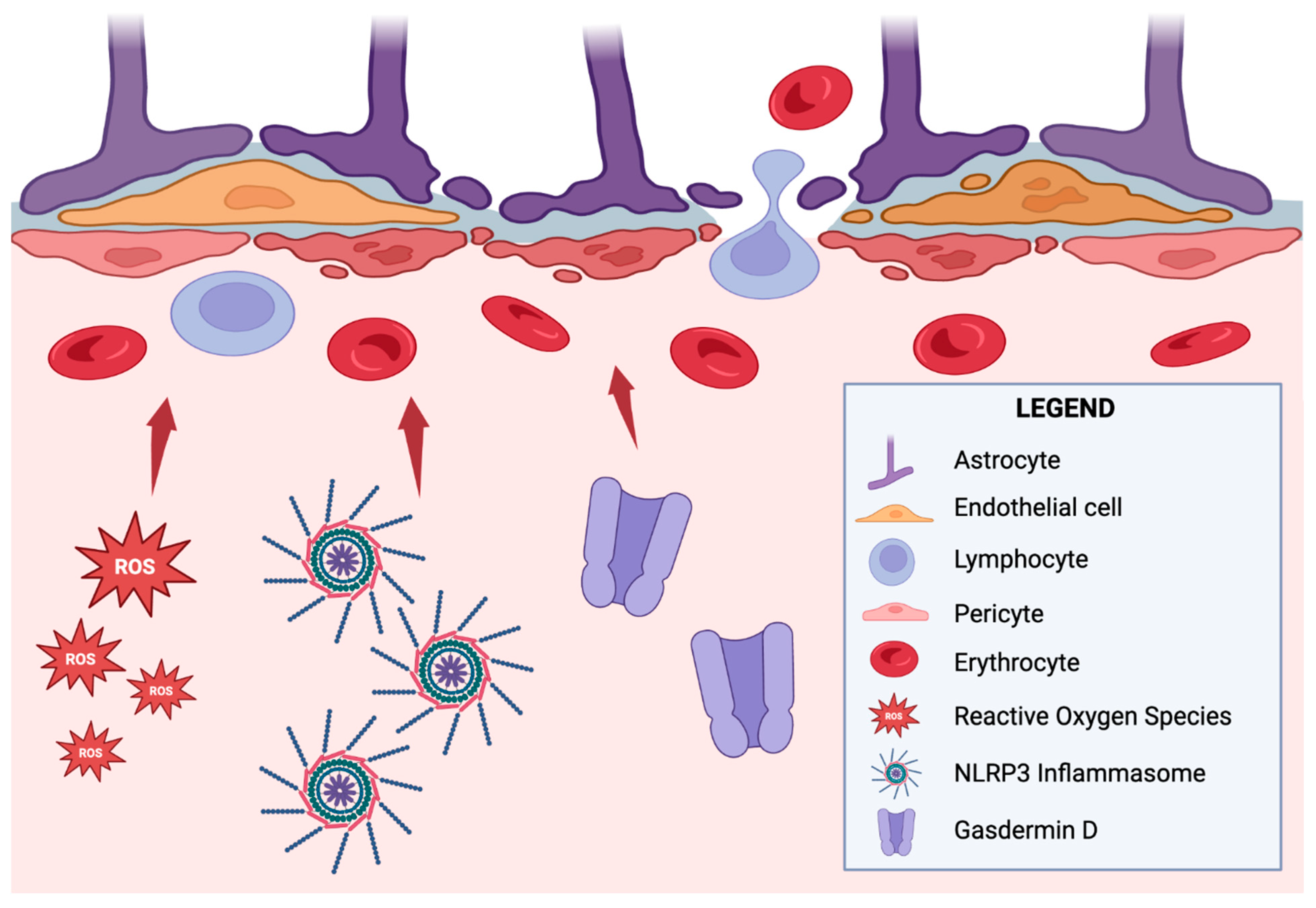
  48. Takata, F.; Nakagawa, S.; Matsumoto, J.; Dohgu, S. Blood-Brain Barrier Dysfunction Amplifies the Development of Neuroinflammation: Understanding of Cellular Events in Brain Microvascular Endothelial Cells for Prevention and Treatment of BBB Dysfunction. Front. Cell. Neurosci. 2021, 15, 661838. [Google Scholar] [CrossRef]
  49. Rochfort, K.D.; Collins, L.E.; Murphy, R.P.; Cummins, P.M. Downregulation of blood-brain barrier phenotype by proinflammatory cytokines involves NADPH oxidase-dependent ROS generation: Consequences for interendothelial adherens and tight junctions. PLoS ONE 2014, 9, e101815. [Google Scholar] [CrossRef]
  50. Wei, C.; Jiang, W.; Wang, R.; Zhong, H.; He, H.; Gao, X.; Zhong, S.; Yu, F.; Guo, Q.; Zhang, L.; et al. Brain endothelial GSDMD activation mediates inflammatory BBB breakdown. Nature 2024, 629, 893–900. [Google Scholar] [CrossRef]
  51. Yoon, S.-H.; Kim, C.Y.; Lee, E.; Lee, C.; Lee, K.-S.; Lee, J.; Park, H.; Choi, B.; Hwang, I.; Kim, J.; et al. Microglial NLRP3-gasdermin D activation impairs blood-brain barrier integrity through interleukin-1β-independent neutrophil chemotaxis upon peripheral inflammation in mice. Nat. Commun. 2025, 16, 699. [Google Scholar] [CrossRef]
  52. Al-Bachari, S.; Naish, J.H.; Parker, G.J.M.; Emsley, H.C.A.; Parkes, L.M. Blood-Brain Barrier Leakage Is Increased in Parkinson’s Disease. Front. Physiol. 2020, 11, 593026. [Google Scholar] [CrossRef]
  53. Laurent, C.; Dorothée, G.; Hunot, S.; Martin, E.; Monnet, Y.; Duchamp, M.; Dong, Y.; Légeron, F.-P.; Leboucher, A.; Burnouf, S.; et al. Hippocampal T cell infiltration promotes neuroinflammation and cognitive decline in a mouse model of tauopathy. Brain 2017, 140, 184–200. [Google Scholar] [CrossRef]
  54. Mou, Y.; Du, Y.; Zhou, L.; Yue, J.; Hu, X.; Liu, Y.; Chen, S.; Lin, X.; Zhang, G.; Xiao, H.; et al. Gut Microbiota Interact With the Brain Through Systemic Chronic Inflammation: Implications on Neuroinflammation, Neurodegeneration, and Aging. Front. Immunol. 2022, 13, 796288. [Google Scholar] [CrossRef]
  55. Kearns, R. Gut-Brain Axis and Neuroinflammation: The Role of Gut Permeability and the Kynurenine Pathway in Neurological Disorders. Cell. Mol. Neurobiol. 2024, 44, 64. [Google Scholar] [CrossRef]
  56. Padhi, P.; Worth, C.; Zenitsky, G.; Jin, H.; Sambamurti, K.; Anantharam, V.; Kanthasamy, A.; Kanthasamy, A.G. Mechanistic Insights Into Gut Microbiome Dysbiosis-Mediated Neuroimmune Dysregulation and Protein Misfolding and Clearance in the Pathogenesis of Chronic Neurodegenerative Disorders. Front. Neurosci. 2022, 16, 836605. [Google Scholar] [CrossRef] [PubMed]
  57. Lukiw, W.J.; Arceneaux, L.; Li, W.; Bond, T.; Zhao, Y. Gastrointestinal (GI)-Tract Microbiome Derived Neurotoxins and their Potential Contribution to Inflammatory Neurodegeneration in Alzheimer’s Disease (AD). J. Alzheimers Dis. Parkinsonism. 2021, 11, 525. [Google Scholar]
  58. Hestad, K.; Alexander, J.; Rootwelt, H.; Aaseth, J.O. The Role of Tryptophan Dysmetabolism and Quinolinic Acid in Depressive and Neurodegenerative Diseases. Biomolecules 2022, 12, 998. [Google Scholar] [CrossRef]
  59. Sanjari Moghaddam, H.; Ghazi Sherbaf, F.; Mojtahed Zadeh, M.; Ashraf-Ganjouei, A.; Aarabi, M.H. Association Between Peripheral Inflammation and DATSCAN Data of the Striatal Nuclei in Different Motor Subtypes of Parkinson Disease. Front. Neurol. 2018, 9, 234. [Google Scholar] [CrossRef]
  60. Kim, R.; Kang, N.; Byun, K.; Park, K.; Jun, J.S. Prognostic significance of peripheral neutrophils and lymphocytes in early untreated Parkinson’s disease: An 8-year follow-up study. J. Neurol. Neurosurg. Psychiatry 2023, 94, 1040–1046. [Google Scholar] [CrossRef]
  61. Zhang, F.; Chen, B.; Ren, W.; Yan, Y.; Zheng, X.; Jin, S.; Chang, Y. Association analysis of dopaminergic degeneration and the neutrophil-to-lymphocyte ratio in Parkinson’s disease. Front. Aging Neurosci. 2024, 16, 1377994. [Google Scholar] [CrossRef] [PubMed]
  62. Muñoz-Delgado, L.; Labrador-Espinosa, M.Á.; Macías-García, D.; Jesús, S.; Zamora, B.B.; Fernández-Rodríguez, P.; Adarmes-Gómez, A.D.; Castillo, M.I.R.; Castro-Labrador, S.; Silva-Rodríguez, J.; et al. Peripheral Inflammation Is Associated with Dopaminergic Degeneration in Parkinson’s Disease. Mov. Disord. 2023, 38, 755–763. [Google Scholar] [CrossRef] [PubMed]
  63. Calabrese, V.; Santoro, A.; Monti, D.; Crupi, R.; di Paola, R.; Latteri, S.; Cuzzocrea, S.; Zappia, M.; Giordano, J.; Calabrese, E.J.; et al. Aging and Parkinson’s Disease: Inflammaging, neuroinflammation and biological remodeling as key factors in pathogenesis. Free Radic. Biol. Med. 2018, 115, 80–91. [Google Scholar] [CrossRef]
  64. Ziabska, K.; Ziemka-Nalecz, M.; Pawelec, P.; Sypecka, J.; Zalewska, T. Aberrant Complement System Activation in Neurological Disorders. Int. J. Mol. Sci. 2021, 22, 4675. [Google Scholar] [CrossRef] [PubMed]
  65. Weiss, F.; Labrador-Garrido, A.; Dzamko, N.; Halliday, G. Immune responses in the Parkinson’s disease brain. Neurobiol. Dis. 2022, 168, 105700. [Google Scholar] [CrossRef]
  66. Rauschenberger, L.; Behnke, J.; Grotemeyer, A.; Knorr, S.; Volkmann, J.; Ip, C.W. Age-dependent neurodegeneration and neuroinflammation in a genetic A30P/A53T double-mutated α-synuclein mouse model of Parkinson’s disease. Neurobiol. Dis. 2022, 171, 105798. [Google Scholar] [CrossRef]
  67. Zhou, Y.; Lu, M.; Du, R.-H.; Qiao, C.; Jiang, C.-Y.; Zhang, K.-Z.; Ding, J.-H.; Hu, G. MicroRNA-7 targets Nod-like receptor protein 3 inflammasome to modulate neuroinflammation in the pathogenesis of Parkinson’s disease. Mol. Neurodegener. 2016, 11, 28. [Google Scholar] [CrossRef]
  68. Chou, J.P.; Effros, R.B. T cell replicative senescence in human aging. Curr. Pharm. Des. 2013, 19, 1680–1698. [Google Scholar] [CrossRef]
  69. Williams-Gray, C.H.; Wijeyekoon, R.S.; Scott, K.M.; Hayat, S.; Barker, R.A.; Jones, J.L. Abnormalities of age-related T cell senescence in Parkinson’s disease. J. Neuroinflamm. 2018, 15, 166. [Google Scholar] [CrossRef]
  70. Rodriguez, M.; Rodriguez-Sabate, C.; Morales, I.; Sanchez, A.; Sabate, M. Parkinson’s disease as a result of aging. Aging Cell 2015, 14, 293–308. [Google Scholar] [CrossRef]
  71. Sakiyama, H.; Baba, K.; Kimura, Y.; Ogawa, K.; Nishiike, U.; Hayakawa, H.; Yoshida, M.; Aguirre, C.; Ikenaka, K.; Nagano, S.; et al. Accelerated senescence exacerbates α-synucleinopathy in senescence-accelerated prone 8 mice via persistent neuroinflammation. Neurochem. Int. 2025, 182, 105906. [Google Scholar] [CrossRef]
  72. Chinta, S.J.; Woods, G.; Demaria, M.; Rane, A.; Zou, Y.; McQuade, A.; Rajagopalan, S.; Limbad, C.; Madden, D.T.; Campisi, J.; et al. Cellular Senescence Is Induced by the Environmental Neurotoxin Paraquat and Contributes to Neuropathology Linked to Parkinson’s Disease. Cell Rep. 2018, 22, 930–940. [Google Scholar] [CrossRef]
  73. Pringsheim, T.; Day, G.S.; Smith, D.B.; Rae-Grant, A.; Licking, N.; Armstrong, M.J.; Roze, E.; Miyasaki, J.M.; Hauser, R.A.; Espay, A.J.; et al. Dopaminergic Therapy for Motor Symptoms in Early Parkinson Disease Practice Guideline Summary: A Report of the AAN Guideline Subcommittee. Neurology 2021, 97, 942–957. [Google Scholar] [CrossRef] [PubMed]
  74. Ferrari, D.P.; Bortolanza, M.; Del Bel, E.A. Interferon-γ Involvement in the Neuroinflammation Associated with Parkinson’s Disease and L-DOPA-Induced Dyskinesia. Neurotox. Res. 2021, 39, 705–719. [Google Scholar] [CrossRef] [PubMed]
  75. Tran, T.N.; Vo, T.N.N.; Frei, K.; Truong, D.D. Levodopa-induced dyskinesia: Clinical features, incidence, and risk factors. J. Neural Transm. 2018, 125, 1109–1117. [Google Scholar] [CrossRef]
  76. Jones-Tabah, J.; Mohammad, H.; Hadj-Youssef, S.; Kim, L.E.H.; Martin, R.D.; Benaliouad, F.; Tanny, J.C.; Clarke, P.B.S.; Hébert, T.E. Dopamine D1 receptor signalling in dyskinetic Parkinsonian rats revealed by fiber photometry using FRET-based biosensors. Sci. Rep. 2020, 10, 14426. [Google Scholar] [CrossRef]
  77. Vega-Angeles, V.T.; Morales-Ruiz, V.; Adalid-Peralta, L.V. Immunomodulation as a treatment for parkinson’s disease in current trials: A systematic review and meta-analysis. Rev. Investig. Clin. 2024, 76, 159–169. [Google Scholar] [CrossRef]
  78. Investigators, N.N.-P. A randomized clinical trial of coenzyme Q10 and GPI-1485 in early Parkinson disease. Neurology 2007, 68, 20–28. [Google Scholar] [CrossRef]
  79. Writing Group for the NINDS Exploratory Trials in Parkinson Disease (NET-PD) Investigators; Kieburtz, K.; Tilley, B.C.; Elm, J.J.; Babcock, D.; Hauser, R.; Ross, G.W.; Augustine, A.H.; Augustine, E.U.; Aminoff, M.J.; et al. Effect of creatine monohydrate on clinical progression in patients with Parkinson disease: A randomized clinical trial. JAMA 2015, 313, 584–593. [Google Scholar] [CrossRef]
  80. Neurol, L. Pioglitazone in early Parkinson’s disease: A phase 2, multicentre, double-blind, randomised trial. Lancet Neurol. 2015, 14, 795–803. [Google Scholar] [CrossRef]
  81. Cankaya, S.; Cankaya, B.; Kilic, U.; Kilic, E.; Yulug, B. The therapeutic role of minocycline in Parkinson’s disease. Drugs Context. 2019, 8, 212553. [Google Scholar] [CrossRef] [PubMed]
  82. Richard, E.; Bloem, B.R. Monoclonal Antibodies in Neurodegenerative Disease May Work, But They Don’t Help: A Perspective from Physicians. J. Parkinsons Dis. 2022, 12, 2289–2291. [Google Scholar] [CrossRef] [PubMed]
  83. Games, D.; Valera, E.; Spencer, B.; Rockenstein, E.; Mante, M.; Adame, A.; Patrick, C.; Ubhi, K.; Nuber, S.; Sacayon, P.; et al. Reducing C-terminal-truncated alpha-synuclein by immunotherapy attenuates neurodegeneration and propagation in Parkinson’s disease-like models. J. Neurosci. 2014, 34, 9441–9454. [Google Scholar] [CrossRef] [PubMed]
  84. Pagano, G.; Taylor, K.I.; Anzures-Cabrera, J.; Marchesi, M.; Simuni, T.; Marek, K.; Postuma, R.B.; Pavese, N.; Stocchi, F.; Azulay, J.-P.; et al. Trial of Prasinezumab in Early-Stage Parkinson’s Disease. N. Engl. J. Med. 2022, 387, 421–432. [Google Scholar] [CrossRef]
  85. Weihofen, A.; Liu, Y.; Arndt, J.W.; Huy, C.; Quan, C.; Smith, B.A.; Baeriswyl, J.-L.; Cavegn, N.; Senn, L.; Su, L.; et al. Development of an aggregate-selective, human-derived α-synuclein antibody BIIB054 that ameliorates disease phenotypes in Parkinson’s disease models. Neurobiol. Dis. 2019, 124, 276–288. [Google Scholar] [CrossRef]
  86. Lang, A.E.; Siderowf, A.D.; Macklin, E.A.; Poewe, W.; Brooks, D.J.; Fernandez, H.H.; Rascol, O.; Giladi, N.; Stocchi, F.; Tanner, C.M.; et al. Trial of Cinpanemab in Early Parkinson’s Disease. N. Engl. J. Med. 2022, 387, 408–420. [Google Scholar] [CrossRef]
  87. Kharel, S.; Ojha, R. Future of Monoclonal Antibody Therapy in Parkinson’s Disease. Ann. Neurosci. 2023, 30, 8–10. [Google Scholar] [CrossRef]
  88. Hickey, P.; Stacy, M. Deep Brain Stimulation: A Paradigm Shifting Approach to Treat Parkinson’s Disease. Front. Neurosci. 2016, 10, 173. [Google Scholar] [CrossRef]
  89. Chiken, S.; Nambu, A. Mechanism of Deep Brain Stimulation: Inhibition, Excitation, or Disruption? Neuroscientist 2016, 22, 313–322. [Google Scholar] [CrossRef]
  90. Chiken, S.; Nambu, A. High-frequency pallidal stimulation disrupts information flow through the pallidum by GABAergic inhibition. J. Neurosci. 2013, 33, 2268–2280. [Google Scholar] [CrossRef]
  91. Reese, R.; Leblois, A.; Steigerwald, F.; Pötter-Nerger, M.; Herzog, J.; Mehdorn, H.M.; Deuschl, G.; Meissner, W.G.; Volkmann, J. Subthalamic deep brain stimulation increases pallidal firing rate and regularity. Exp. Neurol. 2011, 229, 517–521. [Google Scholar] [CrossRef] [PubMed]
  92. Eser, P.; Kocabicak, E.; Bekar, A.; Temel, Y. Insights into neuroinflammatory mechanisms of deep brain stimulation in Parkinson’s disease. Exp. Neurol. 2024, 374, 114684. [Google Scholar] [CrossRef] [PubMed]
  93. Vedam-Mai, V.; Rodgers, C.; Gureck, A.; Vincent, M.; Ippolito, G.; Elkouzi, A.; Yachnis, A.T.; Foote, K.D.; Okun, M.S. Deep Brain Stimulation associated gliosis: A post-mortem study. Parkinsonism Relat. Disord. 2018, 54, 51–55. [Google Scholar] [CrossRef] [PubMed]
  94. Temel, Y.; Visser-Vandewalle, V.; Kaplan, S.; Kozan, R.; Daemen, M.A.; Blokland, A.; Schmitz, C.; Steinbusch, H.W. Protection of nigral cell death by bilateral subthalamic nucleus stimulation. Brain Res. 2006, 1120, 100–105. [Google Scholar] [CrossRef]
  95. Mazumder, S.; Bahar, A.Y.; Shepherd, C.E.; Prasad, A.A. Post-mortem brain histological examination in the substantia nigra and subthalamic nucleus in Parkinson’s disease following deep brain stimulation. Front. Neurosci. 2022, 16, 948523. [Google Scholar] [CrossRef]
  96. Pal, G.D.; Ouyang, B.; Serrano, G.; Shill, H.A.; Goetz, C.; Stebbins, G.; Metman, L.V.; Driver-Dunckley, E.; Mehta, S.H.; Caviness, J.N.; et al. Comparison of neuropathology in Parkinson’s disease subjects with and without deep brain stimulation. Mov. Disord. 2017, 32, 274–277. [Google Scholar] [CrossRef]
  97. Hilker, R.; Voges, J.; Ghaemi, M.; Lehrke, R.; Rudolf, J.; Koulousakis, A.; Herholz, K.; Wienhard, K.; Sturm, V.; Heiss, W. Deep brain stimulation of the subthalamic nucleus does not increase the striatal dopamine concentration in parkinsonian humans. Mov. Disord. 2003, 18, 41–48. [Google Scholar] [CrossRef]
  98. Kwiatek-Majkusiak, J.; Geremek, M.; Koziorowski, D.; Tomasiuk, R.; Szlufik, S.; Friedman, A. Higher serum levels of pro-hepcidin in patients with Parkinson’s disease treated with deep brain stimulation. Neurosci. Lett. 2018, 684, 205–209. [Google Scholar] [CrossRef]
  99. Ownby, R.L. Neuroinflammation and cognitive aging. Curr. Psychiatry Rep. 2010, 12, 39–45. [Google Scholar] [CrossRef]
  100. Zhao, Z.; Fu, Q.; Guo, X.; He, H.; Yang, G. Potential Biomarkers and Treatment of Neuroinflammation in Parkinson’s Disease. Actas Esp. Psiquiatr. 2025, 53, 181–188. [Google Scholar] [CrossRef]
  101. De Lella Ezcurra, A.L.; Chertoff, M.; Ferrari, C.; Graciarena, M.; Pitossi, F. Chronic expression of low levels of tumor necrosis factor-alpha in the substantia nigra elicits progressive neurodegeneration, delayed motor symptoms and microglia/macrophage activation. Neurobiol. Dis. 2010, 37, 630–640. [Google Scholar] [CrossRef]
  102. Tansey, M.G.; Wallings, R.L.; Houser, M.C.; Herrick, M.K.; Keating, C.E.; Joers, V. Inflammation and immune dysfunction in Parkinson disease. Nat. Rev. Immunol. 2022, 22, 657–673. [Google Scholar] [CrossRef] [PubMed]
  103. Ren, L.; Yi, J.; Yang, J.; Li, P.; Cheng, X.; Mao, P. Nonsteroidal anti-inflammatory drugs use and risk of Parkinson disease: A dose-response meta-analysis. Medicine 2018, 97, e12172. [Google Scholar] [CrossRef]
  104. Tansey, M.G.; Romero-Ramos, M. Immune system responses in Parkinson’s disease: Early and dynamic. Eur. J. Neurosci. 2019, 49, 364–383. [Google Scholar] [CrossRef] [PubMed]
  105. Fleury, V.; Zekeridou, A.; Lazarevic, V.; Gaïa, N.; Giannopoulou, C.; Genton, L.; Cancela, J.; Girard, M.; Goldstein, R.; Bally, J.F.; et al. Oral Dysbiosis and Inflammation in Parkinson’s Disease. J. Parkinsons Dis. 2021, 11, 619–631. [Google Scholar] [CrossRef] [PubMed]
  106. Veselý, B.; Dufek, M.; Thon, V.; Brozman, M.; Királová, S.; Halászová, T.; Koriťáková, E.; Rektor, I. Interleukin 6 and complement serum level study in Parkinson’s disease. J. Neural Transm. 2018, 125, 875–881. [Google Scholar] [CrossRef]
  107. Li, N.; Wang, J.X.; Huo, T.T.; Zhao, J.R.; Wang, T.J. Associations of IL-1β and IL-6 gene polymorphisms with Parkinson’s disease. Eur. Rev. Med. Pharmacol. Sci. 2021, 25, 890–897. [Google Scholar] [CrossRef]
  108. Ramírez, J.; Cañete, J.D. Anakinra for the treatment of rheumatoid arthritis: A safety evaluation. Expert Opin. Drug Saf. 2018, 17, 727–732. [Google Scholar] [CrossRef]
  109. Stojakovic, A.; Paz-Filho, G.; Arcos-Burgos, M.; Licinio, J.; Wong, M.L.; Mastronardi, C.A. Role of the IL-1 Pathway in Dopaminergic Neurodegeneration and Decreased Voluntary Movement. Mol. Neurobiol. 2017, 54, 4486–4495. [Google Scholar] [CrossRef]
  110. Sivapalasingam, S.; Lederer, D.J.; Bhore, R.; Hajizadeh, N.; Criner, G.; Hosain, R.; Mahmood, A.; Giannelou, A.; Somersan-Karakaya, S.; O’brien, M.P.; et al. Efficacy and Safety of Sarilumab in Hospitalized Patients With Coronavirus Disease 2019: A Randomized Clinical Trial. Clin. Infect. Dis. 2022, 75, e380–e388. [Google Scholar] [CrossRef]
  111. Miliukhina, I.V.; Usenko, T.S.; Senkevich, K.A.; Nikolaev, M.A.; Timofeeva, A.A.; Agapova, E.A.; Semenov, A.V.; Lubimova, N.E.; Totolyan, A.A.; Pchelina, S.N. Plasma Cytokines Profile in Patients with Parkinson’s Disease Associated with Mutations in GBA Gene. Bull. Exp. Biol. Med. 2020, 168, 423–426. [Google Scholar] [CrossRef] [PubMed]
  112. Boyman, O.; Sprent, J. The role of interleukin-2 during homeostasis and activation of the immune system. Nat. Rev. Immunol. 2012, 12, 180–190. [Google Scholar] [CrossRef]
  113. Si, Y.; Zhang, Y.; Zuloaga, K.; Yang, Q. The role of innate lymphocytes in regulating brain and cognitive function. Neurobiol. Dis. 2023, 179, 106061. [Google Scholar] [CrossRef]
  114. Noble, S.; Goa, K.L. Aldesleukin (recombinant interleukin-2). BioDrugs 1997, 7, 394–422. [Google Scholar] [CrossRef] [PubMed]
  115. Tomasovic, L.M.; Liu, K.; VanDyke, D.; Fabilane, C.S.; Spangler, J.B. Molecular Engineering of Interleukin-2 for Enhanced Therapeutic Activity in Autoimmune Diseases. BioDrugs 2024, 38, 227–248. [Google Scholar] [CrossRef]
  116. Muthuraman, M.; Koirala, N.; Ciolac, D.; Pintea, B.; Glaser, M.; Groppa, S.; Tamás, G.; Groppa, S. Deep Brain Stimulation and L-DOPA Therapy: Concepts of Action and Clinical Applications in Parkinson’s Disease. Front. Neurol. 2018, 9, 711. [Google Scholar] [CrossRef] [PubMed]
  117. Calabresi, P.; Mechelli, A.; Natale, G.; Volpicelli-Daley, L.; Di Lazzaro, G.; Ghiglieri, V. Alpha-synuclein in Parkinson’s disease and other synucleinopathies: From overt neurodegeneration back to early synaptic dysfunction. Cell Death Dis. 2023, 14, 176. [Google Scholar] [CrossRef]
  118. Abbott, A. Fetal-cell revival for Parkinson’s. Nature 2014, 510, 195–196. [Google Scholar] [CrossRef] [PubMed]
  119. Wu, D.; Chen, Q.; Chen, X.; Han, F.; Chen, Z.; Wang, Y. The blood–brain barrier: Structure, regulation and drug delivery. Sig. Transduct. Target. Ther. 2023, 8, 1–27. [Google Scholar] [CrossRef]
  120. Cha, Y.; Park, T.Y.; Leblanc, P.; Kim, K.S. Current Status and Future Perspectives on Stem Cell-Based Therapies for Parkinson’s Disease. J. Mov. Disord. 2023, 16, 22–41. [Google Scholar] [CrossRef]
  121. Jiang, S.; Wang, H.; Yang, C.; Feng, F.; Xu, D.; Zhang, M.; Xie, M.; Cui, R.; Zhu, Z.; Jia, C.; et al. Phase 1 study of safety and preliminary efficacy of intranasal transplantation of human neural stem cells (ANGE-S003) in Parkinson’s disease. J. Neurol. Neurosurg. Psychiatry 2024, 95, 1102–1111. [Google Scholar] [CrossRef] [PubMed]
  122. Madrazo, I.; Kopyov, O.; Ávila-Rodríguez, M.A.; Ostrosky, F.; Carrasco, H.; Kopyov, A.; Avendaño-Estrada, A.; Jiménez, F.; Magallón, E.; Zamorano, C.; et al. Transplantation of Human Neural Progenitor Cells (NPC) into Putamina of Parkinsonian Patients: A Case Series Study, Safety and Efficacy Four Years after Surgery. Cell Transplant. 2019, 28, 269–285. [Google Scholar] [CrossRef] [PubMed]
Figure 1. The interplay of innate and adaptive immune responses between the CNS and PNS has been postulated to result in the neurodegeneration of dopaminergic neurons characteristic of PD pathogenesis. Within the immune-privileged CNS, astrocytes and microglia are responsible for producing pro-inflammatory cytokines, eliciting chronic neuroinflammation, and debilitating the integrity of the BBB. Meanwhile, adaptive immunity, consisting of clonotypic lymphocytes, has been associated with a dysbiosis of the gut microbiota and consequential effects on the gut–brain axis via the vagus nerve. Further studies exploring the intricate communication between the nervous system, immunological responses, and neuroinflammation will prove integral to elucidating novel immunotherapeutics.
Neuroglia 06 00017 g001
Figure 2. Mechanisms of BBB penetration.
Neuroglia 06 00017 g002
Figure 3. Clinical investigation outcomes of immune therapies for PD.
Neuroglia 06 00017 g003
Figure 4. Treatments for aspects of PD. Interventions in green are evidence-supported. Interventions in red are currently unsupported.
Neuroglia 06 00017 g004
Disclaimer/Publisher’s Note: The statements, opinions and data contained in all publications are solely those of the individual author(s) and contributor(s) and not of MDPI and/or the editor(s). MDPI and/or the editor(s) disclaim responsibility for any injury to people or property resulting from any ideas, methods, instructions or products referred to in the content.

