Surgical Treatment of Cavernous Sinus Cavernomas: Evidence from Vietnam


Abstract
1. Introduction
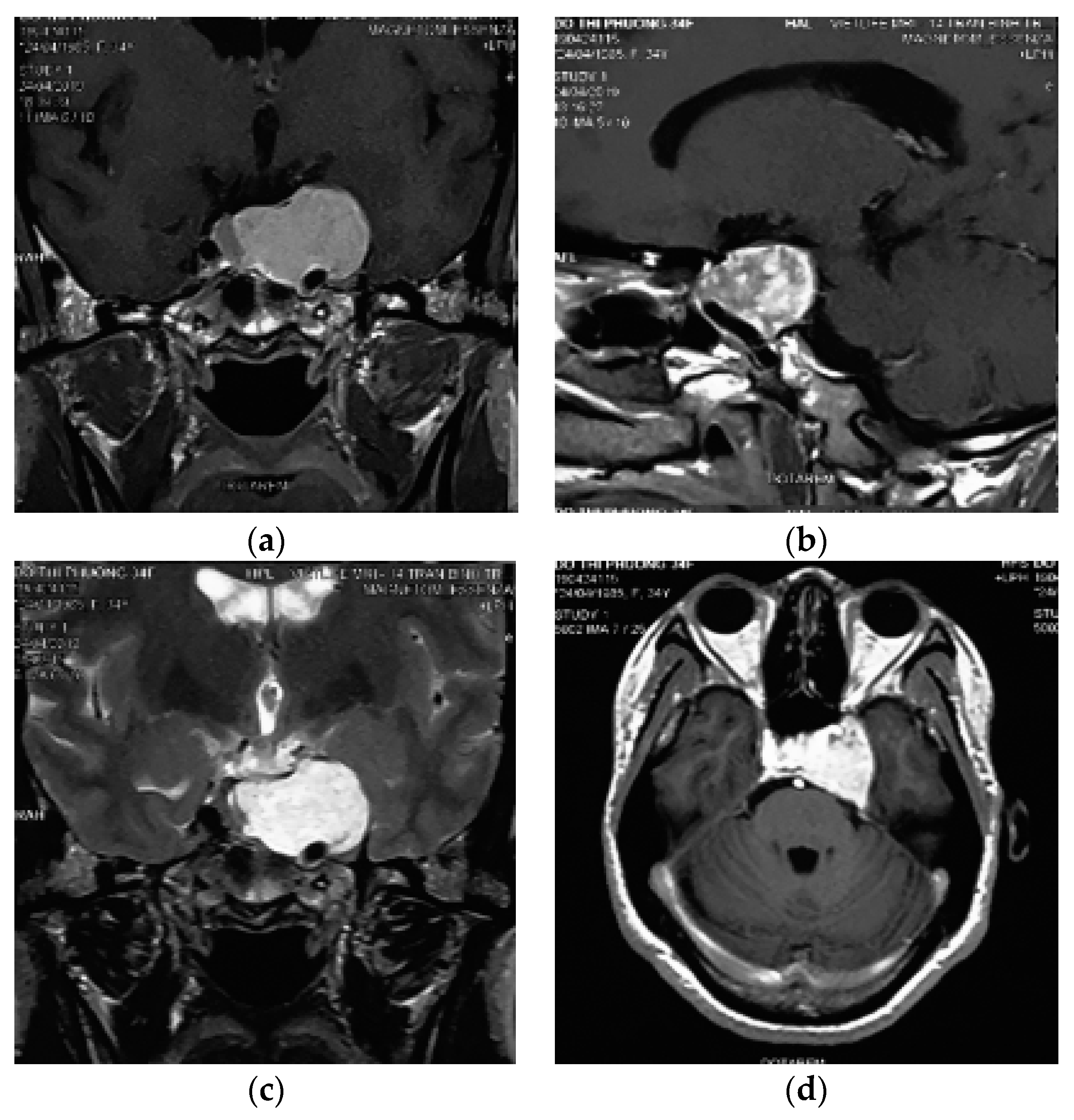
2. Case Presentation
3. Discussion
4. Conclusions
Author Contributions
Funding
Acknowledgments
Conflicts of Interest
References
- Lupret, V.; Negovetić, L.; Smiljanić, D.; Klanfar, Z.; Talan-Hranilović, J. Cavernous haemangioma in cavernous sinus: Case report of rare location. Neurol. Croat. 1992, 41, 241–246. [Google Scholar]
- Rosenblum, B.; Rothman, A.S.; Lanzieri, C.; Song, S. A cavernous sinus cavernous hemangioma: Case report. J. Neurosurg. 1986, 65, 716–718. [Google Scholar] [CrossRef] [PubMed]
- Goto, Y.; Yamabe, K.; Aiko, Y.; Kuromatsu, C.; Fukui, M. Cavernous hemangioma in the cavernous sinus. Neurochirurgia 1993, 36, 93–95. [Google Scholar] [CrossRef] [PubMed]
- Li, M.-H.; Zhao, J.L.; Li, Y.Y.; Zheng, C.H.; Xu, G.S.; Hong, T. Extradural transcavernous approach to cavernous sinus cavernous hemangiomas. Clin. Neurol. Neurosurg. 2015, 136, 110–115. [Google Scholar] [CrossRef]
- Li, H.; Zhang, B.; Wang, W.; Wei, M.H.; Liu, B.Y.; Wu, Z. Clinical Features, Intradural Transcavernous Surgical Management, and Outcomes of Giant Cavernous Sinus Hemangiomas: A Single-Institution Experience. World Neurosurg. 2019, 125, e754–e763. [Google Scholar] [CrossRef]
- Yin, Y.-H.; Yu, X.G.; Xu, B.N.; Zhou, D.B.; Bu, B.; Chen, X.L. Surgical management of large and giant cavernous sinus hemangiomas. J. Clin. Neurosci. 2013, 20, 28–133. [Google Scholar] [CrossRef]
- Lombardi, D.; Giovanelli, M.; De Tribolet, N. Sellar and parasellar extra-axial cavernous hemangiomas. Acta Neurochir. 1994, 130, 47–54. [Google Scholar] [CrossRef]
- Ibrahim, D.; El Fiki, A.; Hafez, M.; Saleem, S. Report of a case of cavernous haemangioma of the cavernous sinus. BJR Case Rep. 2019, 5, 20190031. [Google Scholar] [CrossRef]
- Lee, A.G.; Miller, N.R.; Brazis, P.W.; Benson, M.L. Cavernous sinus hemangioma. Clinical and neuroimaging features. J. Neuroophthalmol. 1995, 15, 225–229. [Google Scholar] [CrossRef]
- Katayama, Y.; Tsubokawa, T.; Miyazaki, S.; Yoshida, K.; Himi, K. Magnetic resonance imaging of cavernous sinus cavernous hemangiomas. Neuroradiology 1991, 33, 118–122. [Google Scholar] [CrossRef]
- Linskey, M.E.; Sekhar, L.N. Cavernous sinus hemangiomas: A series, a review, and an hypothesis. Neurosurgery 1992, 30, 101–1088. [Google Scholar] [CrossRef] [PubMed]
- Namba, S. Extracerebral cavernous hemangioma of the middle cranial fossa. Surg. Neurol. 1983, 19, 379–388. [Google Scholar] [CrossRef]
- Park, C.K.; Choi, S.K.; Kang, I.H.; Choi, M.K.; Park, B.J.; Lim, Y.J. Radiosurgical considerations for cavernous sinus hemangioma: Long-term clinical outcomes. Acta Neurochir. 2016, 158, 313–318. [Google Scholar] [CrossRef] [PubMed]
- Zhou, L.-F.; Mao, Y.; Chen, L. Diagnosis and surgical treatment of cavernous sinus hemangiomas: An experience of 20 cases. Surg. Neurol. 2003, 60, 31–36. [Google Scholar] [CrossRef]
- Mori, K.; Handa, H.; Gi, H. Cavernomas in the middle fossa. Surg. Neurol. 1980, 14, 21–31. [Google Scholar]
- Rigamonti, D.; Pappas, C.T.E.; Spetzler, R.F.; Johnson, P.C. Extracerebral cavernous angiomas of the middle fossa. Neurosurgery 1990, 27, 306–310. [Google Scholar] [CrossRef]
- Nepper-Rasmussen, H.; Bjerre, P.; Jørgensen, K. Endovascular neuroradiologic interventions. Embolization of aneurysms and arteriovenous malformations. Sclerotherapy of cavernous hemangiomas. Ugeskr. Laeger 1993, 155, 3471–3476. [Google Scholar]
- Hashimoto, M.; Yokota, A.; Ohta, H.; Urasaki, E. Intratumoral injection of biobond adhesive for removal of cavernous sinus hemangioma. J. Neurosurg. 2000, 93, 1078–1081. [Google Scholar] [CrossRef]
- Ohata, K.; El-Naggar, A.; Takami, T.; Morino, M.; El-Adawy, Y.; El-Sheik, K.; Inoue, Y.; Hakuba, A. Efficacy of induced hypotension in the surgical treatment of large cavernous sinus cavernomas. J. Neurosurg. 1999, 90, 702–708. [Google Scholar] [CrossRef]
- Li, Z.-H.; Wu, Z.; Zhang, J.T. Surgical Management and Outcomes of Cavernous Sinus Hemangiomas: A Single-Institution Series of 47 Patients. World Neurosurg. 2019, 122, e1181–e1194. [Google Scholar] [CrossRef]
- Goel, A.; Muzumdar, D.; Sharma, P. Extradural approach for cavernous hemangioma of the cavernous sinus: Experience with 13 cases. Neurol. Med. Chir. 2003, 43, 112–119. [Google Scholar] [CrossRef] [PubMed]
- Suri, A.; Ahmad, F.U.; Mahapatra, A.K. Extradural transcavernous approach to cavernous sinus hemangiomas. Neurosurgery 2007, 60, 483–489. [Google Scholar] [CrossRef] [PubMed]
- Goel, A. The extradural approach to lesions involving the cavernous sinus. Br. J. Neurosurg. 1997, 11, 134–138. [Google Scholar] [CrossRef] [PubMed]
- Nadkarni, A.G. Cavernous haemangioma in the cavernous sinus. Br. J. Neurosurg. 1995, 9, 77–80. [Google Scholar] [CrossRef] [PubMed]


© 2020 by the authors. Licensee MDPI, Basel, Switzerland. This article is an open access article distributed under the terms and conditions of the Creative Commons Attribution (CC BY) license (http://creativecommons.org/licenses/by/4.0/).
Share and Cite
Nguyen, D.-A.; Nguyen, H.T.; Duong, T.V.; Pham, B.H.; Vo, H.-L. Surgical Treatment of Cavernous Sinus Cavernomas: Evidence from Vietnam. Reports 2020, 3, 16. https://doi.org/10.3390/reports3020016
Nguyen D-A, Nguyen HT, Duong TV, Pham BH, Vo H-L. Surgical Treatment of Cavernous Sinus Cavernomas: Evidence from Vietnam. Reports. 2020; 3(2):16. https://doi.org/10.3390/reports3020016
Chicago/Turabian StyleNguyen, Duc-Anh, Hao The Nguyen, Thang Van Duong, Binh Hoa Pham, and Hoang-Long Vo. 2020. "Surgical Treatment of Cavernous Sinus Cavernomas: Evidence from Vietnam" Reports 3, no. 2: 16. https://doi.org/10.3390/reports3020016
APA StyleNguyen, D.-A., Nguyen, H. T., Duong, T. V., Pham, B. H., & Vo, H.-L. (2020). Surgical Treatment of Cavernous Sinus Cavernomas: Evidence from Vietnam. Reports, 3(2), 16. https://doi.org/10.3390/reports3020016