Abstract
Four nineteenth-century casts of the decoration on the north side of the exterior of the apse of the Notre-Dame Cathedral in Paris are held in the plaster casts collection at the Victoria and Albert Museum in London. The casts were manufactured by two different nineteenth-century workshops, one run by Jean Pouzadoux and the other by Auguste Malzieux. After an assessment of the condition of the casts, a scientific analysis allowed the characterization of the manufacturing materials and subsequent conservation treatments aimed at ensuring the stability of the casts and removing dirt and grime from the casts’ surfaces. Optical microscopy of the samples taken from the casts allowed the stratigraphy to be studied, which largely consisted of gypsum plaster and a coating layer (oxidized diterpenic resin or shellac) containing silicon and aluminium partially diffused in the porous substrate. These materials were identified by a range of techniques, including X-ray diffraction, scanning electron microscope–energy dispersive X-ray spectroscopy, Fourier transform infrared spectroscopy, and gas chromatography/mass spectrometry. The conservation works returned stability to the panels for redisplay in the galleries and achieved a closer comparative study between the two workshops. The two sets of panels showed numerous differences in manufacturing processes that corresponded to their observed deterioration.
1. Introduction
Plaster casting in the nineteenth century was extremely popular and remunerative [1,2]. Casts of decorative and architectural details, as well as replicas of important sculptural works of art, were traded nationally and internationally, aiming to inspire craftsmen and artists and to educate the public [1]. The relevance of this artisanship in its period and of the historical casts as objects in their own right has been advocated elsewhere [2,3]. Moreover, the importance of historical cast collections has been more recently highlighted as an act of preservation [4,5,6,7] since the originals from which the casts were made often suffer loss of details or even destruction [3,6,7].
The fire that sprung up at the Notre-Dame Cathedral on the 15th of April 2019 once again reminded the world of how crucial it is to record the most important works of art that would be lost without being passed on to future generations. New 3D scanning technologies are nowadays addressing these documentation needs in many cultural institutions. Moreover, historical plaster casts are extremely important as sources of documentation even for what is already lost.
Several plaster casts of decorative details from Notre-Dame Cathedral in Paris are held in the plaster casts collection at the Victoria and Albert Museum (V&A) in London.
The focus of the analysis and conservation treatments described in this paper is on four of these objects, casts of decorative details made by Pierre de Chelles (active about 1287; d. 1320). The originals of the casts were carved in limestone between 1296 and 1316, on the north side of the exterior of the apse of Notre-Dame, Paris. The events of April 2019 renewed interest in these objects, which were coincidentally under investigation and conservation at the time.
The casts were investigated within a PhD project supported by an AHRC CDP studentship in collaboration with Northumbria University [7] and conservation treatments were carried out by conservators Adriana Francescutto Miró, Sayuri Morio, and Sarah Healey-Dilkes. The serendipitous scientific analysis and conservation treatments shed light on the manufacturing techniques and conditions of the casts. This once again reminded us that an interdisciplinary methodology is desirable and effective, as proven in recent approaches to the study of similar materials [8,9,10,11,12,13,14].
1.1. Conservation of Casts at the V&A
The conservation of the V&A Cast Courts has always been a complex task for the museum’s conservators as the collection of casts created by Henry Cole and John Charles Robinson includes an eclectic collection of casts and finishes [6,7]. The recent renovation of the Cast Courts involved the restoration of the roof and the floor, as well as the redecoration of the walls in the original color scheme [15]. Most of the objects have always been on open display rather than in cases. They were thus exposed to pollution caused both by early heating and lighting methods within the Museum and by the general quality of London’s atmospheric air. Humidity and natural light have also had an impact on the ageing of the materials [7,15,16]. The casts have been affected by interventions such as dismantling and re-location, partial or complete coating with paint or resins, and being subjected to a variety of traditional cleaning methods with varying degrees of success. Records of the casts’ makers and their exact processes, as well as the documentation of their previous conservation treatments, were scant. The recent project has allowed the gradual construction of a bigger picture of the collection through close visual observation of the objects and the effects of conservation intervention through limited sampling and analysis and the gathering of written source materials. The Weston Cast Court (Gallery 46b, also known as Italian Cast Court) was renovated from 2010 to 2014 and its twin, The Ruddock Family Cast Court (Gallery 46a, also known as the European Cast Court), reopened in November 2018 [17].
The conservation of the Cast Courts started with an assessment of the condition of the casts. In general, the casts displayed signs of structural problems. Degradation was more evident in the form of cracking and the shrinking of coatings and paint layers. Structural issues were often related to the method of construction. Sealants or coatings appeared discolored and eroded with exposed areas of plaster having darkened considerably due to the absorption and cementation of dirt. A variety of cleaning methods were used during the conservation of the casts at the V&A [15]. New studies and in-depth examinations resulting from the conservation of the collection also provided the opportunity for systematic research into the manufacturing techniques of casts, surface coatings, finishes, and mold making, and allowed the investigation of the groups of casts along with their treatment [7].
1.2. The Nineteenth-Century Casts from the Notre-Dame Cathedral
The plaster casts from the Notre-Dame Cathedral are now located in the south wall of Gallery 46a (The Ruddock Family Cast Court). Two out of the four casts investigated were situated there: Museum’s Accession numbers (Museum numbers are identifiers for the objects and can be used to search the collection [18]): REPRO.1890-80 and REPRO.1890-81 (Figure 1). They were cast by Jean Pouzadoux (1829–1893) and were acquired for £12 (300 francs) each by the V&A in 1890 directly from the workshop at the Museé de Sculpture Compareé where he was the head cast maker. This workshop sold numerous French plaster casts to many different institutions, including the V&A. REPRO.1890-80 (dimensions 152.5 × 160.0 cm) is a plaster cast of a relief of The Assumption of the Virgin (ascending to Heaven in a mandorla surrounded by angels), and REPRO.1890-81 (dimensions 155.0 × 135.0 cm) is a cast of The Coronation of the Virgin.
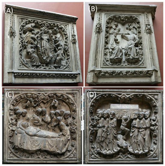
Figure 1.
The Notre-Dame casts at the V&A: The Assumption of the Virgin, REPRO.1890-80 (A), The Coronation of the Virgin, REPRO.1890-81 (B), The death of the Virgin, REPRO.A.1916-3152 (C), and The Entombment of the Virgin, REPRO.A.1916-3153 (D).
REPRO.A.1916-3152 and REPRO.A.1916-3153 (Figure 1) were made ca. 1850–1900 by Auguste Malzieux (1820–1873), whose workshop also sold many French plaster casts to various institutions; a collection of his casts became part of the Architectural Association in London in 1916 including these two panels, eventually becoming part of the V&A collection [19]. REPRO.A.1916-3152 (dimensions 90.0 × 86.0 cm) is a relief plaster cast of The death of the Virgin, and REPRO.A.1916-3153 is a cast of The Entombment of the Virgin (dimensions 61.5 × 53.5 cm).
Jean Pouzadoux and Auguste Malzieux took part in the restoration campaigns of Notre-Dame between 1843 and 1864 directed by the architect Eugène Viollet-le-Duc (1814–1879) [20], the designer of the spire that collapsed during the fire in April 2019.
2. Materials and Methods
2.1. Sampling
Prior to conservation, a total of sixteen samples across the objects were taken from pre-existing areas of loss to investigate the stratigraphy and the methods of manufacture, according to British Standard (BS EN 16085:2012—ISBN 978 0 580 70588 5) [21].
Before the sampling, a careful survey was performed to prevent any risk and to minimize the quantity of sample collected, which was never larger than 1.0 mm across, to maintain the integrity of the object. The sampling areas were determined by many factors, such as accessibility and significance, but also by avoiding foreground areas. The samples were taken from areas of pre-existing loss and undercuts or marginal areas. The utmost attention was given to ensure that the samples were collected while limiting any contamination. Before being stored in vials, the samples were numbered with the museum accession number and progressive sampling numbers, which are used throughout the study to identify the samples as follows: MUSEUM ACCESSION OBJECT NUMBER_PROGRESSIVE SAMPLE NUMBER. A summary of the samples’ locations and descriptions can be seen in Figure A1 and Table A1, and more details can be found in the databases available in the online repository [22,23,24,25].
2.2. Technical Photography
Regular visible photographs were taken with a Panasonic DCM-FZ38 camera under the gallery’s normal illumination (i.e., diffuse lighting, skylight window natural light, and mixed artificial illumination). Color and dimension references were determined by the Past Horizons® Credit Card Photography Scale. The images were processed with Adobe Photoshop® CC 19 and white balanced using the Past Horizons® Credit Card Photography Scale. The objects were also rendered in Autodesk® AutoCAD® 2019 for mapping purposes (Figure A1). Full photographic and AutoCAD® documentation can be found in the databases [22,23,24,25,26].
2.3. Stereomicroscopy
A StereoZoom® LEICA S6D stereomicroscope was used to observe the shape of the samples, including the positions of the layers in the stratigraphy, and therefore defined the processing of the samples. The Leica S6D Stereomicroscope has a 10× eyepiece and an objective magnification range from 0.63× to 4.00×. When possible, samples were split into two parts: one fragment was embedded in polyester resin for cross-sectional study (Section 2.4), and the other was put aside for destructive analyses.
2.4. Samples for Cross-Sectional Analysis
The samples were embedded in Alec TirantiTM Ltd. clear casting resin, which required 48 h to cure and harden. Alec TirantiTM Ltd. clear casting resin consists of styrene and methyl methacrylate/polyester resin (product code: 405-210) and liquid hardener (BUTANOX M-50 methyl ethyl ketone peroxide, solution in dimethyl phthalate—product code: 405-810) in the proportion 4 mL: 1 drop. After curing, the resin-embedded samples were polished with a Metaservice 2000 grinder using Buehler CarmbiMet Carbide SiC abrasive paper, from 180 Grit to 600 and then finishing with 1000 Grit. The final polish was obtained with nylon paper and alumina suspension (Agar Scientific micropolish alumina 0.3 μm—B8226).
2.5. Visible Light Reflectance (VLR) and Ultraviolet Fluorescence (UVf) Optical Microscopy (OM)
Optical microscopy was performed with an Olympus BX51 Metallurgical Microscope equipped with four objectives (magnifications of 5×, 20×, 50× and 100×) and a 10× eyepiece. A total of 50 μL of white spirit was added to the surface of the cross-section to improve the saturation under the microscope. The microscope is equipped with a 6-cube filter turret which allows observation of the samples in reflected visible light (brightfield and darkfield modes) and reflected fluorescence (365 nm) upon excitation using a 100 W mercury lamp.
2.6. X-ray Diffraction (XRD)
The XRD analysis was performed with a Rigaku SmartLab SE equipped with a HyPix-400, a semiconductor hybrid pixel array detector, and Cu X-ray source. The analysis was performed in Bragg–Brentano geometry mode with 40 kV tube voltage and 50 mA tube current. The diffractograms were processed with SmartLab II software. The data were compared to the data available in the RUFFTM database [27] and the Crystallography Open Database (COD) [28].
2.7. Scanning Electron Microscope (SEM)—Energy Dispersive X-ray Spectroscopy (EDS)
The SEM-EDS analysis was performed with a field emission TESCAN MIRA 3 with gigantic chamber. The SEM is equipped with a back-scatter detector (BSE) and back-scatter in-beam detector (In-beam BSE). For EDS analysis, it has an Oxford Instruments setup: software: AztecEnergy, X-ray detector X-Max 150 mm2 and X-ray detector X-Max Extreme, low-energy detector for thin films, high resolution, and low voltage. The samples were analyzed by SEM-EDS in low vacuum mode (10–15 Pa). EDS mapping and data processing were performed with Aztec Oxford software.
2.8. Fourier Transform Infrared Spectroscopy (FTIR) with Focal Plane Array (FPA) Imaging
A Perkin Elmer Frontier SPECTRUM 3 FTIR spectrometer (4000−350 cm−1 with a best resolution of 0.4 cm−1) was used equipped with a UATR Diamond/ZnSe ATR accessory and combined with a Spectrum Spotlight 400 FTIR microscope equipped with a 16 × 1 pixel linear mercury cadmium telluride (MCT) array detector standard with InGaAs array option for optimized NIR imaging. Spectral images from sample areas are possible at pixel resolutions of 6.25, 25, or 50 μm. The Perkin Elmer ATR imaging accessory consists of a germanium crystal for ATR imaging. These run with Perkin Elmer Spectrum 10™ software and with SpectrumIMAGE™ software. Diffuse reflectance, ATR, and imaging were used during the PhD study [7]. The samples discussed herein were analyzed by diffuse reflectance (r-FTIR). Baseline and Kubelka–Munk corrections were applied to the raw data acquired in this mode.
2.9. Gas Chromatography/Mass Spectrometry (GC/MS) and Pyrolysis—Gas Chromatography/Mass Spectrometry (py—GC/MS)
The instrument used for GC/MS was a Thermo Focus Gas Chromatographer with DSQ II single quadrupole mass spec. The column was an Agilent DB5-MS UI column (ID: 0.25 mm, length: 30 m, df: 0.25 μm, Agilent, Santa Clara, CA, USA) fitted with a Pyrola 2000 Platinum filament pyrolyser (PyroLab, Lund, Sweden). The helium carrier gas flow rate was 1.5 mL/min with a split flow of 41 mL/min and split ratio of 27. The temperature of the detector was set at 280 °C and the inlet injector temperature of the GC was kept at 250 °C. The pyrolysis chamber was heated to 175 °C, and pyrolysis was carried out at 600 °C for 2 s.
A range of methods for sample derivatizations, as well as different thermal programmes, were tested during the PhD study [7].
With regard to the samples taken from the casts discussed in this paper and analyzed by py-GC-MS, a pulverized sample of the order of 0.5 mg was directly derivatized in an aliquot of 1 µL of 25 wt% in methanol tetramethylammonium hydroxide (TMAH) and placed on the Pt filament. Methylation has been widely used for the analysis of artists’ media since it is useful for the analysis of both seed oils and natural resins which contain diterpenoid acids or triterpenoid acids, such as moronic acid from mastic [29,30,31,32].
The MS Thermal programme named method (2) was chosen for these samples; it was defined as follows: Seg1 start 2.40 scan events MS, heated zones ion source 250 °C, detector gain 1.21·105 (multiplier voltage 1025 V). Oven: initial temp 40 °C, hold 4 min, ramp 1 10.0 °C/min (rate), 250 °C, hold 45 min. The acquisition was carried out in a total ion count mode where all ions in a range from 40–800 m/z were monitored.
Xcalibur™ 2.2 and PyroLab™ software were used to control the instruments. The former was then supported by the library browser NIST MS Version 2.0 [33] which facilitated data processing.
Assignment of the components was undertaken following [29,30], and a summary of the markers used for this purpose can be found in Table A2.
2.10. Materials for the Conservation Treatments
A range of mechanical tools and chemicals was chosen for the conservation treatments, listed as follows. The treatments are detailed in the results Section 3.3.
Smoke sponges and vulcanized natural rubber sponge were used, cut into smaller pieces, to dry clean the stable surfaces.
Anjusil® latex-based cleaning product with a low ammonia content was used in liquid state, applied to stable surfaces as a poultice, and removed while semi-dry or dry.
Blitzfix® suction polyvinyl alcohol (PVA) block was used for wet cleaning: Pieces of the sponge were submerged in deionized water and then squeezed before rubbing any surface.
Paraloid® B44 methyl methacrylate and ethyl acrylate copolymer was used in low concentrations (1% to 10%) to consolidate surfaces and in higher concentrations for adhering purposes.
Acetone (CAS number 67-64-1) was used as cleaning agent after applying Anjusil® and as solvent for Paraloid® B44.
Industrial methylated spirit (CAS numbers 64-17-5 for ethyl alcohol and 67-56-1 for methyl alcohol) was used in combination with acetone for a Paraloid® B44 solution.
Primal® B60A (CAS number 9036-15-5) aqueous acrylic emulsion with approximately 50% ethyl acrylate and methyl methacrylate copolymer was used in lower concentrations to seal and consolidate powdery surfaces, and in higher concentrations as an adhesive.
Carbon fiber cloth matting (CAS number 7440-44-0) was used for structural reinforcement embedded in resin Paraloid® B44.
Quantofix® was used for sulfate and chloride salt detection.
3. Results
3.1. The Assessment of the Casts
An initial visual assessment of the casts was undertaken by the conservators as part of the surveying process of the refurbishment of the galleries. This activity helped to create a small database of the workshops that made the casts in the Cast Courts galleries. The results showed specific deterioration patterns associated with different workshops and their manufacturing process, which would later inform their conservation treatments.
A later and closer assessment during the treatment phase was carried out in the studio, having access to all sides of the four objects, allowing for thorough condition checking and comparison between the two workshops (Table 1).

Table 1.
Summary of the assessment of Pouzadoux’s and Malzieux’s casts.
The structure of Pouzadoux’s casts, while far more lightweight (Figure 2A) than many others in the V&A collection, was also observed in other casts. The metal stamp (Figure 2B) observed on the proper right lower edge of the front side of the casts shows Pouzadoux’s name as well as the label Musée de Sculpture Comparée. This museum clearly rejected the application of patinas to their casts early on. From 1885, a light patina was applied with the objective of homogenizing the surface of the plaster casts, and by 1908 coatings to mimic the materials of the original works were also applied [34].
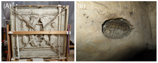
Figure 2.
The back side of REPRO.1980-80 (A) is so thin that shows the reverse image and the wooden armature. A metal stamp (B) with Pouzadoux’s name and the label Musee de Sculpture Comparee du Trocadero can be seen on REPRO.1980-80.
The findings on Malzieux’s casts (Figure 3) relate to other objects acquired through the Architectural Association. They present similar surface colors and salt efflorescence. Malzieux is mentioned in the first catalogue of the Royal Architectural Museum in 1855 under the name Malerieux de Paris and is linked to Alfred Gérente who was acting correspondent to the Architectural Association; in that catalogue, over 150 casts were listed as coming from Notre-Dame among other French Gothic monuments [19]. Other casts from different monuments outside of France show similar color patinas. It is therefore difficult to establish whether Malzieux’s workshop did apply any patina or color on top of the casts or if it was a homogenous finish applied to the entire Architectural Association collection.
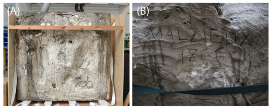
Figure 3.
The back side of REPRO.A.1916-3153 was visibly thicker (A) than what was observed in Pouzadoux’s casts. On the back side of REPRO.A.1916-3152 a rough ‘signature’ by Malzieux and the date of manufacturing of the cast (1855) were found (B).
3.2. The Characterization of the Materials of the Casts
Samples from the plaster casts were taken from pre-existing areas of loss to investigate the stratigraphy and the method of manufacture. A summary of significant scientific results is shown in the following pages and the full database of analysis is available in the online databases [22,23,24,25,26].
A summary and description of the samples, layer by layer, can be seen in Table A3, and a full record is available in the online database [22,23,24,25,26]. Table 2 presents a summary of the results by object.
Table 2.
Summary of results by object *.
All the samples analyzed in this study consistently showed at least three layers in the stratigraphy: the bulk, an interface layer, and a surface or coating layer. Several samples (see, for example, sample REPRO.A.1916-3153_4) showed more layers, in particular showed more layers if they were sampled from areas of repair or with additional varnish layers (for example, REPRO.A.1916-3152_1).
The plaster substrate, which was named layer 0 in most of the samples (Table A3), is made of gypsum plaster (calcium sulfate dihydrate, CaSO4·2H2O). This was confirmed by the presence of calcium, sulfur, and oxygen, overlapping in the EDS maps and spectra, and by the presence of characteristic peaks in the FTIR spectra (the peaks characteristic of the bending and stretching of SO42− at ca. 1005 and 1105 cm−1, and the ν2 H2O related to the sulfate at 1620 and 1680 cm−1), which also show the sulfate overtones in the 2100–2300 cm−1 area (characteristic of reflectance FTIR analysis) [35,36]. Moreover, the ‘tabular’ crystalline structure typical of gypsum plaster can be seen in layer 0 of all the samples in the BSE images, and the XRD analysis also showed the peaks characteristic of gypsum (Table A4) [22,23,24,25,26,27,37]. Aluminium was detected in all the samples and often can be due to 0.3 μm alumina suspension used in samples’ preparation. It was often also observable in combination with silicon (Figure 4), which may imply that it makes up a part of aluminosilicate (clay) minerals. The aluminosilicates are present as few grain inclusions as shown in the elemental mapping (Figure 4). In many samples, manganese, strontium, titanium, and iron were also detected in the bulk (Figure 4). Clay minerals are suggested by the presence of characteristic IR peaks in the 900–1200 cm−1 region [22,23,24,25,26,37]. In REPRO.1890-80 and REPRO.1890-81, aluminium is present overall due to the use of the polishing agent, but along with magnesium and silicon, this could also imply the presence of clay minerals (confirmed EDS mapping). Magnesium is present in the bulk of all the samples, possibly as an exchangeable element in the sulfate variety MgSO4 [38].
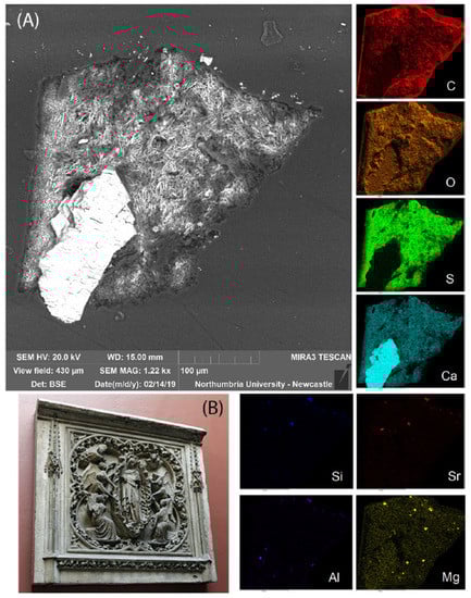
Figure 4.
Sample REPRO.1890-80_1: BSE image (A) shows the tabular mineral structure of the calcium sulfate and the EDS mapping indicates that the sample largely consisted of calcium sulfate (Ca, S, O) and several other inclusions (Al, Mg, Si, Sr). C and O are also present due to the environment and to the casting polyester resin. The sample was taken from REPRO.1890-80 (Assumption of the Virgin, object dimensions 152.5 × 160.0 cm) (B).
The coating layers are not well defined or visible in the stratigraphy. Most often, the outermost layer appears dark under visible illumination, possibly due to the interaction of the portion of the coating that was exposed to the air, pollutants, and UV radiation of the environment, resulting in the oxidation of the coating. A large portion of the coating appeared to be absorbed by the porous plaster bulk and was defined as an interface layer. In most of the samples, the ‘interface layer’, yellow under visible illumination (OM), was observed and possibly consists of a portion of the lower bulk layer soaked with the surface coating(s) and showing characteristics of both layers. In some cases, as in, for example, sample REPRO.1890-80_2 (Figure 5), the BSE image shows that the interface layer appears ‘denser’ than the substrate layer. The crystals of plaster, which appear brighter in the BSE image due to the higher atomic number of calcium in comparison to that of carbon, are more dense in the interface layer. The dark grey areas around the plaster crystals are either voids or filled with the polyester casting resin (mostly made of carbon compounds). In either case, the denser presence of crystals in the interface layer might suggest that the penetration of the coating in the pores of the plaster limited the presence of the casting resin and the subsequent swallowing of the samples.
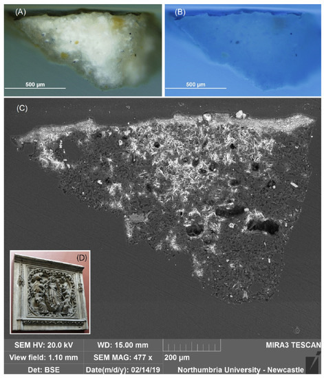
Figure 5.
In sample REPRO.1890-80_2, no differences in the elemental distribution were observed, but the top layer appears denser. VLR (A), UVf (B), and BSE (C) images. The sample was taken from REPRO.1890-80 (D) (Assumption of the Virgin, object dimensions 152.5 × 160.0 cm).
All the samples taken from objects REPRO.1890-80 and REPRO.1890-81 show a dark layer (under visible illumination) which contains silicon, magnesium, and aluminium (detected by EDS). These elements were observed in the EDS mapping as localized in a surface layer, but also sparingly distributed as inclusions in the bulk. In sample REPRO.1890-80_1, these inclusions also contain strontium (naturally occurring in gypsum) [38] and are present in the plaster bulk, but are particularly located in layer 1. Traces of sodium, iron, lead, manganese, potassium, titanium, and chlorine were additionally detected in the surface layers in object REPRO.1890-81.
Barium was detected in all the layers of sample REPRO.A.1916-3153_3 and traces of titanium and were also detected in the surface layer. Both of these elements are known to naturally occur in gypsum [38]. The EDS mapping of samples 1 and 3 of the same object shows that layer 2, dark under visible illumination, consists of aluminium, silicon, and iron.
Samples REPRO.A.1916-3152_1 and _2 show a significant concentration of lead in the bulk but especially in the surface layers. A white layer (layer 1, Table A3) is visible in sample REPRO.A.1916-3152_1 under visible illumination and EDS shows that layer 1 in samples 1 and 2 is made of lead, silicon, and aluminium, as well as traces of chlorine, magnesium, iron, and potassium.
In the same object, the EDS mapping of samples REPRO.A.1916-3152_3 and _4 indicates that the outermost layers in these samples are made mostly of aluminium, silicon, strontium, and traces of barium. Traces of potassium sodium, magnesium, titanium, iron, and chlorine were also detected.
Samples REPRO.1890-80_3 and _4 and REPRO.1980-81_1 and 3 show an additional layer (Table A3), which fluoresces under UV illumination and is consistent with a finishing layer. The presence of the additional varnish only on some of the samples taken from the same object suggests either that a varnish was applied only on the projection areas or that the varnish was applied on all the surfaces but was adsorbed differently depending on local variations in porosity.
In the samples taken from all the objects, a coating made of a resin could be postulated by looking at the FTIR spectra, but the type of resin could not be uniquely determined. The FTIR spectra acquired from the samples are extremely crowded due to the complex composition of the samples and the strong inorganic signal (Appendix B) [37]. The Py-TMAH-GC/MS analyses of samples REPRO.1890-80_4, REPRO.A.1916_3152_3 and REPRO.A.1916_3153_3 suggest that the organic medium in these samples consists of diterpenic resin (rosin or colophony) mixed with drying oil. The Py-TMAH-GC/MS analysis of sample REPRO.1890-81_4 suggests that the organic medium in this sample consists of shellac mixed with linseed oil. Amine fragments were also identified in both the samples, since a minority of protein component is commonly present in the majority of natural ‘non-protein binders’ [29].
Sample REPRO.A.1916-3153_4 shows the same substate of samples 1 and 3 from the same object (layers 0 and 1, Table A3) and, additionally, a layer of repaint (layer 2, white under visible illumination, consisting of barium, zinc, and sodium with traces of iron and chlorine) and a layer of dust (layer 4, dark under visible illumination and made of carbon, oxygen, calcium, sulfur, and silicon).
3.3. The Conservation Treatments
3.3.1. Pouzadoux’s Casts
The conservation treatments of these casts were undertaken in May 2019. REPRO.1890-80—The Assumption of the Virgin—was treated by Adriana Francescutto Miro, as described below. Superficial dust was removed with a vacuum cleaner and soft brushes. The surfaces were cleaned with a smoke sponge. On the front of the panel, the ingrained dirt was removed with the localized use of Anjusil® latex on the darkest areas, carefully selecting appropriate areas rather than using it on all ingrained surfaces. This was followed by using a Blitzfix® sponge lightly dampened in deionized water to rinse the latex solution. All the flaking plaster areas were then consolidated with Paraloid® B44 5%. The paint splashes were inpainted with acrylic paint after all the cleaning treatments were completed. The loose arm of the angel was consolidated using Paraloid® B44 20% in 50:50 IMS–acetone. The areas of plaster loss were consolidated using 25% Primal® B60A and retouched using acrylic paint. The crack on the bottom side of the panel was reinforced by placing carbon fiber matting impregnated with Paraloid® B44 30% placed in the cracking area; this was to ensure the stability of the lower part of the panel on display, as it was to sit on top of a shelf in the gallery.
REPRO-1890-81—The Coronation of the Virgin—was treated by senior sculpture conservator Sarah Healey-Dilkes. Its cleaning treatment was similar to the one undertaken on REPRO.1890-80, but no stabilization was required as the object was structurally stable.
3.3.2. Malzieux’s Casts
REPRO.A.1916-3152—The death of the Virgin—and REPRO.A.1916-3153—The Entombment of the Virgin—were treated in May 2019 by conservator Sayuri Morio. The salt efflorescence observed on the recessed areas, after being removed with a soft brush, left small white spots. The object was dusted with a brush and vacuum. The vertical surface with lighter brown was cleaned with a smoke sponge to remove surface dirt, and sky-facing surfaces in dark brown were cleaned with a Blitzfix® sponge lightly dampened with deionized water. To remove the dark grime on the pronounced area, the latex poultice method was used. For REPRO.A.1916-3153, this was followed by cleaning with a dampened Blitzfix® sponge with deionized water. The splashes of old red museum paint were removed with acetone on cotton swabs. Paint losses and chipped areas were retouched with acrylic paint. For REPRO.A.1916-3153, paint losses and chipped areas were sealed with Paraloid® B44 5% in acetone and IMS and then were retouched with acrylic paint.
4. Discussion
As highlighted during the assessment of the casts, there are many differences in techniques, structures, and surfaces between the two sets of casts. Because of all these material disparities and the different histories behind how they arrived at the V&A, the conservation issues faced were different:
- Pouzadoux’s casts are considerably thinner and lighter and are built around a squared wooden structure (Figure 2). Wood is highly susceptible to hygrometric changes; thus, most of the conservation concerns about these two casts were regarding the movement of the wood causing flaking and the detachment of plaster. This was especially pronounced on the thinner areas of the panel, as well as the small plaster reinforcements on the back side and the lower edge. REPRO.1890-80 had a large crack running through the lower edge of the panel, showing some movement; this was a long-term structural concern caused by the movement of the wood. The surfaces of Pouzadoux’s panels are white and porous, and therefore the cleaning had to be aimed at a deeper level than that of Malzieux’s.
- Malzieux’s casts are heavier and are reinforced with a metallic structure, resulting in thicker, bulkier casts (Figure 3). It is quite possible that, during their time in the Architectural Association collection, they were patinated red, as many other casts acquired through this institution show similar patination and the same deterioration issues. Efflorescent salts on the surface (Figure 6) and ingrained dirt were the main issues flagged regarding the conservation of these panels, which were otherwise structurally stable.
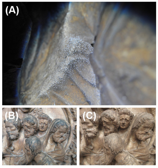 Figure 6. REPRO.A.1916-3152: detail showing salt efflorescence on (A); detail of an area before (B) and after cleaning (C).
The analyses were chosen to ensure an understanding of the coating materials. Priority was given to understanding the surface layers. Being the interface exposed to the environment, this is where deterioration occurs, and conservation is needed. The preliminary analysis of the bodies of the casts suggested that in all the objects the bulk is made of gypsum plaster, which contains several types of inclusions (including silicates and carbonates). Aluminum and silicon are present in the body of casts, but magnesium was only detected in Pouzadoux’s casts and strontium mainly in Malzieux’s. This might suggest a difference in the sourcing and/or processing of the raw material. This hypothesis requires further investigation regarding the mineral composition of the bodies of the casts.
Differences in the surface layers were instead noticeable, as follows:
- -
- Pouzadoux’s casts showed the presence of magnesium, silicon, and aluminium on the surface. The organic medium was identified as diterpenic resin in REPRO.1890-80 and shellac in REPRO.1890-81. In both cases, the additional presence of a drying oil could not be confirmed due to degradation and the characteristics of the samples.
- -
- The surface layers of Malzieux’s casts appear less straightforward. The samples taken from the front-facing side of REPRO.A.1916-3152 showed significant concentrations of lead, suggesting the possibility that these areas were highlighted with lead-based paint (also containing aluminium and silicon). Samples from the same object taken from marginal and recess areas showed the same stratigraphy as the other samples but did not contain lead. Additionally, some surface layers, fluorescing white-blue under UV illumination, were consistent with a layer of varnish. Analysis suggested the presence of diterpenic resin possibly mixed with a non-drying oil, which could have been applied to the surface or also mixed with the paint. The same medium was detected in REPRO.A.1916-3153, but in this case aluminium, silicon, and iron were the main elements on the surface layer. A sample likely taken from a repainted area showed a different layer of zinc–barium white paint.
The presence of silicon and aluminium on the surface layer of all the casts could be due either to the use of clay water as a separating agent [39,40] or to the use of the silicate mineral pigment kaolinite Al2Si2O5(OH)4 to tone the surface [41].
In Pouzadoux’s casts, a significant magnesium component can be also seen in the surface together with aluminium and silicon, suggesting that ‘French chalk’ (talc, mostly containing magnesium and silicon but also aluminium) could have been used to polish the surface [39,41,42].
Particularly important was the identification of organic media and the observation of their penetration in the body of the casts. Organic materials are more susceptible to degradation than inorganic materials, and being able to identify natural substances and their degradation products, particularly in complex mixtures, is a challenge. Organic materials could have undergone various modifications (e.g., oxidation, photodecomposition, and microbial digestion) over time.
The multi-analytical method was designed to ensure an understanding of the coating materials, which is crucial to supporting future approaches to the casts’ surface treatments. Further research on the bulk of the casts is desirable, but at this stage of the research, it was not part of the AHRC project.
The aim of the conservation treatment was to survey the condition of each one of the objects as part of the second phase of the Cast Courts conservation work. After gaining an understanding on the stability of the objects, the main priority was to remediate any structural concerns that arose from the condition checking and secondarily was to reduce the staining on surfaces caused by ingrained dirt that was obscuring the relief of the objects, preventing the correct interpretation of the objects not only as a group but also within the rest of the plaster casts in the gallery.
5. Conclusions
This study achieved a better understanding of the manufacturing practices of two nineteenth-century plaster workshops, those of Pouzadoux and Malzieux, working at the same time during the restoration campaigns of Notre-Dame between 1843 and 1864. The differences identified between the casts, particularly in terms of structural stability and surface finish, influenced the choices of treatment made by the V&A conservators.
Author Contributions
Conceptualization, V.R., A.F.M., S.M. and C.T.; conservation treatments, A.F.M. and S.M.; formal analysis, V.R.; writing—original draft preparation, V.R. and A.F.M.; writing—review and editing, S.M. and C.T.; supervision, C.T.; funding acquisition, C.T. All authors have read and agreed to the published version of the manuscript.
Funding
This research was funded by the Art and Humanities Research Council (2017-21 AHRC/CDP Northumbria University and Victoria and Albert Museum—AH/R00322X/1) and supported by the Henry Moore Foundation (HMF Research and Travel Grant—2018).
Data Availability Statement
All data generated during this study are discussed in this published article and are fully available at the Figshare permanent data links: https://doi.org/10.25398/rd.northumbria.13469925 (accessed on 17 October 2022), https://doi.org/10.25398/rd.northumbria.14040080 (accessed on 17 October 2022), https://doi.org/10.25398/rd.northumbria.14040182 (accessed on 17 October 2022), https://doi.org/10.25398/rd.northumbria.14040224 (accessed on 17 October 2022), https://doi.org/10.25398/rd.northumbria.14040251 (accessed on 17 October 2022).
Acknowledgments
Special thanks to the V&A: Sculpture Conservation team for their insights on their conservation treatments and knowledge of the Cast Collection, and to the Sculpture Curatorial Department for facilitating access to their collection and archival information. Our thanks go to all the parties involved in this work and to Lucia Burgio and Victor Borges for their helpful comments on the article.
Conflicts of Interest
The authors declare no conflict of interest.
Appendix A. Additional Images and Tables

Figure A1.
Graphic representations rendered in Autodesk® AutoCAD® 2019 showing the outline of the casts and the sampling locations. The images are scaled. More details and additional images of the sampling locations can be seen in the online database of results [22,23,24,25,26].
Figure A1.
Graphic representations rendered in Autodesk® AutoCAD® 2019 showing the outline of the casts and the sampling locations. The images are scaled. More details and additional images of the sampling locations can be seen in the online database of results [22,23,24,25,26].

Table A1.
Samples summary and description.
Table A1.
Samples summary and description.
| Object | Sample | Description |
|---|---|---|
| REPRO.1890-80 | 1 | Fragment taken from an area of loss along a crack, PR side. |
| 2 | Fragment taken from an area of loss along a crack, PL side. | |
| 3 | Fragment taken from an area of loss on the PL edge. | |
| 4 | Fragment taken from a large area of loss on the PL side, top rim. | |
| REPRO.1890-81 | 1 | Fragment taken from an area of loss on the PL architectural element. |
| 2 | Fragment taken from an area of loss on the PR, from the frame. | |
| 3 | Fragment taken from an area of loss on the PL, from the frame. | |
| 4 | Fragment taken from an area of loss on the PL, from the frame. | |
| REPRO.A.1916-3152 | 1 | Already detached fragment taken from the PR, under two figures. |
| 2 | Fragment, dust, and efflorescence taken from under the figure’s hand. | |
| 3 | Already detached fragment from an area of crack, bottom PR edge. | |
| 4 | Fragment taken from an area of loss, PR edge, red paint? | |
| REPRO.A.1916-3153 | 1 | Fragment taken from an area of loss on the relief’s PR |
| 2 | Dust and efflorescence taken from the top portion of the relief. | |
| 3 | Fragment taken from an undercut area on the PL of the relief. | |
| 4 | Fragment taken from an area of loss from the PL edge. |
Table A2.
Py-GC/MS markers used to characterize the organic component [29,30,31,32,33].
Table A2.
Py-GC/MS markers used to characterize the organic component [29,30,31,32,33].
| Label | Marker | m/z | RT 1 (min) |
|---|---|---|---|
| Fatty acid | |||
| 1 | Methyl azelate (A) | 42, 58, 74 (100), 87, 120, 138, 152, 171, 185 | 16.68–17.52 |
| 2 | Methyl palmitate (P) | 55, 74(100), 87, 101, 129, 145, 185, 199, 227, 239, 270 | 21.68–21.70 |
| 3 | Methyl stearate (S) | 74 (100), 87, 129, 143, 199, 255, 298 | 23.60–23.62 |
| Diterpene resin | |||
| 4 | Methyl 7-oxo-15-hydroxy-dehydroabietic acid | 58(100), 71, 85, 115, 149, 207, 219, 251, 270, 299, 331 | 22.79 |
| 5 | Methyl 7-Oxodehydroabietic acid | 44, 58, 74, 87, 129, 171, 187, 207, 239, 253(100), 281, 299, 314, 328 | 25.47 |
| 6 | Methyl dehydroabietate | 141, 155, 197, 239(100), 253, 314 | 25.57 |
| 7 | Methyl oxo-dehydroabietic acid | 44(100), 79, 115, 165, 191, 207, 227, 267, 281, 342 | 26.72 |
| Shellac | |||
| 8 | Jalaric acid | 59, 69, 83, 87, 105, 121, 135, 145, 167, 179, 191, 203, 208, 231, 247, 262(100), 275, 307, 322 | 23.35 |
| 9 | Shelloic acid | 55, 71, 97, 109, 137, 169, 201(100), 231, 261, 291, 304, 336 | 23.82 |
| 10 | Shellolic acid | 59, 79, 91, 129, 169, 206, 229, 238, 260, 288, 305, 320(100), 337 | 24.53 |
| 11 | Aleuritic acid | 44, 55, 71(100), 81, 109, 137, 159, 201, 207, 239, 312, 327 | 25.54–26.52 |
| Phthalic compound | |||
| 12 | Phthalic anhydride | 45, 55, 71(100), 85, 101, 111, 142, 147, 156 | 10.67–13.67 |
| 13 | Dimethyl phthalate | 58, 77, 92, 104, 133, 163(100), 194 | 16.35 |
1 Retention times (RT) are specific to the experimental conditions and characteristics of the equipment used. In this study, RT of the same component varies ±0.50 depending on the sample.
Table A3.
Samples’ stratigraphy summary. Color description in this table refers to visible light appearance. In the relevant open access databases [22,23,24,25,26], under the OM-BSE tab, the full record of images is available.
Table A3.
Samples’ stratigraphy summary. Color description in this table refers to visible light appearance. In the relevant open access databases [22,23,24,25,26], under the OM-BSE tab, the full record of images is available.
| Sample | Stratigraphy (Layers) |
|---|---|
| REPRO.1890-80_1 | 1. Dark surface layer with dark particles 0. Calcium sulfate bulk with Sr, Mg, Al, and Si inclusions |
| REPRO.1890-80_2 | 2. Dark surface layer 1. Yellow interface layer 0. Calcium sulfate bulk with Mg, Al, and Si inclusions |
| REPRO.1890-80_3 | 2. Dark surface layer 1. Yellow interface layer 0. Calcium sulfate bulk with Mg, Al, and Si inclusions |
| REPRO.1890-80_4 | 2. Fluorescent coating layer * 1. Dark surface layer * 0. Calcium sulfate bulk with Al and Si inclusions |
| REPRO.1890-81_1 | 3. Fluorescent coating layer 2. Dark surface layer 1. Yellow interface layer 0. Calcium sulfate bulk with Mg, Al, and Si inclusions |
| REPRO.1890-81_2 | 2. Dark surface layer 1. Grey interface layer 0. Calcium sulfate bulk with Mg, Al, and Si inclusions |
| REPRO.1890-81_3 | 3. Fluorescent coating layer 2. Dark surface layer containing Mg and Si 1. Yellow interface layer 0. Calcium sulfate bulk with Mg, Al, and Si inclusions |
| REPRO.1890-81_4 | 2. Dark surface layer containing Mg and Pb ** 1. Yellow interface layer ** 0. Calcium sulfate bulk with Mg, Al, and Si inclusions |
| REPRO.A.1916-3152_1 | 2. Fluorescent coating layer 1. White layer mostly made of Si and Pb 0. Calcium sulfate bulk, rich in Pb and with Al and Si inclusions |
| REPRO.A.1916-3152_2 | 1. Dark surface layer 0. Calcium sulfate bulk, rich in Pb and with Al and Si inclusions |
| REPRO.A.1916-3152_3 | 1. Dark surface layer containing Sr, Al, and Si * 0. Calcium sulfate bulk with Sr, Al, and Si inclusions |
| REPRO.A.1916-3152_4 | 2. Fluorescent coating layer containing Sr, Al, and Si 1. Yellow interface layer with dark inclusions 0. Calcium sulfate bulk with Sr, Al, and Si inclusions |
| REPRO.A.1916-3153_1 | 2. Dark surface containing Al and Si 1. Yellow interface 0. Calcium sulfate bulk with Al and Si inclusions |
| REPRO.A.1916-3153_2 | Dust and efflorescence containing clay minerals, sulfates, and oxalates |
| REPRO.A.1916-3153_3 | 2. Dark surface containing Al, Si, and Fe * 1. Yellow interface * 0. Calcium sulfate bulk with Al and Si inclusions |
| REPRO.A.1916-3153_4 | 3. Dark surface layer 2. White layer made of Na, Zn, and Ba 1. Yellow interface layer with dark inclusions 0. Calcium sulfate bulk |
* Diterpenic resin and possibly oil, ** Shellac and possibly oil.
Table A4.
XRD peaks (2θ, °) identified on the diffractograms acquired from the plaster samples analyzed and relevant references R040029 and R060509 from the RRUFF database [22,23,24,25,26].
Table A4.
XRD peaks (2θ, °) identified on the diffractograms acquired from the plaster samples analyzed and relevant references R040029 and R060509 from the RRUFF database [22,23,24,25,26].
| Sample/Reference | 2θ [°] |
|---|---|
| REPRO.1890-80_2 | 11.63, 20.75, 29.10, 31.10, 33.36, 40.64, 43.31, 47.82, 48.34, 50.34, 51.37, 71.78 |
| REPRO.A.1916-3152_1 | 29.34, 31.39, 33.62, 43.65 |
| R040029 | 11.66, 20.77, 28.15, 29.14, 34.62, 36.00, 36.65, 37.41, 42.21, 44.23, 45.53, 46.46, 47.88, 48.40, 54.47, 55.19, 55.86, 60.36, 66.71 |
| R060509 | 11.69, 20.79, 28.16, 29.16, 36.01, 37.42, 45.55, 47.90, 48.42, 55.22 |
Appendix B. Characterization of the Organic Component in Mostly Inorganic Samples
Identifying a small amount of organic material in historical samples represents a difficult problem for traditional methods of chemical analyses. The situation is even more complicated when an organic mixture of variable composition should be identified in a complex matrix containing a range of other organic and inorganic materials. In this study, FTIR and GC/MS analysis were combined to characterize the organic media in the plaster samples. FTIR spectra were inconclusive due to the broadness of the bands that resulted from the diffuse reflectance signal and the presence of numerous components in the sample. The broad bands did not allow the characterization of specific organic components but were however indicative of the presence of classes of materials. As the results of the EDS suggested the presence of gypsum, calcite, and clay minerals, FTIR reference spectra of these materials [37,43,44] were used to skim the peaks in the spectra acquired from the objects. Reference spectra for the casting resin were also acquired. This was particularly important because, as mentioned in the results section, the porous samples absorbed the casting resin when in a liquid state.
The historical recipes investigated as part of this study and published elsewhere [6,7,45] also helped to select references to navigate our spectra.
Due to the complexity of the FTIR spectra acquired from the samples in this study, the assignment of the broad bands in the spectra was challenging; nonetheless, the spectra were compared, and it was often helpful to note the absence of a characteristic vibration. Just as the presence of functional group bands can be used to select potential matches, the absence of a functional group band is used to eliminate potential groups of materials. It is also important to note that ageing can cause significant changes to the infrared bands of certain binding media.
As salt efflorescence is a common degradation phenomenon observed on the surface of plaster and building materials [46], references for the identification of these products were also taken into account. Calcium oxalate is, for example, suggested as a degradation product [47] but cannot be identified in the FTIR spectra acquired for this study due to the broadness of the bands; some of the characteristic peaks of calcium oxalate (1648 cm−1) are expected to be close to calcium sulfate vibrations (1620 and 1680 cm−1) and others may overlap with organic components.
GC/MS was therefore in this instance particularly important to the identification of organic materials. A mass spectrometer is an excellent tool for clearly identifying the structure of a single compound but is less useful when presented with a mixture [29]. Most often, a full GC pattern that would characterize a fresh or even aged pure sample prepared in the laboratory is not present in the materials sampled from cultural heritage objects. The interpretation of chromatograms and mass spectra, or even the assignment of a few components that may be considered ‘markers’, is key to the characterization of unknown samples. The GC expected of a fresh material was not always detected, but this could be due to the natural degradation of the molecules or to the derivatization processes applied to the already degraded organic material [29].
It has been suggested that the intensity of fatty acid (FA) peaks in chromatograms can change as affected by matrix effects due to the presence of inorganic pigments in organic media [48]. In this study, it is possible that this effect was amplified, as the organic component is significantly less than the inorganic portion. This is likely to have also influenced the small relative abundance of the markers in the chromatograms.
Diterpenic resin was identified by py-GC/MS as its characteristic markers were observed in the GC (Table A2), and the relevant MS spectra were supported by the literature [29]. The presence of methyl palmitate (2) and stearate (3) was detected in all the samples, and the P/S ratio varies significantly from sample to sample [7]. Methyl azelate (1) is not always present in the chromatogram, and when present, it is not very abundant. The resin may have been mixed with an oil or the FAs could be derived from another source of lipids naturally present in the resin [7,29]. Thus, the P/S and A/P ratios cannot be used for the assignment of lipids in this study.
Shellac can be relatively easily identified by UVf OM due to its characteristic pink-orange fluorescence color. Py-TMAH-GC/MS chromatogram markers characteristic of shellac are namely jalaric acid (8), shelloic acid (9), shellolic acid (10), aleuritic acid (11), and other C15 fragments [7,29,30]. Methyl azelate (1), methyl palmitate (2), and stearate (3) were also detected.
References
- Frederiksen, R.; Marchand, E. (Eds.) Plaster Casts: Making, Collecting and Displaying from Classical Antiquity to the Present; Walter de Gruyter: Berlin, Germany, 2010; Volume 18. [Google Scholar]
- Bilbey, D.; Trusted, M. The Question of Casts—Collecting and Later Reassessment of the Cast Collections at South Kensington. In Plaster Casts: Making, Collecting and Displaying from Classical Antiquity to the Present; Frederiksen, R., Marchand, E., Eds.; Walter de Gruyter: Berlin, Germany, 2010; Volume 18, pp. 463–484. [Google Scholar]
- Foster, S.M.; Curtis, N.G.W. The Thing about Replicas—Why Historic Replicas Matter. Eur. J. Archaeol. 2016, 19, 122–148. [Google Scholar] [CrossRef]
- Brown, G.M. Art in the Age of Digital Reproduction. Available online: https://www.ft.com/content/74ffab6e-1b55-11e6-b286-cddde55ca122, (accessed on 20 February 2018).
- Payne, E.M. 3D imaging of the Parthenon sculptures: An assessment of the archaeological value of nineteenth-century plaster casts. Antiquity 2019, 93, 1625–1642. [Google Scholar] [CrossRef]
- Risdonne, V.; Hubbard, C.; Borges, V.H.L.; Theodorakopoulos, C. Materials and Techniques for the Coating of Nineteenth Century Plaster Casts: A Review of Historical Sources. Stud. Conserv. 2022, 67, 186–208. [Google Scholar] [CrossRef]
- Risdonne, V. Materials and Techniques for Coating of the Nineteenth-Century Plaster Casts. A Scientific and Archival Investigation of the Victoria & Albert Museum Cast Collection. Ph.D. Thesis, Northumbria University, Newcastle upon Tyne, UK, 2022. [Google Scholar]
- Kamel, A.M. Dehydration of gypsum component of plasters and stuccos in some Egyptian archaeological buildings and evaluation of K2SO4 activator as a consolidant. Sci. Cult. 2019, 5, 49–59. [Google Scholar] [CrossRef]
- Theologitis, A.; Kapridaki, C.; Kallithrakas-Kontos, N.; Maravelaki-Kalaitzaki, P.; Fotiou, A. Mortar and plaster analysis as a directive to the design of compatible restoration materials in Frangokastello (Crete). Mediterr. Archaeol. Archaeom. 2021, 21, 109–120. [Google Scholar] [CrossRef]
- Quilici, M.; French, C.; Chatzimpaloglou, P. Torba floors from the Maltese islands: A preliminary analytical study. Mediterr. Archaeol. Archaeom. 2022, 22, 183–192. [Google Scholar] [CrossRef]
- Ali, M.F.; Moussa, A.; El-Sayed, S.H. Analytical physicochemical survey of the recently excavated murals at the tomb of Iwrakhy/Hatia at Saqqara, Egypt. Sci. Cult. 2022, 8, 63–79. [Google Scholar] [CrossRef]
- Ashkenazi, D.; Shnabel, R.; Lichtenberger, A.; Tal, O. Chemical composition and microstructure analysis of plaster and pigments retrieved from a decorated house wall at Seleucid Tell Iẓṭabba (Nysa-Scythopolis, Beth She’an, Israel). Mediterr. Archaeol. Archaeom. 2021, 21, 89–122. [Google Scholar] [CrossRef]
- Da Riva, R.; Santos Arévalo, F.J.; Madrid i Fernández, M. The Mortars from rock-cut hydraulic structures of as-Sila (Sela) in Southern Jordan: Mineralogical characterization and Radiocarbon dating. Mediterr. Archaeol. Archaeom. 2021, 21, 37–67. [Google Scholar] [CrossRef]
- Roberto, G.; Eliano, D. Decay of red pigments on a wall painting adorning the church of ‘San Francesco Dei Capuccini’ in Racconigi (Italy): Archaeometric survey and restoration intervention. Mediterr. Archaeol. Archaeom. 2018, 18, 65–80. [Google Scholar] [CrossRef]
- Hubbard, C. Conservation of the Weston Cast Court at the V&A. Available online: https://3rd-dimensionpmsa.org.uk/features/2015-05-10-conservation-of-the-weston-cast-court-at-the-v-a (accessed on 26 October 2017).
- Miramón, J.G. Conservación y Restauración de Esculturas en Yeso en la Real Academia de Bellas Artes de San Fernando. Ph.D. Thesis, Universidad Complutense de Madrid, Madrid, Spain, 2019. [Google Scholar]
- Cast Courts: One Month to Go! Available online: https://www.vam.ac.uk/blog/caring-for-our-collections/cast-courts-one-month-to-go (accessed on 24 September 2022).
- V&A Website—Search the Collections. Available online: https://collections.vam.ac.uk/ (accessed on 10 December 2021).
- Hofman, J.M. Ephémères musées d’archéologie médiévale. La collection de moulages de l’humble M. Malzieux. Situ Rev. Patrim. 2016, 28. [Google Scholar] [CrossRef]
- Hofman, J.M. Rencontre avec un illustre inconnu Jean Pouzadoux (1829–1893), mouleur en plâtre. Conférence du 20 Janvier 2011. Bull. Société Hist. 6e Arrondiss. 2011, 24. [Google Scholar]
- BS EN 16085:2012; Conservation of Cultural Property. Methodology for Sampling from Materials of Cultural Property. General Rules. British Standards Institution (BSI): Loughborough, UK, 2012; ISBN 9780580705885.
- Risdonne, V.; Theodorakopoulos, C. Database of Results. V&A cast of the relief of The Assumption of the Virgin (REPRO1890-80). Northumbria University Figshare repository; v.1. 2021. Available online: https://figshare.northumbria.ac.uk/articles/dataset/Database_of_Results_V_A_cast_of_the_relief_of_The_Assumption_of_the_Virgin_REPRO1890-80_/14040080/1 (accessed on 17 October 2022).
- Risdonne, V.; Theodorakopoulos, C. Database of Results. V&A cast of the relief of The Coronation of the Virgin (REPRO1890-81). Northumbria University Figshare repository; v.1. 2021. Available online: https://figshare.northumbria.ac.uk/articles/dataset/Database_of_Results_V_A_cast_of_the_relief_of_The_Coronation_of_the_Virgin_REPRO1890-81_/14040182/1 (accessed on 17 October 2022).
- Risdonne, V.; Theodorakopoulos, C. Database of Results. V&A cast of the relief of The Death of the Virgin (REPRO.A.1916-3152). Northumbria University Figshare repository; v.1. 2021. Available online: https://figshare.northumbria.ac.uk/articles/dataset/Database_of_Results_V_A_cast_of_the_relief_of_The_Death_of_the_Virgin_REPRO_A_1916-3152_/14040224/1 (accessed on 17 October 2022).
- Risdonne, V.; Theodorakopoulos, C. Database of Results. V&A cast of the relief of The Entombment of the Virgin (REPRO.A.1916-3153). Northumbria University Figshare repository; v.1. 2021. Available online: https://figshare.northumbria.ac.uk/articles/dataset/Database_of_Results_V_A_cast_of_the_relief_of_The_Entombment_of_the_Virgin_REPRO_A_1916-3153_/14040251/1 (accessed on 17 October 2022).
- Risdonne, V.; Theodorakopoulos, C. Database of Results. Materials and techniques for coating of the nineteenth-century plaster casts. A scientific and archival investigation of the Victoria & Albert Museum cast collection. Northumbria University Figshare repository; v.1. 2021. Available online: https://figshare.northumbria.ac.uk/articles/dataset/Database_of_results_Materials_and_techniques_for_coating_of_the_nineteenth-century_plaster_casts_A_scientific_and_archival_investigation_of_the_Victoria_Albert_Museum_cast_collection_/14053985 (accessed on 17 October 2022).
- Lafuente, B.; Downs, R.T.; Yang, H.; Stone, N. The power of databases: The RRUFF project. In Highlights in Mineralogical Crystallography; Walter de Gruyter: Berlin, Germany, 2016; pp. 1–30. [Google Scholar]
- Gražulis, S.; Chateigner, D.; Downs, R.T.; Yokochi, A.F.T.; Quirós, M.; Lutterotti, L.; Manakova, E.; Butkus, J.; Moeck, P.; Le Bail, A. Crystallography Open Database—An open-access collection of crystal structures. J. Appl. Crystallogr. 2009, 42, 726–729. [Google Scholar] [CrossRef]
- Colombini, M.P.; Modugno, F. Organic Mass Spectrometry in Art and Archaeology; John Wiley & Sons: Chichester, UK, 2009. [Google Scholar]
- Singer, B.; McGuigan, R. The simultaneous analysis of proteins, lipids, and diterpenoid resins found in cultural objects. Ann. Di Chim. J. Anal. Environ. Cult. Herit. Chem. 2007, 97, 405–417. [Google Scholar] [CrossRef] [PubMed]
- Challinor, J.M. The development and applications of thermally assisted hydrolysis and methylation reactions. J. Anal. Appl. Pyrolysis 2001, 61, 3–34. [Google Scholar] [CrossRef]
- Challinor, J.M. The scope of pyrolysis methylation reactions. J. Anal. Appl. Pyrolysis 1991, 20, 15–24. [Google Scholar] [CrossRef]
- Linstrom, P.J.; Mallard, W.G. NIST chemistry WebBook, NIST standard reference database number 69, National Institute of Standards and Technology. J. Phys. Chem. Ref. Data Monogr. 1998, 9, 1–1951. [Google Scholar]
- Beauzac, J. L’histoire matérielle des moulages du musée de Sculpture comparée (1897–1927). Situ Rev. Patrim. 2016, 28, 8–12. [Google Scholar] [CrossRef]
- Manfredi, M.; Barberis, E.; Rava, A.; Robotti, E.; Gosetti, F.; Marengo, E. Portable diffuse reflectance infrared Fourier transform (DRIFT) technique for the non-invasive identification of canvas ground: IR spectra reference collection. Anal. Methods 2015, 7, 2313–2322. [Google Scholar] [CrossRef]
- Gariani, G.; Lehuédé, P.; Leroux, L.; Wallez, G.; Goubard, F.; Bouquillon, A.; Bormand, M. First insights on the mineral composition of “stucco” devotional reliefs from Italian Renaissance Masters: Investigating technological practices and raw material sourcing. J. Cult. Herit. 2018, 34, 23–32. [Google Scholar] [CrossRef]
- Price, B.A.; Pretzel, B.; Lomax, S.Q. Infrared and Raman Users Group Spectral Database, 2007 ed.; IRUG: Philadelphia, PA, USA, 2009. [Google Scholar]
- Cox, K.G.; Price, N.B.; Harte, B. An Introduction to the Practical Study of Crystals, Minerals, and Rocks; Halsted Press: London, UK, 1974. [Google Scholar]
- Auerbach, A. Modelled Sculpture and Plaster Casting; Thomas Yoseloff: New York, NY, USA, 1961. [Google Scholar]
- Frederick, F.F. Plaster Casts and How They Are Made; Comstock: New York, NY, USA, 1899. [Google Scholar]
- Millar, W. Plastering, Plain & Decorative. A Practical Treatise on the Art & Craft of Plastering and Modelling, Including Full Description of the Various Tools, Materials, Processes and Appliances Employed; Donhead Publishing Ltd.: Dorset, UK, 1899. [Google Scholar]
- Wager, V.H. Plaster Casting for the Student Sculptor; Alec Tiranti: London, UK, 1963. [Google Scholar]
- Derrick, M.R.; Stulik, D.; Landry, J.M. Infrared Spectroscopy in Conservation Science; Getty Publications: Los Angeles, CA, USA, 2000. [Google Scholar]
- Vahur, S.; Teearu, A.; Peets, P.; Joosu, L.; Leito, I. ATR-FT-IR spectral collection of conservation materials in the extended region of 4000-80 cm–1. Anal. Bioanal. Chem. 2016, 408, 3373–3379. [Google Scholar] [CrossRef] [PubMed]
- Risdonne, V.; Hubbard, C.; Puisto, J.; Theodorakopoulos, C. A multi-analytical study of historical coated plaster surfaces: The examination of a nineteenth-century V&A cast of a tombstone. Herit. Sci. 2021, 9. [Google Scholar] [CrossRef]
- Chelazzi, D.; Poggi, G.; Jaidar, Y.; Toccafondi, N.; Giorgi, R.; Baglioni, P. Hydroxide nanoparticles for cultural heritage: Consolidation and protection of wall paintings and carbonate materials. J. Colloid Interface Sci. 2013, 392, 42–49. [Google Scholar] [CrossRef] [PubMed]
- Toniolo, L.; Zerbi, C.M.; Bugini, R. Black layers on historical architecture. Environ. Sci. Pollut. Res. 2009, 16, 218–226. [Google Scholar] [CrossRef] [PubMed]
- Poli, T.; Piccirillo, A.; Zoccali, A.; Conti, C.; Nervo, M.; Chiantore, O. The role of zinc white pigment on the degradation of shellac resin in artworks. Polym. Degrad. Stab. 2014, 102, 138–144. [Google Scholar] [CrossRef]


Publisher’s Note: MDPI stays neutral with regard to jurisdictional claims in published maps and institutional affiliations. |
© 2022 by the authors. Licensee MDPI, Basel, Switzerland. This article is an open access article distributed under the terms and conditions of the Creative Commons Attribution (CC BY) license (https://creativecommons.org/licenses/by/4.0/).