Review

Visible/Red/NIR Light-Mediated NO Donors for Biological Applications

1
Key Laboratory of Photochemical Conversion and Optoelectronic Materials, Technical Institute of Physics and Chemistry, Chinese Academy of Sciences, Beijing 100190, China
2
University of Chinese Academy of Sciences, Beijing 100190, China
Chemistry 2025, 7(3), 66; https://doi.org/10.3390/chemistry7030066
Submission received: 31 March 2025 / Revised: 15 April 2025 / Accepted: 18 April 2025 / Published: 22 April 2025

Abstract

:
Nitric oxide (NO) is an important messenger molecule in almost all organisms. The diverse biological activities of NO have initiated in-depth research on the development of exogenous NO donors. Light-controlled NO donor can transport NO to specific areas to treat various diseases; thus, light–triggered NO donors are rapidly becoming an important class of compounds for the design of novel potential drugs. This review highlights the recent development of organic small molecule-based light-triggered NO donors and focuses on visible/red/NIR light-mediated NO donors. It contains rational designs of NO donor, NO releasing mechanism and detection, as well as its biological applications. Finally, the advantages, drawbacks, and challenges of this strategy are discussed in view of practical applications.

1. Introduction

Nitric oxide (NO) is an endogenous signal transmitter generated from L-arginine through nitric oxide synthase (NOS) and is involved in numerous physiological and pathological processes [1,2,3,4]. It plays the key regulative roles in many cellular life activities, including vasodilatation, neurotransmission, inflammation, platelet aggregation and adhesion, hormone secretion, and immune response [5,6,7]. It has been demonstrated that NO exhibits remarkable therapeutic abilities in the treatment of cardiovascular disease [8,9], cancers [10,11], wounds [12,13], and bacterial infections [14,15,16]. Moreover, NO-based therapy holds very promising features helpful in cancer treatment because NO can reverse the multidrug resistance of cancer cells and consequently enhance chemotherapeutic effects [17,18]. However, the short half-life of NO and its high sensitivity to biological substances greatly restrict the development of NO-based therapeutic platforms for clinical applications. One the other hand, NO can act as a “double-edged sword” because its biological effects depend on its concentration, tumor microenvironment, and dosage [19,20]; therefore, precise control of intracellular NO delivery and release is a challenge [21,22].
Many efforts have been devoted to exploring NO donors (compounds that can generate NO in situ) as well as their delivery platforms [23,24]. These exogenous NO-releasing systems are able to produce NO when they are triggered by external stimuli, such as pH, heat, or light [23,25]. Among them, the light-triggered NO-releasing platform is the most attractive because it can achieve accurate NO release with precise remote, spatiotemporal control and timing and dosage [26]. This review highlights the recent development of light-triggered NO donors for various biological applications, specifically NO donors based on organic small molecules with visible/red/NIR light triggers. Organic small molecules have several merits, including small size, good biocompatibility, and a highly permeable membrane [27,28,29]. Visible/Red/NIR light-mediated NO release can deepen tissue penetration and reduce cell photodamage [30,31,32,33]. Moreover, NO donors with fluorescence properties are desirable since fluorescence technology can achieve visualized online detection/analysis, especially with turn-on or ratiometric fluorescence [34,35,36,37,38,39,40].

2. Design Strategies for Light-Triggered NO Donors

Many traditional NO donors, such as trinitroglycerin [41], sodium nitroprusside [42], diazeniumdiolates [43], nitrosothiols [44], and isoamyl nitrite [45], do not have controlled release characteristics; they spontaneously release NO in a biological system in the presence of various biological analytes or enzymes, which limits their applications in basic mechanistic studies as well as clinical trials. To achieve controllable release, NO donors with a light-responsive function are excellent candidates, and several notable small molecule systems for light-controlled NO donors such as N-nitrosoamines, nitrobenzenes, and S-nitrosothiols have been developed [46,47].
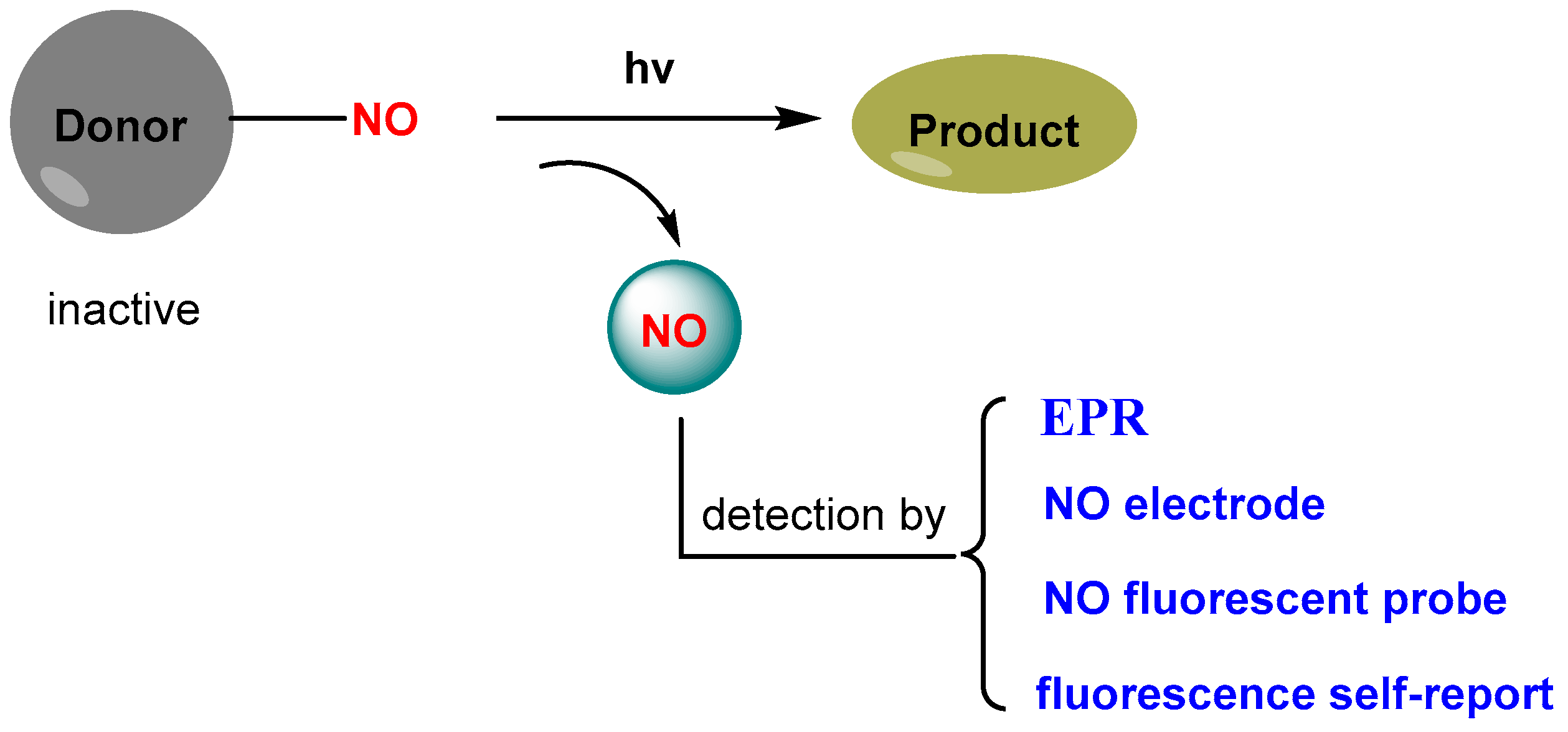
Light-controlled NO release refers to NO donors that remain inactive and nontoxic in the dark and can liberate “caged” NO upon irradiation with light via breaking a covalent bond (Scheme 1). NO is generated only in the region of interest with high precision by the appropriate positioning of the light beam and with accurate control of its doses by tuning the light intensity and the irradiation time. The methods commonly used to detect NO release include EPR (electron paramagnetic resonance) spectroscopy [48,49], NO electrode [50,51], NO fluorescent probe [52,53], and fluorescence self-report [54]; the latter refers to those in which fluorescence is based on a self-calibration mechanism [55,56].
In this review, two kinds of NO donors are involved based on their functions: one is single-function NO donors (Scheme 2A), in which the donors are composed of single pharmacologically active entity and only serve as donors for NO release, and the other is dual-function NO donors (Scheme 2B), in which two pharmacologically active entities are covalently jointed in the same molecular skeleton to enhance the therapeutic effect through multimodal treatment.

3. Light-Triggered NO Release and Their Biological Applications

3.1. Single-Function NO Donor

Red/NIR (near-infrared) light-triggered NO donors are of great significance in practical applications due to deep tissue penetration due to the high optical transmission of hemoglobin and water in the above spectral region [57]. The generation of NO by one-photon excitation in the therapeutic window (650–1300 nm) is highly desirable.
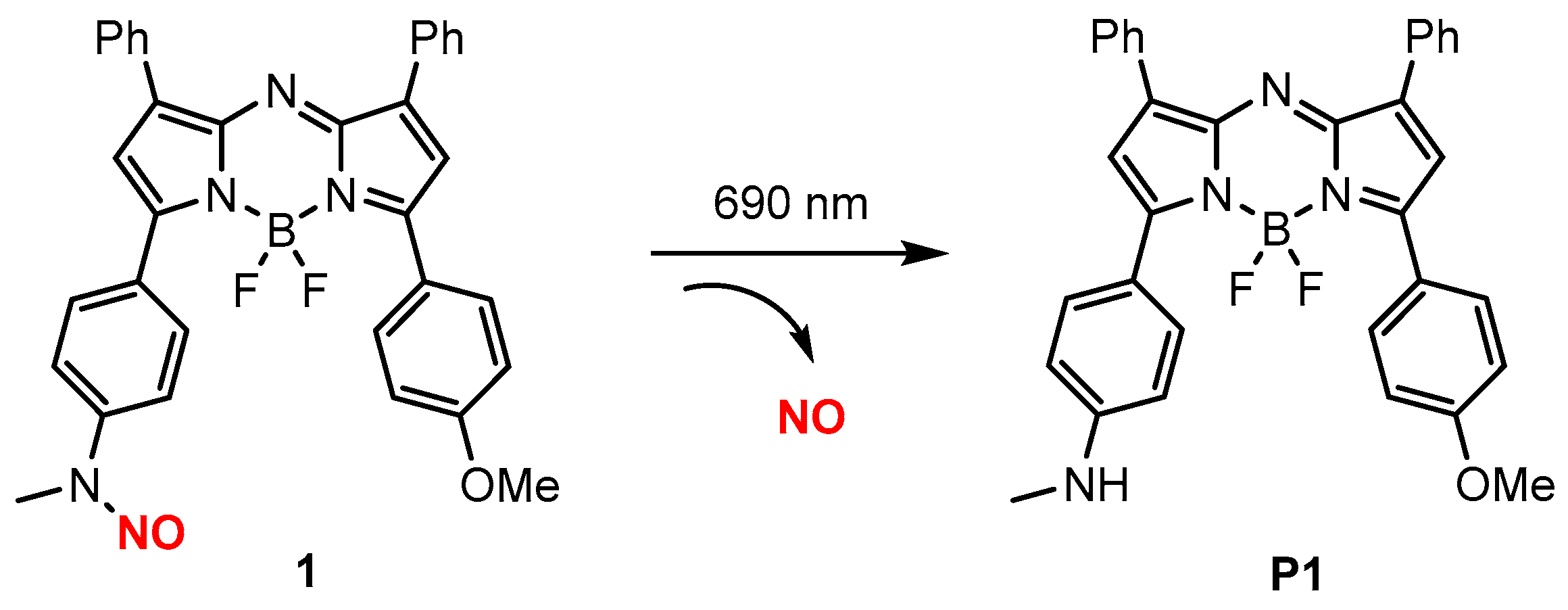
Zhou and co-workers [58] reported an NIR-photoactivatable NO donor using an aza-BODIPY dye as molecular skeleton. Donor 1 (Scheme 3) exhibited absorbance maxima at 680 nm in aqueous buffer and showed good chemostability, pH stability, and biocompatibility. Photo-controlled NO release was confirmed and quantified by EPR (electron paramagnetic resonance) spectra collected after 0, 5, and 40 min of irradiation of 1 with 690 nm irradiation. Additionally, 20.4 μM NO after 5 min irradiation and 30.1 μM NO after 40 min irradiation were obtained.
Donor 1 and its photo-product P1 exhibited absorbance maxima at 680 nm and 733 at, respectively, in aqueous buffer, which is beneficial to PA (photoacoustic) tomography (typically 680–950 nm) [59]. The PA signal is proportional to molar absorptivity, and the relative PA intensity of a molecule can be obtained by its absorbance spectrum at a given wavelength [60]. Photo–triggered release of NO in vivo was validated using pre-injected BALB/c mice. 1 was administered subcutaneously into both flanks of BALB/c mice, and the ratiometric turn-on was monitored with and without 5 min of irradiation at 690 nm. In vivo irradiation of 1 yielded a 1.37-fold ratiometric turn-on (65.6% release).
Modulation of tumor progression with 1 was conducted using BALB/c mice that were implanted with 4T1 tumors as models. The average tumor volumes for vehicle-treated (with and without irradiation) and 1-treated (without irradiation) tumors were approximately 90 mm3. In contrast, the average volume for irradiated tumors in 1-treated mice was only 42 mm3 (Figure 1), which suggests that NO release could modulate tumor progression.
Shen and co-workers [61] employed a photoredox catalyst to achieve red-light-mediated NO release for antibacterial effects. Donor 2 (Scheme 4) was designed based on coumarin containing N-nitrosoamine moiety, and it exhibited maximal absorbance at 328 nm and no evident absorbance above 450 nm. They found that light-triggered NO release of 2 could be performed with 630 nm irradiation using PdTPTBP (Scheme 4) as photoredox catalyst; without PdTPTBP, the NO release could only be completed with 365 nm light irradiation. Mechanism studies showed that (the) NO release of 2 under 630 nm light was activated through a photoinduced electron transfer (PeT) process, in which PdTPTBP used as antennas absorbed 630 nm light to the excited state, and the excited PdTPTBP in its triplet state reduced 2 via the PeT process, while the latter underwent spontaneous release of NO.
Anti-biofilm and anti-bacterial of 2 were conducted by treatment of P. aeruginosa infection. To improve the water solubility, both 2 and PdTPTBP were covalently incorporated into the cores of galactose-based micellar nanoparticles (PGalNP). In vitro anti-biofilm revealed that NO release of PGalNP and ciprofloxacin (Cip)-loaded PGalNP (Cip@PGalNP) led to 70.9% and 96.7% decrease in the biofilm biomass after 630 nm light irradiation for 30 min. It was worth noting that the antibiotic Cip cannot efficiently eradicate the biofilm.
In vivo antibacterial evaluation of PGalNP was conducted by treatment of P. aeruginosa infection in a cutaneous abscess model obtained through subcutaneous injection of P. aeruginosa PAO1 microbes into mice to develop skin abscesses. As shown in Figure 2, negligible changes in the abscess areas were observed with PBS and Cip treatments. PGalNP with 630 nm irradiation, however, distinctly accelerated the lesion healing, and the lesion healing was further augmented with Cip@PGalNP under 630 nm light irradiation.
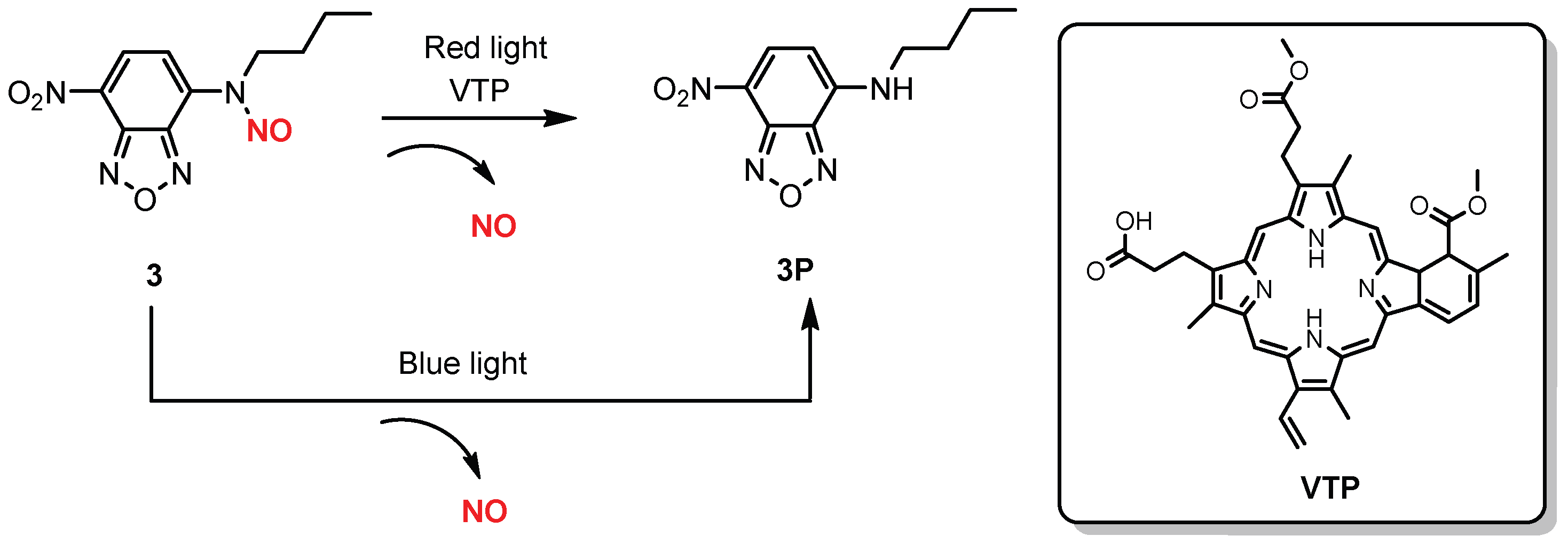
Based on PeT mechanism and Förster resonance energy transfer (FRET) mechanism, Fraix and co-workers [62] developed a red-light-mediated NO release system in which donor 3 (Scheme 5) exhibited a high quantum yield of NO release (φNO = 0.15) [37] with blue light irradiation and was employed as an NO donor and verteporfin (VTP), a clinically approved photosensitizer for photodynamic therapy and used as antennas to absorb red light. It was found that NO release of 3 could be activated upon red light excitation, and mechanism studies confirmed a triplet-mediated photosensitization process of VTP.
In vitro biological application of NO photogeneration from 3 was evaluated with HepG2 hepatocarcinoma and A375 melanoma cancer cell lines. To improve the water solubility of 3 and VTP, polymeric nanoparticles (NPs-3) of poly (ethylene glycol)-block-poly(ε–caprolactone) methyl ether (mPEG-b-PCL) entrapped in 3 and VTP were employed. As shown in Figure 3, the unloaded NPs did not elicit significant changes in cell viability both in the dark and upon light irradiation. NPs-3 were well tolerated in the dark but induced a significant decrease of cell viability upon irradiation with red light.
Nakagawa and co-workers [63] developed a yellow-green-light-controllable NO donor. Donor 4 (Scheme 6) showed absorption maximum at 564 nm in MilliQ water containing 0.1% DMSO. Yellow-green-light (530–590 nm, 100 mW/cm2)-triggered NO release of 4 was confirmed by ESR spin-trapping with Fe-MGD, and quantitative NO release was determined to be 9.8 μM of NO from 10 μM of 4 by fluorescence probe according to the literature [64].
In vitro photo-triggered NO release of 4 was demonstrated in HEK293 cells with DAF-FM DA as the fluorescent probe, in which a significant fluorescence signal was observed when HEK293 cells (pre-treated with 4 and the probe) were irradiated with 530–590 nm for 15 min, while little fluorescence was observed in the absence of 4.
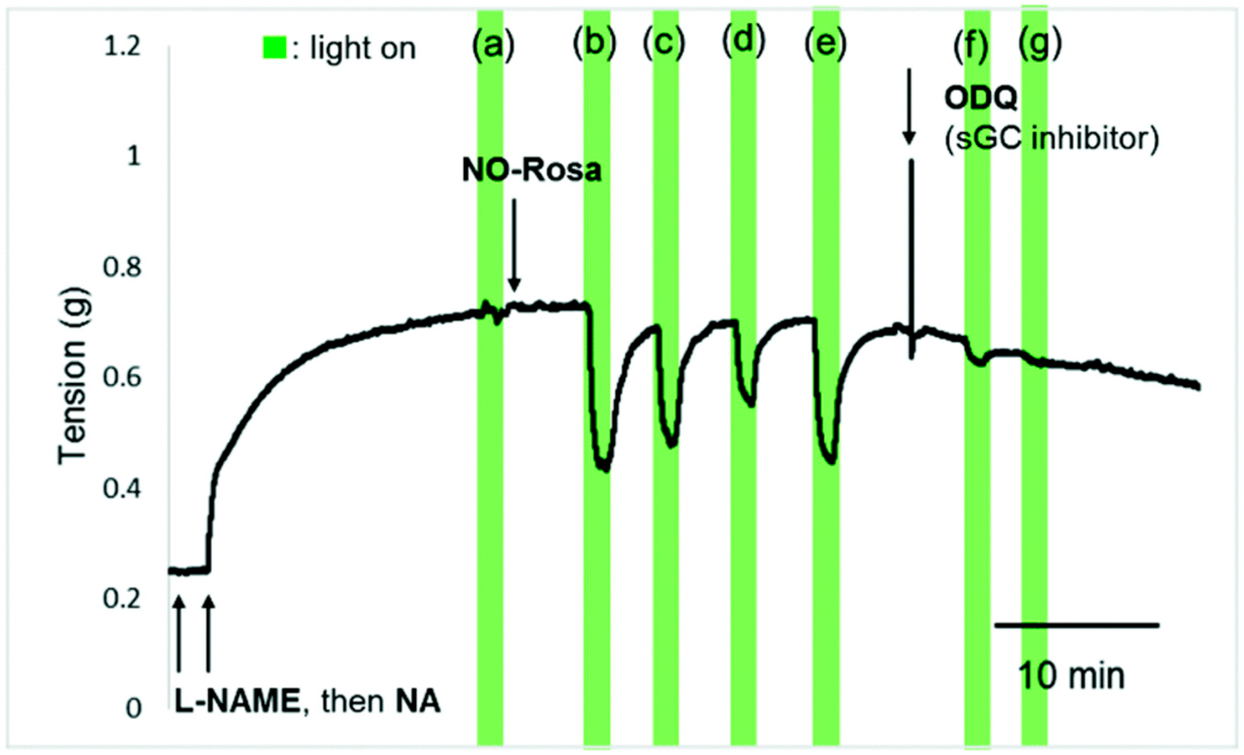
The photo-manipulation of vasodilation with 4 was performed in rat aorta ex vivo. As shown in Figure 4, the vasodilation of rat aorta was induced during photo-irradiation, but the tension was quickly recovered when the light was turned off. Moreover, the vasodilation effect became stronger with the increase of light intensity. Control experiments demonstrated that the vasodilation was completely blocked with the addition of ODQ (sGC inhibitor). These results suggest that NO release from 4 could be controlled by light (530–590 nm) under ex vivo conditions, which induced vasodilation via the NO-sGC-cGMP pathway [65].
Li and co-workers [66] reported a photo-triggered NO donor with turn-on fluorescence for antimicrobial. Donor 5 (Scheme 7) showed fluorescence OFF upon irradiation with 365 nm or 450 nm light; 5 also released NO and simultaneously converted product P5, which exhibited strong fluorescence around at 540 nm. In vitro photo-triggered NO release of NPs-5, which was prepared by directly encapsulating 5 into the amphiphilic copolymer DSPE-PEG2000, was confirmed by fluorescence imaging of RAW 264.7 cells. Antibacterial and anti-biofilm of NPs-5 were induced in E. coli and S. aureus, respectively. Upon irradiation with 450 nm light, significant decrease in the biofilm biomass for both E. coli and S. aureus were observed, and the antibacterial effect of NPs-5 reached 94% for E. coli and 96% for S. aureus.
Shen and co-workers [67] reported a yellow-LED-triggered NO donor for imaging and antitumor effects. Upon irradiation with yellow LED light, donor 6 showed fluorescence OFF, released NO, and converted to P6, which exhibited strong fluorescence at 540 nm (Scheme 8). Donor 6 was proven to be capable of photo-controlled release, and an NO release yield of around 8% was obtained by comparison with other NO donors [68,69].
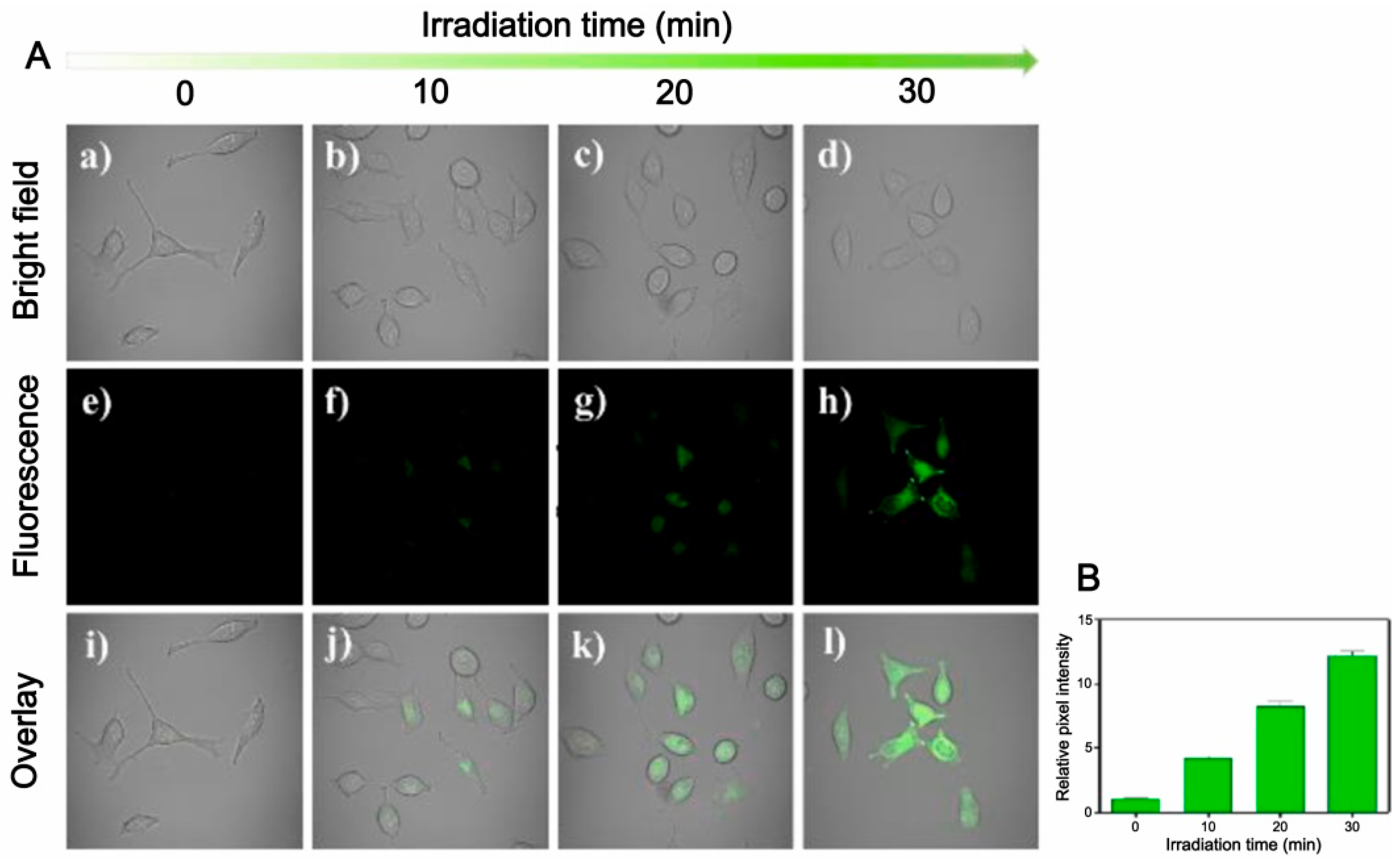
Biological applications of 6 for imaging and apoptosis were evaluated with HeLa cells. Fluorescence confocal imaging was performed in living cells. As shown in Figure 5, significant fluorescence was observed when the HeLa cells pre-incubated with 6 were irradiated with yellow LED light for 30 min. The relative fluorescence intensity also confirmed that the fluorescence increased in cells with irradiation time (Figure 5B). Apoptosis experiments by flow cytometry showed that 95% late apoptotic cells were obtained with 6, whereas P6 and DMSO showed no apoptosis.
NO plays a role in platelet aggregation inhibition [70,71]. He and co-workers [72] developed a green-light-triggered NO donor for inhibition of platelet aggregation. A water-soluble NO donor 7 (Scheme 9) was designed by employing fluorescent rhodamine as scaffold and a sulfonate group as a hydrophilic group to promote aqueous solubility of the donor. 7 showed absorption maximum at 505 nm in PBS (pH 7.4) without fluorescence. Upon irradiation with 532 nm light, 7 released NO and converted to corresponding amine P7, which displayed strong fluorescence at 565 nm.
Inhibition of platelet aggregation with 7 was demonstrated by using adenosine diphosphate (ADP) and collagen as inducers, respectively. As shown in Figure 6, no significant inhibition was detected without 7 and light. In the presence of 7 (10 μM) and green light irradiation, aggregation percentage was, however, down to ca. 50%. Moreover, the inhibition was dose-dependent, and more than 50% inhibition was obtained when the concentration of 7 was increased to 100 μM.

3.2. Dual-Function NO DonorsT

The combination of bioactive species such as clinical drugs and NO provides an appealing strategy in view of multimodal therapeutic systems. Molecular hybrids with covalent conjugation in principle offer more precise control over timing, location, and dosage of the cytotoxic species compared to systems assembled by noncovalent interactions.
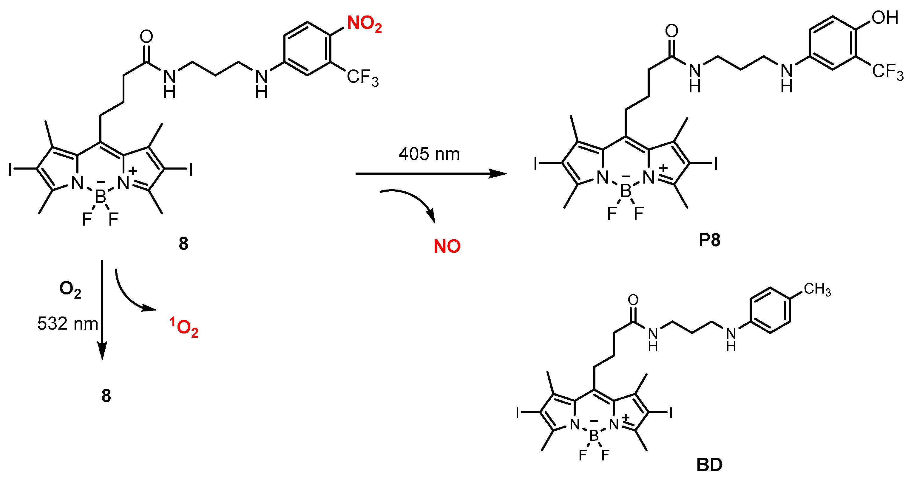
Fraix and co-workers [73] reported a hybrid NO donor in which NO and 1O2 could be generated with a visible light trigger. Donor 8 (Scheme 10) was synthesized by conjugation of a BODIPY unit and an o-CF3 nitrobenzene group via a linker.
1O2 production with 8 was detected and quantified by its typical phosphorescence with green light (532 nm) irradiation, and a high quantum yield for production of 1O2 (φ = 0.78) was obtained with Rose Bengal as standard. The control experiment confirmed that the generation of 1O2 was negligible when the excitation light was commutated to the blue region (420 nm). The NO photo-release of 8 was demonstrated by an ultrasensitive NO electrode employing an amperometric technique. In the dark, 8 is stable, and it releases NO in a photo-regulated fashion upon 405 nm light excitation. Control experiment also confirmed no photo-release of NO was observed when (the) excitation light was switched to 532 nm.
Cell internalization and the phototherapeutic effect with 8 were evaluated by in vitro experiments on A375 cells, a human amelanotic melanoma cell line. It was found that upon excitation at 488 nm, the green fluorescence was noted in the cell cytoplasm, which was attributed to the BODIPY unit of 8. The A375 cells were incubated with 8 and either kept in the dark or irradiated simultaneously with blue and green light. As shown in Figure 7, 8 exhibited low cytotoxicity in the dark, but marked cell mortality occurred under illumination. In particular, the photomortality was significantly greater with 8 than with model compound BD that only demonstrated 1O2 production and no NO release upon irradiation, suggesting that the simultaneous release of NO and 1O2 may play a key role in tumor cells destruction.
One of the most intriguing properties of NO is its capability to inhibit efflux pumps by nitration of critical tyrosine residues and consequently block drug extrusion [74,75]. Doxorubicin (DOX) is widely employed for treating a variety of cancers; its significant resistance has, however, hindered its clinical application. Co-administration of DOX with NO can block drug extrusion; therefore, it has been proposed as valuable approach to overcome resistance [76,77,78].
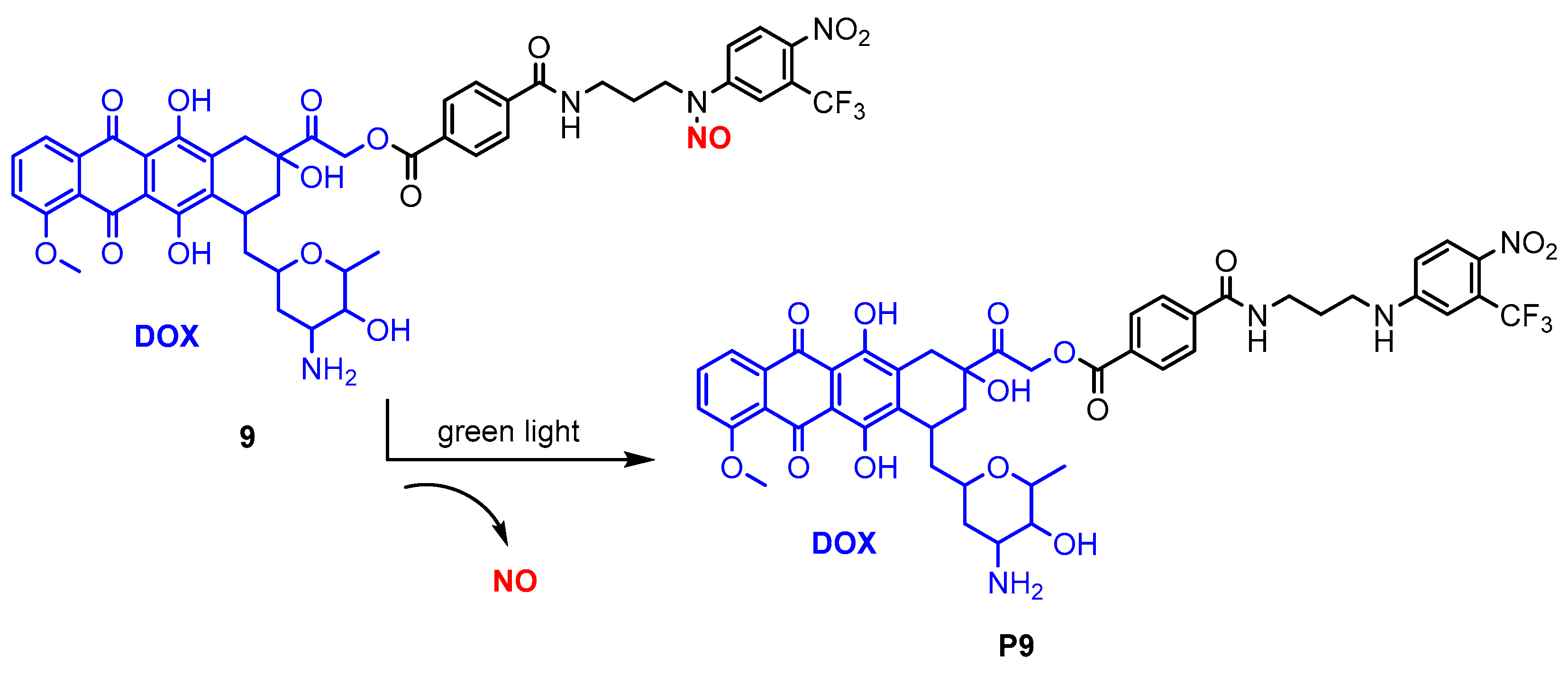
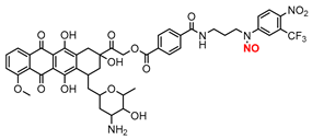
Parisi and co-workers [79] constructed a hybrid NO photodonor for overcoming resistance in breast cancer cells. Donor 9 (Scheme 11) is composed of DOX and an N-nitroso moiety by covalent joint. Intracellular photo-release of NO from 9 was confirmed with the probe DAF-FM, which becomes fluorescent after NO binding. DAF-FM fluorescence exhibited at least a two-fold increase when MDA-MB-231 cells pro-incubated with 9 were irradiated with green light, with respect to the cells in the dark. Moreover, inhibition on the efflux pumps by intracellular NO photo-release with the green light irradiation was also observed.
The antitumor activity of 9 was evaluated with MCF7 and MDA-MB-231 breast cancer cells. Both MCF7 cells and MDA-MB-231 cells were categorized as multidrug resistance (MDR) 1-negative and MDR1-positive, respectively, on the basis of their expression of MDR1 pump. In the dark, although DOX is highly cytotoxic towards MCF7 cells (IC50 7.6 μM), 9 did not show significant cytotoxicity in both cell lines (Figure 8a,c). Upon irradiation with green light, 9 exhibited significant cytotoxicity in both cell lines, and the IC50 values for MDA-MB-231 and MCF7 were 3.9 and 0.46 μM, respectively (Figure 8b,d). Moreover, a photodynamic effect was also observed for DOX, although to a lesser extent than the hybrid. It is worth noting that MDA-MB-231 cells, which showed DOX resistance, exhibited greater photodynamic effect with 9 than with DOX. The authors claimed that they observed that the intracellular accumulation of a MDR-pump substrate significantly increased exclusively when MDA-MB-231 cells were incubated with 9 and exposed to light, indicating the direct inhibition of the pumps upon NO photo-release
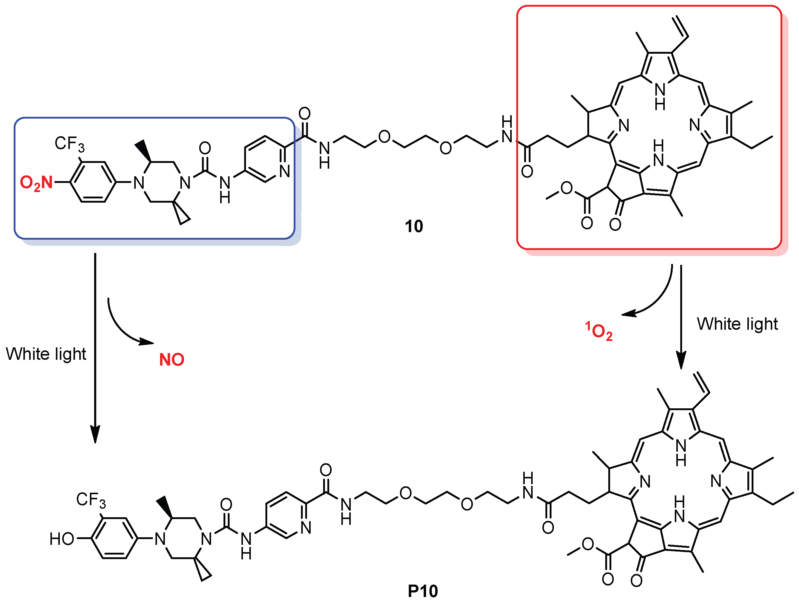
Rapozzi and co-workers [80] developed a hybrid NO photodonor for bimodal photodynamic therapy of prostate cancer in vitro. Donor 10 (Scheme 12) is composed of a pheophorbide a (Pba, photosensitizer) and a nonsteroidal antiandrogen molecule through a small pegylated linker. The antiandrogen molecule acts as an NO donor, and Pba serves as photosensitizer. In vitro uptake and internalization studies confirmed that 10 is able to efficiently bind the androgen receptor (AR) in LNCaP cells, VCaP cells, and malignant PC Cells.
The in vitro photodynamic activity of 10 was investigated against LNCaP, VCaP, and PC3 cells. Upon irradiation with white light for 24 h, cell proliferation was determined by resazurin assay using free Pba as reference. As shown in Figure 9A–C, higher phototoxicity of 10 in all cell lines was observed as compared to Pba alone. Remarkably, the IC50 values of 10 were lower than Pba in all cell lines when using white light. However, no significant difference in photodynamic activity was observed between 10 and Pba (Figure 9D–F) when they were irradiated with red light (only Pba was excited with red light irradiation). Results suggested that under white light irradiation, the o-CF3 nitrobenzene group induced the release of NO species, and Pba generated 1O2; both are synergistic.

4. Conclusions and Prospects

NO plays an important role in a number of signaling pathways, and the interruption in the homeostasis of NOS enzymes may lead to diseases [81]. Thus, therapeutics that either regulate NOS activity or produce NO exogenously have become an important research area [82,83,84,85,86]. Although a number of therapies utilizing gaseous NO or NO donors have been proposed and designed, the knowledge that NO’s effects are concentration-dependent [87,88,89,90,91,92] demands accurate quantification and reporting of the NO release. Traditional approaches such as Griess reaction [93], chemiluminescence [94], and electrochemistry [95,96] for NO release lack the ability to control and monitor the spatiotemporal distribution in live cells and in vivo. Photo-triggered NO donors with turn-on/ratiometric fluorescence can realize not only controllable release but also detection and imaging process in situ, providing a powerful tool for dynamic monitoring and investigating functions.
Despite the remarkable progress in light-mediated NO donors (Table 1), several challenges remain for practical applications, such as achieving excellent stability in the dark, fast release upon irradiation, high SNR (signal to noise ratio) fluorescence, and quantitative detection. In view of clinical demands, biocompatibility, and the cytotoxicity of the NO donor meeting high phototoxicity and low-dark toxicity standards, and excitation light of biological windows remain important issues. In addition, the combination of light-triggered NO release with other treatment modality such as photodynamic therapy is also worth attention since this strategy can increase the effectiveness of therapy and minimize side effects [97,98,99]. It is believed that the integration and development of chemistry and laser medicine technology will provide better therapeutic methods.

Funding

This research received no external funding.

Conflicts of Interest

The author declares no conflicts of interest.

References

  1. Calabrese, V.; Mancuso, C.; Calvani, M.; Rizzarelli, E.; Butterfield, D.A.; Stella, A.M.G. Nitric oxide in the central nervous system: Neuroprotection versus neurotoxicity. Nat. Rev. Neurosci. 2007, 8, 766–775. [Google Scholar] [CrossRef] [PubMed]
  2. Moncada, S.; Higgs, E. The discovery of nitric oxide and its role in vascular biology. Br. J. Pharmacol. 2006, 147, S193–S201. [Google Scholar] [CrossRef] [PubMed]
  3. Garthwaite, J. Concepts of neural nitric oxide-mediated transmission. Eur. J. Neurosci. 2008, 27, 2783–2802. [Google Scholar] [CrossRef]
  4. Xu, W.; Liu, Z.; Loizidou, M.; Ahmed, M.; Charles, I.G. The role of nitric oxide in cancer. Cell Res. 2002, 12, 311–320. [Google Scholar] [CrossRef]
  5. Riccio, D.A.; Schoenfisch, M.H. Nitric oxide release: Part I. Macromolecular scaffolds. Chem. Soc. Rev. 2012, 41, 3731–3741. [Google Scholar] [CrossRef]
  6. Bogdan, C. Nitric oxide and the immune response. Nat. Immunol. 2001, 2, 907–916. [Google Scholar] [CrossRef]
  7. Seabra, A.B.; Duran, N. Nitric oxide-releasing vehicles for biomedical applications. J. Mater. Chem. 2010, 20, 1624–1637. [Google Scholar] [CrossRef]
  8. Carpenter, A.W.; Schoenfisch, M.H. Nitric oxide release: Part II. Therapeutic applications. Chem. Soc. Rev. 2012, 41, 3742–3752. [Google Scholar] [CrossRef]
  9. Kim, J.; Saravanakumar, G.; Choi, H.W.; Park, D.; Kim, W.J. A platform for nitric oxide delivery. J. Mater. Chem. B 2014, 2, 341–356. [Google Scholar] [CrossRef]
  10. Duan, S.F.; Cai, S.; Yang, Q.H.; Forrest, M.L. Multi-arm polymeric nanocarrier as a nitric oxide delivery platform for chemotherapy of head and neck squamous cell carcinoma. Biomaterials 2012, 33, 3243–3253. [Google Scholar] [CrossRef]
  11. Zhang, X.; Tian, G.; Yin, W.Y.; Wang, L.M.; Zheng, X.P.; Yan, L.; Li, J.X.; Su, H.R.; Chen, C.Y.; Gu, Z.J.; et al. Controllable generation of nitric oxide by near-infrared-sensitized upconversion nanoparticles for tumor therapy. Adv. Funct. Mater. 2015, 25, 3049–3056. [Google Scholar] [CrossRef]
  12. Kang, Y.; Kim, J.; Lee, Y.M.; Im, S.; Park, H.; Kim, W.J. Nitric oxide-releasing polymer incorporated ointment for cutaneous wound healing. J. Control. Release 2015, 220, 624–630. [Google Scholar] [CrossRef] [PubMed]
  13. Jeong, H.; Park, J.H.; Shin, J.H.; Yoo, J.C.; Park, C.Y.; Hong, J. Prolonged release period of nitric oxide gas for treatment of bacterial keratitis by amine-rich polymer decoration of nanoparticles. Chem. Mater. 2018, 30, 8528–8537. [Google Scholar] [CrossRef]
  14. Wo, Y.Q.; Brisbois, E.J.; Bartlett, R.H.; Meyerhoff, M.E. Recent advances in thromboresistant and antimicrobial polymers for biomedical applications: Just say yes to nitric oxide (NO). Biomater. Sci. 2016, 4, 1161–1183. [Google Scholar] [CrossRef]
  15. Schairer, D.O.; Chouake, J.S.; Nosanchuk, J.D.; Friedman, A.J. The potential of nitric oxide releasing therapies as antimicrobial agents. Virulence 2012, 3, 271–279. [Google Scholar] [CrossRef]
  16. Sadrearhami, Z.; Nguyen, T.K.; Namivandi-Zangeneh, R.; Jung, K.; Wong, E.H.H.; Boyer, C. Recent advances in nitric oxide delivery for antimicrobial applications using polymer-based systems. J. Mater. Chem. B 2018, 6, 2945–2959. [Google Scholar] [CrossRef]
  17. Chung, M.-F.; Liu, H.-Y.; Lin, K.-J.; Chia, W.-T.; Sung, H.-W. A pH-responsive carrier system that generates NO bubbles to trigger drug release and reverse P-glycoprotein-mediated multidrug resistance. Angew. Chem. Int. Ed. 2015, 54, 9890–9893. [Google Scholar] [CrossRef]
  18. Fan, J.; He, Q.; Liu, Y.; Zhang, F.; Yang, X.; Wang, Z.; Lu, N.; Fan, W.; Lin, L.; Niu, G.; et al. Light-responsive biodegradable nanomedicine overcome es multidrug resistance via NO-enhanced chemosensitization. ACS Appl. Mater. Interfaces 2016, 8, 13804–13811. [Google Scholar] [CrossRef]
  19. Singh, S.; Gupta, A.K. Nitric oxide: Role in tumour biology and iNOS/NO-based anticancer therapies. Cancer Chemother. Pharmacol. 2011, 67, 1211–1224. [Google Scholar] [CrossRef]
  20. Burke, A.J.; Sullivan, F.J.; Giles, F.J.; Glynn, S.A. The yin and yang of nitric oxide in cancer progression. Carcinogenesis 2013, 34, 503–512. [Google Scholar] [CrossRef]
  21. Xu, J.; Zeng, F.; Wu, H.; Hu, C.; Yu, C.; Wu, S. Preparation of a mitochondria-targeted and NO-releasing nanoplatform and its enhanced pro-apoptotic effect on cancer cells. Small 2014, 10, 3750–3760. [Google Scholar] [CrossRef] [PubMed]
  22. Levy, E.S.; Morales, D.P.; Garcia, J.V.; Reich, N.O.; Ford, P.C. Near-IR mediated intracellular uncaging of NO from cell targeted hollow gold nanoparticles. Chem. Commun. 2015, 51, 17692–17695. [Google Scholar] [CrossRef]
  23. Wang, P.G.; Xian, M.; Tang, X.P.; Wu, X.J.; Wen, Z.; Cai, T.W.; Janczuk, A.J. Nitric oxide donors: Chemical activities and biological applications. Chem. Rev. 2002, 102, 1091–1134. [Google Scholar] [CrossRef]
  24. Sharma, K.; Chakrapani, H. Site-directed delivery of nitric oxide to cancers. Nitric Oxide 2014, 43, 8–16. [Google Scholar] [CrossRef]
  25. Eroy-Reveles, A.A.; Leung, Y.; Beavers, C.M.; Olmstead, M.M.; Mascharak, P.K. Near-infrared light activated release of nitric oxide from designed photoactive manganese nitrosyls:  strategy, design, and potential as NO donors. J. Am. Chem. Soc. 2008, 130, 4447–4458. [Google Scholar] [CrossRef]
  26. Sortino, S. Light-controlled nitric oxide delivering molecular assemblies. Chem. Soc. Rev. 2010, 39, 2903–2913. [Google Scholar] [CrossRef]
  27. Meng, T.; Wang, X.; Jiang, S.; Chen, S.; Zhou, S.; Zhu, Y.; Wu, J.; Hu, D.; Yan, Y.; Zhang, G. Delivery of small-molecule drugs and protein drugs by injectable acid-responsive self-assembled COF hydrogels for combinatorial lung cancer treatment. ACS App. Mater. Interfaces 2023, 15, 42354–42368. [Google Scholar] [CrossRef]
  28. Li, S.; Xiao, Y.; Chen, C.; Jia, L. Recent progress in organic small-molecule fluorescent probe detection of hydrogen peroxide. ACS Omega 2022, 7, 15267–15274. [Google Scholar] [CrossRef]
  29. Dong, S.; He, J.; Sun, Y.; Li, D.; Li, L.; Zhang, M.; Ni, P. Efficient click synthesis of a protonized and reduction-sensitive amphiphilic small-molecule prodrug containing camptothecin and gemcitabine for a drug self-delivery system. Mol. Pharm. 2019, 16, 3770–3779. [Google Scholar] [CrossRef]
  30. Jia, S.; Sletten, E.M. Spatiotemporal control of biology: Synthetic photochemistry toolbox with far-red and near-infrared light. ACS Chem. Biol. 2022, 17, 3255–3269. [Google Scholar] [CrossRef]
  31. Weinstain, R.; Slanina, T.; Kand, D.; Klán, P. Visible-to-NIR-light activated release: From small molecules to nanomaterials. Chem. Rev. 2020, 120, 13135–13272. [Google Scholar] [CrossRef] [PubMed]
  32. Yang, Y.; Jiang, Q.; Zhang, F. Nanocrystals for deep-tissue in vivo luminescence imaging in the near-infrared region. Chem. Rev. 2024, 124, 554–628. [Google Scholar] [CrossRef] [PubMed]
  33. Lin, J.; Cao, M.; Wang, S.; Wu, X.; Pan, Y.; Dai, Z.; Xu, N.; Zuo, L.; Liu, J.; Wang, Y.; et al. Deep red-light-mediated nitric oxide and photodynamic synergistic antibacterial therapy for the treatment of drug-resistant bacterial infections. Small 2025, 21, e2408759. [Google Scholar] [CrossRef] [PubMed]
  34. Laneri, F.; Parisi, C.; Seggio, M.; Fraix, A.; Longobardi, G.; Catanzano, O.; Quaglia, F.; Sortino, S. Supramolecular red-light-photosensitized nitric oxide release with fluorescence self-reporting within biocompatible nanocarriers. J. Mater. Chem. B 2024, 12, 6500–6508. [Google Scholar] [CrossRef]
  35. Chen, G.; Yu, J.; Wu, L.; Ji, X.; Xu, J.; Wang, C.; Ma, S.; Miao, Q.; Wang, L.; Wang, C.; et al. Fluorescent small molecule donors. Chem. Soc. Rev. 2024, 53, 6345–6398. [Google Scholar] [CrossRef]
  36. Wang, L.; Zhang, J.; An, X.; Duan, H. Recent progress on the organic and metal complex-based fluorescent probes for monitoring nitric oxide in living biological systems. Org. Biomol. Chem. 2020, 18, 1522–1549. [Google Scholar] [CrossRef]
  37. Parisi, C.; Seggio, M.; Fraix, A.; Sortino, S. A high-performing metal-free photoactivatable nitric oxide donor with a green fluorescent reporter. ChemPhotoChem 2020, 4, 742–748. [Google Scholar] [CrossRef]
  38. Chen, C.; Tian, R.; Zeng, Y.; Chu, C.; Liu, G. Activatable fluorescence probes for “turn-on” and ratiometric biosensing and bioimaging: From NIR-I to NIR-II. Bioconjug. Chem. 2020, 31, 276–292. [Google Scholar] [CrossRef]
  39. Huang, X.; Song, J.; Yung, B.C.; Huang, X.; Xiong, Y.; Chen, X. Ratiometric optical nanoprobes enable accurate molecular detection and imaging. Chem. Soc. Rev. 2018, 47, 2873–2920. [Google Scholar] [CrossRef]
  40. Wu, L.; Huang, C.; Emery, B.P.; Sedgwick, A.C.; Bull, S.D.; He, X.-P.; Tian, H.; Yoon, J.; Sessler, J.L.; James, T.D. Förster resonance energy transfer (FRET)-based small-molecule sensors and imaging agents. Chem. Soc. Rev. 2020, 49, 5110–5139. [Google Scholar] [CrossRef]
  41. Murrell, W. Nitro-glycerine as a remedy for angina pectoris. Lancet 1879, 113, 113–115. [Google Scholar] [CrossRef]
  42. Friederich, J.A.; Butterworth, J.F. Sodium nitroprusside: Twenty years and counting. Anesth. Analg. 1995, 81, 152–162. [Google Scholar] [PubMed]
  43. Hrabie, J.A.; Keefer, L.K. Chemistry of the nitric oxide-releasing diazeniumdiolate (“nitrosohydroxylamine”) functional group and its oxygen-substituted derivatives. Chem. Rev. 2002, 102, 1135–1154. [Google Scholar] [CrossRef]
  44. Al-sadoni, H.; Ferro, A. S-Nitrosothiols: A class of nitric oxide-donor drugs. Clin. Sci. 2000, 98, 507–520. [Google Scholar] [CrossRef]
  45. Fye, W.B.T. Lauder Brunton and amyl nitrite: A Victorian vasodilator. Circulation 1986, 74, 222–229. [Google Scholar] [CrossRef]
  46. Seymour, C.P.; Tohda, R.; Tsubaki, M.; Hayashi, M.; Matsubara, R. Photosensitization of fluorofuroxans and its application to the development of visible light-triggered nitric oxide donor. J. Org. Chem. 2017, 82, 9647–9654. [Google Scholar] [CrossRef]
  47. Wang, Z.; Jin, A.; Yang, Z.; Huang, W. Advanced nitric oxide generating nanomedicine for therapeutic applications. ACS Nano 2023, 17, 8935–8965. [Google Scholar] [CrossRef]
  48. McGarvey, B.R.; Ferro, A.A.; Tfouni, E.; Bezerra, C.W.B.; Bagatin, I.; Franco, D.W. Detection of the EPR spectra of NO• in ruthenium(II) complexes. Inorg. Chem. 2000, 39, 3577–3581. [Google Scholar] [CrossRef]
  49. Zheng, Q.; Bonoiu, A.; Ohulchanskyy, T.Y.; He, G.S.; Prasad, P.N. Water-soluble two-photon absorbing nitrosyl complex for light-activated therapy through nitric oxide release. Mol. Pharm. 2008, 5, 389–398. [Google Scholar] [CrossRef]
  50. Kandoth, N.; Mosinger, J.; Gref, R.; Sortino, S. A NO photoreleasing supramolecular hydrogel with bactericidal action. J. Mater. Chem. B 2013, 1, 3458–3463. [Google Scholar] [CrossRef]
  51. Zhao, X.; Wang, K.; Li, B.; Wang, C.; Ding, Y.; Li, C.; Mao, L.; Lin, Y. Fabrication of a flexible and stretchable nanostructured gold electrode using a facile ultraviolet-irradiation approach for the detection of nitric oxide released from cells. Anal. Chem. 2018, 90, 7158–7163. [Google Scholar] [CrossRef]
  52. Kojima, H.; Nakatsubo, N.; Kikuchi, K.; Kawahara, S.; Kirino, Y.; Nagoshi, H.; Hirata, Y.; Nagano, T. Detection and imaging of nitric oxide with novel fluorescent indicators: Diaminofluoresceins. Anal. Chem. 1998, 70, 2446–2453. [Google Scholar] [CrossRef] [PubMed]
  53. Harbie, J.A.; Klose, J.R.; Wink, D.A.; Keefer, L.K. New nitric oxide-releasing zwitterions derived from polyamines. J. Org. Chem. 1993, 58, 1472–1476. [Google Scholar]
  54. Zhang, Z.; Wu, J.; Shang, Z.; Wang, C.; Cheng, J.; Qian, X.; Xiao, Y.; Xu, Z.; Yang, Y. Photocalibrated NO release from N-nitrosated napthalimides upon one-photon or two-photon irradiation. Anal. Chem. 2016, 88, 7274–7280. [Google Scholar] [CrossRef]
  55. Chen, X.; Liu, J.; Chen, M.; Zhou, J.; Zhang, Y.; Hu, X.; Geng, W.; Mao, Q.; Kitagishi, H.; Chen, J.; et al. Green-light-triggered and self-calibrated cascade release of nitric oxide and carbon monoxide for synergistic glaucoma therapy. J. Am. Chem. Soc. 2024, 146, 30361–30371. [Google Scholar] [CrossRef] [PubMed]
  56. Xu, Y.; Li, H.; Xu, S.; Liu, X.; Lin, J.; Chen, H.; Yuan, Z. Light-triggered fluorescence self-reporting nitric oxide release from coumarin analogues for accelerating wound healing and synergistic antimicrobial applications. J. Med. Chem. 2022, 65, 424–435. [Google Scholar] [CrossRef]
  57. Weissleder, R. A clearer vision for in vivo imaging. Nat. Biotechnol. 2001, 19, 316–317. [Google Scholar] [CrossRef]
  58. Zhou, E.Y.; Knox, H.J.; Reinhardt, C.J.; Partipilo, G.; Nilges, M.J.; Chan, J. Near-infrared photoactivatable nitric oxide donors with integrated photoacoustic monitoring. J. Am. Chem. Soc. 2018, 140, 11686–11697. [Google Scholar] [CrossRef]
  59. Knox, H.J.; Kim, T.W.; Zhu, Z.; Chan, J. Photophysical tuning of N-oxide-based probes enables ratiometric photoacoustic imaging of tumor hypoxia. ACS Chem. Biol. 2018, 13, 1838–1843. [Google Scholar] [CrossRef]
  60. Weber, J.; Beard, P.C.; Bohndiek, S.E. Contrast agents for molecular photoacoustic imaging. Nat. Methods 2016, 13, 639–650. [Google Scholar] [CrossRef]
  61. Shen, Z.; Zheng, S.; Xiao, S.; Shen, R.; Liu, S.; Hu, J. Red-light-mediated photoredox catalysis enables self-reporting nitric oxide release for efficient antibacterial treatment. Angew. Chem. Int. Ed. 2021, 60, 20452–20460. [Google Scholar] [CrossRef] [PubMed]
  62. Fraix, A.; Parisi, C.; Longobardi, G.; Conte, C.; Pastore, A.; Stornaiuolo, M.; Graziano, A.C.E.; Alberto, M.E.; Francés-Monerris, A.; Quaglia, F.; et al. Red-light-photosensitized NO release and its monitoring in cancer cells with biodegradable polymeric nanoparticles. Biomacromolecules 2023, 24, 3887–3897. [Google Scholar] [CrossRef] [PubMed]
  63. Okuno, H.; Ieda, N.; Hotta, Y.; Kawaguchi, M.; Kimura, K.; Nakagawa, H. A yellowish-green-light-controllable nitric oxide donor based on N-nitrosoaminophenol applicable for photocontrolled vasodilation. Org. Biomol. Chem. 2017, 15, 2791–2796. [Google Scholar] [CrossRef] [PubMed]
  64. Damiani, P.; Burini, G. Fluorometric determination of nitrite. Talanta 1986, 33, 649–652. [Google Scholar] [CrossRef]
  65. Ignarro, L.J.; Buga, G.M.; Wood, K.S.; Byrns, R.E.; Chaudhuri, G. Endothelium-derived relaxing factor produced and released from artery and vein is nitric oxide. Proc. Natl. Acad. Sci. USA 1987, 84, 9265–9269. [Google Scholar] [CrossRef]
  66. Li, L.; Lin, Z.; Lu, X.; Chen, C.; Xie, A.; Tang, Y.; Zhang, Z. Photo-controlled and photo-calibrated nanoparticle enabled nitric oxide release for antibacterial and anti-biofilm applications. RSC Adv. 2022, 12, 33358–33364. [Google Scholar] [CrossRef]
  67. Shen, W.; Yuan, Y.; Liu, X.; Jiang, J.; Yu, S.; Zhou, H.; Zhu, Q. A fluorogenic nitric oxide donor induced by yellow LED light for cells proliferation inhibition and imaging. Nitric Oxide 2024, 145, 1–7. [Google Scholar] [CrossRef]
  68. Suzuki, T.; Nagae, O.; Kato, Y.; Nakagawa, H.; Fukuhara, K.; Miyata, N. Photoinduced nitric oxide release from nitrobenzene derivatives. J. Am. Chem. Soc. 2005, 127, 11720–11726. [Google Scholar] [CrossRef]
  69. Hishikawa, K.; Nakagawa, H.; Furuta, T.; Fukuhara, K.; Tsumoto, H.; Suzuki, T.; Miyata, N. Photoinduced nitric oxide release from a hindered nitrobenzene derivative by two-photon excitation. J. Am. Chem. Soc. 2009, 131, 7488–7489. [Google Scholar] [CrossRef]
  70. Radomski, M.W.; Palmer, R.M.J.; Moncada, S. The role of nitric oxide and cGMP in platelet adhesion to vascular endothelium. Biochem. Biophys. Res. Commun. 1987, 148, 1482–1489. [Google Scholar] [CrossRef]
  71. Sogo, N.; Magid, K.S.; Shaw, C.A.; Webb, D.J.; Megson, I.L. Inhibition of human platelet aggregation by nitric oxide donor drugs: Relative contribution of cGMP-independent mechanisms. Biochem. Biophys. Res. Commun. 2000, 279, 412–419. [Google Scholar] [CrossRef] [PubMed]
  72. He, H.; Xia, Y.; Qi, Y.; Wang, H.-Y.; Wang, Z.; Bao, J.; Zhang, Z.; Wu, F.-G.; Wang, H.; Chen, D.; et al. A water-soluble; green-light triggered, and photo-calibrated nitric oxide donor for biological applications. Bioconjug. Chem. 2018, 29, 1194–1198. [Google Scholar] [CrossRef] [PubMed]
  73. Fraix, A.; Blangetti, M.; Guglielmo, S.; Lazzarato, L.; Marino, N.; Cardile, V.; Graziano, A.C.E.; Manet, I.; Fruttero, R.; Gasco, A.; et al. Light-tunable generation of singlet oxygen and nitric oxide with a bichromophoric molecular hybrid: A bimodal approach to killing cancer cells. ChemMedChem 2016, 11, 1371–1379. [Google Scholar] [CrossRef] [PubMed]
  74. Bonavida, B.; Khineche, S.; Huerta-Yepez, S.; Garban, H. Therapeutic potential of nitric oxide in cancer. Drug Resist. Updates 2006, 9, 157–173. [Google Scholar] [CrossRef]
  75. Riganti, C.; Miraglia, E.; Viarisio, D.; Costamagna, C.; Pescarmona, G.; Ghigo, D.; Bosia, A. Nitric oxide reverts the resistance to doxorubicin in human colon cancer cells by inhibiting the drug efflux. Cancer Res. 2005, 65, 516–525. [Google Scholar] [CrossRef]
  76. De Boo, S.; Kopecka, J.; Brusa, D.; Gazzano, E.; Matera, L.; Ghigo, D.; Bosia, A.; Riganti, C. iNOS activity is necessary for the cytotoxic and immunogenic effects of doxorubicin in human colon cancer cells. Mol. Cancer 2009, 8, e108. [Google Scholar] [CrossRef]
  77. Chegaev, K.; Riganti, C.; Lazzarato, L.; Rolando, B.; Guglielmo, S.; Campia, I.; Fruttero, R.; Bosia, A.; Gasco, A. Nitric oxide donor doxorubicin conjugates accumulate into doxorubicin resistant human colon cancer cells inducing cytotoxicity. ACS Med. Chem. Lett. 2011, 2, 494–497. [Google Scholar] [CrossRef]
  78. Riganti, C.; Rolando, B.; Kopecka, J.; Campia, I.; Chegaev, K.; Lazzarato, L.; Federico, A.; Fruttero, R.; Ghigo, D. Mitochondrialtargeting nitrooxy-doxorubicin: A new approach to overcome drug resistance. Mol. Pharm. 2013, 10, 161–174. [Google Scholar] [CrossRef]
  79. Parisi, C.; Moret, F.; Fraix, A.; Menilli, L.; Failla, M.; Sodano, F.; Conte, C.; Quaglia, F.; Reddi, E.; Sortino, S. Doxorubicin–NO releaser molecular hybrid activatable by green light to overcome resistance in breast cancer cells. ACS Omega 2022, 7, 7452–7459. [Google Scholar] [CrossRef]
  80. Rapozzi, V.; Ragno, D.; Guerrini, A.; Ferroni, C.; della Pietra, E.; Cesselli, D.; Castoria, G.; Di Donato, M.; Saracino, E.; Benfenati, V.; et al. Androgen receptor targeted conjugate for bimodal photodynamic therapy of prostate cancer in vitro. Bioconjug. Chem. 2015, 26, 1662–1671. [Google Scholar] [CrossRef]
  81. Thomas, D.D.; Ridnour, L.A.; Isenberg, J.S.; Flores-Santana, W.; Switzer, C.H.; Donzelli, S.; Hussain, P.; Vecoli, C.; Paolocci, N.; Ambs, S.; et al. The chemical biology of nitric oxide: Implications in cellular signaling. Free Radic. Biol. Med. 2008, 45, 18–31. [Google Scholar] [CrossRef] [PubMed]
  82. Zhu, Y.; Silverman, R.B. Revisiting heme mechanisms. A perspective on the mechanisms of nitric oxide synthase (NOS), heme oxygenase (HO), and cytochrome P450s (CYP450s). Biochemistry 2008, 47, 2231–2243. [Google Scholar] [CrossRef] [PubMed]
  83. Tiburcio-Félix, R.; Cisneros, B.; Hernández-Kelly, L.C.R.; Hernández-Contreras, M.A.; Luna-Herrera, J.; Rea-Hernández, I.; Jiménez-Aguilar, R.; Olivares-Bañuelos, T.N.; Ortega, A. Neuronal nitric oxide synthase in cultured cerebellar bergmann glia: Glutamate-dependent regulation. ACS Chem. Neurosci. 2019, 10, 2668–2675. [Google Scholar] [CrossRef] [PubMed]
  84. Tse, J.K.Y. Gut microbiota; nitric oxide, and microglia as prerequisites for neurodegenerative disorders. ACS Chem. Neurosci. 2017, 8, 1438–1447. [Google Scholar] [CrossRef]
  85. Arora, D.P.; Hossain, S.; Xu, Y.; Boon, E.M. Nitric oxide regulation of bacterial biofilms. Biochemistry 2015, 54, 3717–3728. [Google Scholar] [CrossRef]
  86. Chug, M.K.; Feit, C.; Brisbois, E.J. Increasing the Lifetime of Insulin Cannula with Antifouling and Nitric Oxide Releasing Properties. ACS Appl. Bio Mater. 2019, 2, 5965–5975. [Google Scholar] [CrossRef]
  87. Thomas, D.D.; Espey, M.G.; Ridnour, L.A.; Hofseth, L.J.; Mancardi, D.; Harris, C.C.; Wink, D.A. Hypoxic inducible factor 1α, extracellular signal-regulated kinase, and p53 are regulated by distinct threshold concentrations of nitric oxide. Proc. Natl. Acad. Sci. USA 2004, 101, 8894–8899. [Google Scholar] [CrossRef]
  88. Thomas, D.D.; Ridnour, L.A.; Espey, M.G.; Donzelli, S.; Ambs, S.; Hussain, S.P.; Harris, C.C.; DeGraff, W.; Roberts, D.D.; Mitchell, J.B.; et al. Superoxide fluxes limit nitric oxide-induced signaling. J. Biol. Chem. 2006, 281, 25984–25993. [Google Scholar] [CrossRef]
  89. Pervin, S.; Singh, R.; Hernandez, E.; Wu, G.; Chaudhuri, G. Nitric oxide in physiologic concentrations targets the translational machinery to increase the proliferation of human breast cancer cells: Involvement of mammalian target of rapamycin/eIF4E pathway. Cancer Res. 2007, 67, 289–299. [Google Scholar] [CrossRef]
  90. Pervin, S.; Singh, R.; Freije, W.A.; Chaudhuri, G. MKP-1-induced dephosphorylation of extracellular signal-regulated kinase is essential for triggering nitric oxide-induced apoptosis in human breast cancer cell lines. Cancer Res. 2003, 63, 8853–8860. [Google Scholar]
  91. Paul, S.A.; Simons, J.W.; Mabjeesh, N.J. HIF at the crossroads between ischemia and carcinogenesis. J. Cell. Physiol. 2004, 200, 20–30. [Google Scholar] [CrossRef] [PubMed]
  92. Borutaite, V.; Brown, G.C. S-nitrosothiol inhibition of mitochondrial complex I causes a reversible increase in mitochondrial hydrogen peroxide production. Biochim. Biophys. Acta Bioenerg. 2006, 1757, 562–566. [Google Scholar] [CrossRef] [PubMed]
  93. Hetrick, E.M.; Schoenfisch, M.H. Analytical chemistry of nitric oxide. Annu. Rev. Anal. Chem. 2009, 2, 409–433. [Google Scholar] [CrossRef] [PubMed]
  94. Bates, J.N. Nitric oxide measurement by chemiluminescence detection. Neuroprotocols 1992, 1, 141–149. [Google Scholar] [CrossRef]
  95. Bedioui, F.; Villeneuve, N. Electrochemical nitric oxide sensors for biological samples–principle, selected examples and applications. Electroanalysis 2003, 15, 5–18. [Google Scholar] [CrossRef]
  96. Privett, B.J.; Shin, J.H.; Schoenfisch, M.H. Electrochemical nitric oxide sensors for physiological measurements. Chem. Soc. Rev. 2010, 39, 1925–1935. [Google Scholar] [CrossRef]
  97. Huang, W.; Zhang, J.; Luo, L.; Yu, Y.; Sun, T. Nitric oxide and tumors: From small-molecule donor to combination therapy. ACS Biomater. Sci. Eng. 2023, 9, 139–152. [Google Scholar] [CrossRef]
  98. Benkovics, G.; Perez-Lloret, M.; Afonso, D.; Darcsi, A.; Béni, S.; Fenyvesi, É.; Malanga, M.; Sortino, S. A multifunctional β-cyclodextrin-conjugate photodelivering nitric oxide with fluorescence reporting. Int. J. Pharm. 2017, 531, 614–620. [Google Scholar] [CrossRef]
  99. Fraix, A.; Manet, I.; Ballestri, M.; Guerrini, A.; Dambruoso, P.; Sotgiu, G.; Varchi, G.; Camerin, M.; Coppellotti, O.; Sortino, S. Polymer nanoparticles with electrostatically loaded multicargo for combined cancer phototherapy. J. Mater. Chem. B 2015, 3, 3001–3010. [Google Scholar] [CrossRef]
Scheme 1. Light–triggered NO release and NO detection methods.
Chemistry 07 00066 sch001
Scheme 2. Light–triggered NO release from single-function NO donors (A) or dual-function NO donors (B) and their biological applications.
Chemistry 07 00066 sch002
Scheme 3. Chemical structure of donor 1 and its photo–triggered NO release.
Chemistry 07 00066 sch003
Figure 1. Schematic illustration of light–triggered NO release and detection methods. (A) PA images acquired before/after a 5 min period with/without irradiation 4 h following systemic administration of 1 (1 = photoNOD-1) (1.2 mg/kg, 150 μL, and 20% DMSO in sterile saline). Both sets of images have been adjusted to identical contrast for comparison. (B) Tumor volumes measured by calipers on each day of treatment. (C) Tumor volumes measured by calipers on day 7 of treatment. Scale bar represents 2.0 mm. Data presented as mean ± SD (n ≥ 3). * p < 0.05. Reproduced with permission from reference [58]. Copyright 2018 American Chemical Society.
Chemistry 07 00066 g001
Scheme 4. Chemical structure of donor 2 and its photo–triggered NO release.
Chemistry 07 00066 sch004
Figure 2. Schematic illustration of light–triggered NO release and detection methods. (A) Representative images of the abscess during the treatment process and (B) quantitative analysis of the infected areas receiving different treatments. ** p < 0.01 and *** p < 0.001 compared with the PBS group. (C) Photographs of bacterial colonies on the agar plates of the abscess tissues with varying treatments. (D) Bacterial colony-forming unit separated from abscess tissues with varying treatments. * p < 0.05, *** p < 0.001, and **** p < 0.0001 compared with the group receiving Cip@PGalNP (+hv) treatment on day 7. (E) Changes of body weights of P. aeruginosa biofilm-infected mice after different treatments. * p < 0.05. In all cases, the Cip and micelle concentrations were 10 μg/mL and 0.2 g/L, respectively. Reproduced with permission from reference [61]. Copyright 2021 Wiley-VCH.
Chemistry 07 00066 g002
Scheme 5. Chemical structure of donor 3 and its photo–triggered NO release.
Chemistry 07 00066 sch005
Figure 3. Cell viability of HepG2 and A375 cancer cells incubated with unloaded NPs and NPs-3 (NPs-3 = NPs-1) for 4 h and either kept in the dark or irradiated with a LED red light (λexc = 620–630 nm; 7 mW/cm). [Unloaded NPs] = [NPs-3] = 0.4 mg/mL; [3] = 20 μM, [VTP] = 1.7 μM. Reproduced with permission from reference [62]. Copyright 2023 American Chemical Society.
Chemistry 07 00066 g003
Scheme 6. Chemical structure of donor 4 and its photo–triggered NO release.
Chemistry 07 00066 sch006
Figure 4. Changes in tension of rat aorta ex vivo induced by yellowish-green-light-triggered NO release from 4 (4 = NO-Rosa). The strip was pretreated with L-NAME (10 μM) and noradrenaline (10 μM) before 4 (10 μM) was added. The strip was irradiated with 530–590 nm for 1 min each time. After several cycles of irradiation, ODQ (10 μM) was added, and the irradiation was performed again. Light intensity (mW/cm2): (a) 97, (b) 32, (c) 12, (d) 4, (e) 32, (f) 32, and (g) 32. Reproduced with permission from reference [63]. Copyright 2017 Royal Society Chemistry.
Chemistry 07 00066 g004
Scheme 7. Chemical structure of donor 5 and its photo–triggered NO release.
Chemistry 07 00066 sch007
Scheme 8. Chemical structure of donor 6 and its photo–triggered NO release.
Chemistry 07 00066 sch008
Figure 5. (A) Confocal fluorescence imaging of HeLa cells treated with 6 (20 μM) and irradiation for 0–30 min with yellow LED light. (B) Relative fluorescence intensity of imaging with different light irradiation time. Reproduced with permission from reference [67]. Copyright 2024 Elsevier.
Chemistry 07 00066 g005
Scheme 9. Chemical structure of donor 7 and its photo–triggered NO release.
Chemistry 07 00066 sch009
Figure 6. Inhibition of 7 on platelet aggregation in the presence of ADP (10 μM) and collagen (5 μg/mL). The arrow indicates the time when adenosine diphosphate and collagen were added into the platelet-rich plasma to induce aggregation. NC: negative control without 7 or 7P or light irradiation. Light: 3 W green LED light. Light duration: 10 min. Reproduced with permission from reference [72]. Copyright 2018 American Chemical Society.
Chemistry 07 00066 g006
Scheme 10. Chemical structures of 8 and model compound BD as well as photo–triggered NO release of 8.
Chemistry 07 00066 sch010
Figure 7. Viability of A375 melanoma cells incubated for 1 h with 8 and BD and either kept in the dark or irradiated for 5 or 10 min with simultaneous blue light (420 nm: 7 mW/cm2) and green light (528 nm: 0.5 mW/cm2). [8] = [BD] = 10 μM. Significance of 8 vs. BD at 5 min: * p < 0.002; significance of 8 vs. BD at 10 min: ** p < 0.001. Reproduced with permission from reference [73]. Copyright 2016 Wiley-VCH.
Chemistry 07 00066 g007
Scheme 11. Chemical structure of donor 9 and its NO photo–release.
Chemistry 07 00066 sch011
Figure 8. Decrease of viability in MCF7 (a,b) and MDA-MB-231 (c,d) breast cancer cells after incubation with 9 (9 = DXNO-GR) or DOX in the dark (a,c) or after green light irradiation (72 J/cm2) (b,d). Cells were incubated for 24 h before assessing cell viability. Data are expressed as mean percentage ± SD of at least three independent experiments, carried out in triplicate. * p < 0.05; ** p < 0.01; *** p < 0.001. Reproduced with permission from reference [79]. Copyright 2022 American Chemical Society.
Chemistry 07 00066 g008
Scheme 12. Chemical structure of donor 10 and its NO photo–release.
Chemistry 07 00066 sch012
Figure 9. Metabolic activity (%) of PC3, VCaP, and LNCaP cells treated with increasing amounts of Pba and 10 (10 = DR2). After 6 h incubation in the dark, the cells were irradiated (AC) with a white halogen lamp (fluence 7.2 J/cm2) and (DF) with the same lamp equipped with a red filter (fluence 0.84 J/cm2). A resazurin assay was carried out 24 h after irradiation. Data represent mean values ± SD of three independent experiments. Student’s t-test analysis of different treatments (Pba and 10) vs. NT: a. p < 0.05 and a. p < 0.01; 10 vs. Pba: b. p < 0.05 and b. p < 0.01. Reproduced with permission from reference [80]. Copyright 2015 American Chemical Society.
Chemistry 07 00066 g009
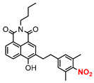
Table 1. Highlights of NO donors included in this review for biological applications.
DonorChemical
Structure
Class aIrradiationTesting ModeBiological ApplicationRef b
1Chemistry 07 00066 i001A690 nmIn vivoAntitumor[58]
2Chemistry 07 00066 i002A630 nmIn vivoAntimicrobial[61]
3Chemistry 07 00066 i003A620–630 nmIn vitroAntitumor[62]
4Chemistry 07 00066 i004A530–590 nmEx vivoVasodilation[63]
5Chemistry 07 00066 i005A450 nmIn vitroAntimicrobial[66]
6Chemistry 07 00066 i006AYellow LEDIn vitroAntitumor[67]
7Chemistry 07 00066 i007A532 nmIn vitroPlatelet aggregation[72]
8Chemistry 07 00066 i008B405 and 532 nmIn vitroAntitumor[73]
9Chemistry 07 00066 i009BGreen lightIn vitroAntitumor[79]
10Chemistry 07 00066 i010BWhite lightIn vitroAntitumor[80]
a Class: A, single-function NO donor; B dual-function NO donor. b Ref: reference.
Disclaimer/Publisher’s Note: The statements, opinions and data contained in all publications are solely those of the individual author(s) and contributor(s) and not of MDPI and/or the editor(s). MDPI and/or the editor(s) disclaim responsibility for any injury to people or property resulting from any ideas, methods, instructions or products referred to in the content.