Review

Syntheses, Properties, and Applications of ZnS-Based Nanomaterials

by
Amartya Chakrabarti
* and
Emily Alessandri
Department of Physical Sciences, Dominican University, River Forest, IL 60305, USA
*
Author to whom correspondence should be addressed.
Appl. Nano 2024, 5(3), 116-142; https://doi.org/10.3390/applnano5030010
Submission received: 2 July 2024 / Revised: 22 August 2024 / Accepted: 23 August 2024 / Published: 26 August 2024
(This article belongs to the Collection Review Papers for Applied Nano Science and Technology)

Abstract

:
ZnS is a II-VI semiconductor with a wide bandgap. ZnS-based nanomaterials have been produced in a variety of morphologies with unique properties and characteristic features. An extensive collection of research activities is available on various synthetic methodologies to produce such a wide variety of ZnS-based nanomaterials. In this comprehensive review, we thoroughly covered all the different synthetic techniques employed by researchers across the globe to produce zero-dimensional, one-dimensional, two-dimensional, and three-dimensional ZnS-based nanomaterials. Depending on their morphologies and properties, ZnS-based nanomaterials have found many applications, including optoelectronics, sensors, catalysts, batteries, solar cells, and biomedical fields. The properties and applications of ZnS-based nanostructures are described, and the scope of the future direction is highlighted.

1. Introduction

Nanomaterials are emerging as the most researched materials in today’s world by virtue of their unique properties, which in turn have led to their applications in diverse fields. According to the Web of Science, out of 2,759,830 research articles published worldwide in 2023, 244,938 are articles related to nanotechnology [1]. According to the National Science and Technology Council (NCST), any material that measures between 1 and 100 nm in at least one of its dimensions can be considered a nanomaterial. The rapid growth of and technological advancements in characterization tools, e.g., electron microscopes, undoubtedly contributed to the discovery of a variety of nanomaterials over the past few decades. The progress in nanomaterials and nanotechnology has also been guided by the market demand, specifically in high-performance electronics, automotives, and sustainable energy requirements. Nanomaterials have made a significant impact in the medicinal field as well, along with other various consumer products, including beauty and health, sports, construction, paints, etc. In this comprehensive review, we limit our focus to a very specific type of nanomaterial, zinc sulfide (ZnS)-based nanomaterials, which is one of the first semiconductors to be discovered in the late 1940s [2]. The versatile application areas of ZnS-based nanomaterials have been expanding exponentially since then (Figure 1).
ZnS has been long known for its application in light-emitting materials [3], photocatalysts [4], and pigments [5]. We find ZnS in two common polymorphic forms, sphalerite (the cubic crystalline Zn blende structure) and wurtzite (the hexagonal crystals), which is less abundant. In general, ZnS is stable under normal environmental conditions and considered to be nontoxic in nature. ZnS is one of the wide bandgap II-VI semiconductors, which has applications in high-performance light-emitting diodes and sensors. With the rapid advancement of nanotechnology, a wide variety of ZnS-based nanomaterials have been synthesized within the last twenty years. ZnS nanomaterials are now being synthesized as nanotubes, nanosheets, nanowires, nanoribbons, nanobelts, nanoparticles, and quantum dots using a variety of techniques. The most common synthesis techniques include chemical vapor deposition, pulsed laser vaporization, and thermochemical, solvothermal, and other chemical synthetic methodologies. Although there are a few publications available in the literature, a thorough review of the different nanomorphologies of ZnS and their applications is highly desired.
While nanomaterials can be classified in different ways, the most common practice remains categorizing them in terms of their dimensions [6]. Typically, zero-dimensional nanomaterials include nanoparticles, nanocrystals, and quantum dots, where the length, width, and depth of such materials fall under 100 nm. One-dimensional nanomaterials include nanotubes, nanowires, nanorods, nanoribbons, and nanofibers, and two out of three of their dimensions are under the scale of 100 nm. Usually, these nanostructures have larger lengths compared to their depth and width. On the contrary, two-dimensional nanomaterials have at least one dimension under 100 nm, and most likely their thickness is on that scale. Nanosheets, nanoplates, and nanoflakes are different forms of two-dimensional nanostructures. The least common form is three-dimensional nanomaterials, where nanodimensional building blocks make bulk materials and neither of their dimensions are confined to within the 100 nm scale. Nanocomposites, the dispersion of nanoparticles, and multilayered thin films are considered to fall in this category. ZnS-based nanomaterials have been synthesized and are currently available in almost all the aforesaid varieties of nanostructures. In the following segments, we will describe the synthesis and applications of zero-dimensional, one-dimensional, two-dimensional, and three-dimensional nanomaterials of ZnS.

2. Syntheses of ZnS Nanomaterials

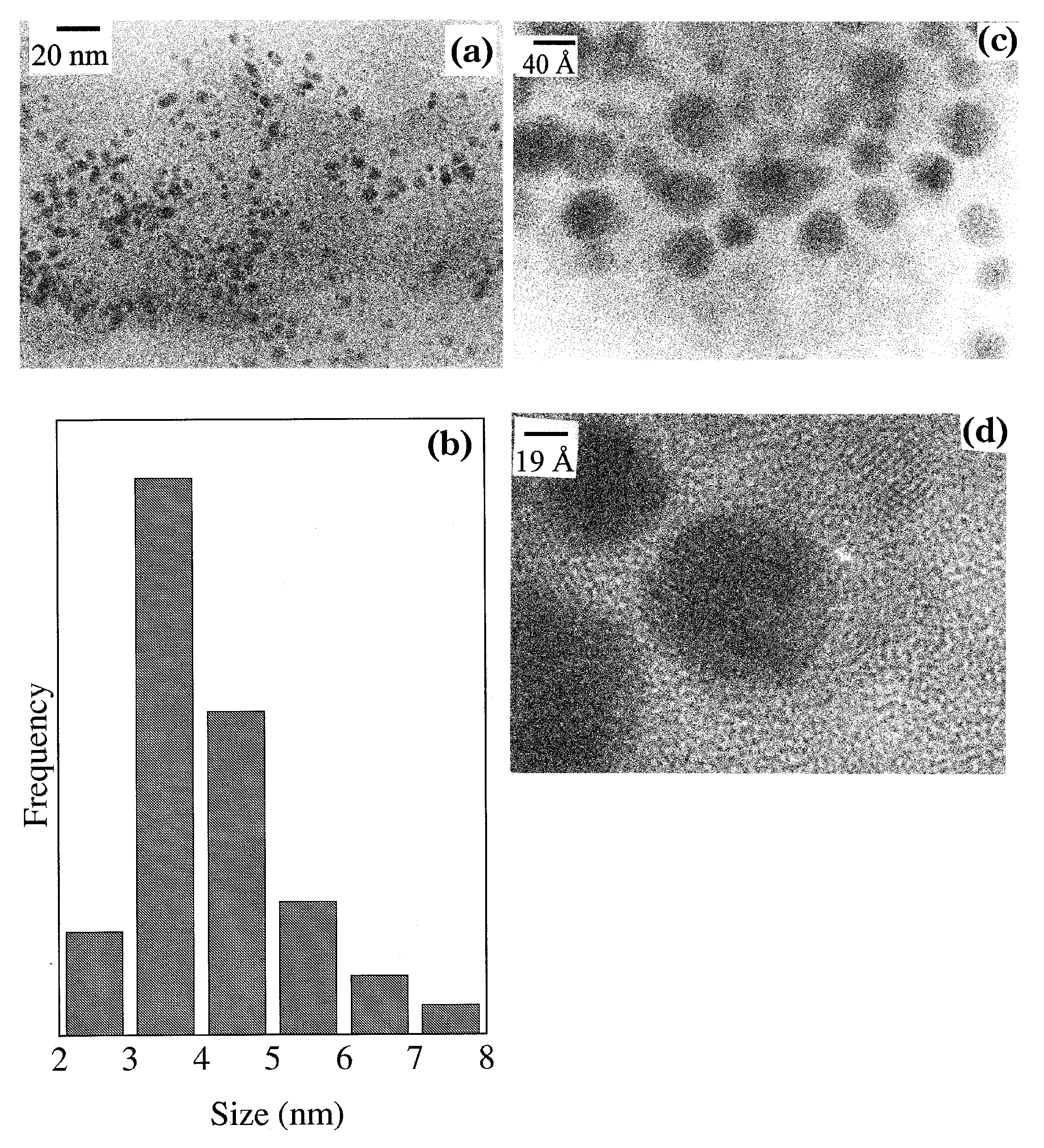
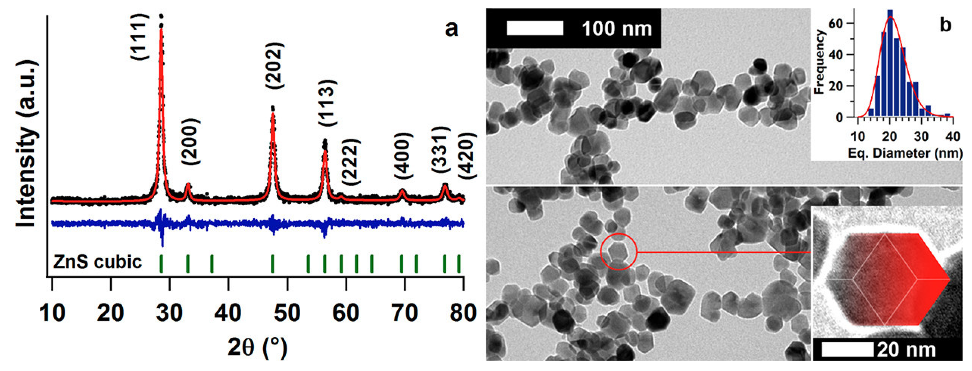
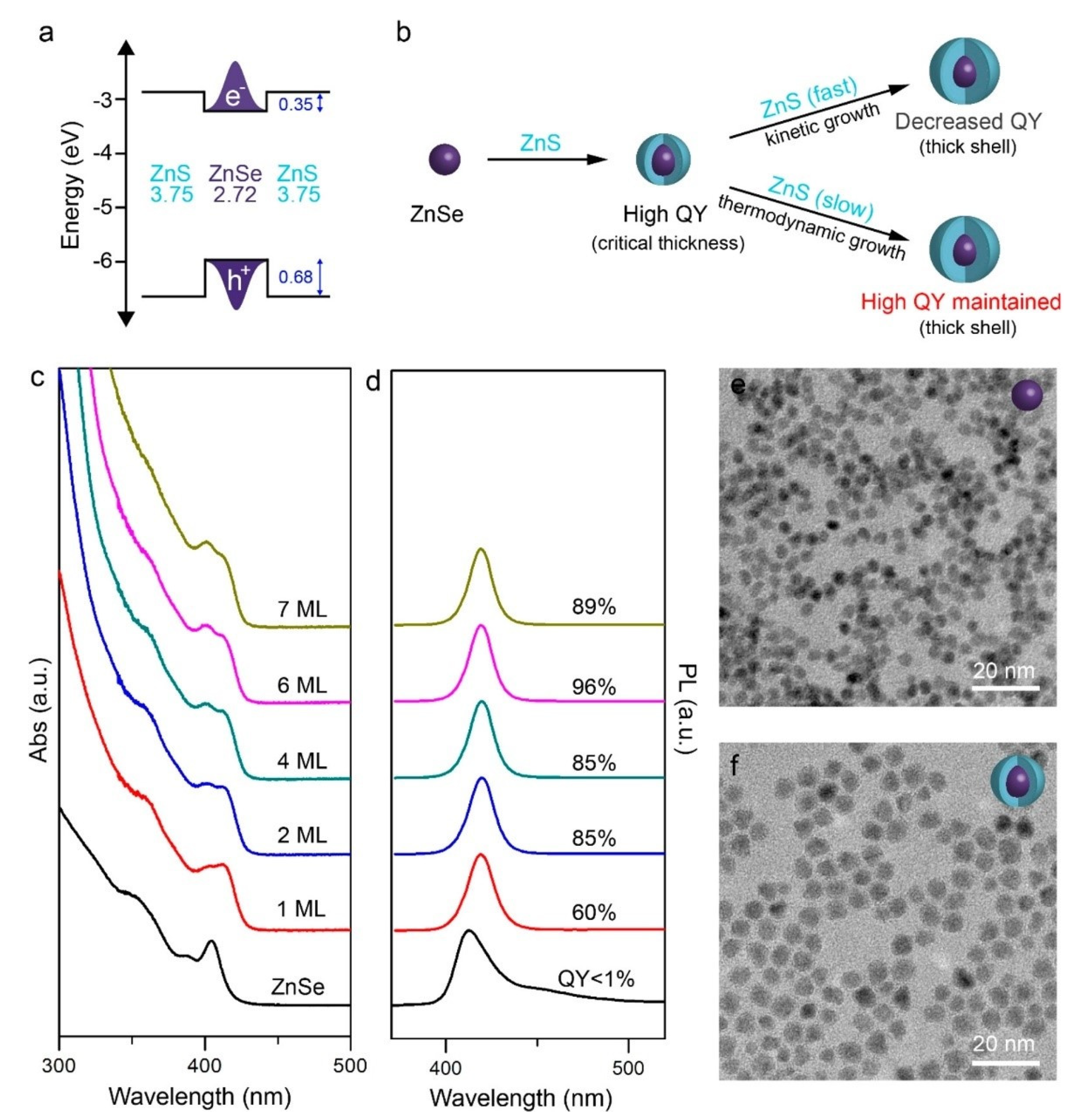
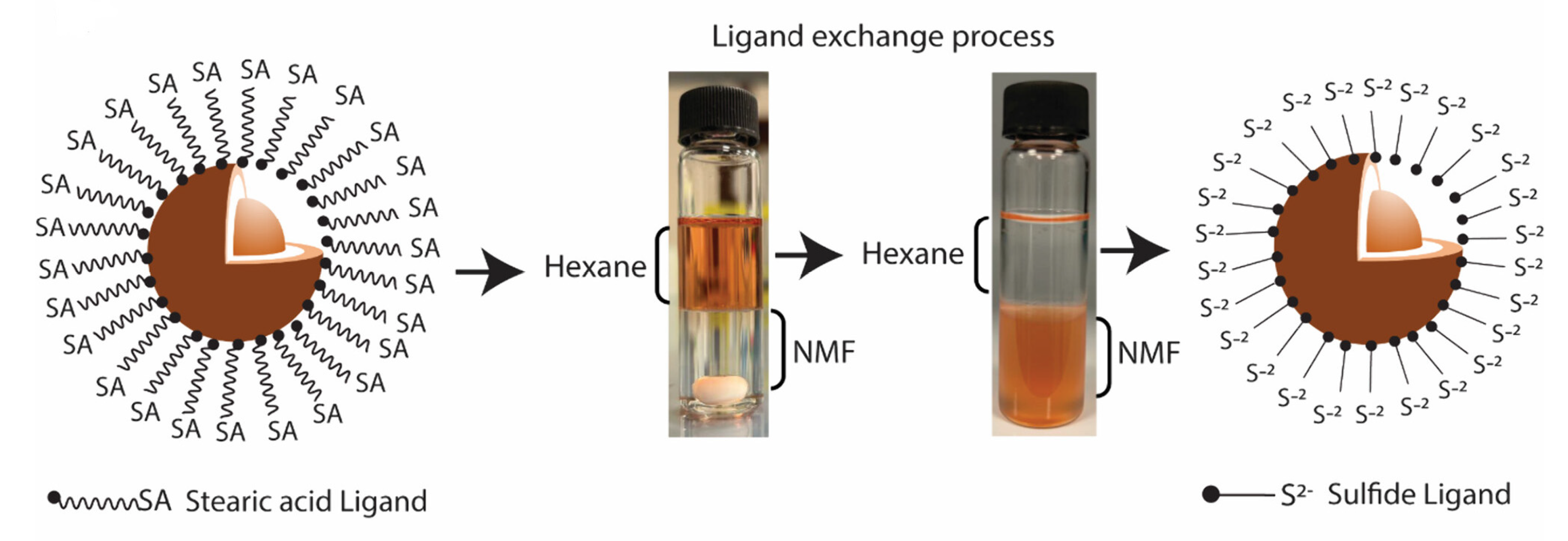
2.1. Zero-Dimensional ZnS

ZnS is one of the most widely investigated nanocrystalline semiconductors, and a variety of methods have been employed so far to develop ZnS nanoparticles and nanocrystallites. ZnS quantum dots (QDs) are also emerging as novel materials with potential applications in electronics and light-emitting devices by virtue of their comparatively lower toxicity and excellent electrical, optical, and magnetic properties [7,8,9]. A nanomaterial that exhibits quantum confinement effects by restricting the mobility of any charge carriers in all three dimensions is termed a QD [10]. Synthesis of nanomaterials comes with different challenges, and ZnS nanomaterials are no exception to that. The major obstacle of synthesizing any nanostructures includes confining the structural growth to the desired dimension. The most common way of handling such an issue is the introduction of various capping or stabilizing agents, which helps with restricting crystal growth. The other important parameters being investigated include the pH of the reaction medium, solvents, and reaction temperature. In Table 1, we list the most common capping agents used in the synthesis of ZnS nanostructures.
While the chemical precipitation of ZnS from different solvent systems is mostly cited as the preferred method of ZnS synthesis, a typical process of synthesizing ZnS nanomaterials begins with nucleation, followed by crystallization and aggregation steps. It has been noted that the inclusion of appropriate capping agents can suitably interfere in the aggregation process, hindering the growth of the crystal structure. Among different capping agents, amino acid cysteine has been reported to create nanocrystalline ZnS by several research groups. Bae and coworkers reported that cysteine-capped ZnS nanocrystals can be formed within a 6 nm-diameter range [11,12]. Their unique approach involved capping the metal prior to the introduction of the S source. At pH 6, they observed the formation of ZnS nanocrystals. Similarly, Lau et al. reported that thiol-containing ligands cysteine and thioglycolate were effective in restricting the agglomeration of large particles (>0.2 μm) within the first 90 min of precipitation of ZnS nanoparticles from the reaction medium [13]. 1-Thioglycerol (GSH) is considered to be another useful capping agent to facilitate the synthesis of ZnS nanoparticles. Nakaoka et al. published their accomplishments in synthesizing quantized ZnS (Q-ZnS) by utilizing GSH as the stabilizing agent [14]. An aqueous mixture of zinc perchlorate and GSH was exposed to 5% hydrogen sulfide (H2S), and the Q-ZnS products were precipitated using 2-propanol after concentrating the reaction mixture. The products were identified as sphalerite or zinc blende in cubic form. A slightly modified version of Nakaoka’s experiments was adapted by Nanda et al. to prepare and report GSH-capped ZnS nanocrystallites (NCs) [15]. According to their procedure, refluxing zinc acetate and GSH along with sodium sulfide while maintaining the pH at 8 for 10–12 h resulted in ZnS NCs. The products were precipitated from the concentrated reaction mixture either by acetone or by alcohol, and high-resolution transmission electron microscopy (HRTEM) exhibited the particle size to be 1.8 nm. They also varied the reaction stoichiometry to obtain ZnS NCs with larger diameters, varying from 2.5 to 3.5 nm (Figure 2). Mercaptoethanol was successfully used as a capping agent by Vogel and coworkers to synthesize stabilized nanoparticles (NPs) of ZnS of 1.4 nm diameter [16]. A room temperature reaction between zinc chloride and sodium sulfide in the presence of mercaptoethanol produced ZnS NPs. The pH of the reaction was maintained at 10.8 and the precipitation was carried out by adding methanol/2-propanol (1:2 vol %) after dialyzing the reaction mixture for a prolonged period of nearly 40 h to produce ZnS NCs. They also investigated the effect of annealing on the structure of the nanoparticles. Samples of as-prepared ZnS NCs were annealed up to 740 °C. The effect of thermally induced growth of nanoparticles became prominent above 310 °C, as the surfactant molecules tended to decompose at elevated temperatures, helping with the coalescence of the nanoparticles. Wide-angle X-ray scattering (WAXS) confirmed the average particle size of >3.8 nm of the vacuum-annealed ZnS NPs at 1013K.
Many water-soluble polymers have acted successfully as capping agents in ZnS nanoparticle synthesis. Polyvinylpyrrolidone (PVP), polyvinylalcohol (PVA), and polyethyleneglycol (PEG) were utilized by Ayodhya and coworkers to prepare ZnS NPs in a coprecipitation method [17]. One wt% loading of the capping agents resulted in NPs with a dimension of between 2.60 and 3.25 nm, which was measured by X-ray diffraction (XRD). Allehyani et al. reported synthesis of ZnS NPs using PEG (MW 8000) capping agents with a higher loading of 5 wt% [18]. The negatively charged hydroxyl groups of PEG molecules interacted with positive surface ions of ZnS crystals, and these van der Waals interactions became evident in FTIR studies. The dimensions of NPs were calculated using XRD and HRTEM and the particle sizes were found to be within the range of 2.9–3.3 nm.
In a recent study, Ajibade and coworkers reported synthesis of ZnS NPs using a metal complex precursor and different capping agents, including dodecylamine (DDA), hexadecylamine (HDA), and octadecylamine (ODA) [19]. Bis(diallydithiocarbamato) Zn precursor (Zn2(dalldtc)4) was prepared by stirring an aqueous solution of potassium diallyldithiocarbamate and zinc chloride at room temperature for 4 h. Then, Zn2(dalldtc)4 was dispersed in oleic acid (OA) and injected into the preheated capping agents, and thermolyzed at 200 °C for 1 h under N2. The resulting solution was cooled and ZnS NPs were precipitated using methanol. The size of the ZnS NPs was determined using HRTEM, and they had particle sizes ranging from 1.98 to 5.49 nm. The influence of the capping agents on the morphologies of NPs was captured using scanning electron microscope (SEM) imaging. The DDA-capped ZnS NPs exhibited flake-like morphology, the HDA-capped ZnS NPs showed sponge-like morphologies, and the ODA-capped NPs turned out to have rough flake-like surface morphologies (Figure 3).
The effect of pH on particle size has also been thoroughly investigated by different research groups. Zhang and coworkers studied and reported the formation of the two different phases of ZnS, with wurtzite and sphalerite being governed by the change in pH of the reaction medium [20]. The formation of sphalerite ZnS was favored at a pH higher than 9.5, while a mixed phase was generated at the acidic medium (pH ~ 6.6). The higher reaction rate was attributed to the formation of the sphalerite phase, which is the face-centered cubic structure. It was concluded that a basic reaction medium caused faster precipitation, which eventually helped the cubic stacking, leading towards the formation of the sphalerite phase. Wang et al. observed the effect of pH on the surface properties of ZnS nanomaterials, which in turn affected their ability to adsorb certain capping agents like xanthates [21]. Kaur and coworkers reported a direct correlation between the pH of the reaction medium and the particle size of ZnS nanomaterials [22]. They produced ZnS NPs using zinc acetate and sodium sulfide in the presence of 2-mercaptoethnol. NaOH was used to control the pH of the solution. Utilizing the dynamic light scattering (DLS) technique, they observed an increment in the particle size of ZnS NPs in the range of 4.5–25.6 nm to 22.7–104.1 nm as the pH of the reaction medium changed from 8 to 12. While the formation of Zn(OH)2 at the elevated pH may have contributed to the increase in particle size, dissolution of smaller NPs at higher pH could also have affected the size distribution. Yin et al. reported a systematic study on pH dependent size variation in ZnS nanostructures [23]. As depicted in Figure 4, we can observe how the particle size distribution was impacted by the pH of the reaction medium. The precipitation rate and the ammonia content were identified as the factors contributing the most to the varying size of the nanocrystals. At a higher pH, the rate of nucleation was proven to be higher than the rate of crystallization, resulting in smaller crystal size. On the contrary, a lower pH provided a lower rate of nucleation, which in turn created larger particles. As ammonia was used to increase the pH of the medium, it was anticipated that ammonia could be absorbed on the ZnS nanocrystal’s surface while ZnS was nucleated. The coordination bond formed between ammonia and metallic zinc could have efficiently lowered the surface energy, and the crystal growth was restricted.
The effect of solvents on the formation of ZnS nanoparticles has been investigated and published by researchers across the globe. Nanocrystalline ZnS of particle size 3.0 ± 0.3 nm has been reported, and the synthesis involved room temperature preparation of ZnS nanocrystals in anhydrous methanol [24]. Interestingly, it was observed that the addition of water to the methanol solution transformed the crystal structure of ZnS. With the help of molecular dynamics simulation, the authors exhibited that the introduction of water molecules increased the crystallinity in ZnS nanoforms, which was initially not present in the structures obtained directly in methanol. It was further demonstrated that this impact could be reversed by the addition of methanol to the mixture. Balansteva et al. reported that surface chemistry can significantly impact the growth of ZnS nanocrystals [25]. Zinc acetate dihydrate and thioacetamide (TAA) precursors were used in the synthesis with a 1:1 methanol/ethanol mixture as the solvent system. The authors utilized a thermal treatment of the as-synthesized samples to monitor the effect of temperature on the particle size and the surface properties of ZnS NPs. Transmission electron microscope (TEM) imaging exhibited a narrow size distribution of 2.6 ± 0.4 nm of the as-prepared samples, and a significant increase in the particle size (35.0 ± 6.1 nm) was obtained at 700 °C under vacuum. They further investigated the surface properties of ZnS NCs using CO adsorption and found that the Lewis acidity of the Zn2+ ions on the surface of ZnS nanostructures was significantly different than that of the bulk ZnS surface ions. In another report, ZnS nanocrystal formation was obtained using tetracosane (TCA) and octadecene (ODE) solvent mixture. The Zn precursor, zinc stearate, was dissolved in the given solvent system and heated to 340 °C under an Ar flow in a three-neck flask to become activated. Sulfur was used as the S source, which was introduced to the hot mixture via injection as a solution in ODE. The nanocrystals were reported to be grown at 300 °C [26]. The diameter of the particles ranged between 10 and 12 nm, which was determined by photoluminescence (PL) spectroscopy, and the effect of noncoordinating fatty acid ligands on the photoluminescence of ZnS NCs was manifested. Pyridine and triphenylphophite (TPP) were chosen by Rath and coauthors in the synthesis of ZnS NCs [27]. Zinc acetate dihydrate and TAA precursors were allowed to react in two different ways, conventional heating at 130 °C and via the AntonPaar microwave reaction system Synthos 3000 (800 W, 2.54 GHz). The presence of TPP in the reaction mixture produced NCs with smaller dimensions. SAXS measurements revealed nanoparticles with dimensions of 1–7 nm. However, as the NCs tended to aggregate, the SEM images exhibited larger particulates within the 650 and 1200 nm range. Other than the simplicity of the experimental setup and shorter reaction time, the microwave assisted synthesis did not provide any better advantages, as the products were of larger dimensions obtained via this route.
Deionized water is a preferred solvent medium for many synthetic approaches of ZnS NPs. A coprecipitation technique was used by Ali et al. to prepare ZnS in aqueous medium [28]. NCs of about 8–10 nm were produced by reacting zinc chloride and sodium sulfide in the presence of NaOH at the pH of 12.82. A hydrothermal synthesis of ZnS NPs was published in which the authors simply mixed the aqueous solutions of zinc sulfate and sodium sulfide without any capping agents [29]. The reaction temperature was maintained at 220 °C. While the size of the NPs was calculated from the XRD data to be between 13.08 and 28.06 nm, a change in molar ratio of the precursors played a certain role in the observed particle size. A higher S ratio resulted in larger particle diameter, so to obtain low dimensional NPs, a Zn-to-S ratio of 1:0.7 was required. Another hydrothermal method at a relatively low temperature (135 °C) was accomplished by Dengo et al. [30]. The preparation involved zinc acetate and sodium sulfide mixed in water and reacted over a period of 24 h at the given temperature. TEM micrographs exhibited an average particle size of around 21 ± 5 nm (Figure 5). John and Florence reported synthesis of nanocolloids of ZnS by a simple precipitation reaction using zinc chloride and sodium sulfide in aqueous medium [31]. The authors calculated the particle size of the bean-like nanostructures using the Debye–Scherrer formula, a common method being used by researchers that utilizes the XRD peaks of materials. The average particle size from the most intense XRD peak was found to be 12 nm.
Stabilizing ZnS nanoparticles in selective solvent systems has also been a popular choice among researchers. While the stabilizing agents may not directly impact the crystal structure or dimension of the nanoparticles, they provide stability of the suspended nanoparticles in the respective solvents. Shi et al. reported Cyanex 302 (di-(2,4,4-trimethylpentyl) monothiophosphinic acid) as a stabilizing agent [32]. A very high loading concentration of 24.4 g L−1 in gasoline was reported, and the suspension was stable for up to 6 months. Dodecanethiol-functionalized ZnS NPs showed excellent stability in polyalphaolefin (PAO) lubricating oils [33]. Zinc acetate precursors previously dissolved in oleylamine (OAm) were mixed with 1-dodecanethiol and heated at 285 °C for 3 h in an Ar atmosphere. The resulting NPs were then precipitated from the solution using ethanol. Using the dynamic light-scattering technique, the average particle size of ZnS NPs in PAO base oil was found to be 10 ± 1 nm and 14 ± 2 nm. Laponite® XLG is a layered clay material that was used as a stabilizer in synthesizing ZnS nanoparticles [34]. A typical size distribution within the range of 2 to 6 nm was obtained for the nanoparticles.
More recent studies have focused on the synthesis of ZnS QDs. Colloidal QDs prepared from semiconducting materials that can demonstrate quantum confinement effect are gaining overwhelming attraction in scientific communities [35]. As we discussed previously, capping agents play a significant role in producing and tuning the dimension of QDs. 3-Mercaptopropionic acid (MPA) and (3-mercaptopropyl)trimethoxysilane (MPS)-capped ZnS QDs were first synthesized by Li and coworkers [36]. The synthetic procedure involved the preparation of MPA-capped ZnS first, followed by the replacement of some MPA by MPS. The resulting ZnS QDs were measured to have an average particle size of 5 nm. The capping molecule replacement provided better stability to the QDs. A protein-aided biofabrication of ZnS QDs has also been reported [37]. In this study, Zhou and coworkers identified ZnS binding peptides and successfully used a constructed variant of E. coli thioredoxin 1 (Trx A) to restrict the growth of the crystal structure during the mineralization process. Those protein-coated QDs were reported to have diameters in the range of ~4 nm and exhibited a long shelf life. ZnS QDs were synthesized by Cooper et al. using an aliphatic primary amine as the capping agent [38]. They used ODA as capping agent in the relatively high temperature (300 °C) synthesis of ZnS QDs. Zinc stearate and elemental S were precursors, while ODE and tributyl phosphate (TBP) served as the solvent systems. A thorough cleaning of the QDs was then carried out by the researchers, where the crude reaction mixture was treated with a solvent mixture of dichloromethane and methanol. The methanol phase was discarded, and the process was repeated twice more. Finally, ZnS QDs were precipitated using acetone and washed repeatedly before dispersing back in a mixture of hexanes and DCM or toluene. The average particle size was determined to be 4 nm using TEM imaging. Mandal and coworkers reported a green synthesis of ZnS QDs using thiolactic acid (TLA) as the capping agent [39]. ZnS QDs with a particle size of around 3.5 nm were prepared by reacting anhydrous zinc acetate and sodium sulfide in aqueous medium at room temperature. Synthesis of pure ZnS QDs from a single-source precursor was reported by Lu et al. [40]. QDs with average sizes ranging between 1.6 and 4.5 nm were prepared by treating zinc ethylxanthate at various temperatures (90 °C to 210 °C) in the presence of OA, OAm, and tri-n-octylamine (TOA). While TOA was the solvent, OA and OAm acted as the capping agents. This unique approach utilized a single precursor that could be thermolyzed to create Zn-S bonds at elevated temperatures. Hosseinzadeh et al. extensively investigated different capping agents in the synthesis of ZnS QDs and their effect on the surface of the QDs as they interact with insulin [41]. 2-Mercaptoethanol (ME), cysteamine hydrochloride (CA), thioglycolic acid (TGA), L-cysteine, mercaptosuccinic acid (MSA), mercaptopropionic acid (MPA), 6-mercaptohexanoic acid (MHA), and 11-mercaptoundecanoic acid (MUA) are capping agents with various functional groups that were included in the study. A pH of 10 was maintained during the synthetic process, where aqueous solutions of zinc acetate and sodium sulfide reacted at 50 °C. As at a pH lower than 7, hydrolysis of sulfide ion takes place, and it was important to maintain a higher pH using NaOH solution. The particle sizes of different products with varying capping agents were measured using XRD and found to be between 3.5 nm and 7.9 nm, with MUA producing the smallest crystallites and ME the largest ones.
While there was significant advancement towards synthesis of pure ZnS QDs, research on preparing ZnS core/shell QDs with a variety of other semiconductors progressed equally. It was anticipated that development of a core/shell model by growing a semiconductor shell surrounding other semiconducting QDs may passivate the surface atoms of the QDs, which would improve the optical properties of these novel materials. Colloidal cadmium selenide CdSe/ZnS QDs surely exhibited such improvements in the optical properties, as reported by Yu et al. [42]. In a core/shell synthesis, typically, the core materials are synthesized prior to the shell materials, which are then prepared in the presence of the core. In their attempt to make CdSe/ZnS core/shell structures, Yu and coworkers followed a similar yet complex protocol. Trioctylphosphine oxide (TOPO), hexadecylamine (HAD), and trioctylphosphine (TOP) were mixed in a pot and degassed at 110 °C. Cadmium 2,4-pentanedionate and hexadecanediol were mixed with TOP in a separate vial, with degassing at 110 °C. Elemental Se was dissolved in TOP and injected into the vial after cooling the vial to room temperature. The Se- and Cd-containing mixture was then added to the pot, which was maintained at a temperature of 360 °C under Ar. The temperature of the pot was reduced to 270 °C for several minutes. The formation of CdSe was confirmed by finding the core emission of aliquots of the sample at ~570 nm. Quenching of the reaction was conducted by bringing the pot to room temperature. CdSe QDs were precipitated from the reaction mixture via the addition of methanol and butanol and purified by centrifugation. The purified product was redispersed in hexane before being overcoated with ZnS. TOPO and hexylphosphonic acid (HPA) were taken in a pot and degassed at 140 °C. The hexane solution of CdSe was then added to the pot, followed by evaporation of hexane at 80 °C. A small amount of decylamine was added to the pot and it was heated to 155 °C. A solution of diethyl zinc, dimethyl cadmium, and TOP, and a second solution of hexamethyldisilathiane and TOP, were slowly injected into the pot at a rate of 2 mL/h for about 2 h. The reaction continued overnight at 80 °C. The core/shell CdSe/ZnS QDs were then precipitated twice using the same technique. Scanning transmission electron microscope (STEM) analysis revealed that the average size of the CdSe/ZnS QDs increased to 4.3 nm from that of the original 3.4 nm CdSe core.
Among many other core/shell QDs, Ji and coworkers reported synthesis of zinc selenide (ZnSe)/ZnS core/shell QDs [43]. The core ZnSe QDs were synthesized using a previous report [44]. To synthesize ZnS shell on the core, zinc oleate, which was synthesized in-house, and 1-octanethiol were introduced to the previously dispersed ZnSe in hexane solution in the presence of ODE, and the reaction temperature was kept at 310 °C for 5 h. The product was purified by precipitation with hexane and ethanol. The authors investigated the effect of shell growth under thermodynamic conditions on the photoluminescence (PL) quantum yields (QYs) (Figure 6). In order to predominate the thermodynamic growth conditions over the kinetic regime, they manipulated the molar ratio of Zn and OA while synthesizing zinc oleate. A high OA-to-Zn ratio limited the availability of zinc, increasing the solubility of zinc oleate and reducing the reactivity of the precursor.
In a very recent study, indium phosphide (InP)/ZnS core/shell (InP/ZnS) QDs (particle size of 3.75 nm) were carefully engineered, with the InP core covered by a thin shell of ZnS [45]. The green process involved InP as a non-toxic replacement for Cd-based semiconductors. InP core was synthesized using a previously reported method [46]. The InP core was then covered by ZnS shell using a controlled synthetic method by reaction of zinc stearate dissolved in ODE and elemental S dissolved in TOP at 240 °C for 30 min (Figure 7). As the reaction was finished, the product was purified by centrifugation using a mixture of hexane and ethanol. The InP/ZnS core/shell products were then subjected to a ligand exchange process (Figure 7), where the long-chain fatty acid was replaced by short-chain ligands, e.g., S2−, Cl, and MPA, in order to enhance the solubility of the product. The higher solubility improved the antibacterial properties of the QDs.

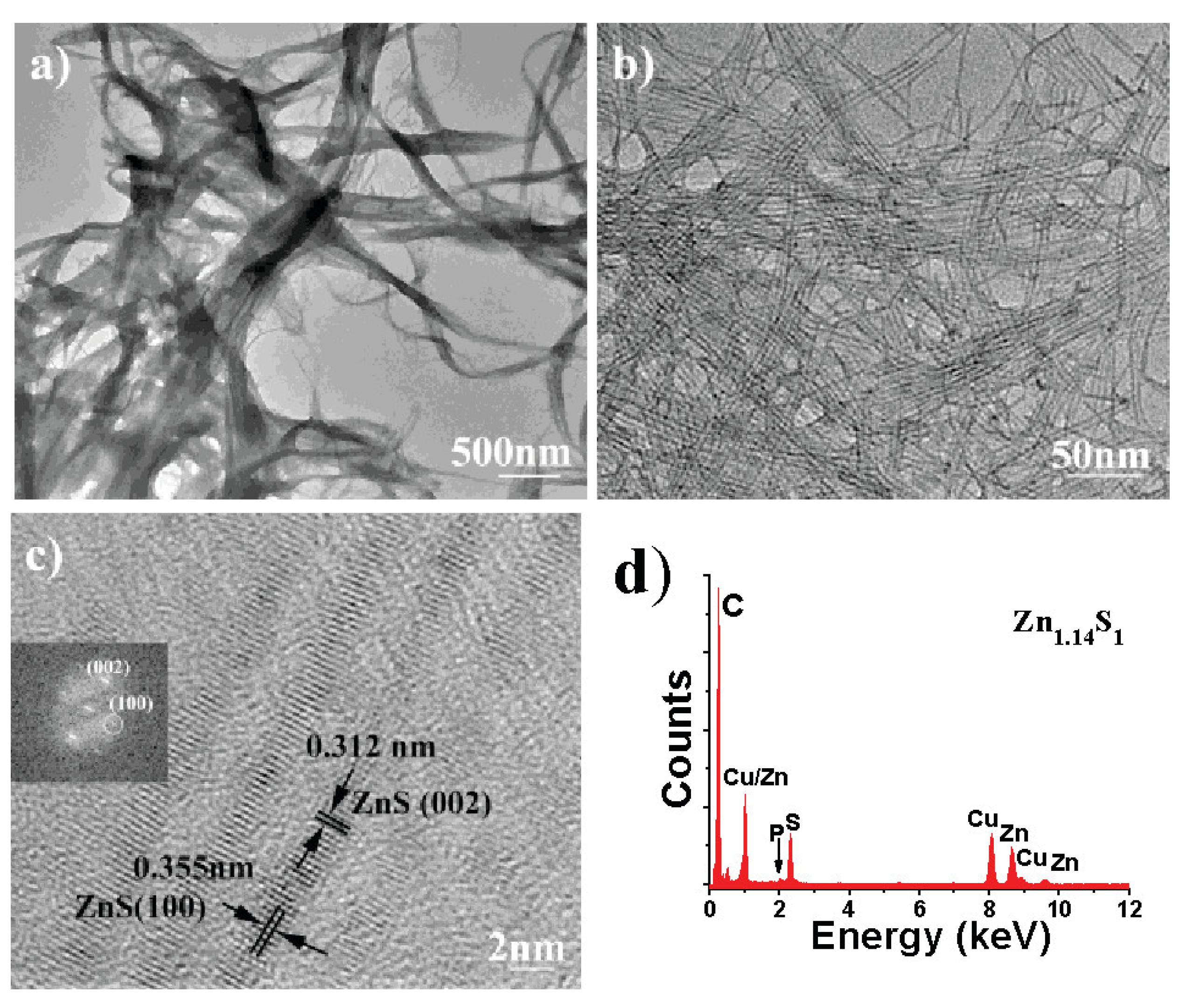
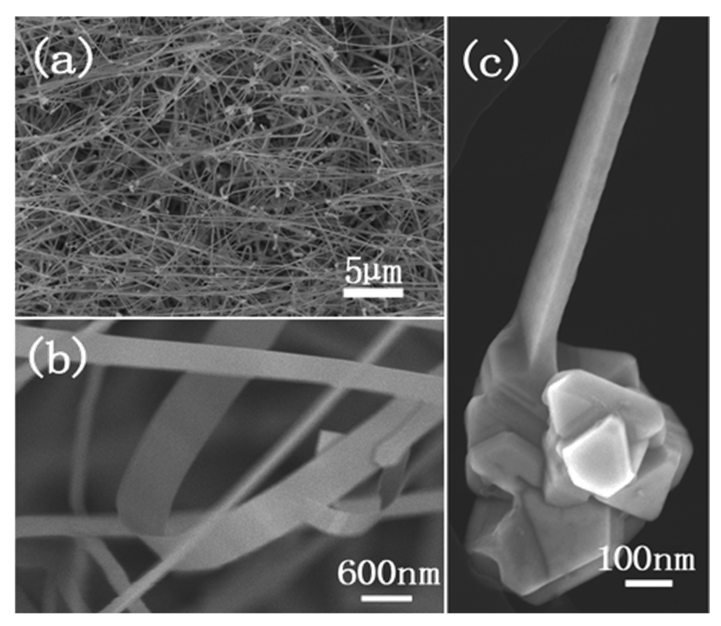
2.2. One-Dimensional ZnS

One-dimensional nanomaterials are an important class of materials gaining attention for their potential applications in different nanodevices. They are different from zero-dimensional nanomaterials because they are considered to be the smallest-dimension structure that is capable of transporting electrons and optical excitation [47]. This means that they are crucial for the function and integration of nanoscale devices. Even though one-dimensional nanomaterials are sufficient in these devices, there is still much to learn about one-dimensional nanostructures. Nanowires, nanotubes, nanorods, and nanocables are categorized as different types of one-dimensional nanomaterials.
Typically, synthesis of one-dimensional nanostructures requires higher temperature and intervention of catalysts. A catalyst-free thermolysis process was reported by Zhu and coauthors to produce ZnS nanowires [48]. A zinc dibutyldithiocarbamate complex precursor was prepared and thermolyzed at 280 °C in the mixed solvent of dodecylamine and triphenyl phosphine. The resulting product exhibited an ultrathin nanowire of ZnS 2 nm in width and with a length of up to 10 microns (Figure 8). Moore et al. demonstrated how to use a two-step thermal evaporation process via a horizontal furnace to produce ZnS nanowires [49]. First, CdSe powder was placed at the center of the furnace and vacuumed to remove traces of oxygen from the chambers. Then, the temperature was elevated, and N2 was introduced into the system as the carrier gas. Once the system was cooled down, the CdSe powder was replaced with ZnS powder. Next, the temperature was increased with the introduction of N2 again. The system remained at these conditions for an hour to collect the sample downstream from the source of ZnS. Characterization of the products via SEM showed that the formed nanowires were grouped into bundles and grew uniformly. In this process, CdSe acted as a substrate for the epitaxial growth of the ZnS growth. In another study, conducted by Lin et al., a thermal evaporation process was used [50]. ZnS powder was placed in a vacuum and placed under high temperature. The powder was evaporated onto an Au-coated silicon (Si) substrate. Imaging with a SEM showed that the lengths of the products were about 10 µm, while the diameter was between 80 and 100 nm.
ZnS nanowires were also successfully synthesized by Shen et al. as ZnS sheathed Zn-Cd heterojunctions [51]. These structures were produced via a one-step thermochemical process of evaporating ZnS and cadmium sulfide (CdS) powders in a vertical induction furnace. Once the powders were placed in the furnace tube, air was evacuated, and Ar was introduced into the system. The system was heated, and these conditions were sustained for an hour before being terminated and cooled. Characterization of the synthesized product was conducted via SEM and TEM. The length of the nanowires was concluded to be several tens of µm, and the diameter ranged from 30 to 70 nm. In a similar way, Shen and coworkers successfully synthesized graphitic carbon-coated ZnS nanowires [52]. This work was accomplished by a two-stage temperature-controlled process in a vertical induction furnace with the help of Sn catalysts. Once the ZnS nanowires were prepared by an Sn-catalytic vapor–liquid–solid (VLS) mechanism, they were allowed to react with the carbon of the graphitic crucible at 1300 °C to produce carbon disulfide (CS2) gas. The generated CS2 gas was then transported to a low-temperature region of the furnace, and it was absorbed onto the surface of the ZnS nanowires. Upon decomposition, CS2 left a graphitic coating over the ZnS nanowires. The coating thickness was found to be 2 nm, while the nanowire’s diameter was in the range of 50 to 120 nm. They were several micrometers long.
Zhu et al. demonstrated how nanocables can be synthesized through a thermochemical process using ZnS powder [53]. In this method, graphite powder and (ZnS) powder were placed in different crucibles with N2 and water streaming through. Both crucibles were placed under high pressure as well. Using TEM imaging, the diameter of the nanocables was about 25 nm and the thickness about 8 nm. Zhu et al. also synthesized nanotubes with the same process and produced tubes with a diameter of 35 nm and a thickness of 7 nm.
Zhao et al. demonstrated synthesis of ZnS nanorods [54]. In this study, they utilized a hydrothermal synthetic route using a triblock copolymer surfactant, Pluronic P123. A solution of zinc nitrate, thiourea, and the surfactant was sonicated for 40 min and heated at 105 °C for 72 h inside a Teflon-lined hydrothermal reactor. Characterization of the samples was conducted using a TEM, which showed that the nanorods had a length ranging from 10 to 30 nm and a diameter of 2 to 5 nm. Nanorods were also synthesized by a surfactant-assisted soft chemistry method, as Zhao et al. revealed in a study [55]. In this case, zinc chloride and thiourea were dissolved in water. Then, a solution of ethylenediamine and 1-dodecanethiol was added to the first solution and was then refluxed at a high temperature for a total of 8 h before being brought back to room temperature. TEM images showed that the nanorods had a length of 1 to 1.5 µm and an average diameter of about 40 nm.
ZnS nanorods have also been produced as manganese (Mn)-incorporated ZnS nanorods via a solvothermal process, as demonstrated by Biswas et al. [56]. This study used zinc nitrate hexahydrate, manganese acetate, and thiourea, and they were mixed in a Teflon-lined chamber that was also filled with ethylenediamine. After stirring for half an hour, the chamber was placed into the furnace at a high temperature for 12 h before being brought back to room temperature. The resulting product with manganese was characterized with a TEM, and it showed that the width of the nanorods was about 100 nm, while the length ranged from 100 to 200 nm.

2.3. Two-Dimensional ZnS

Two-dimensional semiconductor nanomaterials, like nanoribbons, nanosheets, and nanobelts, are gaining attraction as research interests due to their high aspect ratio, unique surface chemistry, and diverse potential applications [57]. Two-dimensional nanomaterials can vary in size, shape, thickness, and composition, with applications in different nanodevices. The growth of 2D morphology is not thermodynamically favorable, hence requiring kinetic control of the growth pathway in order to promote 2D nanomaterial growth. Moreover, high pressure plays a critical role in the growth direction of the nanostructures. Thus, high temperature and high pressure remain the preferred reaction conditions in most of the synthetic techniques.
A study conducted by Zhang et al. synthesized ZnS nanoribbons in a two-temperature-zone furnace [58]. Here, potassium chloride (KCl) and Si wafers acted as substrates. S powder, the substrate, and Zn powder were added to a quartz tube. The Zn powder and substrates were placed in a higher-temperature zone, while the S powder was placed in the lower-temperature zone. Ar was introduced into the system as the carrier gas while the reaction continued for 3 h. The produced nanoribbons were characterized with a field emission scanning electron microscope (FESEM), which showed that the product had a width of several hundred nanometers and a length of about tens of micrometers (Figure 9).
For the synthesis of nanosheets, Li et al. used a high-pressure synchrotron angle dispersive X-ray diffraction (ADXD) process with solvothermal methods to prepare them [59]. This method produced ZnS nanosheets using zinc nitrate and thiourea as the precursors, where the nanosheets ranged from 20 to 50 nm in size. This process was performed under high pressure in a mixture of methanol/ethanol in a 4:1 ratio. In their published work, Verma et al. prepared ZnS nanosheets through a sol-gel co-precipitation method [60]. This process included mixing zinc sulfate monohydrate and water with a solution of thiourea and water. The next step entailed adding NaOH dropwise to the first solution until it reached a pH of 7. The nanomaterial produced in this work had an average size of about 7 nm, which was determined using XRD.
ZnS nanotowers were synthesized through a vapor deposition in a horizontal tube furnace [61]. ZnS powder was placed into the ceramic tube with the substrate, a Si wafer that was spin-coated with thin wurtzite ZnS nanoparticle film. Oxygen was completely eliminated from the tube by purging with Ar, and the reactor was then heated to 1200 °C for 30 min. At the peak temperature, Ar was re-entered into the system to act as a carrier gas. SEM characterization determined that the product had a length of between 400 and 600 nm and a diameter of about 200 to 300 nm. The diameter slowly decreased from the bottom of the structure to the top, which formed the appearance of a tower, which was due to the alternating two-dimensional and one-dimensional stacking. Additionally, the nanotowers demonstrated layers that each had a thickness of about 30 to 40 nm. In another work, conducted by Liang et al., it was shown that a high-temperature horizontal tube furnace could synthesize a variety of nanostructures like nanobelts (or nanoribbons), nanosheets, and nanorods [62]. In this experiment, a sapphire substrate coated in a thin Au film and ZnS powder was added to the tube. Again, Ar, the carrier gas, was added, and the system was heated and placed under pressure. Using SEM imaging, it was found that the produced nanobelts had a length of between 10 and 100 µm and was hundreds of nanometers in width. On the other hand, the nanosheets were shorter in length but larger in width.
One of the more common synthesis methods to produce ZnS nanobelts is thermal evaporation, as shown by Ma et al. [63]. In their work, KCl and Si wafers were used as the substrates, which were both coated in a thin film of Au. Zn powder and the substrates were placed in one zone, while the S powder was placed in another. As seen previously, Ar was used as the carrier gas. This method produced nanoribbons with a width of several hundred nanometers and a length of about tens of micrometers, which was analyzed using a FESEM. In other studies, Li et al. synthesized ZnS nanobelts through pulsed laser vaporization (PLV) by using ZnS powder as the precursor [64]. These nanobelts had a recorded length that ranged between 50 and 670 nm, as determined from TEM imaging.

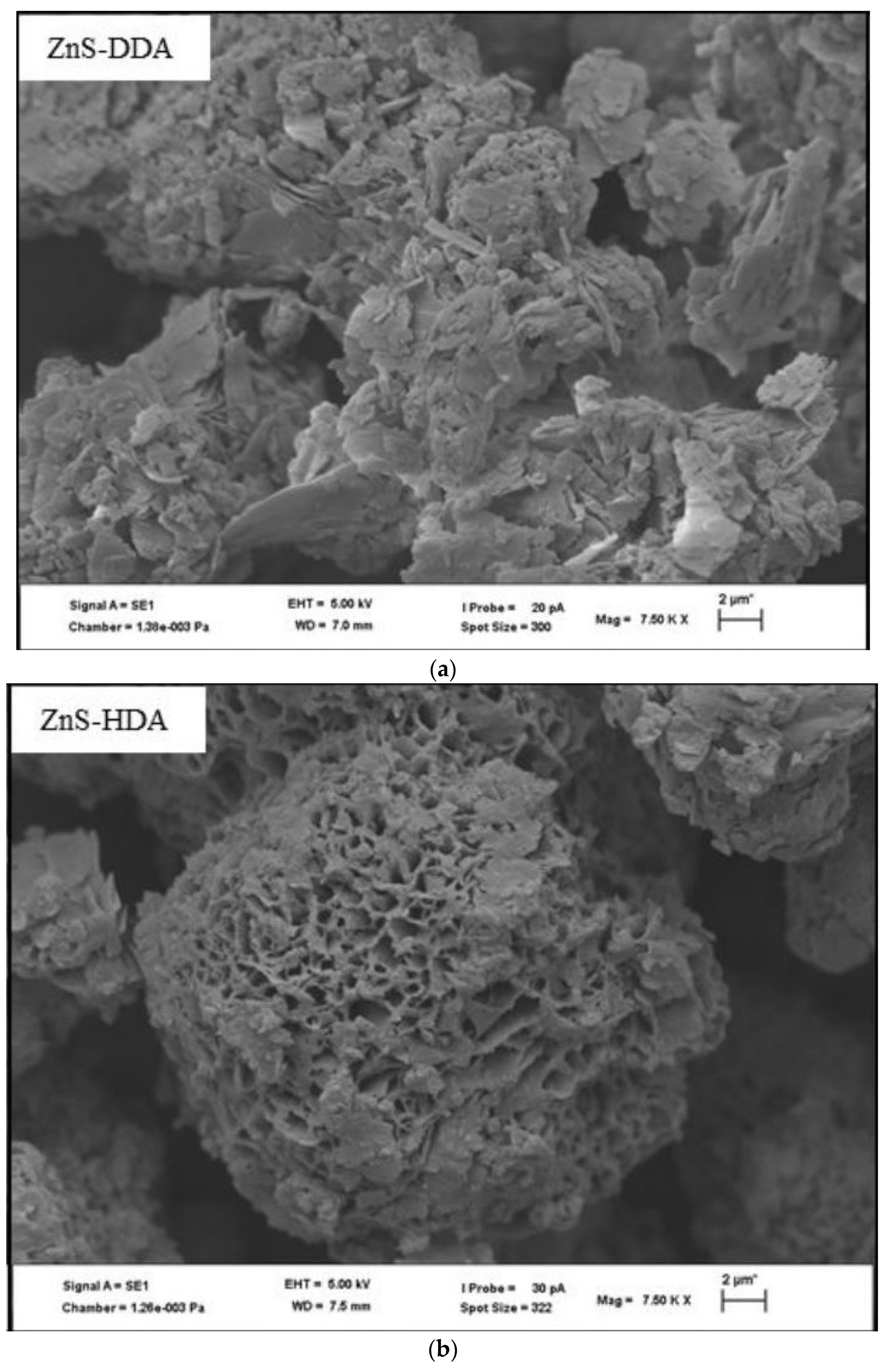
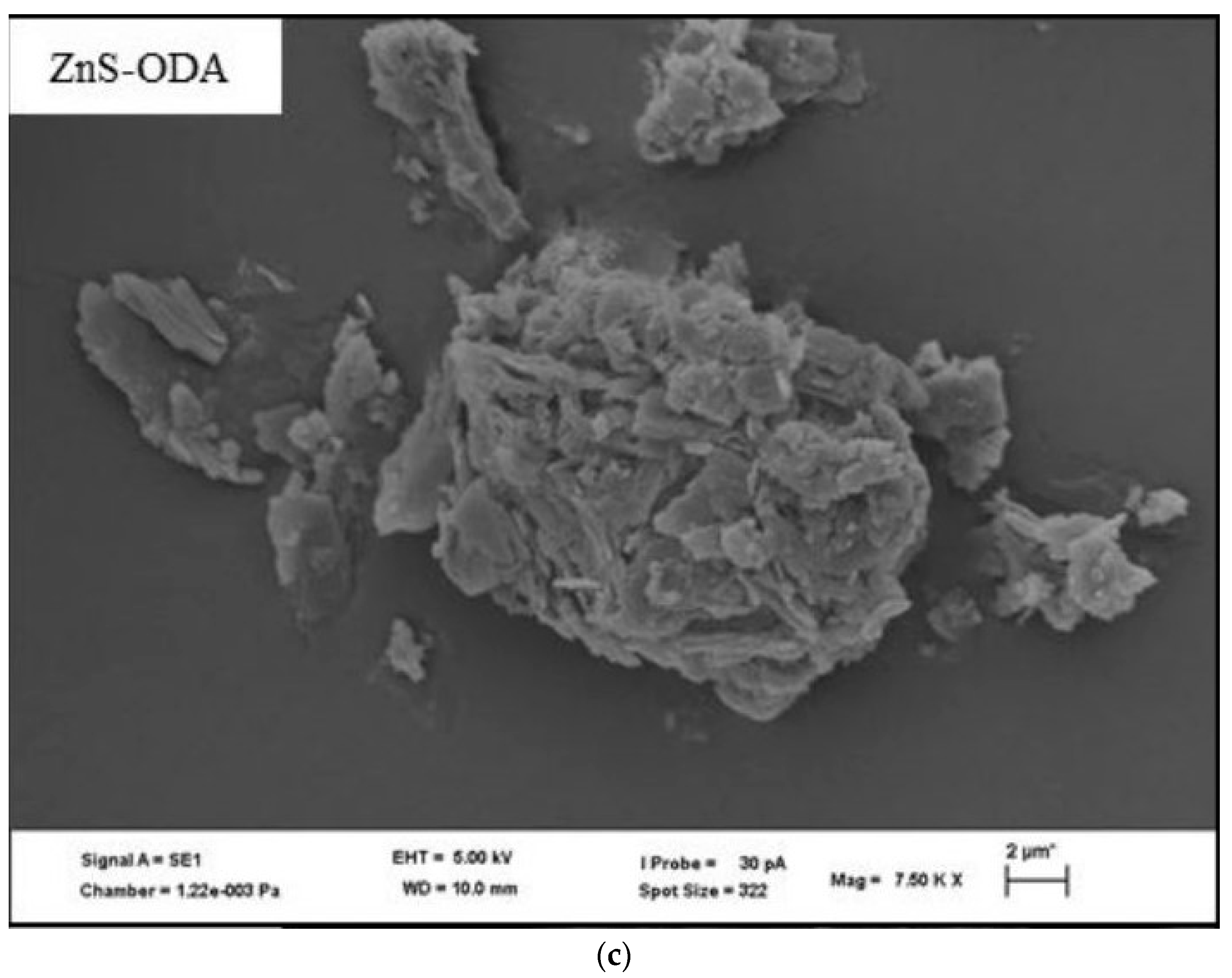
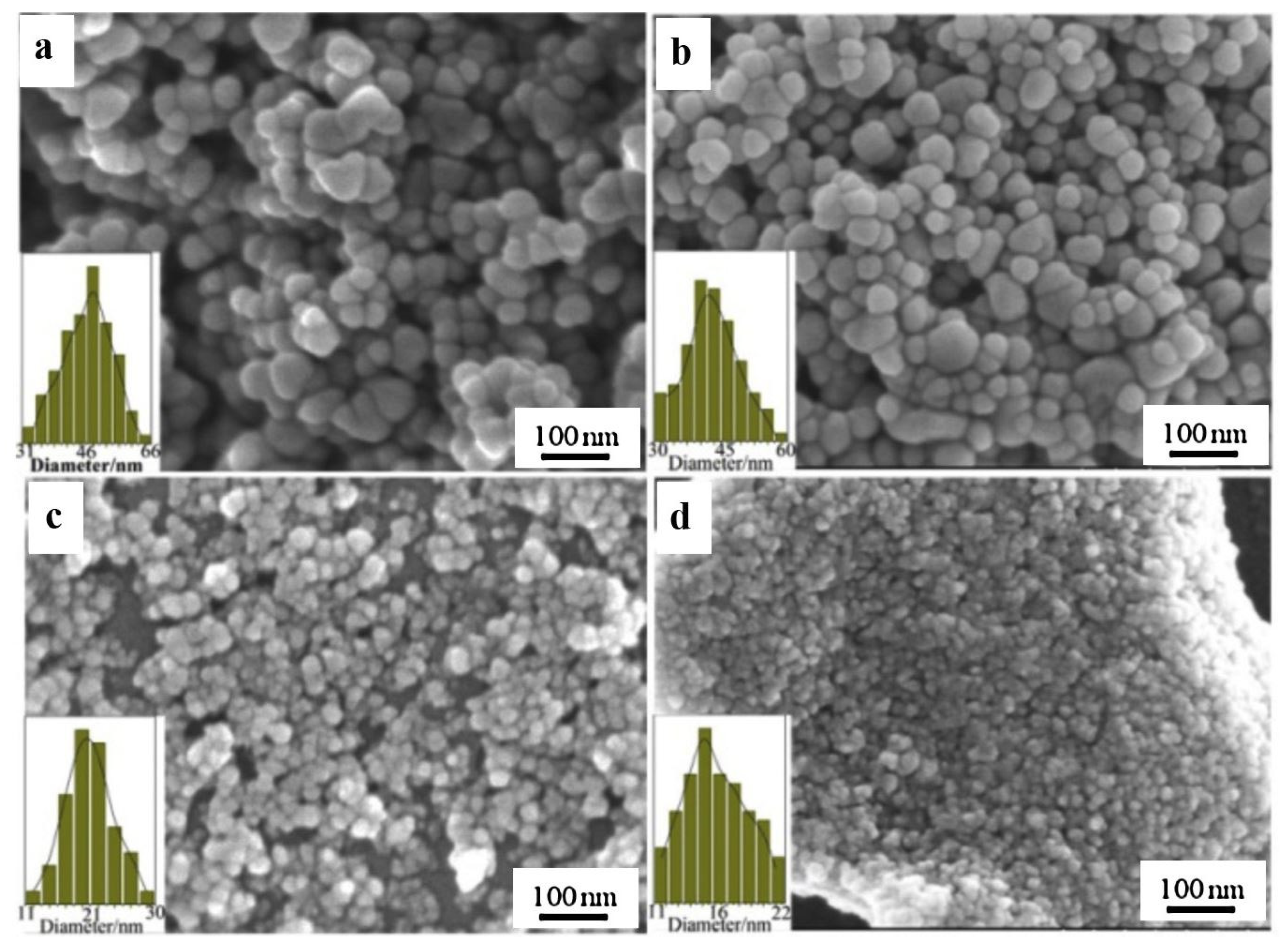
2.4. Three-Dimensional ZnS

Nanocomposites are considered three-dimensional structures that contain two or more nanomaterial components. In nanocomposite structures, there is a continuous matrix phase reinforced by a non-continuous material [65]. There are certain applications that demand the utilization of nanomaterials in composite form. Catalytic degradation of different waste materials in water [66,67] or the preparation of sensors for the detection of certain metal ions [68] may require ZnS nanocomposites in different forms and morphologies. The non-cytotoxicity of ZnS has prompted biomedical applications of their nanocomposites as well [69]. In the following segment, we will describe the different synthetic methodologies employed to produce a variety of ZnS nanocomposite structures.
Molybdenum disulfide (MoS2)/ZnS nanocomposites have also been synthesized through a hydrothermal reaction, as shown in a study by Gusain et al. [66]. The MoS2 nanosheet precursors were synthesized previously using a hydrothermal process. Zinc acetate dihydrate and MoS2 were added to ethanol and sonicated for an hour. A solution of PVP in ethanol was then added to that solution. After stirring for 15 min, a solution of NaOH, water, and ethanol was added and stirred for another 30 min. Then, the solution was heated for 24 h and then cooled back to room temperature. The process is depicted in Figure 10. Characterization using HRTEM showed that the average diameter of ZnS nanoparticles were about 0.48 nm when attached to molybdenum disulfide (MoS2) nanosheets in the nanocomposites.
A study by Xaba showed the synthesis of ZnS nanoparticles by mixing zinc acetate in 50% methanol and 50% methanolic solution [67]. The mixture was then refluxed in a water bath for an hour, and a starch or PVA solution was added to the first solution. Then, the pH of the solution was adjusted to be equivalent to 1 by using ammonium hydroxide. The ZnS-PVA-capped nanoparticles were then added to water and filtered before adding the nanoparticles to a 0.5% chitosan solution, prepared previously from dilute acetic acid. The mixture was then sealed with foil and placed in an ultrasonic bath for 4 h to produce ZnS-chitosan nanocomposites.
Wang et al. demonstrated how ZnS/copper sulfide (CuS) nanocomposites can be synthesized through a combination of biomimetic synthesis and an ion-exchange strategy [69]. In their study, bovine serum albumin (BSA)-conjugated ZnS nanoparticles were prepared by mixing zinc sulfate with a BSA solution at room temperature for 12 h. Then, TAA was added to the solution above and maintained at room temperature for 3 days. Additionally, the BSA-conjugated ZnS/CuS nanocomposites were synthesized by using the BSA-conjugated ZnS nanoparticles as the precursor for the ion-exchange method. This precursor was added to water and copper sulfate pentahydrate and stirred for 8 h. These nanocomposites were characterized using a HRTEM and it was found that the diameter of these products was 31.5 ± 5.6 nm.
In another study by Dutta et al., nanocomposites were synthesized by way of a one-pot, green synthesis protocol [70]. Here, reduced graphene oxide (RGO) ZnS nanocomposites were synthesized by adding zinc nitrate to the sulfur precursor, a cysteamine hydrochloride solution that was prepared using a graphene oxide (GO) solution. This solution was stirred for 2 h at room temperature. After this step, the solution was placed in a Teflon liner and was autoclaved for 14 h at 160 °C. TEM characterization found that the RGO-ZnS nanocomposites had a crystallite size of about 4 nm. Jawad et al. also prepared ZnS/RGO nanocomposites [71]. In this study, ZnS discs were placed inside a GO solution to be exposed to laser pulses. There were 600 laser pulses, which had a duration of 10 nanoseconds each with an energy of 800 mJ and a frequency of 6 Hz. Characterization through XRD spectra showed that the average diameter was about 25.2 nm.
Reduced graphene oxide-ZnS-CuS (RGO-ZCS) nanocomposites have been synthesized by an in situ microwave method, as demonstrated by Mahalingam et al. [72]. A solution of GO in water was ultrasonicated for 90 min before adding zinc sulfate heptahydrate and copper sulfate pentahydrate to the solution to stir for 2 h. Next, the solution was placed in a microwave oven and was microwaved at 950 W for 10 min. Then, thiourea was added to the solution and was stirred for 2 h. The solution was then microwaved again with the same steps as previously to produce sphere-like morphology with a particle size range within 10 and 30 nm.
Yu et al. also demonstrated how to produce CuS/ZnS nanocomposites via an ion-exchange method [73]. First, monodispersed ZnS colloidal nanospheres were prepared by mixing zinc nitrate and TAA in hot water before being quenched in an ice-water bath. Next, the CuS/ZnS nanocomposites were prepared by adding the ZnS nanospheres previously prepared to an ethanolic solution of copper (II) nitrate. A color change to a deep green indicated that the CuS was produced. Then, the solution was placed in a Teflon-lined autoclave for 4 h at 80 °C. TEM imaging showed that the average diameter of the nanocomposites was about 255 nm.

3. Properties and Applications

The uniqueness of different nanomaterials lies in the fact that their properties are controlled by many physical parameters, e.g., dimensions, morphology, crystal structure, etc. There have been significant studies conducted to establish the structure–property relationship within a variety of nanomaterials. ZnS nanomaterials have shown similar trends in the relationship between size and properties, mainly optical and electrical. However, ZnS nanomaterials have applications in other fields, including photocatalysis, chemical and biosensors, thermoelectric, and many more. In the following section, we will describe the different properties of ZnS nanomaterials as relevant to their respective morphologies, along with selected potential applications. Table 2 lists various applications of ZnS nanomaterials categorized by their respective morphologies.
The major applications of ZnS nanomaterials are found to be in optoelectronics, as ZnS is an important wide-bandgap (~3.6–3.7 eV) semiconductor. Upon photoillumination, these extremely small-sized nanomaterials can generate electron–hole pairs resulting in quantum confinement. With the decrease in particle size, and as the radius approaches the exciton Bohr radius, the quantum size effect becomes more and more prominent. We can observe a blue shift of the optical absorption peak as the energy gap increases with the decrease in grain size. Several studies have been conducted to establish such a relationship between the dimensions of NPs and their optical properties. Kafel et al. conducted a theoretical study recently and depicted the changes in quantum confinement energies as a function of particle size along with the correlation between the energy gap and the confinement energy (Figure 11) [74]. As reported, a very strong quantum confinement was observed when the particle size was 10 nm, and the confinement energy varied between 0.1 eV and 1.21 eV as the particle size decreased. The energy gap decreased as the confinement energy decreased, which is typically observed in most semiconducting materials.
Bulk ZnS materials exhibit an optical absorption peak around 340 nm, corresponding to a band gap energy of 3.68 eV [15]. Due to the quantum effect, there is a shift of nearly 1.1–1.4 eV in the band gap towards blue light [34], which in turn is indicative of the formation of nanostructured materials. Most research groups extensively working in this field have observed and reported such a shift, and this size-tunable variation of optical properties opens a wide range of applications of ZnS nanomaterials. The photoluminescence peaks of ZnS vary depending on the wavelength of the excitation energy, as well as the size of the nanomaterials. With the 295 nm excitation wavelength, emission peaks for ZnS nanomaterials are found to be in the 400 nm range. Many times, for semiconducting nanomaterials, two emission peaks are observed: a sharper peak, which can be attributed to the exciton luminescence, and a broader peak at higher wavelength due to the trapped luminescence [75]. The capping agents also contribute to altering the emission properties, as reported by many investigators. Li and coauthors conducted studies on ZnS nanocrystals, and prepared by fatty acid ligands and amine-terminated ligands. The fatty acid-capped ZnS NCs produced PL spectra almost 10 times brighter than the ones with amine-terminated ligands as capping agents. Mansur and coworkers found that phosphoethanolamine-terminated ZnS QDs exhibited PL emissions in the violet–green range [76]. While comparing the effect of capping agents on ZnS QDs, Li et al. observed blue trap-state emissions at around 415 nm for the QDs, and the (3-mercaptopropyl) trimethoxysilane-capped ZnS QDs showed stronger emissions that the 3-mercaptopropionic acid-capped ZnS QDs [36]. The lesser emissions in the MPA-capped QDs were attributed to the absorption of light by the excess MPA present in the colloidal suspensions, which in turn reduced the emissions by that solution. Studies were also conducted to investigate the dependency of the emission properties on pH, and Bae et al. reported a red shift in the emission peak with increasing pH [11].
Doped ZnS nanomaterials exhibited different PL properties depending on the type of metal doping agents. Terbium (Tb3+)-doped ZnS NPs exhibited the conventional 400 nm emission peak upon excitation by a 295 nm wavelength, as well as sharp peaks at 490, 545, 585, and 620 nm, which are characteristic peaks for Tb3+ species [77]. The authors confirmed that the peaks at the higher wavelengths were generated by the NPs and not the free Tb3+ ions present in the solution. Since the concentration of any free Tb3+ ions present in the solutions was in the micromolar range, it was not possible for the free Tb3+ ions to generate emission peaks, as to generate emissions, millimolar concentrations are required. Bose and coauthors successfully synthesized bluish-green-emitting Au-Zn-S NPs [78]. Although short lived, the narrow and sharp emission peak at 470 nm was attributed to the Au-doped ZnS NCs (Figure 12). Cu-doped ZnS NCs were reported to produce sub-bandgap red, green, and blue PL emission bands. A distinct PL emission peak at 670 nm was observed, whose intensity was noted to increase with an increment in the Cu:Zn ratio. Similarly, Mukherjee et al. employed black (4.1 eV), blue (3.2 eV), and red 2.9 (eV) excitation energies to obtain emissions at the black (3.3 eV), blue (2.78 eV), and red (2.47 eV, 0.54 eV) regions in Cu zinc sulfide (CZS) NCs [79]. Srivastava and coworkers synthesized Mn2+-doped ZnS NCs, which exhibited significantly red-shifted emissions with more than 50% quantum yield (QY) [80]. They noted that a higher sulfur ratio in the reactant mixture slowed down the incorporation of Mn2+ ion as dopants and reduced the QY drastically.
The utilization of ZnS-based QDs in light-emitting diodes (LEDs) has long been reported in many accounts and was effectively compiled by Talapin et al. in their review article [81]. As depicted in Figure 13, a very thin layer of QD nanomaterial was sandwiched between the hole transport layer (HTL) and electron transport layer (ETL) to construct the LEDs. By varying the size of the NCs, one can fine tune the emission from the UV to the near-infrared (IR) region. Moreover, NC-based LEDs are characterized by their high color purity because of the narrow emission bands. ZnS-based core shell nanostructures were investigated and utilized as thin-layer QDs in many QLEDs. In a very recent study, Feng and coauthors reported ZnSeTe/ZnSe/ZnS QDs with an adjustable energy band structure and blue emissions from 455 to 470 nm [82]. Efficient near-IR light-emitting diodes were developed by Zhang and coworkers, with PL peaks located at 705, 719, and 728 nm, along with respective external quantum efficiencies of 3.0%, 4.0%, and 2.5% [83]. They employed Zn:CuInSe2/ZnS//ZnS QDs to obtain heavy metal-free QLEDs with outstanding quantum efficiencies comparable to those of QLEDs based on a PbS/CdS system. Solid-state white light-emitting diodes (WLEDs) have also gained popularity since their conception in early 2000 because of their longer lifetime, higher efficiency, and lower thermal resistance [84]. Hasanirokh et al. reported the production of WLED based on thin and uniform CdS/ZnS/CdS/ZnS colloidal QD film, which combined the blue emissions from the CdS core and green/orange emissions from the ZnS shell to produce the white light [85].
Many metal ions provided a quenching effect to the emission properties of ZnS nanomaterials. Thus, ZnS nanomaterials can serve as sensor devices in the detection of such metal ions. An interesting investigation conducted by Mandal and coworkers revealed that in the presence of Ag+ ion, the PL emission of ZnS QDs was quenched significantly [39]. With 0.5–1.0 µM Ag+, ZnS QDs showed almost zero emissions, and such findings can be successfully utilized to detect the presence of Ag+ ions in a solution, as the authors aptly suggested. Another report published by Asadi and coworkers revealed utilization of PEG-capped ZnS QDs for the detection of Cu2+ ions [68]. The quenching of the fluorescence emission peak in the presence of Cu2+ ions was recorded, and a sensor was successfully devised based on the PEG-capped ZnS QDs that can detect Cu2+ ions as low as 0.96 nM in concentration. The quenching effect was attributed to the ability of the Cu2+ ions to form complexes with the hydroxyl functional groups of the capping agents. Consequently, there was electron transfer taking place from the excited ZnS QDs to Cu2+, reducing the fluorescence intensity of the composite to a great extent.
Among other applications, ZnS nanomaterials can be used as photocatalysts as well. ZnS has shown a great potential in degrading many organic molecules in water-based systems, which in turn make them a valid contender as a catalyst to be used in wastewater treatment. In their experiment Kho et al. showed that in the presence of low-power UV excitation of ZnS nanocrystals, p-nitrophenol (pNP), a model organic pollutant compound, was degraded at an excellent rate [12]. They demonstrated that cysteine-capped ZnS NPs are capable of degrading pNPs by more than 90% within 10 min, and that the NPs can retain such properties even after a storage life of 30 months. A similar study was conducted by Ayodhya and coworkers to exhibit the photocatalytic activity of ZnS NPs [17]. They have shown that xylenol orange (3,3′-bis[N,N-bis(carboxymethyl)aminomethyl]-o-cresolsulfonephthalein), an organic dye, can be degraded using PVA-capped ZnS NPs by up to 87.24% in 120 min under exposure to sunlight. Yin et al. reported photocatalytic degradation of methyl orange (MO) dye by ZnS nanocrystallites under UV radiation (365 nm) [23]. In 30 min, 99.5% MO was degraded upon irradiation with the UV light supplied by a high-pressure mercury lamp (300 W). They prepared nanostructures of varying sizes and demonstrated that the particles with the smallest size showed the maximum efficiency of the photocatalytic activity. The smaller size with a higher surface area can be attributed to the higher degradation efficiency. Moreover, the narrower bandgap of the ZnS nanostructures may have contributed positively to improvement in the catalytic activity. In a more recent study, ZnS NPs capped with DDA, HDA, and ODA were found to be 80.20, 43.21, and 91.91% efficient in degrading bromothymol dyes, respectively, after 180 min of irradiation under visible light [19].
ZnS-based nanocomposites have also been explored as photocatalysts for degrading organic pollutants, reducing CO2 via degradation, as well as producing H2 through photocatalytic water splitting. While Entradas and coauthors published their report on the photocatalytic activity of a nanocomposite based on titanate nanofibers (TNFs) with nanocrystalline ZnS and Bi2S3 semiconductors [86], Naudin and coworkers fabricated a nanocomposite by mixing titanate nanorods (TNR) with ZnS nanocrystallites to obtain up to 77% photodegradation of safranine-T, a model organic pollutant [87]. More recently, Dabbous et al. reported synthesis of hybrid a CdSe/ZnS QD–Au nanoparticle composite to be used as photoredox system with a high turnover number (TON) [88]. Li and coworkers fabricated a ZnS/ZnO heterojunction for photocatalytic reduction of CO2 [89]. Zhang et al. demonstrated increased photocatalytic hydrogen production via water splitting by Au-nanoparticle-decorated ZnS nanocomposite materials [90]. However, a recent study conducted by Lange and coworkers posed a valid question about the efficiency of ZnS as a photocatalyst since ZnS suffers photodegradation itself [91].
Table 2. Various applications of ZnS nanomaterials.
ZnS NanostructureApplicationReference Number(s)
0D, 1D, 2D, 3DBiomedical/Biotechnology[45,54,69,88]
0D, 1D, 3DCatalyst [29,48,54,66,72,73]
0D, 1D, 2DElectroluminescence[28,34,48,56]
1DElectronics[50,56]
2DInfrared devices[56]
0D, 1D, 2DNanodevices/Nanoelectronics[20,48,50,52,54,61,62,63]
0DOptical[13,18,29,30,31]
0D, 1D, 2D, 3DOptoelectronic devices[15,18,28,29,31,33,54,59,62,63,73]
0DPhotovoltaic/Photoconductor[15,29,31]
0D, 1D, 2DSensors[18,28,29,30,31,34,56,59,62]
3DWater and air treatment[67,68,69,71]
The magnetic properties of ZnS nanostructures were also investigated. Zhu and coworkers used a superconducting quantum interference device (SQUID) to conduct such an investigation on ultrathin ZnS nanowires and observed ferromagnetic behavior [48]. Theoretical studies conducted by Xiao et al. support this finding, as they observed similar properties of ZnS QDs [9]. The magnetic behavior of the NCs was attributed to the p-like dangling electrons in the 3p orbitals of S atoms in ZnS. Proshchenko and coworkers used the surface-bulk model (SB) to investigate nanoferromagnetism in ZnS QDs and nanowires [92]. Their studies concluded that there was a less than 10% discrepancy in Exact DFT and SB calculations for the QD magnetic moment and the mechanism. They attributed the d0 ferromagnetism of ZnS nanowires and QDs to the unpaired sulfur electrons in the tetrahedral crystal field.
ZnS nanomaterials also find their usage in different energy applications. With the emergence of lithium sulfur (Li-S) batteries, nanostructured ZnS showed great potential as a stable resource for sulfur cathodes [93]. The generic low conductivity of bulk ZnS may hinder the application of ZnS as cathode materials. Liu and coworkers coated a carbon cloth with a thin (50–70 nm thick) nanolayer of ZnS, and the Li-S battery performance was heavily improved [94]. While ZnS acted as a catalyst to boost the conversion reaction of lithium polysulfides, the conductive carbon cloth helped with electrical conductivity. Another potential application of ZnS nanomaterials is in sodium-ion batteries (SIBs). The high theoretical capacity of ZnS (550 mAh g−1) made them an excellent candidate as an anode material in SIBs [95]. Encapsulated ZnS nanocrystals by nitrogen-doped carbon shells were designed and developed by Zhao et al. [96]. The improved anode provided a high-rate capability and excellent cycling stability. ZnS-based composites are also useful in solar cell applications. A comprehensive review by Ummartyotin and Infahsaeng listed potential application areas of ZnS in a variety of solar cells, including dye-sensitized solar cells (DSSCs), quantum dot-sensitized solar cells (QDSCs), Cu(In,Ga)Se2 (CIGS)-based thin-film solar cells, and organic–inorganic hybrid solar cells [97].
ZnS NPs, being non-cytotoxic [7], have proven their efficiency in many biological and biomedical applications. Khan et al. demonstrated the antibacterial properties of InP/ZnS core/shell QDs [45]. These QD nanostructures were found to be inhibitors to the growth of both E. coli and multidrug-resistant S. aureus bacteria under light. Wang and coauthors showed biological activities of ZnS-CuS nanocomposites, where rat pheochromocytoma (PC12) cells upon exposure to the nanocomposites exhibited a very high inhibition rate (54.1 ± 5.3%) [69]. In a recent study, Liang et al. demonstrated in-vitro transfection of miR-26a plasmid into HepG2 cells by polyethyleneimine-coated ZnS-CdSe QDs [98]. Their findings may open pathways for the application of ZnS QDs in drug deliveries, gene therapies, and many other bioimaging applications.

4. Conclusions

In recent years, there has been an increase in the interest and work being conducted on ZnS-based nanomaterials. Unlike many other nanomaterials, ZnS exhibits zero-dimensional, one-dimensional, two-dimensional, and three-dimensional morphologies in distinct shape and dimensions. Various synthetic techniques are employed to create such a variety of unique structures. Hydrothermal, solvothermal, thermal evaporations, and sol-gel co-precipitation processes are a few. The major challenges of synthesizing zero-dimensional material can be to keep the size within its limited dimensions. Some ways to combat this struggle are to vary the conditions through different solvent systems, pH, reaction temperature, and capping agents. Additionally, much of the work conducted on one-dimensional materials uses diverse thermochemical reactions. On the other hand, two-dimensional materials typically require kinetic control of the growth pathway to maintain the dimensions within the confinement of two-dimensional materials. Three-dimensional materials are synthesized by first producing the ZnS precursor and then embedding or coating it with another material to form a nanocomposite. We have included a thorough and detailed review of the various synthetic techniques.
We have discussed different properties of ZnS and their potential applications based on such unique properties. While the photoluminescence and electroluminescence of ZnS-based nanomaterials make them useful in many optoelectronic devices, their emission properties can also be used in devising sensors for a variety of metal ions. ZnS-based nanomaterials have proven their utility as photocatalysts despite some reports with minor contradiction. Nanocomposites of ZnS were utilized in battery applications and solar cells. Moreover, the non-cytotoxicity of ZnS makes them potential candidates in many biomedical applications, including bioimaging, gene therapy, and drug deliveries.
While research involving synthetic investigations of the zero-dimensional and one-dimensional nanostructures of ZnS materials have made significant progress, methods of making two-dimensional ZnS nanomaterials are comparatively less reported. The pressure induced growth and formation of two-dimensional ZnS nanomorphologies need to be explored largely since the need for new battery materials is increasing day by day. As sulfur batteries are gaining more attention in both the scientific community and the corporate sector, ZnS-based nanostructures will also be in great demand. Fabrication of two-dimensional ZnS as anode materials for sulfur batteries will be less challenging and more beneficial. The photocatalytic activity of ZnS-based nanomaterials may need further scrutiny, as some research exhibited photodegradation of ZnS itself, which may result in lesser efficiency of ZnS as a photocatalyst. ZnS-based nanomaterials have also become more visible in biomedical application fields, as more research is being conducted and published in this area. The non-cytotoxicity and relatively lower cost of ZnS should be the major driving force to appeal more funding sources in this direction. Overall, a steady growth of research focused on synthesis and application of ZnS-based nanomaterials has been noted over recent years, and we are extremely hopeful about the sustainability of such progress.

Author Contributions

Conceptualization, A.C. investigation, A.C. and E.A.; writing—original draft preparation, A.C. and E.A.; writing—review and editing, A.C.; visualization, A.C. and E.A.; supervision, A.C.; project administration, A.C. All authors have read and agreed to the published version of the manuscript.

Funding

This research received no external funding.

Conflicts of Interest

The authors declare no conflicts of interest.

References

  1. Nanotechnology Publications (Article). Available online: https://statnano.com/report/s29 (accessed on 8 April 2024).
  2. Davidson, W.L. X-ray diffraction evidence for ZnS formation in zinc activated rubber vulcanizates. Phys Rev. 1948, 74, 116–117. [Google Scholar]
  3. Garlick, F.J.; Gibson, A.F. The Luminescence of Photo-Conducting Phosphors. J. Opt. Soc. Am. 1949, 39, 935–941. [Google Scholar] [CrossRef] [PubMed]
  4. Yanagida, S.; Yoshiya, M.; Shiragami, T.; Pac, C.; Mori, H.; Fujita, H. Semiconductor photocatalysis. I. Quantitative photoreduction of aliphatic ketones to alcohols using defect-free zinc sulfide quantum crystallites. J. Phys. Chem. 1990, 94, 3104–3111. [Google Scholar] [CrossRef]
  5. Work, L.; Odell, H. Mechanism of Development of Pigment Value in Zinc Sulfide. I. Theory of Development of Pigment Value and Methods of Test. Ind. Eng. Chem. 1933, 25, 411–416. [Google Scholar] [CrossRef]
  6. Mekuye, B.; Abera, B. Nanomaterials: An overview of synthesis, classification, characterization, and applications. Nano Sel. 2023, 4, 486–501. [Google Scholar] [CrossRef]
  7. Li, H.; Li, M.; Shih, W.; Lelkes, P.I.; Shih, W.-H. Cytotoxicity Tests of Water Soluble ZnS and CdS Quantum Dots. J. Nanosci. Nanotechnol. 2011, 11, 3543–3551. [Google Scholar] [CrossRef]
  8. Fang, X.; Zhai, T.; Gautam, U.K.; Li, L.; Wu, L.; Bando, Y.; Golberg, D. ZnS nanostructures: From synthesis to applications. Prog. Mater. Sci. 2011, 56, 175–287. [Google Scholar] [CrossRef]
  9. Xiao, W.-Z.; Wang, L.-L.; Rong, Q.-Y.; Xiao, G.; Meng, B. Magnetism in undoped ZnS studied from density functional theory. J. Appl. Phys. 2014, 115, 213905. [Google Scholar] [CrossRef]
  10. Azpiroz, J.M.; Lopez, X.; Ugalde, J.M.; Infante, I. Modeling Surface Passivation of ZnS Quantum Dots. J. Phys. Chem. C 2012, 116, 2740–2750. [Google Scholar] [CrossRef]
  11. Bae, W.; Mehra, R.K. Cysteine-capped ZnS nanocrystallites: Preparation and Characterization. J. Inorg. Biochem. 1998, 70, 125–135. [Google Scholar] [CrossRef]
  12. Kho, R.; Torres-Martínez, C.L.; Mehra, R.K. A Simple Colloidal Synthesis for Gram-Quantity Production of Water-Soluble ZnS Nanocrystal Powders. J. Colloid Interface Sci. 2000, 227, 561–566. [Google Scholar] [CrossRef]
  13. Lau, B.L.T.; Hsu-Kim, H. Precipitation and Growth of Zinc Sulfide Nanoparticles in the Presence of Thiol-Containing Natural Organic Ligands. Environ. Sci. Technol. 2008, 42, 7236–7241. [Google Scholar] [CrossRef] [PubMed]
  14. Nakaoka, Y.; Nosaka, Y. Electron Spin Resonance Study of Radicals Produced by Photoirradiation on Quantized and Bulk ZnS Particles. Langmuir 1997, 13, 708–713. [Google Scholar] [CrossRef]
  15. Nanda, J.; Sapra, S.; Sarma, D.; Chandrasekharan, N.; Hodes, G. Size-Selected Zinc Sulfide Nanocrystallites: Synthesis, Structure, and Optical Studies. Chem. Mater. 2000, 12, 1018–1024. [Google Scholar] [CrossRef]
  16. Vogel, W.; Borse, P.H.; Deshmukh, N.; Kulkarni, S.K. Structure and Stability of Monodisperse 1.4-nm ZnS Particles Stabilized by Mercaptoethanol. Langmuir 2000, 16, 2032–2037. [Google Scholar] [CrossRef]
  17. Ayodhya, D.; Venkatesham, M.; Kumari, A.; Mangatayaru, K.; Veerabhadram, G. Synthesis, Characterization of ZnS nanoparticles by Coprecipitation method using various capping agents—Photocatalytic activity and Kinetic study. IOSR J. Appl. Chem. 2016, 6, 1–9. [Google Scholar] [CrossRef]
  18. Allehyani, A.H.A.; Seoudi, R.; Said, D.A.; Lashin, A.R.; Abouelsayed, A. Synthesis, Characterization, and Size Control of Zinc Sulfide Nanoparticles Capped by Poly(ethylene glycol). J. Electron. Mater. 2015, 44, 4227–4235. [Google Scholar] [CrossRef]
  19. Ajibade, P.A.; Mbuyazi, T.B.; Paca, A.M. Synthesis and Crystal Structures of Bis(diallydithiocarbamato)zinc(II) and Silver(I) Complexes: Precursors for Zinc Sulfide and Silver Sulfide Nanophotocatalysts. ACS Omega 2023, 8, 24750–24760. [Google Scholar] [CrossRef]
  20. Zhang, H.; Chen, B.; Gilbert, B.; Banfield, J.F. Kinetically controlled formation of a novel nanoparticulate ZnS with mixed cubic and hexagonal stacking. J. Mater. Chem. 2006, 16, 249–254. [Google Scholar] [CrossRef]
  21. Wang, M.; Zhang, Q.; Hao, W.; Sun, Z.-X. Surface stoichiometry of zinc sulfide and its effect on the adsorption behaviors of xanthate. Chem. Cent. J. 2011, 5, 73. [Google Scholar] [CrossRef]
  22. Kaur, J.; Sharma, M.; Pandey, O.P. Effect of pH on size of ZnS nanoparticles and its application for dye degradation. Part. Sci. Technol. 2015, 33, 184–188. [Google Scholar] [CrossRef]
  23. Yin, L.; Zhang, D.; Wang, D.; Kong, X.; Huang, J.; Wang, F.; Wu, Y. Size dependent photocatalytic activity of ZnS nanostructures prepared by a facile precipitation method. Mater. Sci. Eng. B 2016, 208, 15–21. [Google Scholar] [CrossRef]
  24. Zhang, H.; Gilbert, B.; Huang, F.; Banfield, J.F. Water-driven structure transformation in nanoparticles at room temperature. Nature 2003, 424, 1025–1029. [Google Scholar] [CrossRef] [PubMed]
  25. Balantseva, E.; Berlier, G.; Camino, B.; Lessio, M.; Ferrari, A.M. Surface Properties of ZnS Nanoparticles: A Combined DFT and Experimental Study. J. Phys. Chem. C 2014, 118, 23853–23862. [Google Scholar] [CrossRef]
  26. Li, S.; Pradhan, N.; Wang, Y.; Peng, X. High Quality ZnSe and ZnS Nanocrystals Formed by Activating Zinc Carboxylate Precursors. Nano Lett. 2004, 4, 2261–2264. [Google Scholar] [CrossRef]
  27. Rath, T.; Kunert, B.; Resel, R.; Fritz-Popovski, G.; Saf, R.; Trimmel, G. Investigation of Primary Crystallite Sizes in Nanocrystalline ZnS Powders: Comparison of Microwave Assisted with Conventional Synthesis Routes. Inorg. Chem. 2008, 47, 3014–3022. [Google Scholar] [CrossRef] [PubMed]
  28. Ali, A.H.; Hashem, H.A.-E.; Elfalaky, A. Preparation, Properties, and Characterization of ZnS Nanoparticles. Eng. Proc. 2023, 31, 74. [Google Scholar]
  29. Hoa, T.T.Q.; van Vu, L.; Canh, T.D.; Long, N.N. Preparation of ZnS nanoparticles by hydrothermal method. J. Phys. Conf. Ser. 2009, 187, 012081. [Google Scholar] [CrossRef]
  30. Dengo, N.; Vittadini, A.; Natile, M.; Gross, S. In-Depth Study of ZnS Nanoparticle Surface Properties with a Combined Experimental and Theoretical Approach. J. Phys. Chem. C 2020, 124, 7777–7789. [Google Scholar] [CrossRef]
  31. John, R.; Florence, S. Optical, Structural and Morphological Studies of Bean- Like ZnS Nanostructures by Aqueous Chemical Method. Chalcogenide Lett. 2010, 7, 269–273. [Google Scholar]
  32. Shi, H.; Zhou, X.; Fu, X.; Gao, F.; Hu, Z. Preparation of organic fluids containing Cyanex 302-modified ZnS particles with high loading concentration and their tribological properties. Colloids Surf. A 2008, 313, 482–489. [Google Scholar] [CrossRef]
  33. Kumara, C.; Armstrong, B.; Lyo, I.; Lee, H.W.; Qu, J. Organic-modified ZnS nanoparticles as a high-performance lubricant additive. RSC Adv. 2023, 13, 7009–7019. [Google Scholar] [CrossRef] [PubMed]
  34. Arao, Y.; Hirooka, Y.; Tsuchiya, K.; Mori, Y. Structure and Photoluminescence Properties of Zinc Sulfide Nanoparticles Prepared in a Clay Suspension. J. Phys. Chem. C 2009, 113, 894–899. [Google Scholar] [CrossRef]
  35. Alivisatos, A.P. Semiconductor Clusters, Nanocrystals, and Quantum Dots. Science 1996, 271, 933–937. [Google Scholar] [CrossRef]
  36. Li, H.; Shih, W.Y.; Shih, W.-H. Highly Photoluminescent and Stable Aqueous ZnS Quantum Dots. Ind. Eng. Chem. Res. 2010, 49, 578–582. [Google Scholar] [CrossRef]
  37. Zhou, W.; Schwartz, D.T.; Baneyx, F. Single-Pot Biofabrication of Zinc Sulfide Immuno-Quantum Dots. J. Am. Chem. Soc. 2010, 132, 4731–4738. [Google Scholar] [CrossRef]
  38. Cooper, J.K.; Franco, A.M.; Gul, S.; Corrado, C.; Zhang, J.Z. Characterization of Primary Amine Capped CdSe, ZnSe, and ZnS Quantum Dots by FT-IR: Determination of Surface Bonding Interaction and Identification of Selective Desorption. Langmuir 2011, 27, 8486–8493. [Google Scholar] [CrossRef]
  39. Mandal, A.; Dandapat, A.; De, G. Magic sized ZnS quantum dots as a highly sensitive and selective fluorescence sensor probe for Ag+ ions. The Analyst 2012, 137, 765–772. [Google Scholar] [CrossRef]
  40. Lu, S.; Chen, T.; Wang, A.; Wu, Z.; Wang, Y. Lattice and optical property evolution of ultra-small ZnS quantum dots grown from a single-source precursor. Appl. Surf. Sci. 2014, 299, 116–122. [Google Scholar] [CrossRef]
  41. Hosseinzadeh, G.; Maghari, A.; Farniya, S.M.F.; Keihan, A.H.; Moosavi-Movahedi, A.A. Interaction of insulin with colloidal ZnS quantum dots functionalized by various surface capping agents. Mater. Sci. Eng. C 2017, 77, 836–845. [Google Scholar] [CrossRef]
  42. Yu, Z.; Guo, L.; Du, H.; Krauss, T.; Silcox, J. Shell Distribution on Colloidal CdSe/ZnS Quantum Dots. Nano Lett. 2005, 5, 565–570. [Google Scholar] [CrossRef] [PubMed]
  43. Ji, B.; Koley, S.; Slobodkin, I.; Remennik, S.; Banin, U. ZnSe/ZnS Core/Shell Quantum Dots with Superior Optical Properties through Thermodynamic Shell Growth. Nano Lett. 2020, 20, 2387–2395. [Google Scholar] [CrossRef] [PubMed]
  44. Hines, M.A.; Guyot-Sionnest, P. Bright UV-Blue Luminescent Colloidal ZnSe Nanocrystals. J. Phys. Chem. B 1998, 102, 3655–3657. [Google Scholar] [CrossRef]
  45. Khan, S.U.; Surme, S.; Eren, G.O.; Almammadov, T.; Pehlivan, Ç.; Kaya, L.; Hassnain, M.; Onal, A.; Balamur, R.; Şahin, A.; et al. Sulfide-Capped InP/ZnS Quantum Dot Nanoassemblies for a Photoactive Antibacterial Surface. ACS Appl. Nano Mater. 2024, 7, 5922–5932. [Google Scholar] [CrossRef]
  46. Karatum, O.; Aria, M.M.; Eren, G.O.; Yildiz, E.; Melikov, R.; Srivastava, S.B.; Surme, S.; Dogru, I.B.; Jalali, H.B.; Ulgut, B.; et al. Nanoengineering InP quantum dot-based photoactive biointerfaces for optical control of neurons. Front. Neurosci. 2021, 15, 652608. [Google Scholar] [CrossRef]
  47. Hu, J.; Odom, T.W.; Lieber, C.M. Chemistry and Physics in One Dimension: Synthesis and Properties of Nanowires and Nanotubes. Acc. Chem. Res. 1999, 32, 435–445. [Google Scholar] [CrossRef]
  48. Zhu, G.; Zhang, S.; Xu, Z.; Ma, J.; Shen, X. Ultrathin ZnS Single Crystal Nanowires: Controlled Synthesis and Room-Temperature Ferromagnetism Properties. J. Am. Chem. Soc. 2011, 133, 15605–15612. [Google Scholar] [CrossRef]
  49. Moore, D.F.; Ding, Y.; Wang, J.L. Crystal Orientation-Ordered ZnS Nanowire Bundles. J. Am. Chem. Soc. 2004, 126, 14372–14373. [Google Scholar] [CrossRef]
  50. Lin, M.; Zhang, J.; Boothroyd, C.; Foo, Y.L.; Yeadon, M.; Loh, K.P. Hollowing Mechanism of Zinc Sulfide Nanowires in Vacuum Induced by an Atomic Oxygen Beam. J. Phys. Chem. B 2004, 108, 9631–9637. [Google Scholar] [CrossRef]
  51. Shen, G.; Bando, Y.; Gao, Y.; Goldberg, D. Synthesis and Interface of Zinc Sulfide Sheathed Zinc-Cadmium Nanowire Heterojunctions. J. Phys. Chem. B 2006, 110, 14123–14127. [Google Scholar] [CrossRef]
  52. Shen, G.; Bando, Y.; Golberg, D. Carbon-Coated Single-Crystalline Zinc Sulfide Nanowires. J. Phys. Chem. B. 2006, 110, 20777–20780. [Google Scholar] [CrossRef]
  53. Zhu, Y.C.; Bando, Y.; Uemura, Y. ZnS-Zn Nanocables and ZnS Nanotubes. Chem. Comm. 2003, 836–837. [Google Scholar] [CrossRef]
  54. Zhao, Z.G.; Geng, F.X.; Cong, H.T.; Bai, J.; Cheng, H.-M. A simple solution route to controlled synthesis of ZnS submicrospheres, nanosheets and nanorods. Nanotechnology 2006, 17, 4731–4735. [Google Scholar] [CrossRef] [PubMed]
  55. Zhao, Q.; Hou, L.; Huang, R. Synthesis of ZnS nanorods by a surfactant-assisted soft chemistry method. Inorg. Chem. Comm. 2003, 6, 971–973. [Google Scholar] [CrossRef]
  56. Biswas, S.; Kar, S.; Chaudhuri, S. Optical and Magnetic Properties of Manganese-Incorporated Zinc Sulfide Nanorods Synthesized by a Solvothermal Process. J. Phys. Chem. B 2005, 109, 17523–17530. [Google Scholar] [CrossRef] [PubMed]
  57. Chen, Y.; Fan, Z.; Zhang, Z.; Niu, W.; Li, C.; Yang, N.; Chen, B.; Zhang, H. Two-Dimensional Metal Nanomaterials: Synthesis, Properties, and Applications. Chem. Rev. 2018, 118, 6409–6455. [Google Scholar] [CrossRef]
  58. Zhang, Z.; Wang, J.; Yuan, H.; Gao, Y.; Lin, D.; Song, L.; Xiang, Y.; Zhao, X.; Liu, L.; Luo, S.; et al. Low-Temperature Growth and Photoluminescence Property of ZnS Nanoribbons. J. Phys. Chem. B 2005, 109, 18352–18355. [Google Scholar] [CrossRef]
  59. Li, Z.; Wang, J.; Liu, B.; Liu, J. Phase Transition for Zinc Sulfide Nanosheets under High Pressure. J. Phys. Chem. C 2016, 120, 781–785. [Google Scholar] [CrossRef]
  60. Verma, A.; Chaudhary, P.; Singh, A.; Tripathi, R.K.; Yadav, B.C. ZnS Nanosheets in a Polyaniline Matrix as Metallopolymer Nanohybrids for Flexible and Biofriendly Photodetectors. ACS Appl. Nano Mater. 2022, 5, 4860–4874. [Google Scholar] [CrossRef]
  61. Zhang, Y.; Lu, F.; Wang, Z.; Wang, H.; Kong, M.; Zhu, X.; Zhang, L. ZnS Nanoparticle-Assisted Synthesis and Optical Properties of ZnS Nanotowers. Cryst. Growth Des. 2007, 7, 1459–1462. [Google Scholar] [CrossRef]
  62. Liang, C.; Shimizu, Y.; Sasaki, T.; Umehara, H.; Koshizaki, N. Au-Mediated Growth of Wurtzite ZnS Nanobelts, Nanosheets, and Nanorods via Thermal Evaporation. J. Phys. Chem. B 2004, 108, 9728–9733. [Google Scholar] [CrossRef]
  63. Ma, C.; Moore, D.; Ding, Y.; Li, J.; Wang, Z.L. Nanobelt and nanosaw structures of II-IV semiconductors. Int. J. Nanotechnol. 2004, 1, 431–451. [Google Scholar] [CrossRef]
  64. Li, X.; Wang, X.; Xiong, Q.; Eklund, P.C. Mechanical Properties of ZnS Nanobelts. Nano Lett. 2005, 5, 1982–1986. [Google Scholar] [CrossRef] [PubMed]
  65. Ates, B.; Koytepe, S.; Ulu, A.; Gurses, C.; Thakur, V.K. Chemistry, Structures, and Advanced Applications of Nanocomposites from Biorenewable Resources. Chem. Rev. 2020, 120, 9304–9362. [Google Scholar] [CrossRef]
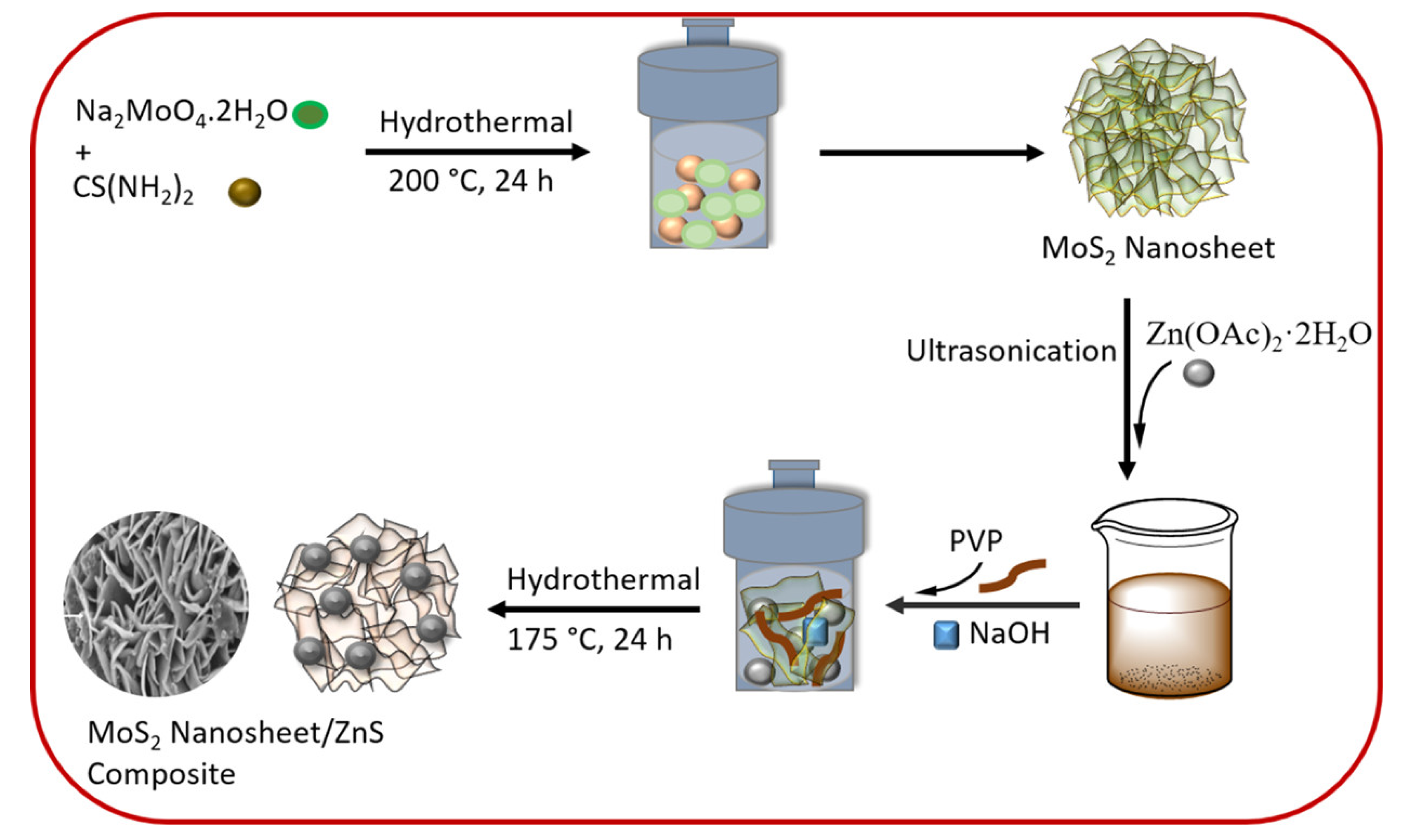
  66. Gusain, R.; Kumar, N.; Opoku, F.; Poomani Govender, P.; Sinha Ray, S. MoS2 Nanosheet/ZnS Composites for the Visible-Light-Assisted Photocatalytic Degradation of Oxytetracycline. ACS Appl. Nano Mater. 2021, 4, 4721–4734. [Google Scholar] [CrossRef]
  67. Xaba, T. Green synthesis of ZnS nanoparticles and fabrication of ZnS-chitosan nanocomposites for the removal of Cr(VI) ion from wastewater. Green Process. Synth. 2021, 10, 374–383. [Google Scholar] [CrossRef]
  68. Asadi, F.; Azizi, S.N.; Chaichi, M.J. Green synthesis of fluorescent PEG-ZnS QDs encapsulated into Co-MOFs as an effective sensor for ultrasensitive detection of copper ions in tap water. Mater. Sci. Eng. C 2019, 105, 110058. [Google Scholar] [CrossRef] [PubMed]
  69. Wang, H.-J.; Yu, X.-H.; Wang, C.-F.; Cao, Y. Cooperative cytotoxic activity of Zn and Cu in bovine serum albumin-conjugated ZnS/CuS nano-composites in PC12 cancer cells. J. Nanopart. Res. 2013, 15, 2020. [Google Scholar] [CrossRef]
  70. Dutta, S.; Chatterjee, S.; Mukherjee, I.; Saha, R.; Singh, B.P. Fabrication of ZnS Hollow Spheres and RGO-ZnS Nanocomposite Using Cysteamine as Novel Sulfur Source: Photocatalytic Performance on Industrial Dyes and Effluent. Ind. Eng. Chem. Res. 2017, 56, 4768–4778. [Google Scholar] [CrossRef]
  71. Jawad, W.; Jader, M. Preparing and Studying the Combination of ZnS/ rGO Nano Composite by Laser Ablation Method. J. Phys. Conf. Ser. 2021, 1999, 012156. [Google Scholar] [CrossRef]
  72. Mahalingam, S.; Neelan, Y.D.; Bakthavatchalam, S.; Al-Humaid, L.A.; Al-Dahmash, N.D.; Santhanam, H.; Yang, T.-Y.; Hossain, N.; Park, S.H.; Kim, J. Effective Visible-Light-Driven Photocatalytic Degradation of Harmful Antibiotics Using Reduced Graphene Oxide-Zinc Sulfide-Copper Sulfide Nanocomposites as a Catalyst. ACS Omega 2023, 8, 32817–32827. [Google Scholar] [CrossRef] [PubMed]
  73. Yu, J.; Zhang, J.; Liu, S. Ion-Exchange Synthesis and Enhanced Visible-Light Photoactivity of CuS/ZnS Nanocomposite Hollow Spheres. J. Phys, Chem. C 2010, 114, 13642–13649. [Google Scholar] [CrossRef]
  74. Kafel, A.; Al-Rashid, S.N.T. Examining the impact of quantum confinement energy on the optical characteristics of zinc sulfide and gallium nitrate in the ultraviolet spectral range. Chalcogenide Lett. 2023, 20, 423–429. [Google Scholar] [CrossRef]
  75. Geng, B.; Ma, J.; Zhan, F. A solution phase thermal decomposition molecule precursors route to ZnS:Cu2+ nanorods and their optical properties. Mater. Chem. Phys. 2009, 113, 534–538. [Google Scholar] [CrossRef]
  76. Mansur, A.A.P.; Mansur, H.S.; Carvalho, S.M.; Lobato, Z.I.P.; de Fátima Leite, M.; Mansur, L.L. Fluorescent ZnS Quantum Dot—Phosphoethanolamine Nanoconjugates for Bioimaging Live Cells in Cancer Research. ACS Omega 2018, 3, 15679–15691. [Google Scholar] [CrossRef]
  77. Debnath, G.H.; Chakraborty, A.; Ghatak, A.; Mandal, M.; Mukherjee, P. Controlled Terbium(III) Luminescence in Zinc Sulfide Nanoparticles: An Assessment of Competitive Photophysical Processes. J. Phys. Chem. C 2015, 119, 24132–24141. [Google Scholar] [CrossRef]
  78. Bose, R.; Thupakula, U.; Bal, J.K.; Pradhan, N. Short-Lived, Intense and Narrow Bluish-Green Emitting Gold Zinc Sulfide Semiconducting Nanocrystals. J. Phys. Chem. C 2012, 116, 16680–16686. [Google Scholar] [CrossRef]
  79. Mukherjee, A.; Bhattacharyya, B.; Rajasekar, G.P.; Narayan, A.; Pandey, A. Optical Properties and Electronic Structure of Copper Zinc Sulfide Nanocrystals. J. Phys. Chem. C. 2021, 125, 17890–17896. [Google Scholar] [CrossRef]
  80. Srivastava, B.B.; Jana, S.; Karan, N.S.; Paria, S.; Jana, N.R.; Sarma, D.D.; Pradhan, N. Highly Luminescent Mn-Doped ZnS Nanocrystals: Gram-Scale Synthesis. J. Phys. Chem. Lett. 2010, 1, 1454–1458. [Google Scholar] [CrossRef]
  81. Talapin, D.V.; Lee, J.-S.; Kovalenko, M.V.; Shevchenko, E.V. Prospects of Colloidal Nanocrystals for Electronic and Optoelectronic Applications. Chem. Rev. 2010, 110, 389–458. [Google Scholar] [CrossRef]
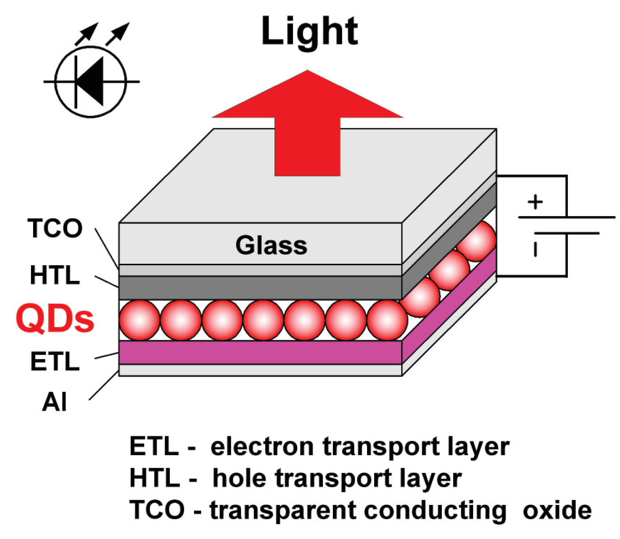
  82. Feng, J.; Jiang, M.; Li, D.; Zhang, Y.; Pei, C.; Zhou, L.; Chen, Z.; Li, Y.; Li, X.; Xu, X. Top-Emission ZnSeTe/ZnSe/ZnS-Based Blue Quantum Dot Light-Emitting Diodes with Enhanced Chroma Efficiency. J. Phys. Chem. Lett. 2023, 14, 2526–2532. [Google Scholar] [CrossRef] [PubMed]
  83. Zhang, X.; Wang, T.; Lin, Q.; Chen, F.; Wang, L.; Du, Z. Highly efficient near-infrared light-emitting diodes based on Zn:CuInSe2/ZnS//ZnS quantum dots with double shell engineering. Opt. Express 2022, 30, 29449–29460. [Google Scholar] [CrossRef] [PubMed]
  84. Schubert, E.F.; Kim, J.K. Solid-state light sources getting smart. Science 2005, 308, 1274–1278. [Google Scholar] [CrossRef]
  85. Hasanirokh, K.; Asgari, A.; Mohammadi, S. Fabrication of a light-emitting device based on the CdS/ZnS spherical quantum dots. J. Eur. Opt. Soc.-Rapid Publ. 2021, 17, 26. [Google Scholar] [CrossRef]
  86. Entradas, T.J.; Cabrita, J.F.; Barrocas, B.; Nunes, M.R.; Silvestre, A.J.; Monteiro, O.C. Synthesis of titanate nanofibers co-sensitized with ZnS and Bi2S3 nanocrystallites and their application on pollutants removal. Mater. Res. Bull. 2015, 72, 20–28. [Google Scholar] [CrossRef]
  87. Naudin, G.; Entradas, T.; Barrocas, B.; Monteiro, O.C. Titanate Nanorods Modified with Nanocrystalline ZnS Particles and Their Photocatalytic Activity on Pollutant Removal. J. Mater. Sci. Technol. 2016, 32, 1122–1128. [Google Scholar] [CrossRef]
  88. Dabbous, A.; Bauer, P.; Marcucci, C.; Périé, S.; Gahlot, S.; Lombard, C.; Caillat, S.; Ravanat, J.-L.; Mouesca, J.-M.; Kodjikian, S.; et al. Hybrid CdSe/ZnS Quantum Dot—Gold Nanoparticle Composites Assembled by Click Chemistry: Toward Affordable and Efficient Redox Photocatalysts Working with Visible Light. ACS Appl. Mater. Interfaces 2023, 15, 56167–56180. [Google Scholar] [CrossRef]
  89. Li, D.; Zhang, H.; Xie, S.; Zhang, H.; Wang, H.; Ma, X.; Gao, D.; Qi, J.; You, F. Lattice Distortion in a Confined Structured ZnS/ZnO Heterojunction for Efficient Photocatalytic CO2 Reduction. Energy Environ. Catal. Appl. 2023, 15, 36324–36333. [Google Scholar] [CrossRef]
  90. Zhang, J.; Wang, Y.; Zhang, J.; Lin, Z.; Huang, F.; Yu, J. Enhanced Photocatalytic Hydrogen Production Activities of Au-Loaded ZnS Flowers. ACS Appl. Mater. Interfaces 2013, 5, 1031–1037. [Google Scholar] [CrossRef]
  91. Lange, T.; Reichenberger, S.; Ristig, S.; Rohe, M.; Strunk, J.; Barcikowski, S.; Schlögl, R. Zinc sulfide for photocatalysis: White angel or black sheep? Prog. Mater. Sci. 2022, 124, 100865. [Google Scholar] [CrossRef]
  92. Proshchenko, V.; Karanovich, A.; Dahnovsky, Y. Surface-Bulk Model for d0 Ferromagnetism in ZnS Quantum Dots and Wires. J. Phys. Chem. C. 2016, 120, 11253–11261. [Google Scholar] [CrossRef]
  93. Xu, J.; Zhang, W.; Fan, H.; Cheng, F.; Su, D.; Wang, G. Promoting lithium polysulfide/sulfide redox kinetics by the catalyzing of zinc sulfide for high performance lithium-sulfur battery. Nano Energy 2018, 51, 73–82. [Google Scholar] [CrossRef]
  94. Liu, R.; Tao, W.; Du, Y.; Wu, C.; Ye, H.; Fan, M.; Chen, S.; Chen, G.; Mao, J.; Xin, S.; et al. ZnS-Nanoparticle-Coated Carbon Cloth as an Efficient Interlayer for High-Performance Li-S Batteries. ACS Appl. Energy Mater. 2022, 5, 12408–12414. [Google Scholar] [CrossRef]
  95. Fan, A.; Hou, T.; Sun, X.; Xie, D.; Li, X.; Zhang, N.; Guo, J.; Jin, S.; Zhou, Y.; Cai, S.; et al. One-Pot Hydrothermal Synthesis of ZnS Nanospheres Anchored on 3D Conductive MWCNTs Networks as High-Rate and Cold-Resistant Anode Materials for Sodium-Ion Batteries. ChemElectroChem 2020, 7, 1904–1913. [Google Scholar] [CrossRef]
  96. Zhao, L.; Yin, J.; Lin, J.; Chen, C.; Chen, L.; Qiu, X.; Alshareef, H.N.; Zhang, W. Highly Stable ZnS Anodes for Sodium-Ion Batteries Enabled by Structure and Electrolyte Engineering. ACS Nano 2024, 18, 3763–3774. [Google Scholar] [CrossRef]
  97. Ummartyotin, S.; Infahsaeng, Y. A comprehensive review on ZnS: From synthesis to an approach on solar cell. Renew. Sustain. Energy Rev. 2016, 55, 17–24. [Google Scholar] [CrossRef]
  98. Liang, G.; Li, Y.; Feng, W.; Wang, X.; Jing, A.; Li, J.; Ma, K. Polyethyleneimine-coated quantum dots for miRNAdelivery and its enhanced suppression in HepG2 cells. Int. J. Nanomed. 2016, 11, 6079–6088. [Google Scholar] [CrossRef] [PubMed]
Figure 1. Common applications of ZnS nanomaterials.
Applnano 05 00010 g001
Figure 2. (a) Low-magnification TEM picture of ZnS-3.5 nanocrystallites. (b) Histogram showing the nanocrystallite size distribution. (c) High-resolution picture of ZnS-3.5 nanocrystallites showing lattice-resolved planes. (d) Magnified single ZnS-3.5 nanocrystallite showing twinning. Reprinted with permission from reference [15]. Copyright [2000] American Chemical Society.
Applnano 05 00010 g002
Figure 3. SEM images of ZnS nanoparticles capped with capping agents; (a) DDA; (b) HAD; (c) ODA. Reprinted with permission from reference [19]. Copyright [2023] American Chemical Society.
Applnano 05 00010 g003aApplnano 05 00010 g003b
Figure 4. SEM images of the as-prepared ZnS nanocrystallites prepared at different pH values; (a) pH = 3; (b) pH = 5; (c) pH = 7; (d) pH = 9. Reprinted from reference [23]. Copyright [2016], with permission from Elsevier.
Applnano 05 00010 g004
Figure 5. (a) XRD diffraction pattern, experimental (dots), fitted (red line), and residuals (blue line). (b) TEM micrograph with (inset, top panel) the size distribution obtained from the segmentation analysis fitted with a log-normal distribution and (inset, bottom panel) superimposition of a 2D projection of a rhombic dodecahedron with a particle of the sample. Reprinted with permission from reference [30]. Copyright [2020] American Chemical Society.
Applnano 05 00010 g005
Figure 6. (a) Potential energy scheme for ZnSe/ZnS core/shell QDs; (b) schematic illustration of the controlled shell growth of ZnS on a spherical ZnSe QD. Stage I, initial ZnS shell growth produces ZnSe/ZnS QDs with high QYs when below a critical thickness. Stage II, ZnSe QDs with thick ZnS shells grown in a kinetic regime display decreased QYs versus maintained high QYs for shell growth in a thermodynamic regime. The thermodynamic growth is realized by using Zn precursor with low reactivity. Dependence of absorption spectra (c) and emission spectra (PL QYs indicated) (d) upon shell thickness (in monolayers, MLs) when using C36H66O4Zn (molar ratio of Zn/OA, 1/10) as shell precursor for thermodynamic growth; (e) TEM image of the 4.0 nm ZnSe core QDs; (f) TEM image of ZnSe/ZnS core/shell QDs with 4 MLs of ZnS shell. Reprinted with permission from reference [43]. Copyright [2020] American Chemical Society.
Applnano 05 00010 g006
Figure 7. Ligand exchange by transferring stearic acid-capped InP/ZnS QDs in hexane to sulfide-capped InP/ZnS QDs in N-methylformamide (NMF). Reprinted with permission from reference [45]. Copyright [2024] American Chemical Society.
Applnano 05 00010 g007
Figure 8. (a,b) TEM images of the as-prepared ZnS nanowires. (c) HRTEM image of ZnS nanowires. The inset in panel (c) is the fast Fourier transformation (FFT) pattern of the single nanowire. (d) The EDS spectrum of the ZnS nanowires: The copper and carbon signals come from the copper grid coated by carbon film, while phosphorus comes from the capping agent on the surface of ZnS nanowires. Reprinted with permission from reference [48]. Copyright [2011] American Chemical Society.
Applnano 05 00010 g008
Figure 9. Representative FESEM images of the ZnS nanoribbons: (a) low-magnification FESEM image of the ZnS nanoribbons showing the product in high yield, (b) high-magnification FESEM image, and (c) high-magnification FESEM image of a representative ZnS nanoribbon with many nanocrystals on the bottom. Reprinted with permission from reference [58]. Copyright [2005] American Chemical Society.
Applnano 05 00010 g009
Figure 10. Synthetic steps for the preparation of MoS2/ZnS nanocomposites. Reprinted with permission from reference [66]. Copyright [2021] American Chemical Society.
Applnano 05 00010 g010
Figure 11. (a) The change in the energy of quantum confinement as a function of nanoparticle size. (b) The variation in the energy gap of semiconductor materials (ZnS) is depicted as a function of the quantum confinement energy. Reproduced with permission from reference [74], copyright © Authors.
Applnano 05 00010 g011
Figure 12. (a) Schematic presentation of the synthesis of bluish-green-emitting gold zinc sulfide nanocrystals from yellow-emitting Au(III)−MPA mixture. (b) Successive evolution of UV−visible and PL spectra during the formation of bluish-green-emitting gold zinc sulfide nanocrystals. Excitation wavelength is 320 nm. Reprinted with permission from reference [78]. Copyright [2012] American Chemical Society.
Applnano 05 00010 g012
Figure 13. Schematic diagram and a typical structure of a thin-film LED utilizing semiconductor NCs. Reprinted with permission from reference [81]. Copyright [2006] American Chemical Society.
Applnano 05 00010 g013
Table 1. Different capping agents for ZnS synthesis. (* NR = not reported).
Zinc
Precursor
Sulfur
Precursor
Capping AgentpHParticle Size/
Crystallite Size (nm)
Method Used to Determine Particle SizeReference Number
Zinc
sulfate
Sodium sulfideCysteine62.4UV-Vis absorption
spectroscopy
[11]
Zinc
sulfate
Sodium sulfideCysteine7.66.08 ± 0.74HRTEM[12]
Zinc
nitrate
Sodium sulfideThioglycolate7.6>200 nmDynamic light scattering[13]
Zinc
perchlorate
Hydrogen sulfide (5%)1-Thioglycerol111.5 ± 0.2XRD[14]
Zinc
acetate
Sodium sulfide1-Thioglycerol81.8HRTEM[15]
Zinc
chloride
Sodium sulfideMercaptoethanol10.21.4HRTEM[16]
Zinc
acetate
dihydrate
Sodium sulfidePVPNR *2.45XRD[17]
Zinc
acetate
dihydrate
Sodium sulfidePVANR *2.60XRD[17]
Zinc
acetate
dihydrate
Sodium sulfide PEG-4000NR *3.25XRD[17]
Zinc
chloride
Sodium sulfidePEG-8000NR *2.8–3.3XRD[18]
Zinc
chloride
Bis(diallydithiocarbamato)HDANR1.98–2.81HRTEM[19]
Zinc
chloride
Bis(diallydithiocarbamato)DDANR2.17–5.49HRTEM[19]
Zinc
chloride
Bis(diallydithiocarbamato)ODANR2.03–3.51HRTEM[19]
Disclaimer/Publisher’s Note: The statements, opinions and data contained in all publications are solely those of the individual author(s) and contributor(s) and not of MDPI and/or the editor(s). MDPI and/or the editor(s) disclaim responsibility for any injury to people or property resulting from any ideas, methods, instructions or products referred to in the content.

