Article

Antihypertensive Amaranth Protein Hydrolysates Encapsulation in Alginate/Pectin Beads: Influence on Bioactive Properties upon In Vitro Digestion

by
Dora Elisa Cruz-Casas
1,
Rodolfo Ramos-González
2,
Lilia Arely Prado-Barragán
3,
Cristóbal N. Aguilar
1,
Raúl Rodríguez-Herrera
1,
Anna Iliná
4,
Sandra Cecilia Esparza-González
5 and
Adriana Carolina Flores-Gallegos
1,*
1
School of Chemistry, Universidad Autónoma de Coahuila, Boulevard Venustiano Carranza e Ing. José Cárdenas Valdés s/n Col. República, Saltillo 25280, Mexico
2
CONAHCYT, Nanobioscience Research Group, School of Chemistry, Universidad Autónoma de Coahuila, Boulevard Venustiano Carranza e Ing. José Cárdenas Valdés s/n Col. República, Saltillo 25280, Mexico
3
Biotechnology Department, Biological, Health Sciences Division, Universidad Autónoma Metropolitana, Iztapalapa Campus, Mexico City 09340, Mexico
4
Nanobioscience Research Group, School of Chemistry, Universidad Autónoma de Coahuila, Boulevard Venustiano Carranza e Ing. José Cárdenas Valdés s/n Col. República, Saltillo 25280, Mexico
5
Odontology Faculty, Universidad Autónoma de Coahuila, Saltillo 25125, Mexico
*
Author to whom correspondence should be addressed.
Polysaccharides 2024, 5(3), 450-462; https://doi.org/10.3390/polysaccharides5030028
Submission received: 20 July 2024 / Revised: 29 August 2024 / Accepted: 30 August 2024 / Published: 5 September 2024

Abstract

:
Protein hydrolysates containing bioactive peptides have emerged as therapeutic agents. However, these peptides may lose this bioactivity under gastrointestinal conditions. Encapsulation in edible biopolymers is a solution to this problem. Protein hydrolysates with ACE-I inhibitory activity, obtained previously, were encapsulated. A 1% solution of the biopolymers alginate (AG) and pectin (PC) in various ratios was prepared. The beads formed were evaluated in both wet and dry states for size, roundness, thermal gravimetric analysis (TGA), encapsulation efficiency, and ACE-I inhibitory activity. Selected samples underwent in vitro digestion, after which peptide release and ACE-I inhibitory activity were determined. Size analysis revealed that increasing the PC content increased the bead size, with 100% PC beads showing total deformation and reduced roundness. TGA indicated that wet beads had lower thermal stability compared to dry beads. The highest encapsulation efficiency (95.57% ± 0.49) was observed with 100% AG beads. The 75% AG 25% PC beads exhibited the highest ACE-I inhibitory activity (97.97% ± 1.01). Encapsulated protein hydrolysates retained their ACE-I inhibitory activity after simulated digestion, whereas non-encapsulated hydrolysates lost their bioactivity. Encapsulation of amaranth protein hydrolysates with AG and PC thus preserves antihypertensive activity even after in vitro digestion.

1. Introduction

Amaranth, a pseudocereal belonging to the Amaranthaceae family, is notable for its high nutritional value, containing significant amounts of dietary fiber, lipids, and protein [1,2]. It also stores essential vitamins (vitamin B6, C, carotene, and folate) and contains high levels of calcium, sodium, iron, zinc, and magnesium. It is gluten-free, making it safe for individuals with celiac disease [3,4]. The FAO considers amaranth one of the most promising crops for feeding the global population [5]. In recent years, its high protein content (13–19%), high protein quality (90% digestibility), and balanced amino acid profile have been of great interest [6]. Due to its nutritional composition, it has been identified as a potential source of protein hydrolysates [7].
Protein hydrolysates are a mixture of oligopeptides, peptides, and free amino acids [8]. Peptides, sequences of 2 to 20 amino acids, stand out due to their significant health benefits, such as antihypertensive activity [9]. Small peptides (<3 kDa) with aromatic rings or cycles and aliphatic chains exhibit the greatest antihypertensive effects by inhibiting the angiotensin I-converting enzyme (ACE-I) [10,11]. Inhibition of this enzyme is crucial because it catalyzes the conversion of angiotensin I to angiotensin II, leading to increased vasoconstriction and inflammation, and consequently contributing to elevated blood pressure [11].
The composition of protein hydrolysates, as with the mixture of peptides that compose them, is influenced by several factors; one of these is the method used for hydrolysis [12]. The most common methods are enzymatic hydrolysis and microbial fermentation; both have several advantages, including their Generally Recognized As Safe (GRAS) character [13]. In recent years, microbial fermentation of amaranth has gained attention for producing protein hydrolysates with high percentages of ACE-I inhibition [7,14]. This method is favored for its diverse microbial proteases, high protease activity, and cost-effectiveness compared to enzymatic hydrolysis [15,16].
However, protein hydrolysates may lose their bioactivity upon consumption due to extreme pH conditions and the digestive enzyme complex in the gastrointestinal tract [17,18]. Additionally, they have an undesirable or bitter taste, low solubility, poor stability during processing, and poor oral bioavailability [19]. One strategy to protect these molecules and improve some of their characteristics is encapsulation.
Encapsulation is the process used to trap a substance within another substance, known as an encapsulant, coating, or wall material [20]. This process protects molecules such as protein hydrolysates from adverse environmental or gastrointestinal conditions and improves their stability [21]. Encapsulation of protein hydrolysates using natural biopolymers employs biodegradable, biocompatible, and less toxic materials [22]. Pectin and alginate are biopolymeric polysaccharides that are considered GRAS. Alginate is widely used to protect protein hydrolysates, while pectin has shown favorable results in the controlled release of various bioactive ingredients [23,24]. Combining or using these two polysaccharides individually can improve the unfavorable characteristics of protein hydrolysates that inhibit ACE-I.
The aim of this study was to develop alginate/pectin beads to encapsulate amaranth protein hydrolysates with ACE-I inhibitory activity and evaluate their stability in retaining this bioactivity even after in vitro digestion.

2. Materials and Methods

2.1. Materials

Sodium alginate (AG) was purchased from Cape Crystal Brands (Summit, NJ, USA), and low methoxyl amidated pectin (PC) was purchased from Modernist Pantry (Portsmouth, NH, USA). Alpha-amylase (M2871, 165,000 BAU/g) was purchased from MP Biomedicals (Santa Ana, CA, USA). Pepsin from porcine gastric mucosa (P-7012, ≥2500 units/mg) and pancreatin from porcine pancreas (P-1750) were obtained from Sigma-Aldrich (St. Louis, MO, USA). Enterococcus faecium-LR9 was donated by the Food Research Department of the Universidad Autónoma de Coahuila.

2.2. Encapsulation by Ionic Gelation

Amaranth protein hydrolysates were obtained from the fermentation of amaranth protein with Enterococcus faecium-LR9, following previous research conducted by the working group. The fermentation process was conducted at 37 °C for 24 h with a cell density of 7.0 log CFU/mL, according to the conditions reported by Ayala-Niño et al. [25]. The samples were then centrifuged at 6000× g for 15 min at 4 °C and subsequently freeze-dried.
The AG-PC protein hydrolysates were encapsulated using the ionic gelation method described by Pamunuwa et al. [26] with some modifications. Different mixtures of AG-PC were made at different concentrations: 100% AG, 75% AG 25% PC, 50% AG 50% PC, 25% AG 75% PC, and 100% PC. An aqueous solution of the polymers (1% w/v) was prepared and homogenized by stirring. Subsequently, the lyophilized protein hydrolysate (1% w/v) was added, and this solution was stirred again for 30 min at 1500 rpm. The polymers and protein hydrolysates solution were dripped using a 22G-gauge syringe, 10 cm high, over 50 mL of 0.1 M CaCl2. The formed beads were washed three times with distilled water.
The effect of bead drying was evaluated by comparing wet (undried) beads with beads dried using two different techniques: air-drying and freeze-drying. For the air-drying process, the beads were dried in a forced air circulation oven at 37 °C until a constant weight was achieved. The freeze-drying process was conducted using a freeze-dryer (Ecoshel, ECO-FD10PT, PHARR, TX, USA) at −60 °C and a minimum pressure of 2 Pa for 24 h.

2.3. Particle Size and Roundness Percentage

Particle size and percent roundness were evaluated as described by Zhao et al. [27] with some modifications. For particle size, the longest (Dmax) and shortest (Dmin) diameters of 100 beads were measured randomly with a digital vernier caliper (0–150 mm, accuracy 0.02 mm, Shanghai, Beijing), and the average of the values obtained was calculated. The percent roundness (% RD) was calculated according to the following equation:
%   R D = D m i n D m a x × 100
where Dmin is the minimum diameter and Dmax is the maximum diameter of the beads.

2.4. Encapsulation Efficiency

Encapsulation efficiency was determined as the concentration of peptides trapped in the core or surface compared to the initial amount added [28]. The o-phthaldialdehyde (OPA) method of Church et al. [29], with some modifications by Muhialdin et al. [30], was used to calculate the concentration of peptides. Briefly, to prepare the OPA solution, 25 mL of 100 mmol/L sodium tetraborate, 2.5 mL of 20% (wt/wt) SDS, 40 mg of OPA (dissolved in 1 mL of methanol), and 100 μL of β-mercaptoethanol were mixed and diluted to a final volume of 50 mL with deionized water. To a 96-well microplate, 36 μL of the sample and 270 μL of OPA reagent were added. A standard curve with tryptone (0.25 to 1.5 mg/mL) was used as a reference. The solution was incubated in the dark for 3 min at room temperature, and the absorbance was measured at 340 nm (BioTek, EPOCH2, Bad Friedrichshall, Germany).
The following equation was used to calculate the percentage encapsulation efficiency (% EE) in the wet gel beads:
%   E E = P I P S b P I × 100
where PI is the initial peptide concentration and PSb is the peptide concentration found in the supernatant.
To calculate the % EE of the dried beads, they were dissolved in 0.1 M sodium citrate, and the following equation was used:
%   E E = P f P I × 100
where Pf is the peptide concentration in the dissolved bead sample and PI is the initial peptide concentration.

2.5. Thermogravimetric Analysis

The beads’ thermal stability was evaluated via thermogravimetric analysis under controlled N2 atmospheres using Pyris 1 TGA (PerkinElmer, Norwalk, CT, USA). The samples were subjected to a temperature range of 30–800 °C, with a heating rate of 30 °C/min, and the mass loss was recorded.

2.6. Inhibitory Activity of ACE-I In Vitro

The ACE-I inhibitory activity of the selected beads was determined by dissolving them in 0.1 M sodium citrate. A fluorescent method was used with the ACE-I activity assay kit (Catalog No. CS0002, Sigma-Aldrich® (St. Louis, MO, USA)) according to the protocol described by the manufacturer. All reagents were diluted in the assay buffer according to the manufacturer’s instructions. Ten µL of the assay buffer (control) or samples are mixed with 40 µL of ACE. Then, 50 µL of the fluorogenic substrate, previously heated to 37 °C, was added to the experimental samples, controls, and blank [31]. A standard curve between 0 and 8 nmol was also prepared to calculate the enzymatic activity. ACE inhibitory activity was measured using a fluorescence plate reader, with excitation and emission wavelengths of 320 and 405 nm, respectively. The % inhibition of ACE was calculated using the equation:
%   A C E = 100 × ( m U C m U B ) ( m U S m U B s ) ( m U C m U B )
where mUC (control) is mU after the action of ACE on substrate in the absence of inhibitor; mUS (sample) is mU after the action of ACE on substrate in the presence of inhibitory sample; mUB (blank) is mU of substrate; and mUBs (blank sample) is mU of substrate and sample.

2.7. Simulated Gastrointestinal Digestion of Encapsulated Protein Hydrolysates

The selected beads and non-encapsulated protein hydrolysate (at the same concentration as the encapsulated protein hydrolysate) were subjected to in vitro digestion according to the standardized INFOGEST protocol [32]. The digestion consisted of three simulated phases: oral, gastric, and intestinal.
In the oral phase, 1 g of the beads were weighed, and 1 mL of salivary liquid solution (SSF), salivary alpha-amylase enzyme at a concentration of 75 U/mL, and 1.5 mmol/L CaCl2 × 2 H2O were added. The samples were placed in a water bath at 37 °C for 2 min with constant agitation at 120 rpm. Porcine pepsin was added to the samples at a final activity of 2000 U/mL, in addition to a solution of gastric fluid (SGF) and CaCl2 × 2 H2O 0.15 mmol/L to continue the simulation of the gastric phase. The pH was adjusted to 3 with 1M HCl. Samples were incubated at 37 °C for 2 h with constant agitation at 120 rpm. In the intestinal phase, a solution of intestinal fluid (SIF), porcine pancreatin at a final activity of 100 U/mL, bovine bile 10 mmol/L, and CaCl2(H2O)2 at 0.6 mmol/L were added. The pH was adjusted to 7 with NaOH 1M. Samples were incubated at 37 °C for 2 h with constant agitation at 120 rpm. Samples were taken at the end of each in vitro digestion step. The enzymes were inactivated by heating at 90 °C for 5 min. All digested products of each stage were centrifuged at 500× g for 15 min. The supernatants were taken and centrifuged at 10,000× g for 5 min.
Each digest was evaluated for peptide release using the OPA method and ACE-I inhibitory activity.

2.8. Statistical Analysis

Data were subjected to one-way ANOVA; when needed, pair-comparison of treatment means was achieved by Tukey’s procedure at p ˂ 0.05, using the statistical software Statistica (Statistica7.0 per Windows).

3. Results and Discussion

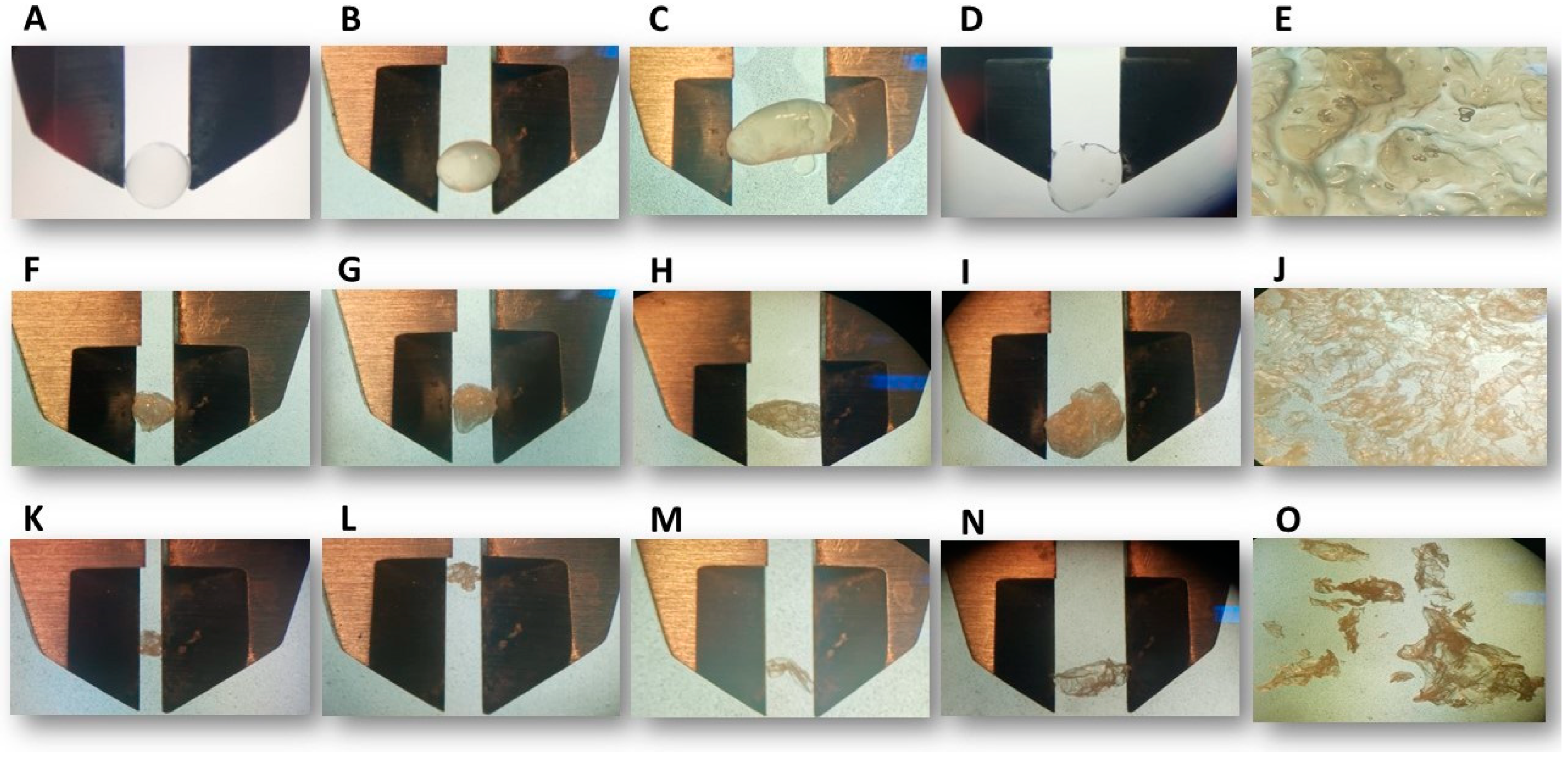
3.1. Particle Size and Roundness Percentage

The particle size and % RD of the evaluated beads are presented in Table 1. The graphical visualization of the evaluated beads is shown in Figure 1. In all the beads, the addition of PC influenced the bead size. Higher PC concentrations resulted in larger bead sizes. The beads made with 100% PC did not have a defined shape, making their size unmeasurable (Figure 1E,J,O). This is because the galacturonic acid in PC is randomly distributed, and when it binds to Ca2+ ions, it forms a structure resembling an egg-box in “dot mode”. This results in faster, non-uniform gelation, producing larger beads with larger pores compared to those with higher AG concentrations. In contrast, the guluronic and mannuronic acid in AG are distributed in a more structured manner, forming an egg-box structure in “zipper mode” when bound to Ca2+ ions, leading to smaller bead sizes [26]. This behavior is similar to that described by Pamunuwa et al. [26], where the size of folic acid beads increased with higher PC concentrations.
The size also varied between wet and dry beads, where air-drying beads (Figure 1K–O) presented a smaller size than freeze-dried (Figure 1F–J) and wet beads (Figure 1A–E). The size difference between dried and wet beads is due to water loss, which weakens the gelatinous structure, causing shrinkage and collapse of the encapsulate surface [33]. These results are similar to those reported by Stachowiak et al. [34], who produced PC-AG polymer encapsulates and dried them by freeze-drying. They also correspond with the findings of Santagapita et al. [35], who evaluated the effect of different drying methods (freeze-drying, vacuum drying, and air-drying) on the encapsulation of invertase with AG.
According to Table 1, the wet beads with 100% AG had the highest % RD. When more PC is added to the beads, the % RD decreases, as shown in Figure 1. This occurs because PC forms irregular and large pores, which affect the size and shape of the beads. Drying also affected the % RD, with wet beads showing better % RD than dry ones (Figure 1). However, the freeze-drying process better preserved the roundness of the beads compared to air-drying. During freeze-drying, the beads were frozen, and water was removed by sublimation, which minimized shrinkage and collapse. In contrast, air-drying subjected the beads to temperature and oxygen, leading to more severe shrinkage and collapse.

3.2. Encapsulation Efficiency (EE)

Results for the evaluated % EE of amaranth protein hydrolysate in beads are displayed in Figure 2. The highest % EE (95.57% ± 0.49) was observed with the 100% AG wet beads. This value is higher than that reported by Alvarado et al. [36], who found a 70% EE of whey protein peptides in AG beads. The differences may be attributed to the different protein sources from which the peptides come, and the varying parameters used for encapsulation and % EE determination.
The concentration of PC in the encapsulation formulation impacts % EE. Specifically, higher concentrations of PC lead to lower % EE, likely due to the properties of the materials used, such as their entrapment and binding capacity [36]. AG is composed of guluronic acid and mannuronic acid arranged in a clockwise manner. The egg-box structure in “zipper mode” produces a three-dimensional network with greater stability, resulting in better entrapment [26]. In contrast, PC forms smaller egg-box dots, leading to lower mechanical stability compared to AG. Therefore, the % EE was lower [37]. However, forced air-dried beads with 25% AG 75% PC demonstrated high % EE despite the high PC concentration. This may be attributed to interactions between the protein hydrolysates and the specific concentrations of AG and PC, where PC was more available to interact with the protein hydrolysate. Additionally, the effect of forced air-drying at this temperature may have enhanced these interactions, resulting in a higher % EE compared to the wet and freeze-dried beads.
The drying process negatively affects the % EE of the beads. This can be caused by the formation of crystals during freeze-drying, which can damage the bioactive compound, as noted by Vivek et al. [38]. On the other hand, the presence of oxygen and temperature during forced air drying can also lead to changes in % EE compared to wet beads. Additionally, the drying process can increase porosity, leading to the release of protein hydrolysates [34]. Between the two drying methods, forced air-drying results in a greater decrease in % EE compared to freeze-drying.

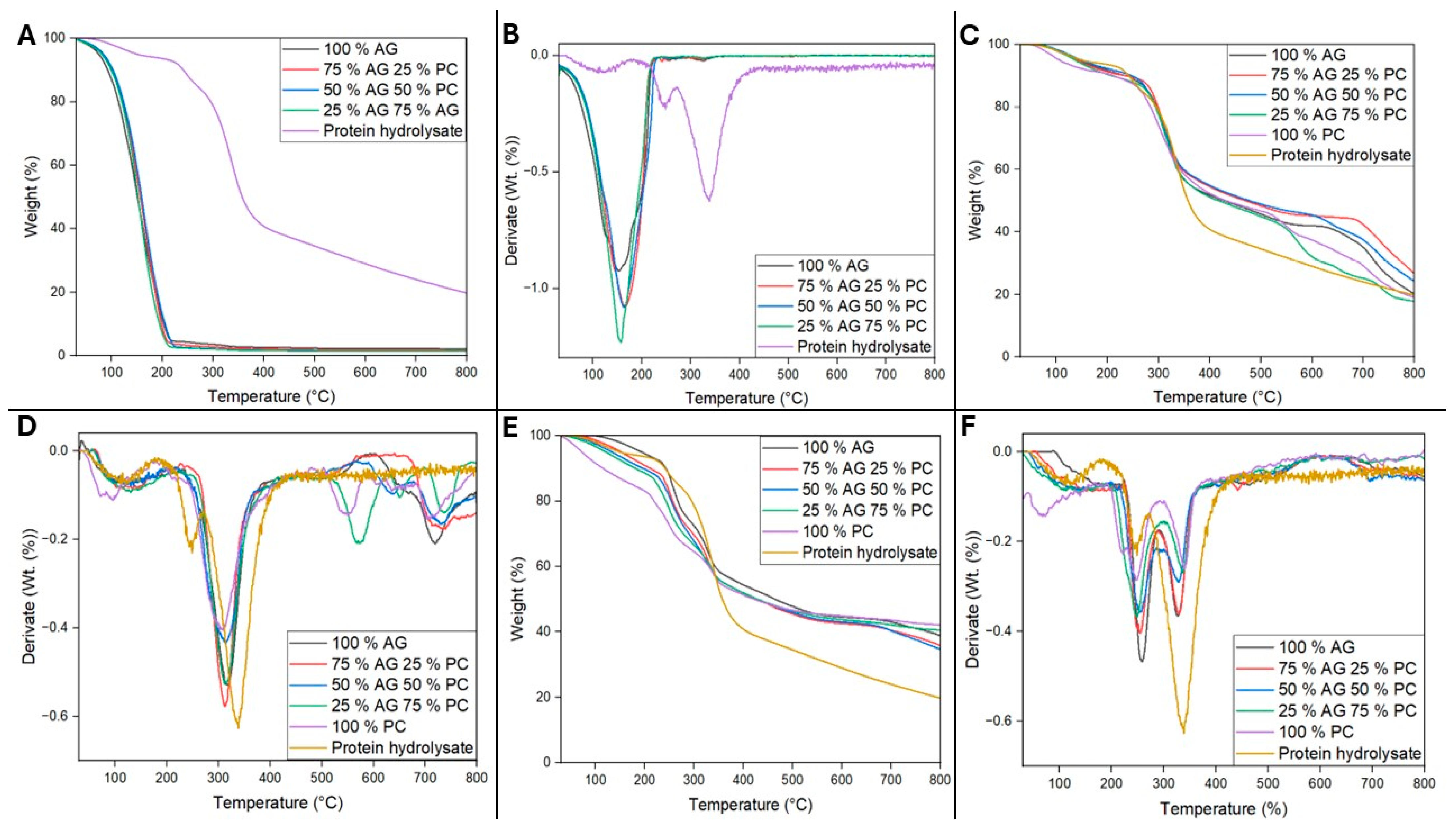
3.3. Thermogravimetric Analysis

TGA was performed, and DTG curves were calculated to evaluate the thermal stability of the non-encapsulated amaranth protein hydrolysate and the prepared beads. The results are presented in Figure 3. The protein hydrolysate exhibited three stages of weight loss. The first stage, occurring between 44 and 181 °C, showed a 6.1% weight loss, which is attributed to the release of free water or water weakly bound to the protein molecules [39]. In the second stage, the non-encapsulated amaranth protein hydrolysate lost 11.14% of its weight between 219 and 270 °C, likely due to the volatilization of peptides [40]. The most significant weight loss, 44.11%, was observed between 270 and 415 °C, which can be attributed to the slow decomposition of peptides from the previous stage, as complex decomposition reactions occur with increasing temperature [39].
In contrast, the wet beads exhibited a 98.48% weight loss between 50 and 226 °C (Figure 3A,B), primarily due to the evaporation of their high water content. Similar findings have been reported by Paswan et al. [41], where the bioactive compound demonstrated higher thermal stability without encapsulation. This may be due to the addition of AG and PC, which have lower decomposition temperatures, and the high water content of the beads.
Although the wet beads displayed lower thermal stability compared to the non-encapsulated amaranth protein hydrolysate, it is essential to consider the other advantages of the encapsulation strategy. Additionally, the weight loss of the wet beads occurs at temperatures higher than those typically encountered during storage and some types of cooking.
Figure 3C,D show the TGA and DTG results for the freeze-dried beads. The highest weight loss (30.58%) occurred between 236 and 370 °C, indicating that the beads no longer contain water. This temperature range is associated with the decomposition of AG carbon chains and the formation of sodium carbonate [42]. For PC, the galacturonic acid chains undergo thermal degradation, leading to the formation of solid carbon [43]. Subsequently, as observed in the DTG curves (Figure 3D), additional weight-loss events were observed at higher temperatures. Beads with a higher percentage of AG showed weight losses of 14.79% for 100% AG beads and 15.43% for 75% AG 25% PC beads between 667–765 °C, attributed to the decomposition of sodium carbonate [42]. The 50% AG and 50% PC beads had two additional weight loss events between 589–676 °C and between 682–760 °C. The 25% AG 75% PC beads had weight losses between 524–616 °C and 707–767. On the other hand, the 100% PC beads showed weight losses between 504–581 °C and 685–738 °C, which is attributed to the partial destruction of solid carbon [43]. The freeze-dried beads exhibit slightly lower thermal stability compared to the non-encapsulated amaranth protein hydrolysate, as they do not contain the high water content present in the wet beads.
TGA and DTG results for air-dried beads are shown in Figure 3E,F. Two distinct weight loss events were observed at 220–287 °C and 294–350 °C, corresponding to the issues described above. The air-dried beads exhibit slightly lower thermal stability compared to the non-encapsulated amaranth protein hydrolysate, as they do not contain the high water content present in the wet beads.

3.4. ACE-I Inhibitory Activity

Figure 4 presents the ACE-I inhibitory activity of the selected beads (wet beads), which were chosen because they showed better results in size, % RD, and % EE. The amaranth protein hydrolysates maintained their ACE-I inhibitory activity after encapsulation using the ionic gelation technique. This technique is effective in protecting bioactive compounds from temperature/pH variations as well as light/oxygen exposure, thereby retaining their bioactivity [44].
Although there was no significant difference among the treatments, the 75% AG 25% PC beads had a greater tendency towards a higher percentage of inhibition of ACE-I (96.97% ± 1.01). This could be due to synergistic effects between AG and PC concentrations, which could help maintain the appropriate amino acid sequences with the right structure, charge, size, or composition necessary for this bioactivity.
While the 75% AG 25% PC beads had a high % EE, the 100% AG beads had the highest % EE. However, the latter may not demonstrate a high trend in ACE-I inhibition percentage, as % EE is based on the concentration of encapsulated peptides. It is possible that some encapsulated peptides do not possess the essential characteristics required to inhibit ACE-I.
The 100% AG beads and the 75% AG 25% PC beads showed the most favorable results in terms of characterization, % EE, and a high tendency towards ACE-I inhibition. Therefore, these beads were selected to evaluate the stability of bioavailability after undergoing in vitro digestion.

3.5. Simulated Gastrointestinal Digestion of Encapsulated Protein Hydrolysates

Peptide release (monitored as protein concentration) and ACE-I inhibitory activity were determined during oral, gastric, and intestinal phases simulation for the selected samples listed in Table 2.
In all tested samples, the highest release of peptides occurred during the intestinal phase, which is favorable as peptides need to reach the intestine to be absorbed. On the other hand, the lowest peptide release was observed during the oral phase. Additionally, the amaranth protein hydrolysate without encapsulation showed a higher tendency to release peptides compared to the encapsulated samples. This is undesirable, as peptide release is ideally targeted to occur during the intestinal phase. This is attributed to the encapsulating material protecting the samples during different phases of digestion, thus preventing a higher release of peptides. During the oral phase, both AG and PC protect the protein hydrolysates because amylase does not target these biopolymers. Additionally, the near-neutral pH (~6.8–7.2) of the oral environment prevents significant deprotonation of the carboxyl groups in AG, and PC remains stable at this pH. In the gastric phase, the acidic environment (pH 3) activates pepsin, which specifically targets proteins and does not degrade AG or PC. The acidic pH does not substantially affect the stability of these biopolymers. In the intestinal phase, the pH increases to a more neutral level (pH 7), leading to the deprotonation of carboxyl groups in AG. While PC is generally more resistant to changes in pH, it forms less stable structures compared to AG, potentially affecting its integrity [45,46]. These findings are similar to those reported by Alvarado et al. [36].
The non-encapsulated amaranth protein hydrolysate did not exhibit ACE-I inhibitory activity during any stage of the digestion process (Table 2). This lack of activity is attributed to the production of inactive peptides under the conditions and enzymes present during the in vitro digestion phases. Previous studies have also reported the instability and rapid degradation of non-encapsulated protein hydrolysates, leading to a loss of biological activity during digestion [36,47].
During the oral phase, the 100% AG beads demonstrated a low percentage of ACE-I inhibition (9.97% ± 11.33). However, this bioactivity was not observed in the gastric phase (0% ± 0), likely due to the production of inactive peptides. In the intestinal phase, bioactivity reappeared (19.93% ± 6.30), probably due to the favorable conditions and enzymes present in this phase, which resulted in the production of peptides with the necessary characteristics to inhibit ACE-I. Additionally, more peptides were released from the encapsulation during this phase, contributing to the observed bioactivity. Despite this, the percentage of ACE-I inhibition in the intestinal phase remained low (19.93% ± 6.30).
In the case of beads composed of 75% AG and 25% PC, as mentioned earlier, a low release of peptides was observed during the oral phase (0.007 ± 0.003), which is advantageous as the goal is to release these peptides primarily in the intestinal phase. Notably, these beads exhibited a higher tendency to inhibit ACE-I during the oral phase (23.68 ± 10.05) compared to the gastric (17.46 ± 5.03) and intestinal 16.10 ± 8.77 phases. This could be attributed to the modification of the released peptides by the amylase enzyme, which may have imparted structural characteristics conducive to a higher level of ACE-I inhibition. However, this bioactivity decreased during the gastric and intestinal phases, where more peptides were released. The increased hydrolysis and pH changes in these later phases might have contributed to the production of inactive peptides.
Differences in ACE-I inhibitory activity were observed between the various phases of the digestion process for the 100% AG beads and the 75% AG 25% PC beads. These differences are likely attributable to the varying concentrations of AG and the addition of PC, as also reflected in the % EE, as these parameters had a significant influence. The bioactivity observed in each digestion phase may result from the release of peptides with different structures in each bead (100% AG and 75% AG 25% PC). Depending on the structure of the peptides, complexes may or may not form with each bead, influenced by the differences in the concentrations of the biopolymer (AG) and the addition of another biopolymer (PC). This could explain why, during the intestinal phase, the 100% AG beads favor the production of ACE-I inhibitory peptides, while the 75% AG 25% PC beads tend to produce inactive peptides.
When comparing amaranth protein hydrolysates with and without encapsulation, it was found that beads encapsulated with AG and PC preserved bioactivity during in vitro digestion, but only to a limited extent. The study showed that the 100% AG beads and 75% AG 25% PC beads contained 87.33% ± 4.22 and 96.97% ± 1.01 ACE-I inhibition, respectively, before in vitro digestion. However, the formation of complexes between these biopolymers and the protein hydrolysates may prevent the release of the ACE-I inhibitory peptides, resulting in low inhibition percentages.

4. Conclusions

A study was conducted to encapsulate amaranth protein hydrolysates using the ionic gelation method with different concentrations of AG and PC. The findings indicated that beads with higher PC concentration were unsuitable due to their larger size, being less round, and having lower encapsulation efficiency. Additionally, drying the beads negatively impacted encapsulation efficiency. The best results during characterization were obtained with 100% wet AG beads. However, the better preservation of ACE-I inhibitory activity was observed with beads containing 75% AG 25% PC. Consequently, beads composed of 100% AG and 75% AG 25% PC were selected for evaluation in a simulated gastrointestinal digestion. The encapsulation of amaranth protein hydrolysates using these selected concentrations of AG and PC preserved ACE inhibitory activity, whereas non-encapsulated protein hydrolysates lost bioactivity during gastrointestinal digestion simulation. However, the ACE-I inhibitory activity observed during the intestinal phase was very low, indicating that other biopolymers and concentrations need to be evaluated to enhance protein hydrolysate stability.

Author Contributions

Conceptualization, D.E.C.-C. and A.C.F.-G.; methodology, D.E.C.-C., R.R.-H. and A.C.F.-G.; software, R.R.-G.; validation, A.I. and A.C.F.-G.; formal analysis, D.E.C.-C.; investigation D.E.C.-C. and A.C.F.-G.; resources, R.R.-H. and A.C.F.-G.; writing—original draft preparation, D.E.C.-C.; writing—review and editing, R.R.-G., C.N.A., L.A.P.-B., A.I., S.C.E.-G. and A.C.F.-G.; visualization, D.E.C.-C. and A.C.F.-G.; supervision, A.C.F.-G.; project administration, A.C.F.-G.; funding acquisition, A.C.F.-G. All authors have read and agreed to the published version of the manuscript.

Funding

D.E.C.-C. received a postgraduate scholarship grant from the National Council of Humanities, Sciences, and Technologies (CONAHCYT) of Mexico, grant number 9994499.

Institutional Review Board Statement

Not applicable.

Data Availability Statement

The raw data supporting the conclusions of this article will be made available by the authors on request.

Acknowledgments

The authors thank the National Council of Humanities, Sciences, and Technologies (CONAHCYT) of Mexico and the Department of Food Research of the Universidad Autónoma de Coahuila.

Conflicts of Interest

The authors declare no conflicts of interest.

References

  1. Bekkering, C.S.; Tian, L. Thinking Outside of the Cereal Box: Breeding Underutilized (Pseudo)Cereals for Improved Human Nutrition. Front. Genet. 2019, 10, 1289. [Google Scholar] [CrossRef] [PubMed]
  2. Taniya, M.S.; Reshma, M.V.; Shanimol, P.S.; Krishnan, G.; Priya, S. Bioactive Peptides from Amaranth Seed Protein Hydrolysates Induced Apoptosis and Antimigratory Effects in Breast Cancer Cells. Food Biosci. 2020, 35, 100588. [Google Scholar] [CrossRef]
  3. Mekonnen, G.; Woldesenbet, M.; Teshale, T.; Biru, T. Amaranthus Caudatus Production and Nutrition Contents for Food Security and Healthy Living in Menit Shasha, Menit Goldya and Maji Districts of Bench Maji Zone, South Western Ethiopia. Nutr. Food Sci. Int. J. 2018, 7, 1–7. [Google Scholar] [CrossRef]
  4. Graziano, S.; Marando, S.; Prandi, B.; Boukid, F.; Marmiroli, N.; Francia, E.; Pecchioni, N.; Sforza, S.; Visioli, G.; Gullì, M. Technological Quality and Nutritional Value of Two Durum Wheat Varieties Depend on Both Genetic and Environmental Factors. J. Agric. Food Chem. 2019, 67, 2384–2395. [Google Scholar] [CrossRef] [PubMed]
  5. Jan, N.; Hussain, S.Z.; Naseer, B.; Bhat, T.A. Amaranth and Quinoa as Potential Nutraceuticals: A Review of Anti-Nutritional Factors, Health Benefits and Their Applications in Food, Medicinal and Cosmetic Sectors. Food Chem. X 2023, 18, 100687. [Google Scholar] [CrossRef]
  6. Kurek, M.A.; Karp, S.; Wyrwisz, J.; Niu, Y. Physicochemical Properties of Dietary Fibers Extracted from Gluten-Free Sources: Quinoa (Chenopodium quinoa), Amaranth (Amaranthus caudatus) and Millet (Panicum miliaceum). Food Hydrocoll. 2018, 85, 321–330. [Google Scholar] [CrossRef]
  7. Cruz-Casas, D.E.; Aguilar, C.N.; Ascacio-Valdés, J.A.; Rodríguez-Herrera, R.; Chávez-González, M.L.; Flores-Gallegos, A.C. Bioactive Protein Hydrolysates Obtained from Amaranth by Fermentation with Lactic Acid Bacteria and Bacillus Species. Heliyon 2023, 9, e13491. [Google Scholar] [CrossRef]
  8. Garbacz, K.; Wawrzykowski, J.; Czelej, M.; Czernecki, T.; Waśko, A. Recent Trends in the Application of Oilseed-Derived Protein Hydrolysates as Functional Foods. Foods 2023, 12, 3861. [Google Scholar] [CrossRef]
  9. Nikhita, R.; Sachindra, N.M. Optimization of Chemical and Enzymatic Hydrolysis for Production of Chicken Blood Protein Hydrolysate Rich in Angiotensin-I Converting Enzyme Inhibitory and Antioxidant Activity. Poult. Sci. 2021, 100, 101047. [Google Scholar] [CrossRef]
  10. Samaei, S.P.; Martini, S.; Tagliazucchi, D.; Gianotti, A.; Babini, E. Antioxidant and Angiotensin I-Converting Enzyme (ACE) Inhibitory Peptides Obtained from Alcalase Protein Hydrolysate Fractions of Hemp (Cannabis sativa L.) Bran. J. Agric. Food Chem. 2021, 69, 9220–9228. [Google Scholar] [CrossRef]
  11. Cruz-Chamorro, I.; Santos-Sánchez, G.; Álvarez-López, A.I.; Pedroche, J.; Lardone, P.J.; Arnoldi, A.; Lammi, C.; Carrillo-Vico, A. Pleiotropic Biological Effects of Lupinus Spp. Protein Hydrolysates. Trends Food Sci. Technol. 2023, 133, 244–266. [Google Scholar] [CrossRef]
  12. Heffernan, S.; Giblin, L.; O’Brien, N. Assessment of the Biological Activity of Fish Muscle Protein Hydrolysates Using In Vitro Model Systems. Food Chem. 2021, 359, 129852. [Google Scholar] [CrossRef] [PubMed]
  13. He, Y.; Pan, X.; Chi, C.F.; Sun, K.L.; Wang, B. Ten New Pentapeptides from Protein Hydrolysate of Miiuy Croaker (Miichthys miiuy) Muscle: Preparation, Identification, and Antioxidant Activity Evaluation. LWT-Food Sci. Technol. 2019, 105, 1–8. [Google Scholar] [CrossRef]
  14. Sánchez-López, F.; Robles-Olvera, V.J.; Hidalgo-Morales, M.; Tsopmo, A. Angiotensin-I Converting Enzyme Inhibitory Activity of Amaranthus hypochondriacus Seed Protein Hydrolysates Produced with Lactic Bacteria and Their Peptidomic Profiles. Food Chem. 2021, 363, 130320. [Google Scholar] [CrossRef]
  15. Tamang, J.P.; Cotter, P.D.; Endo, A.; Han, N.S.; Kort, R.; Liu, S.Q.; Mayo, B.; Westerik, N.; Hutkins, R. Fermented Foods in a Global Age: East Meets West. Compr. Rev. Food Sci. Food Saf. 2020, 19, 184–217. [Google Scholar] [CrossRef]
  16. Ozyurt, G.; Boga, M.; Uçar, Y.; Boga, E.K.; Polat, A. Chemical, Bioactive Properties and in Vitro Digestibility of Spray-Dried Fish Silages: Comparison of Two Discard Fish (Equulites klunzingeri and Carassius gibelio) Silages. Aquac. Nutr. 2018, 24, 998–1005. [Google Scholar] [CrossRef]
  17. Bao, C.; Jiang, P.; Chai, J.; Jiang, Y.; Li, D.; Bao, W.; Liu, B.; Liu, B.; Norde, W.; Li, Y. The Delivery of Sensitive Food Bioactive Ingredients: Absorption Mechanisms, Influencing Factors, Encapsulation Techniques and Evaluation Models. Food Res. Int. 2019, 120, 130–140. [Google Scholar] [CrossRef] [PubMed]
  18. Han, Y.; Gao, Z.; Chen, L.; Kang, L.; Huang, W.; Jin, M.; Wang, Q.; Bae, Y.H. Multifunctional Oral Delivery Systems for Enhanced Bioavailability of Therapeutic Peptides/Proteins. Acta Pharm. Sin. B 2019, 9, 902–922. [Google Scholar] [CrossRef]
  19. Devaraju, R.; Pushpadass, H.A.; Emerald, F.M.E.; Padaki, N.V.; Nath, B.S. Nanoencapsulation of Casein-Derived Peptides within Electrospun Nanofibres. J. Sci. Food Agric. 2021, 102, 1684–1698. [Google Scholar] [CrossRef]
  20. Yun, P.; Devahastin, S.; Chiewchan, N. Microstructures of Encapsulates and Their Relations with Encapsulation Efficiency and Controlled Release of Bioactive Constituents: A Review. Compr. Rev. Food Sci. Food Saf. 2021, 20, 1768–1799. [Google Scholar] [CrossRef]
  21. Bodade, R.G.; Bodade, A.G. Microencapsulation of Bioactive Compounds and Enzymes for Therapeutic Applications; Elsevier Inc.: Amsterdam, The Netherlands, 2020; ISBN 9780128168981. [Google Scholar]
  22. Gheorghita, R.; Anchidin-Norocel, L.; Filip, R.; Dimian, M.; Covasa, M. Applications of Biopolymers for Drugs and Probiotics Delivery. Polymers 2021, 13, 2729. [Google Scholar] [CrossRef]
  23. Das, S. Pectin Based Multi-Particulate Carriers for Colon-Specific Delivery of Therapeutic Agents. Int. J. Pharm. 2021, 605, 120814. [Google Scholar] [CrossRef] [PubMed]
  24. Riseh, R.S.; Skorik, Y.A.; Thakur, V.K.; Pour, M.M.; Tamanadar, E.; Noghabi, S.S. Encapsulation of Plant Biocontrol Bacteria with Alginate as a Main Polymer Material. Int. J. Mol. Sci. 2021, 22, 11165. [Google Scholar] [CrossRef] [PubMed]
  25. Ayala-Niño, A.; Rodríguez-Serrano, G.M.; Jiménez-Alvarado, R.; Bautista-Avila, M.; Sánchez-Franco, J.A.; González-Olivares, L.G.; Cepeda-Saez, A. Bioactivity of Peptides Released during Lactic Fermentation of Amaranth Proteins with Potential Cardiovascular Protective Effect: An In Vitro Study. J. Med. Food 2019, 22, 976–981. [Google Scholar] [CrossRef] [PubMed]
  26. Pamunuwa, G.; Anjalee, N.; Kukulewa, D.; Edirisinghe, C.; Shakoor, F.; Karunaratne, D.N. Tailoring of Release Properties of Folic Acid Encapsulated Nanoparticles via Changing Alginate and Pectin Composition in the Matrix. Carbohydr. Polym. Technol. Appl. 2020, 1, 100008. [Google Scholar] [CrossRef]
  27. Zhao, Q.Q.; Zhang, X.Y.; Tang, X.F.; Qiao, H. A Novel and Oral Colon Targeted Isoliquiritigenin Delivery System: Development, Optimization, Characterization and in Vitro Evaluation. J. Drug Deliv. Sci. Technol. 2021, 66, 102777. [Google Scholar] [CrossRef]
  28. Garzón, A.G.; Cian, R.E.; Drago, S.R. Effects of Agar-Carrageenan Wall Materials and Core-to-Wall Material Ratio on Physicochemical Properties and in Vitro Bioaccessibility of Microencapsulated Bioactive Peptides. Food Hydrocoll. 2023, 139, 108570. [Google Scholar] [CrossRef]
  29. Church, F.C.; Swaisgood, H.E.; Porter, D.H.; Catignani, G.L. Spectrophotometric Assay Using O-Phthaldialdehyde for Determination of Proteolysis in Milk and Isolated Milk Proteins. J. Dairy Sci. 1983, 66, 1219–1227. [Google Scholar] [CrossRef]
  30. Muhialdin, B.J.; Abdul Rani, N.F.; Meor Hussin, A.S. Identification of Antioxidant and Antibacterial Activities for the Bioactive Peptides Generated from Bitter Beans (Parkia speciosa) via Boiling and Fermentation Processes. LWT-Food Sci. Technol. 2020, 131, 109776. [Google Scholar] [CrossRef]
  31. Díaz-Gómez, J.L.; Neundorf, I.; López-Castillo, L.M.; Castorena-Torres, F.; Serna-Saldívar, S.O.; García-Lara, S. In Silico Analysis and In Vitro Characterization of the Bioactive Profile of Three Novel Peptides Identified from 19 KDa α-Zein Sequences of Maize. Molecules 2020, 25, 5405. [Google Scholar] [CrossRef]
  32. Brodkorb, A.; Egger, L.; Alminger, M.; Alvito, P.; Assunção, R.; Ballance, S.; Bohn, T.; Bourlieu-Lacanal, C.; Boutrou, R.; Carrière, F.; et al. INFOGEST Static in Vitro Simulation of Gastrointestinal Food Digestion. Nat. Protoc. 2019, 14, 991–1014. [Google Scholar] [CrossRef] [PubMed]
  33. Sampaio, G.L.A.; Pacheco, S.; Ribeiro, A.P.O.; Galdeano, M.C.; Gomes, F.S.; Tonon, R.V. Encapsulation of a Lycopene-Rich Watermelon Concentrate in Alginate and Pectin Beads: Characterization and Stability. LWT 2019, 116, 108589. [Google Scholar] [CrossRef]
  34. Stachowiak, N.; Kowalonek, J.; Kozlowska, J. Freeze-Dried Matrices Composed of Degradable Polymers with Surfactant-Loaded Microparticles Based on Pectin and Sodium Alginate. Materials 2021, 14, 3044. [Google Scholar] [CrossRef]
  35. Santagapita, P.R.; Mazzobre, M.F.; Buera, M. del P. Invertase Stability in Alginate Beads. Effect of Trehalose and Chitosan Inclusion and of Drying Methods. Food Res. Int. 2012, 47, 321–330. [Google Scholar] [CrossRef]
  36. Alvarado, Y.; Muro, C.; Illescas, J.; del Carmen Díaz, M.; Riera, F. Encapsulation of Antihypertensive Peptides from Whey Proteins and Their Releasing in Gastrointestinal Conditions. Biomolecules 2019, 9, 164. [Google Scholar] [CrossRef] [PubMed]
  37. Fang, Y.; Al-Assaf, S.; Phillips, G.O.; Nishinari, K.; Funami, T.; Williams, P.A. Binding Behavior of Calcium to Polyuronates: Comparison of Pectin with Alginate. Carbohydr. Polym. 2008, 72, 334–341. [Google Scholar] [CrossRef]
  38. Vivek, K.; Mishra, S.; Pradhan, R.C.; Nagarajan, M.; Kumar, P.K.; Singh, S.S.; Manvi, D.; Gowda, N.N. A Comprehensive Review on Microencapsulation of Probiotics: Technology, Carriers and Current Trends. Appl. Food Res. 2023, 3, 100248. [Google Scholar] [CrossRef]
  39. López, D.N.; Ingrassia, R.; Busti, P.; Bonino, J.; Delgado, J.F.; Wagner, J.; Boeris, V.; Spelzini, D. Structural Characterization of Protein Isolates Obtained from Chia (Salvia hispanica L.) Seeds. LWT 2018, 90, 396–402. [Google Scholar] [CrossRef]
  40. Ricci, L.; Umiltà, E.; Righetti, M.C.; Messina, T.; Zurlini, C.; Montanari, A.; Bronco, S.; Bertoldo, M. On the Thermal Behavior of Protein Isolated from Different Legumes Investigated by DSC and TGA. J. Sci. Food Agric. 2018, 98, 5368–5377. [Google Scholar] [CrossRef]
  41. Paswan, M.; Chandel, A.K.S.; Malek, N.I.; Dholakiya, B.Z. Preparation of Sodium Alginate/Cur-PLA Hydrogel Beads for Curcumin Encapsulation. Int. J. Biol. Macromol. 2024, 254, 128005. [Google Scholar] [CrossRef]
  42. Ayouch, I.; Barrak, I.; Kassab, Z.; El Achaby, M.; Barhoun, A.; Draoui, K. Impact of the Drying Process on the Efficiency of Alginate Beads for Cadmium Removal from Water: Kinetic, Isotherm and Thermodynamic Study. Environ. Technol. Innov. 2020, 20, 101157. [Google Scholar] [CrossRef]
  43. Wang, W.; Ma, X.; Jiang, P.; Hu, L.; Zhi, Z.; Chen, J.; Ding, T.; Ye, X.; Liu, D. Characterization of Pectin from Grapefruit Peel: A Comparison of Ultrasound-Assisted and Conventional Heating Extractions. Food Hydrocoll. 2016, 61, 730–739. [Google Scholar] [CrossRef]
  44. Fasolin, L.H.; Santana, R.C.; Cunha, R.L. Influence of Organic Acids on Surfactant Self-Assemblies in Surfactant/Oil/Water Systems. Colloids Surf. A Physicochem. Eng. Asp. 2014, 459, 290–297. [Google Scholar] [CrossRef]
  45. Freitas, C.M.P.; Coimbra, J.S.R.; Souza, V.G.L.; Sousa, R.C.S. Structure and Applications of Pectin in Food, Biomedical, and Pharmaceutical Industry: A Review. Coatings 2021, 11, 922. [Google Scholar] [CrossRef]
  46. Sen, O.; Manna, S.; Nandi, G.; Jana, S.; Jana, S. Recent Advances in Alginate Based Gastroretentive Technologies for Drug Delivery Applications. Med. Nov. Technol. Devices 2023, 18, 100236. [Google Scholar] [CrossRef]
  47. Alvarado-Pérez, Y.; Muro-Urista, C.; Illescas-Martínez, J.; Díaz-Nava, M.D.C.; Riera-Rodríguez, F.A. Functionalized Polymers for Enhance Oral Bioavailability of Sensitive Molecules. Polymers 2016, 8, 214. [Google Scholar] [CrossRef]
Figure 1. Wet, freeze-dried, and air-dried amaranth protein hydrolysates beads with different treatments. (A): 100% AG wet, (B): 75% AG 25% PC wet, (C): 50% AG 50% PC wet, (D): 25% AG 75% PC wet, (E): 100% PC wet, (F): 100% AG freeze-dried, (G): 75% AG 25% PC freeze-dried, (H): 50% AG 50% PC freeze-dried, (I): 25% AG 75% PC freeze-dried, (J): 100% PC freeze-dried, (K): 100% AG air-drying, (L): 75% AG 25% PC air-drying, (M): 50% AG 50% PC air-drying, (N): 25% AG 75% PC air-drying, and (O): 100% PC air-drying. AG: Alginate, PC: Pectin.
Polysaccharides 05 00028 g001
Figure 2. Percentage encapsulation efficiency quantified for wet and dried (with freeze-dried and air-drying) beads with amaranth protein hydrolysate. The bars represent the mean ± SD of three independent experiments (n = 3). Different letters show significant differences between group means (p < 0.05).
Polysaccharides 05 00028 g002
Figure 3. TGA results obtained with non-encapsulated amaranth protein hydrolysate and amaranth protein hydrolysate beads, (A): wet, (C): dried by freeze-drying, and (E): dried by air-drying. DTG results obtained with amaranth protein hydrolysate beads, (B): wet, (D): dried by freeze-drying, and (F): dried by air-drying. AG: Alginate, PC: Pectin.
Polysaccharides 05 00028 g003
Figure 4. The percentage of ACE-I inhibition with the beads contained amaranth protein hydrolysate obtained with the selected beads (wet beads). No significant differences are observed. AG: Alginate, PC: Pectin.
Polysaccharides 05 00028 g004
Table 1. Size and degree of roundness of wet and dry amaranth protein hydrolysates beads with the different treatments.
SampleEncapsulating Biopolymer Treatment (%)Dmax (mm)Dmin (mm)Mean (mm)RD (%)
Wet100 AG3.7 ± 0.23.5 ± 0.23.6 ± 0.2 a94.0 ± 4.3 a
75 AG 25 PC4.2 ± 0.43.5 ± 0.33.9 ± 0.3 ab85.0 ± 8.4 ab
50 AG 50 PC4.4 ± 0.73.6 ± 0.54.0 ± 0.5 b82.1 ± 12.8 bc
25 AG 75 PC6.7 ± 1.14.9 ± 1.05.84 ± 0.90 c73.9 ± 15.0 cd
100 PC----
Freeze-dried100 AG3.0 ± 0.62.5 ± 0.92.7 ± 0.5 a83.8 ± 10.6 a
75 AG 25 PC3.6 ± 0.82.6 ± 0.63.1 ± 0.6 a72.5 ± 14.5 bc
50 AG 50 PC4.7 ± 1.13.1 ± 0.73.9 ± 0.8 bc68.0 ± 18.2 c
25 AG 75 PC5.2 ± 1.33.7 ± 1.04.4 ± 0.9 c73.1 ± 18.4 bc
100 PC----
Air-drying100 AG2.0 ± 0.31.5 ± 0.31.8 ± 0.2 a71.7 ± 15.2 a
75 AG 25 PC1.9 ± 0.31.3 ± 0.21.6 ± 0.2 a68.7 ± 15.1 a
50 AG 50 PC3.1 ± 0.81.9 ± 0.62.5 ± 0.5 b65.0 ± 21.5 a
25 AG 75 PC3.1 ± 1.12.4 ± 0.83.0 ± 0.9 c65.6 ± 16.0 a
100 PC----
Different letters show significant differences between group means (p < 0.05). AG: Alginate, PC: Pectin.
Table 2. Peptide release and ACE-I inhibitory activity of selected beads (wet beads) and non-encapsulated amaranth protein hydrolysate under simulation of gastrointestinal digestion.
Digestion PhasePeptide Release (mg/mL)ACE-I Inhibition (%)
100% AG75% AG 25% PCNon-Encapsulated100% AG75% AG 25% PCNon-Encapsulated
Oral0.005 ± 0.001 c0.007 ± 0.003 b0.050 ± 0.003 c9.97 ± 11.3323.68 ± 10.050 ± 0
Gastric0.142 ± 0.005 b0.108 ± 0.006 b0.126 ± 0.007 b0 ± 017.46 ± 5.030 ± 0
Intestinal0.532 ± 0.091 a0.548 ± 0.015 a0.515 ± 0.059 a19.93 ± 6.3016.10 ± 8.770 ± 0
Total0.679 ± 0.0970.663 ± 0.0240.691 ± 0.06929.9 ± 17.6357.24 ± 23.850 ± 0
Different letters indicate a significant difference between the means of each digestive phase. AG: Alginate, PC: Pectin.
Disclaimer/Publisher’s Note: The statements, opinions and data contained in all publications are solely those of the individual author(s) and contributor(s) and not of MDPI and/or the editor(s). MDPI and/or the editor(s) disclaim responsibility for any injury to people or property resulting from any ideas, methods, instructions or products referred to in the content.

