Article

Chondroitin Sulfate/Cyanocobalamin–Chitosan Polyelectrolyte Complexes for Improved Oral Delivery of Colistin

by
Natallia V. Dubashynskaya
1,
Andrey Y. Borovskoy
1,
Anton N. Bokatyi
1,
Tatiana S. Sall
2,
Tatiana S. Egorova
3,
Elena V. Demyanova
3,
Ekaterina A. Murashko
4 and
Yury A. Skorik
1,*
1
Branch of Petersburg Nuclear Physics Institute named by B.P. Konstantinov of National Research Centre Kurchatov Institute—Institute of Macromolecular Compounds, Bolshoi VO 31, 199004 St. Petersburg, Russia
2
Institute of Experimental Medicine, Acad. Pavlov 12, 197022 St. Petersburg, Russia
3
State Research Institute of Highly Pure Biopreparations, Pudozhsakya 7, 197110 St. Petersburg, Russia
4
Almazov National Medical Research Centre, Akkuratova 2, 197341 St. Petersburg, Russia
*
Author to whom correspondence should be addressed.
Polysaccharides 2025, 6(1), 21; https://doi.org/10.3390/polysaccharides6010021
Submission received: 7 December 2024 / Revised: 29 January 2025 / Accepted: 5 March 2025 / Published: 7 March 2025

Abstract

:
Introduction. The rise of multidrug resistance in Gram-negative ESKAPE pathogens is a critical challenge for modern healthcare. Colistin (CT), a peptide antibiotic, remains a last-resort treatment for infections caused by these superbugs due to its potent activity against Gram-negative bacteria and the rarity of resistance. However, its clinical use is severely limited by high nephro- and neurotoxicity, low oral bioavailability, and other adverse effects. A promising strategy to improve the biopharmaceutical properties and safety profile of antibiotics is the development of biopolymer-based delivery systems, also known as nanoantibiotics. Objective. The aim of this study was to develop polyelectrolyte complexes (PECs) for the oral delivery of CT to overcome its major limitations, such as poor bioavailability and toxicity. Methods. PECs were formulated using chondroitin sulfate (CHS) and a cyanocobalamin–chitosan conjugate (CSB12). Vitamin B12 was incorporated as a targeting ligand to enhance intestinal permeability through receptor-mediated transport. The resulting complexes (CHS-CT-CSB12) were characterized for particle size, ζ-potential, encapsulation efficiency, and drug release profile under simulated gastrointestinal conditions (pH 1.6, 6.5, and 7.4). The antimicrobial activity of the encapsulated CT was evaluated in vitro against Pseudomonas aeruginosa. Results. The CHS-CT-CSB12 PECs exhibited a hydrodynamic diameter of 446 nm and a ζ-potential of +28.2 mV. The encapsulation efficiency of CT reached 100% at a drug loading of 200 µg/mg. In vitro release studies showed that approximately 70% of the drug was released within 1 h at pH 1.6 (simulating gastric conditions), while a cumulative CT release of 80% over 6 h was observed at pH 6.5 and 7.4 (simulating intestinal conditions). This release profile suggests the potential use of enteric-coated capsules or specific administration guidelines, such as taking the drug on an empty stomach with plenty of water. The antimicrobial activity of encapsulated CT against P. aeruginosa was comparable to that of the free drug, with a minimum inhibitory concentration of 1 µg/mL for both. The inclusion of vitamin B12 in the PECs significantly improved intestinal permeability, as evidenced by an apparent permeability coefficient (Papp) of 1.1 × 10⁻⁶ cm/s for CT. Discussion. The developed PECs offer several advantages over conventional CT formulations. The use of vitamin B12 as a targeting ligand enhances drug absorption across the intestinal barrier, potentially increasing oral bioavailability. In addition, the controlled release of CT in the intestinal environment reduces the risk of systemic toxicity, particularly nephro- and neurotoxicity. These findings highlight the potential of CHS-CT-CSB12 PECs as a nanotechnology-based platform for improving the delivery of CT and other challenging antibiotics. Conclusions. This study demonstrates the promising potential of CHS-CT-CSB12 PECs as an innovative oral delivery system for CT that addresses its major limitations and improves its therapeutic efficacy. Future work will focus on in vivo evaluation of the safety and efficacy of the system, as well as exploring its applicability for delivery of other antibiotics with similar challenges.

1. Introduction

The worldwide increase in microbial resistance is a major global health challenge [1,2,3,4]. Currently, the spread of microorganisms resistant to most known antimicrobial drugs (so-called superbacteria) has reached extremely high levels, as reported by the World Health Organization (WHO. Antimicrobial resistance. https://www.who.int/news-room/fact-sheets/detail/antimicrobial-resistance (accessed on 4 March 2025); WHO publishes list of bacteria for which new antibiotics are urgently needed. https://www.who.int/news-room/detail/27-02-2017-who-publishes-list-of-bacteria-for-which-new-antibiotics-are-urgently-needed (accessed on 4 March 2025); https://www.who.int/news-room/spotlight/ten-threats-to-global-health-in-2019 (accessed on 4 March 2025). Mortality from infectious diseases (including nosocomial infections) caused by superbacteria is the third leading cause of death worldwide [5,6,7]. For example, the Center for Disease Control and Prevention’s Antibiotic Resistance Threat Report 2019 found that there are more than 2.8 million antibiotic-resistant infections and more than 35,000 deaths each year in the United States (https://www.cdc.gov/antimicrobial-resistance/data-research/threats/index.html (accessed on 4 March 2025)). The most critical group of microorganisms are Gram-negative superbacteria, namely ESKAPE pathogens (Enterococcus faecium, Staphylococcus aureus, Klebsiella pneumoniae, Acinetobacter baumannii, Pseudomonas aeruginosa and Enterobacter spp. [8]), which have multidrug resistance, including resistance to last generation cephalosporins and carbapenems [8,9,10]. P. aeruginosa is an example of such Gram-negative superbacteria that can rapidly evolve resistance to latest-generation antibiotics and cause serious nosocomial infections, including pneumonia and bacteremia [9,11,12]. P. aeruginosa is responsible for 10% of all nosocomial infections. In-hospital mortality due to bloodstream infections caused by P. aeruginosa is 25–39%. The increasing resistance of clinical isolates of P. aeruginosa to carbapenems is a serious concern, as the average 30-day mortality of carbapenem-resistant bacteremia caused by carbapenem-resistant P. aeruginosa is approximately 41%, and these isolates are also capable of becoming cross-resistant to other conventional antibiotics [13,14].
The rise of multidrug-resistant microorganisms to modern antimicrobial agents has led to the comeback of old drugs, such as polymyxins, that were considered too toxic for clinical use [15]. The peptide antibiotic polymyxin E, or colistin (CT), is an example of an antimicrobial agent to which bacterial resistance rarely develops due to its specific mechanism of antibacterial action, which is to damage the membrane of the bacterial cell, making CT the drug of last resort for the treatment of infections caused by Gram-negative microorganisms [16,17,18]. However, CT has several disadvantages, including the need for high doses and increased nephro- and neurotoxicity [19,20,21], as well as virtually no absorption in the gastrointestinal tract, which limits the clinical use of the drug [22,23,24]. In addition, a plasmid containing a CT resistance gene (MCR-1) has emerged due to inappropriate use of the drug [25,26,27,28]. Nephrotoxicity is one of the most common side effects of CT, occurring in 60% of patients following intravenous administration of the antibiotic. CT is able to dramatically accumulate in the renal proximal tubules via receptor-mediated endocytosis [29]. It is important to note that oral application of antibiotics is the most common route of administration due to its safety, simplicity, and convenience for patients [30]. However, due to its low bioavailability in the gastrointestinal tract, oral CT is not used for systemic administration of the antibiotic, although this route of administration can significantly reduce its adverse effects. Increasing the intestinal permeability of CT, improving its pharmacological activity, reducing toxicity, and protecting it from microbial resistance is an urgent task of medicinal chemistry.
One of the strategies to improve the biopharmaceutical properties of antimicrobial agents and their safety profile is the development of biopolymeric delivery systems for antibiotics, also known as nanoantibiotics [31,32,33,34]. The use of nanoantibiotics is certainly a new pharmacotherapeutic paradigm in the treatment of bacterial infections. Chemical conjugation or physical encapsulation of an antibiotic with a biopolymeric carrier modifies drug release, improves drug biodistribution, reduces toxicity, and increases intestinal permeability and oral bioavailability [35,36,37,38]. In addition, biopolymers such as chitosan (CS) and its derivatives, chondroitin sulfate (CHS), etc., are non-toxic, biocompatible, biodegradable, and safe for medical use [39,40].
Recently, we developed CT-containing polyelectrolyte complexes (PECs) based on hyaluronic acid and CS derivatives, diethylaminoethyl chitosan and cyanocobalamin–chitosan conjugate (CSB12), and demonstrated their potential for modeling biopharmaceutical antibiotic properties [41,42,43]. It was shown that encapsulation of CT in interpolymer complexes with hyaluronic acid (CT/hyaluronic acid mass ratio was 1:5) resulted in the formation of particles with sizes of 210–250 nm and a negative ζ-potential of about −19 mV. At the same time, hyaluronic acid excellently bound CT (encapsulation efficiency was 100%) and modified its release (45% and 85% of the drug was released in 15 and 60 min, respectively [41]). PECs based on hyaluronic acid and CSB12 loaded with CT (the mass ratio of CT/hyaluronic acid/CSB12 was 1:5:15) were prepared to obtain enteric formulations. The resulting particles had a size of about 284 nm and a positive ζ-potential of about 26 mV, but the encapsulation efficiency decreased to 88% due to competition between the amino groups of CT and CSB12 for binding to the carboxylate groups of hyaluronic acid. The resulting systems also exhibited modified release (approximately 50% of CT was released in 0.5–1 h, and then 60% of the drug was cumulatively released in 5 h) [43]. Both systems retained antimicrobial activity against P. aeruginosa at the level of free CT (minimum inhibitory concentration was 1–2 µg/mL). B12-containing PECs also had potential for intestinal absorption, as evidenced by a CT apparent permeability coefficient (Papp) value of 2.4 × 10⁻⁶ cm/s. Thus, the use of B12-modified CS prolonged CT release but reduced the ability of hyaluronic acid to encapsulate CT.
To overcome these drawbacks, in this work we developed CT-containing PECs based on CHS, which contains a carboxylate (pKa~3–4) or sulfate (pKa~1.0–2.5) group in each monomeric unit, in contrast to hyaluronic acid, which contain one carboxylate group per two monomeric units. The use of a sulfated polysaccharide containing a more acidic sulfate group may allow the formation of stronger PECs with high CT binding capacity and modified release of CT bound to both the carboxyl and sulfate groups of CHS [44,45]. We used B12-modified CS as the polycation, which has shown efficacy in improving the intestinal permeability of peptides [46,47,48], because the cyanocobalamin moiety has its own mechanism of absorption in the ileum through specific transport proteins [49,50,51,52]. Thus, the aim of this study was to obtain PECs based on CHS and CSB12 to improve the biopharmaceutical properties of the peptide antibiotic CT.

2. Materials and Methods

2.1. Materials and Reagents

In this study, we used chondroitin-4-O-sulfate (pharmaceutical grade) from bovine cartilage (Bioberica S.A., Barcelona, Spain) with a viscosity average molecular weight of 1.8 × 10⁴ and a sulfur content of 7% [53], as well as crab shell CS with a viscosity average molecular weight of 3.7 × 10⁴ and a degree of deacetylation of 74% [54]. The MWs of both polymers were determined by viscometry using the Ubbelohde viscometer (Design Bureau, Pushchino, Russia).
The synthesis of CSB12 with a B12 content of 29.8 wt% was performed according to the previously developed methodology [43,47]. A two-step synthesis was used to graft vitamin B12 onto the CS macromolecule. First, the succinyl-B12 (SucB12) was synthesized as described previously [47]. The resulting SucB12 was then grafted onto CS using a carbodiimide activation reaction as described in [43].
CT sulfate (Sigma-Aldrich, St. Louis, MO, USA) contained 40% and 60% of CT A and CT B, respectively, as determined by liquid chromatography-mass spectrometry (LC-MS) using a previously developed protocol [55]. HPLC-grade reagents, including acetonitrile, trifluoroacetic acid, and sodium formate, were supplied by Sigma-Aldrich (St. Louis, MO, USA). Deionized HPLC-grade water was obtained using a Milli-Q system (Millipore, Milford, MA, USA). B12 and PBS were purchased from Sigma-Aldrich (St. Louis, MO, USA), and glacial acetic acid and hydrochloric acid were purchased from Acros Organics (Geel, Belgium).
All reagents were of analytical grade and were used without further purification.

2.2. Preparation of PECs

To obtain PECs, stock solutions of CHS (1 mg/mL) and a CT solution (1 mg/mL) were prepared in bidistilled water. CSB12 solutions at concentrations of 0.5, 1, 2, 3, and 5 mg/mL were prepared in acetic acid solution at two pHs, 3.5 and 5.0.
To prepare CHS-CSB12 PECs, an equal volume of CSB12 solution at different concentrations and pH values was added to the CHS solution according to Table 1.
To study the binding of CT to CHS, the CT solution was added to the CHS solution at different CT/CHS mass ratios (from 0.2 to 1).
To obtain CHS-CT-CSB12 PECs, the CHS solution was mixed with the CT solution (the mass ratio of components was 1:1), and then the CSB12 solution was added at a concentration of 3 mg/mL and pH 3.5 (the CHS/CT/CSB12 mass ratio was 1:1:3). The tri-component PEC formation scheme is shown in Figure 1.
All solutions were added dropwise through a 23G needle under ultrasonic conditions at 20 W in pulse mode (one pulse of 3 s on, 7 s off for a total of 180 s) using an ultrasonic sonicator (Bandelin Sonopuls mini 20; Bandelin Electronics, Berlin, Germany) to facilitate the formation of PECs. The ultrasound treatment conditions were chosen considering that the rate of polymer degradation depends on the intensity of the ultrasound and the duration of the ultrasound exposure [56]. Subsequently, the PECs were lyophilized using a 10 N lyophilizer (Fanbolun, Guangzhou, China).

2.3. Characterization of PECs

To determine the hydrodynamic diameter (Dh) and electrokinetic potential (ζ-potential) of the formed PECs, we used dynamic and electrophoretic light scattering, respectively. The apparent hydrodynamic diameters and ζ-potentials of the PECs were determined using a Photocor Compact-Z instrument (Photocor, Zelenograd, Russia) with a He-Ne laser (wavelength 659.7 nm) at a detection angle of 90° and a power of 25 mV. The autocorrelation function of the scattered light was approximated with a multi-exponential decay using the DynaLS software, version 1.0 (Photocor, Zelenograd, Russia).
The morphology of PECs was analyzed using scanning electron microscopy (SEM) on a Tescan Mira 3 instrument (Tescan, Brno, Czech Republic), as detailed in reference [57].

2.4. Encapsulation Efficiency and CT Content

The encapsulation efficiency (EE, %) and CT content (μg/mg) were determined by measuring the concentration of unloaded CT (indirect method). To this end, the PEC dispersion was concentrated by ultrafiltration using the JetSpin centrifugal filter with a pore size of 30,000 MWCO (Jet Bio-Filtration, Guangzhou, China), with the objective of separating unloaded CT. The quantity of encapsulated CT was calculated as the difference between the total mass of CT and the mass of CT in the filtrate. The concentration of CT in the filtrate was determined by LC-MS, as previously described [42]. The results were calculated in accordance with the following equations:
EE   ( % ) = C T   m a s s   t o t a l C T   m a s s   i n   t h e   f i l t r a t e C T   m a s s   t o t a l × 100
CT   content   ( μ g / mg ) = C T   m a s s   t o t a l C T   m a s s   i n   t h e   f i l t r a t e P E C   m a s s × 1000

2.5. In Vitro CT Release Kinetics

To study the kinetics of CT release, 1 mg of sample was placed in the JetSpinTM centrifugal filter (MWCO = 30,000), and 1 mL of release medium was added. Samples were incubated at 37 °C, and at regular intervals, the medium was completely ultracentrifuged at 4500 rpm and replaced with 1 mL of fresh release medium. The release media used were pH 1.6 buffer (time intervals of 0.5 and 1 h), pH 6.5 buffer (time interval of 2 h), and PBS (time intervals of 3 and 6 h). Each point was replicated three times.
The pH 1.6 buffer was prepared as follows: 1.999 g of sodium chloride, and 29.10 g of 1 M hydrochloric acid were dissolved in approximately 900 mL of distilled water, and the pH was adjusted to 1.6 with 1 M hydrochloric acid or 1 M sodium hydroxide; then the volume of the solution was made up to 1000 mL with distilled water.
The pH 6.5 buffer was prepared as follows: The solution was prepared by dissolving 0.420 g sodium hydroxide pellets, 3.438 g anhydrous sodium phosphate monobasic, and 6.186 g sodium chloride in approximately 900 mL of distilled water. The pH was then adjusted to 6.5 with 1 M hydrochloric acid or 1 M sodium hydroxide. The solution was then filled up to 1000 mL of distilled water. The phosphate-buffered saline (PBS) solution at pH 7.4 was prepared using commercially available PBS tablets (Sigma-Aldrich, St. Louis, MO, USA). The concentration of CT in the solution after filtration was determined by LC-MS as described previously [42].

2.6. Mucoadhesion Properties

The mucoadhesiveness of the polymer particles was determined by assessing their ability to absorb mucin using the two-step periodic acid/Schiff colorimetric method [58], as previously described [57]. The mucoadhesiveness was calculated using the following equation:
M u c o a d h e s i v e n e s s % = m 0 m s m 0 × 100
where m0 is the initial mucin weight and ms is the free mucin weight.

2.7. Caco-2 Cell Permeability

Caco-2 cell permeability assays were performed using a Caco-2 cell model (human colon adenocarcinoma cell line) as previously reported [43,47]. The Caco-2 cell line was obtained from the Russian Cell Culture Collection (Institute of Cytology, Russian Academy of Sciences, St. Petersburg, Russia). The apparent permeability coefficient (Papp) was calculated using the following equation [59]:
P app   =   d Q d t 1 A C 0
where Papp is the apparent permeability coefficient (cm/s), dQ/dt is the permeation rate (µg/s), A is a monolayer area (0.3 cm2), and C0 is a CT concentration in the apical chamber at the initial moment of time (µg/mL).

2.8. Antimicrobial Activity of PECs Against P. aeruginosa

The antibacterial activity of PECs against P. aeruginosa (ATCC 27853 culture from the Museum of Microbiological Cultures, State Research Institute of Highly Pure Biopreparations, St. Petersburg, Russia) was determined by the microtiter broth dilution method, as previously described [42]. Relative bacterial growth (%) was calculated according to the following equation:
Relative   bacterial   growth   ( % ) = O D 630   s a m p l e O D 630   c o n t r o l × 100
where OD630 sample is the optical density at 630 nm at each concentration of the test sample (CT-containing PECs, CT-free PECs, and pure CT at equivalent concentrations); each sample was tested three times in three independent series (n = 9); OD630 control is the optical density at 630 nm of the control (0 μg/mL).

3. Results

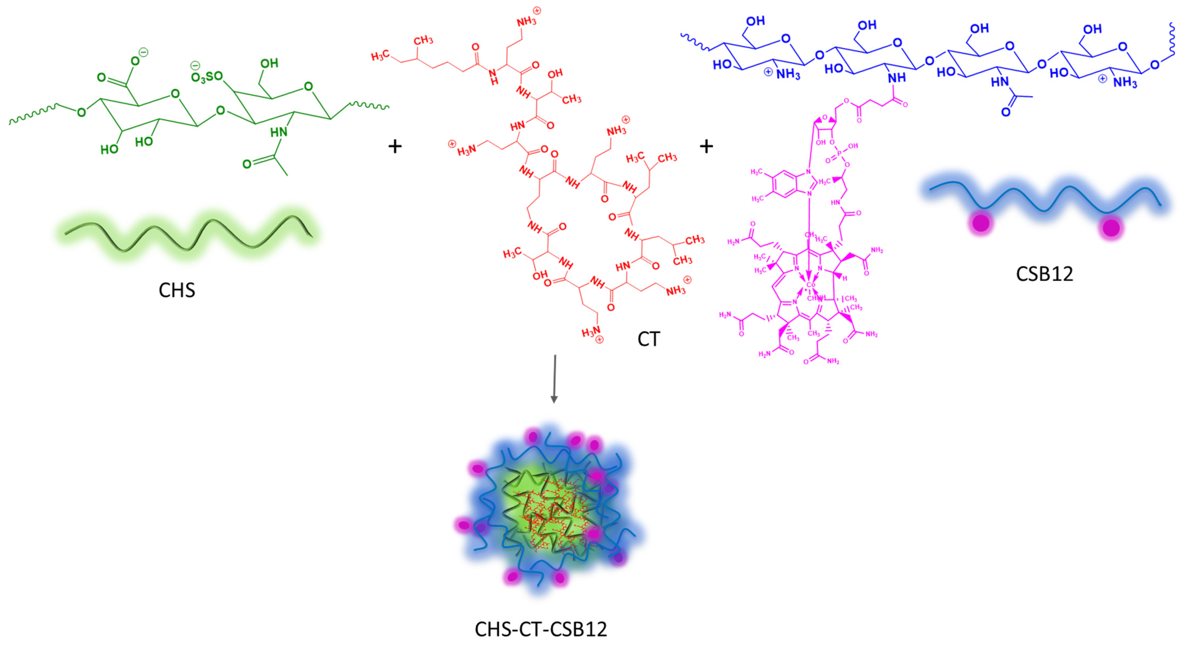
3.1. Formation and Characterization of Polyelectrolyte Complexes Based on CT, CHS, and CSB12

Oppositely charged macromolecules can form PEC particles by polyelectrolyte self-assembly. When CSB12 interacts with CHS, amino groups (pKa~6.5) of CSB12 form ionic bonds with sulfate (pKa~1.0–2.5) and carboxylate (pKa~3–4) groups of CHS, allowing the formation of PECs with a pH-dependent release profile [44,60].
We studied the PEC formation when CSB12 solution was added to CHS solution at different mass ratios (CSB12/CHS of 0.5, 1, 3, and 5) at pH 3.5 and pH 5.0. The different pH values were chosen to vary the degree of polyelectrolyte ionization. Obviously, the CSB12 molecules at pH 3.5 have more protonated amino groups (higher positive charge) than at pH 5.0. Conversely, the carboxyl groups of CHS are more deprotonated (higher negative charge) at pH 5.0 than at pH 3.5. These differences affect the electrostatic interactions between the polymers, making pH critical for optimizing PEC formation. As a result, when small amounts of polycation are added to the CHS solution (ζ-potential of −22.3 mV), the ζ-potential of the system is higher at pH 3.5 (−9.1 mV) than at pH 5.0 (−32.7 mV). Charge compensation using a solution of CSB12 at pH 3.5 and at pH 5.0 occurs at CSB12/CHS mass ratios of about 1.1 and 1.5, respectively. Upon further addition of polycation, the ζ-potentials change independently of the pH of the CSB12 solution and reach values of 27–31 mV at CSB12/CHS mass ratios of 2–5. The decrease of the ζ-potential to −32.7 mV when adding 0.5 pbw (parts by weight) of CSB12 in the form of a solution with pH 5.0 to the CHS solution indicates that polymer particles with negative charge are formed under these conditions (Figure 2a). At the same time, with increasing CSB12 content from 0.5 to 5 pbw, the Dh of the formed PECs gradually increases from 154 to 518 nm and from 120 to 638 nm at pH 3.5 and pH 5.0, respectively. The exception is the particle size of about 450 nm formed at a CSB12/CHS mass ratio of 1 at pH 3.5, which is due to the fact that the ζ-potential is close to 0 mV under these conditions (Figure 2b). As a result, PECs with the Dh of 384 nm and a ζ-potential of 26.9 mV, obtained using a CSB12/CHS mass ratio of 3 and pH 3.5, were selected for CT loading.
The CT molecule has five positively charged amino groups that interact with the carboxylate and sulfate groups of CHS to form intrapolymer complexes by ionic bonding. To study the formation of CHS-CT complexes, CT solutions were added to the CHS solution at various mass ratios (0.2–1). It was shown that when 0.2 pbw CT was added to 1 pbw CHS, particles with a broad size distribution (446 ± 214 nm) were formed; as the CT content in the system increased to 1 pbw, Dh decreased to 175 ± 52 nm. The ζ-potential of the polymer particles also increased from −26.6 to −11.6 mV with increasing CT content (Figure 2c).
To obtain a tri-component PEC, we used the CHS/CT/CSB12 mass ratio of 1:1:3. To load CT into the tri-component PECs, the CT solution was first added to the CHS solution, and then an acetic acid solution of CSB12 at pH 3.5 was added to the CHS-CT complex (sample CHS-CT-CSB12).
We also obtained analogous PECs by changing CSB12 to native CS, resulting in sample CHS-CT-CS, which was used in the Caco-2 experiment to evaluate the effect of B12 on the intestinal permeability of CT incorporated into polyelectrolyte particles. The properties of the obtained particles as well as their unloaded analogs (blank samples CHS-CSB12 and CHS-CS) are presented in Table 2. The obtained tri-component particles CHS-CT-CSB12 had the Dh and ζ-potentials of 446 nm and 28.2 mV, respectively. Meanwhile, polymeric particles based on native CS were characterized by a larger Dh of about 950 nm.
The encapsulation efficiency (EE) for both PECs with CSB12 and with the original CS was 100%.
Our previous studies show that when CT binds to hyaluronic acid in a 1:5 ratio, the EE is also 100%; however, when cationic CS derivatives, namely, diethylaminoethyl chitosan [42] or CSB12 [43], were added, the EE decreased with increasing polycation content in the system, which is due to the competition between CT and polycation for binding to the –COO groups of hyaluronic acid. In contrast to hyaluronic acid, which has one carboxylate group per two monomer units, CHS has one carboxylate or sulfate group in each monomer unit, which apparently increases its capacity to bind cationic molecules.
SEM revealed the presence of particles with an average size of about 350 nm, consistent with dynamic light scattering data (Figure 3).

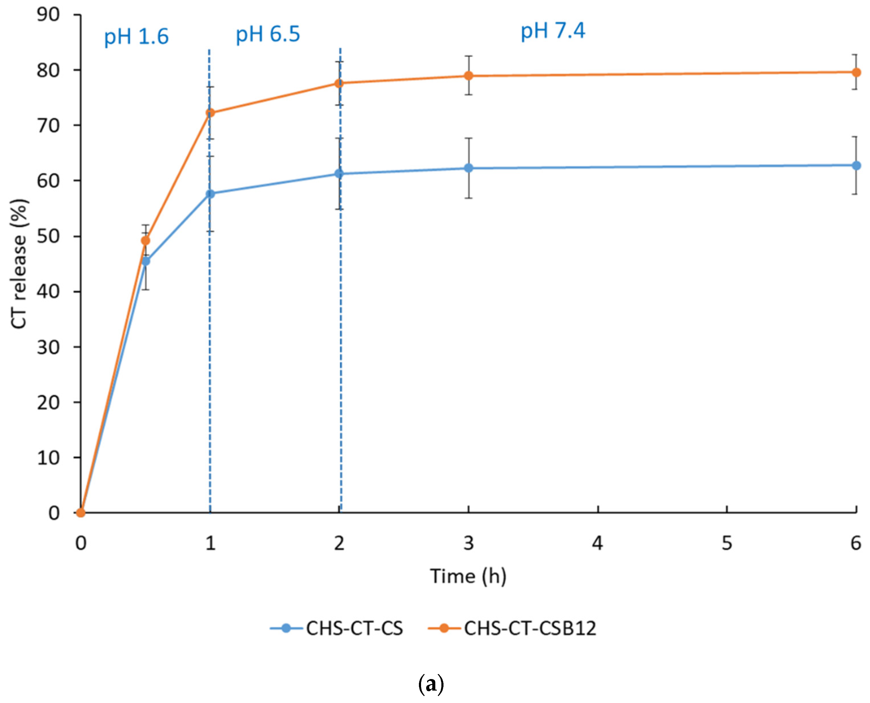
3.2. In Vitro CT Release Profile

It is known that vitamin B12 is absorbed by active transport in the ileum, where the average pH is about 7.4–7.5). Specific transport proteins, including transcobalamin I (haptocorrin), intrinsic factor, and transcobalamin II, are used to actively transport vitamin B12 across the intestinal wall [46,49]. Due to the presence of this intrinsic transport system, vitamin B12 can be used as a targeting ligand to improve the oral bioavailability of peptides and proteins [48,50,61]. For successful absorption, the nanoformulation must be stable during transit from the stomach (fasted pH of 1.5–2.0; residence time of 5 min to 2 h) to different parts of the small intestine, namely to the duodenum (fasted pH of about 6.5) and then to the ileum (total residence time in the small intestine is 3–4 h) [62,63]. In maintaining the colloidal stability of polymer particles, the ζ-potential plays an important role, and it should be about |30| mV and higher. However, under physiological conditions, due to the influence of pH and ionic strength, ionic interactions between PEC components weaken and antibiotic release occurs [41].
The kinetics of CT release from the obtained PECs were studied using different media and time intervals simulating the conditions of passage of the formulation through different parts of the gastrointestinal tract (namely, buffer solutions with pH 1.6, 6.5 and 7.4, simulating the environment of the fasting stomach, fasting small intestine and ileum). As shown in Figure 4a, the obtained systems released about 55 and 70% of the CT (pH 1.6; 1 h) and 60 and 80% of the CT (pH 6.5 and 7.4; 6 h) of CHS-CT-CS PECs and CHS-CT-CSB12 PECs, respectively. The obtained PECs did not show stability in acidic conditions and released CT, apparently due to protonation of -COO- groups. PECs based on the original CS (CHS-CT-CS) showed a delayed release profile compared to PECs based on CSB12 (CHS-CT-CSB12), which may be due to their greater stability due to the presence of a greater number of amino groups in the original CS compared to CSB12, as well as their larger size.

3.3. Antimicrobial Activity

The pharmacological activity of the developed PECs was evaluated against P. aeruginosa. It was shown that the MICs for both CHS-CT-CSB12 and free CT were 1 μg/mL, indicating that the antimicrobial activity of the encapsulated CT was retained. However, the MIC of CHS-CT-CS was 2-fold higher than that of free CT, indicating a decrease in antimicrobial activity. At a concentration of 0.25 μg/mL of CT, CHS-CT-CSB12 reduced relative bacterial growth by approximately 50% compared to free CT, which may be due to the enhanced antimicrobial effect of the uptake of PECs by the microbial cell and is a beneficial factor in the prevention of microbial resistance. In general, the patterns of antimicrobial activity correlate with the results of CT in vitro release. In contrast, CT-free particles had no antibacterial activity under the conditions of this experiment (Figure 4b). Thus, CT-containing CSB12-based PECs have a pronounced antimicrobial effect comparable to that of free CT.

3.4. Mucoadhesive Properties

The ability of CS-based polymeric particles to adhere to and remain on the surface of the gastrointestinal mucosa has been widely exploited to develop oral dosage forms with modified drug release and prolonged therapeutic effects [64,65,66]. The mucoadhesive properties of CS are mainly due to the ability of partially protonated amino groups to bind to negatively charged mucin groups through ionic bonds under physiological conditions; CS mucoadhesion is also achieved through hydrogen bonding and hydrophobic interactions [67]. We investigated the mucoadhesiveness of the developed polyelectrolyte particles. It was shown (Figure 4c) that the obtained PECs with CSB12 as the polycation had excellent mucoadhesive properties, although the mucoadhesion of CSB12-containing PECs (84%) was lower than that of PECs based on native CS (97%). When N-substituted CS derivatives are synthesized, as in the case of obtaining CSB12 by conjugating CS with succinyl-B12 by carbodiimide activation [43], hydrogen bonds are reorganized, and the availability of functional groups, including protonated amino groups, as well as the conformation (flexibility) of the macromolecule, is changed. The combination of these factors can affect both chemical (ionic and hydrogen bonds, van der Waals interactions, etc.) and physical (e.g., entanglement of macromolecular chains) interactions of the CS derivative with mucins and reduce the mucoadhesive properties of the polymer. The mucoadhesive nature of the developed PECs may be a beneficial factor to ensure their adhesion to the mucosal layer as well as further absorption through the intestinal wall into the systemic bloodstream.

3.5. Caco-2 Cell Permeability Assay

The Caco-2 (human colon adenocarcinoma) cell line is widely used in pharmacological studies for qualitative assessment of intestinal permeability to various drugs and macromolecules [59]. Vitamin B12 ingested per os is absorbed in the ileum by an active transport system involving specific transporter proteins called transcobalamins [46]. Because of its own transport system, vitamin B12 can be used as a target ligand to improve the oral bioavailability of drugs [52,61,68]. In addition, CS and its derivatives can improve paracellular permeability and intestinal absorption of drugs by opening tight junctions between cells (the distance between epithelial cells is 20 nm when tight junctions are fully open) [69,70].
In vitro experiments in the Caco-2 cell model using PECs based on both CSB12 and native CS showed that modification of the polymer with the targeting ligand B12 had a significant effect on the intestinal permeability of encapsulated CT. Typically, oral absorption ranges from 50 to 100% for compounds with in vitro Papp greater than 1 × 10⁻⁶ cm/s. The CHS-CT-CSB12 PECs provided a CT Papp of 1.1 × 10⁻⁶ cm/s, which was close to the absorption of pure B12 (Papp of 3.5 × 10⁻⁶ cm/s). However, CT loaded in CHS-CT-CS PECs did not reach acceptable values of Papp due to the large size of the PECs and their inability for transcellular transport. Nevertheless, the presence of CS increased the Papp compared to pure CT (2.1 × 10⁻⁷ cm/s versus 4.0 × 10⁻⁸ cm/s), but in general the contribution of CS to the oral absorption of CT is not significant (Table 3).

4. Discussion and Outlook

The study focused on the formation of PECs using the polyanion CHS and the polycation CSB12 at different mass ratios and pH values (3.5 and 5.0). It was shown that particles with a stable positive ζ-potential (27–31 mV) were formed regardless of the pH of the medium when the mass ratio of CSB12 to CHS ranged from 2 to 5. However, the pH of the medium significantly affected the hydrodynamic diameter of the PECs. At pH 3.5, the particles formed at CSB12/CHS mass ratios of 2 and 3 were 1.6 times smaller than those formed at pH 5.0. This observation suggests that the higher degree of polyelectrolyte ionization at pH 3.5 promotes the formation of more compact and dense polymer particles, probably due to enhanced electrostatic interactions between the oppositely charged polyelectrolytes.
Further investigation of the interaction of CHS with CT revealed that intrapolymer complexes with different Dh (175–446 nm) and ζ-potentials (ranging from −26.6 to −11.6 mV) could be formed at different mass ratios of CT to CHS (from 0.2 to 1). These intrapolymer complexes were successfully used to prepare tri-component systems based on CHS, CT, and CSB12 with a mass ratio of 1:1:3. The resulting CHS-CT-CSB12 PECs exhibited a Dh of 446 nm and a ζ-potential of 28.2 mV. Notably, these particles exhibited an exceptional ability to bind CT, with an encapsulation efficiency of 100% at a CHS:CT mass ratio of 1:1 when 3 pbw of polycationic CSB12 was added. This remarkable binding efficiency can be attributed to the high negative charge density of the CHS macromolecule, which contains anionic groups (carboxylate or sulfate) in each monomer unit, facilitating strong electrostatic interactions with the positively charged CT.
Despite their promising properties, the particles were found to be unstable at the fasting gastric pH of 1.6, which poses a challenge for oral administration. To overcome this, encapsulation in enterosoluble capsules or microcapsules along with specific administration guidelines (e.g., take the formulation on an empty stomach with plenty of water) could be recommended to protect the particles from the acidic environment of the stomach. Nevertheless, the CT-containing biopolymer particles exhibited several beneficial properties. First, the CT loaded into the PECs retained its antimicrobial activity at levels comparable to pure CT. For example, CHS-CT-CSB12 PECs at a concentration of 0.25 μg/mL (relative to pure CT) reduced bacterial growth by approximately 50%, suggesting their potential to mitigate the development of bacterial resistance. Second, the PECs exhibited high mucoadhesion, a critical property for ensuring attachment to the mucosal lining and facilitating intestinal permeability. Finally, the presence of the vitamin B12 fragment in the CSB12 macromolecule contributed to an apparent permeability coefficient of CT of 1.1 × 10⁻⁶ cm/s, indicating the potential for improved oral bioavailability of CT when delivered via these nanotechnology-based systems.
The use of sulfated polysaccharides, such as CHS, in the development of stable CT-containing polyelectrolyte particles holds great promise for improving their physicochemical, pharmacological, and biopharmaceutical properties. Given the encouraging results, further research is warranted to explore the full potential of these systems. In particular, in vivo animal studies should be conducted to evaluate the efficacy, safety, and pharmacokinetics of the developed polysaccharide-based CT delivery systems following oral administration. Such studies would provide critical insight into their therapeutic potential and pave the way for their application as advanced drug delivery systems with improved bioavailability and targeted release properties. In addition, future work could explore the optimization of particle stability in acidic environments, the scalability of the manufacturing process, and the potential for combining CT with other therapeutic agents to enhance synergistic effects. Overall, this research highlights the exciting possibilities of using polyelectrolyte complexes to develop next-generation drug delivery systems.

5. Conclusions

The main results of this study are as follows:
(i)
The optimal conditions and component mass ratios for the formation of colloidally stable PECs based on CHS and CSB12 were identified. Polymer particles with suitable hydrodynamic sizes (330 and 384 nm) and ζ-potentials (25–27 mV) were formed at pH 3.5 and CSB12/CHS mass ratios of 2 and 3.
(ii)
Two-component intrapolymer complexes based on CHS and CT were prepared, demonstrating extremely effective encapsulation efficiencies (EE) of 100% over various CS:CT mass ratios up to 1:1. Subsequently, stable tri-component systems based on CHS, CT, and CSB12 were obtained at a component mass ratio of 1:1:3, with a hydrodynamic diameter of 446 nm and a ζ-potential of 28.2 mV.
(iii)
The developed CHS-CT-CSB12 PECs retained antimicrobial activity against P. aeruginosa comparable to that of pure CT. In addition, they exhibited an apparent permeability coefficient similar to that of vitamin B12. Combined with high mucoadhesion, these properties make the resulting formulations promising candidates for improved oral delivery of CT.
(iv)
Another important aspect of this study is the potential use of sulfated polysaccharides to create polymer complexes for enhanced delivery of polymyxins.

Author Contributions

Conceptualization, N.V.D. and Y.A.S.; methodology, N.V.D., T.S.S., E.V.D. and E.A.M.; investigation, N.V.D., A.Y.B., A.N.B., T.S.S., T.S.E., E.V.D. and E.A.M.; writing—original draft preparation, N.V.D.; writing—review and editing, Y.A.S.; supervision, Y.A.S.; project administration, Y.A.S.; funding acquisition, Y.A.S. All authors have read and agreed to the published version of the manuscript.

Funding

This research was funded by the Russian Science Foundation (grant number 19-73-20157-P).

Institutional Review Board Statement

The Caco-2 cell line was obtained from the Russian Cell Culture Collection (Institute of Cytology, Russian Academy of Sciences, St. Petersburg, Russia).

Data Availability Statement

Data are contained within the article and available upon request.

Conflicts of Interest

The authors declare no conflicts of interest.

References

  1. Hernando-Amado, S.; Coque, T.M.; Baquero, F.; Martínez, J.L. Defining and combating antibiotic resistance from one health and global health perspectives. Nat. Microbiol. 2019, 4, 1432–1442. [Google Scholar] [CrossRef]
  2. Roope, L.S.; Smith, R.D.; Pouwels, K.B.; Buchanan, J.; Abel, L.; Eibich, P.; Butler, C.C.; Tan, P.S.; Walker, A.S.; Robotham, J.V. The challenge of antimicrobial resistance: What economics can contribute. Science 2019, 364, eaau4679. [Google Scholar] [CrossRef]
  3. Coque, T.M.; Cantón, R.; Pérez-Cobas, A.E.; Fernández-de-Bobadilla, M.D.; Baquero, F. Antimicrobial resistance in the global health network: Known unknowns and challenges for efficient responses in the 21st century. Microorganisms 2023, 11, 1050. [Google Scholar] [CrossRef] [PubMed]
  4. Larsson, D.; Flach, C.-F. Antibiotic resistance in the environment. Nat. Rev. Microbiol. 2022, 20, 257–269. [Google Scholar] [CrossRef] [PubMed]
  5. Murray, C.J.; Ikuta, K.S.; Sharara, F.; Swetschinski, L.; Aguilar, G.R.; Gray, A.; Han, C.; Bisignano, C.; Rao, P.; Wool, E. Global burden of bacterial antimicrobial resistance in 2019: A systematic analysis. Lancet 2022, 399, 629–655. [Google Scholar] [CrossRef]
  6. Ma, Y.X.; Wang, C.Y.; Li, Y.Y.; Li, J.; Wan, Q.Q.; Chen, J.H.; Tay, F.R.; Niu, L.N. Considerations and caveats in combating eskape pathogens against nosocomial infections. Adv. Sci. 2020, 7, 1901872. [Google Scholar] [CrossRef]
  7. Aljeldah, M.M. Antimicrobial resistance and its spread is a global threat. Antibiotics 2022, 11, 1082. [Google Scholar] [CrossRef]
  8. Miller, W.R.; Arias, C.A. Eskape pathogens: Antimicrobial resistance, epidemiology, clinical impact and therapeutics. Nature Rev. Microbiol. 2024, 22, 598–616. [Google Scholar] [CrossRef] [PubMed]
  9. Mulani, M.S.; Kamble, E.E.; Kumkar, S.N.; Tawre, M.S.; Pardesi, K.R. Emerging strategies to combat eskape pathogens in the era of antimicrobial resistance: A review. Front. Microbiol. 2019, 10, 539. [Google Scholar] [CrossRef]
  10. Singh, A.; Tanwar, M.; Singh, T.; Sharma, S.; Sharma, P. An escape from eskape pathogens: A comprehensive review on current and emerging therapeutics against antibiotic resistance. Int. J. Biol. Macromol. 2024, 279, 135253. [Google Scholar] [CrossRef]
  11. Shah, S.N.; Bhat, M.A.; Bhat, M.A.; Jan, A.T. Antimicrobial resistance: An overview. In Nanotechnology Based Strategies for Combating Antimicrobial Resistance; Wani, M.Y., Wani, I.A., Rai, A., Eds.; Springer: Singapore, 2024; pp. 1–44. [Google Scholar] [CrossRef]
  12. Mancuso, G.; Midiri, A.; Gerace, E.; Biondo, C. Bacterial antibiotic resistance: The most critical pathogens. Pathogens 2021, 10, 1310. [Google Scholar] [CrossRef] [PubMed]
  13. Lee, C.-H.; Su, T.-Y.; Ye, J.-J.; Hsu, P.-C.; Kuo, A.-J.; Chia, J.-H.; Lee, M.-H. Risk factors and clinical significance of bacteremia caused by pseudomonas aeruginosa resistant only to carbapenems. J. Microbiol. Immunol. Infect. 2017, 50, 677–683. [Google Scholar] [CrossRef]
  14. De Oliveira, D.M.; Forde, B.M.; Kidd, T.J.; Harris, P.N.; Schembri, M.A.; Beatson, S.A.; Paterson, D.L.; Walker, M.J. Antimicrobial resistance in eskape pathogens. Clin. Microbiol. Rev. 2020, 33, e00181-19. [Google Scholar] [CrossRef] [PubMed]
  15. El-Sayed Ahmed, M.A.E.-G.; Zhong, L.-L.; Shen, C.; Yang, Y.; Doi, Y.; Tian, G.-B. Colistin and its role in the era of antibiotic resistance: An extended review (2000–2019). Emerg. Microbes Infect. 2020, 9, 868–885. [Google Scholar] [CrossRef]
  16. Band, V.I.; Weiss, D.S. Mechanisms of antimicrobial peptide resistance in gram-negative bacteria. Antibiotics 2014, 4, 18–41. [Google Scholar] [CrossRef]
  17. Grégoire, N.; Aranzana-Climent, V.; Magréault, S.; Marchand, S.; Couet, W. Clinical pharmacokinetics and pharmacodynamics of colistin. Clin. Pharmacokinet. 2017, 56, 1441–1460. [Google Scholar] [CrossRef]
  18. Vaara, M. Polymyxins and their potential next generation as therapeutic antibiotics. Front. Microbiol. 2019, 10, 1689. [Google Scholar]
  19. Slingerland, C.J.; Martin, N.I. Recent advances in the development of polymyxin antibiotics: 2010–2023. ACS Infect. Dis. 2024, 10, 1056–1079. [Google Scholar] [CrossRef]
  20. Nation, R.L.; Rigatto, M.H.P.; Falci, D.R.; Zavascki, A.P. Polymyxin acute kidney injury: Dosing and other strategies to reduce toxicity. Antibiotics 2019, 8, 24. [Google Scholar] [CrossRef]
  21. Doymaz, M.Z.; Karaaslan, E. Comparison of antibacterial activities of polymyxin b and colistin against multidrug resistant gram negative bacteria. Infect. Dis. 2019, 51, 676–682. [Google Scholar] [CrossRef]
  22. Aboumanei, M.H.; Mahmoud, A.F.; Motaleb, M. Formulation of chitosan coated nanoliposomes for the oral delivery of colistin sulfate: In vitro characterization, 99mtc-radiolabeling and in vivo biodistribution studies. Drug Dev. Ind. Pharm. 2021, 47, 626–635. [Google Scholar] [CrossRef] [PubMed]
  23. Ardebili, A.; Izanloo, A.; Rastegar, M. Polymyxin combination therapy for multidrug-resistant, extensively-drug resistant, and difficult-to-treat drug-resistant gram-negative infections: Is it superior to polymyxin monotherapy? Expert Rev. Anti-Infect. Ther. 2023, 21, 387–429. [Google Scholar] [CrossRef]
  24. Nation, R.L. Polymyxins. In Kucers’ the Use of Antibiotics; CRC Press: Cambridge, UK, 2017; pp. 1420–1449. [Google Scholar]
  25. Wang, R.; van Dorp, L.; Shaw, L.P.; Bradley, P.; Wang, Q.; Wang, X.; Jin, L.; Zhang, Q.; Liu, Y.; Rieux, A.; et al. The global distribution and spread of the mobilized colistin resistance gene mcr-1. Nat. Commun. 2018, 9, 1179. [Google Scholar] [CrossRef]
  26. Olaitan, A.O.; Chabou, S.; Okdah, L.; Morand, S.; Rolain, J.-M. Dissemination of the mcr-1 colistin resistance gene. Lancet Infect. Dis. 2016, 16, 147. [Google Scholar] [CrossRef]
  27. Hamel, M.; Rolain, J.-M.; Baron, S.A. The history of colistin resistance mechanisms in bacteria: Progress and challenges. Microorganisms 2021, 9, 442. [Google Scholar] [CrossRef] [PubMed]
  28. Anyanwu, M.U.; Jaja, I.F.; Nwobi, O.C. Occurrence and characteristics of mobile colistin resistance (mcr) gene-containing isolates from the environment: A review. Int. J. Environ. Res. Public Health 2020, 17, 1028. [Google Scholar] [CrossRef]
  29. Azad, M.A.; Nation, R.L.; Velkov, T.; Li, J. Mechanisms of polymyxin-induced nephrotoxicity. In Polymyxin Antibiotics: From Laboratory Bench to Bedside. Advances in Experimental Medicine and Biology, Vol 1145; Li, J., Nation, R., Kaye, K., Eds.; Springer: Cham, Switzerland, 2019; pp. 305–319. [Google Scholar] [CrossRef]
  30. Alqahtani, M.S.; Kazi, M.; Alsenaidy, M.A.; Ahmad, M.Z. Advances in oral drug delivery. Front. Pharmacol. 2021, 12, 618411. [Google Scholar] [CrossRef]
  31. Hung, Y.-P.; Chen, Y.-F.; Tsai, P.-J.; Huang, I.-H.; Ko, W.-C.; Jan, J.-S. Advances in the application of nanomaterials as treatments for bacterial infectious diseases. Pharmaceutics 2021, 13, 1913. [Google Scholar] [CrossRef] [PubMed]
  32. Jampilek, J.; Kralova, K. Advances in nanostructures for antimicrobial therapy. Materials 2022, 15, 2388. [Google Scholar] [CrossRef]
  33. Brown, P.; Dawson, M.J. Development of new polymyxin derivatives for multi-drug resistant gram-negative infections. J. Antibiot. 2017, 70, 386–394. [Google Scholar] [CrossRef]
  34. Dubashynskaya, N.V.; Skorik, Y.A. Polymyxin delivery systems: Recent advances and challenges. Pharmaceuticals 2020, 13, 83. [Google Scholar] [CrossRef] [PubMed]
  35. Gao, P.; Nie, X.; Zou, M.; Shi, Y.; Cheng, G. Recent advances in materials for extended-release antibiotic delivery system. J. Antibiot. 2011, 64, 625–634. [Google Scholar] [CrossRef]
  36. Dubashynskaya, N.V.; Bokatyi, A.N.; Dobrodumov, A.V.; Kudryavtsev, I.V.; Trulioff, A.S.; Rubinstein, A.A.; Aquino, A.D.; Dubrovskii, Y.A.; Knyazeva, E.S.; Demyanova, E.V. Succinyl chitosan-colistin conjugates as promising drug delivery systems. Int. J. Mol. Sci. 2022, 24, 166. [Google Scholar] [CrossRef]
  37. Le, H.; Karakasyan, C.; Jouenne, T.; Le Cerf, D.; Dé, E. Application of polymeric nanocarriers for enhancing the bioavailability of antibiotics at the target site and overcoming antimicrobial resistance. Appl. Sci. 2021, 11, 10695. [Google Scholar] [CrossRef]
  38. Skwarczynski, M.; Bashiri, S.; Yuan, Y.; Ziora, Z.M.; Nabil, O.; Masuda, K.; Khongkow, M.; Rimsueb, N.; Cabral, H.; Ruktanonchai, U. Antimicrobial activity enhancers: Towards smart delivery of antimicrobial agents. Antibiotics 2022, 11, 412. [Google Scholar] [CrossRef]
  39. Sharma, S.; Bhende, M. An overview: Non-toxic and eco-friendly polysaccharides—Its classification, properties, and diverse applications. Polym. Bull. 2024, 81, 12383–12429. [Google Scholar] [CrossRef]
  40. Pal, D.; Saha, S. Chondroitin: A natural biomarker with immense biomedical applications. RSC Adv. 2019, 9, 28061–28077. [Google Scholar] [CrossRef]
  41. Dubashynskaya, N.V.; Raik, S.V.; Dubrovskii, Y.A.; Shcherbakova, E.S.; Demyanova, E.V.; Shasherina, A.Y.; Anufrikov, Y.A.; Poshina, D.N.; Dobrodumov, A.V.; Skorik, Y.A. Hyaluronan/colistin polyelectrolyte complexes: Promising antiinfective drug delivery systems. Int. J. Biol. Macromol. 2021, 187, 157–165. [Google Scholar] [CrossRef]
  42. Dubashynskaya, N.V.; Raik, S.V.; Dubrovskii, Y.A.; Demyanova, E.V.; Shcherbakova, E.S.; Poshina, D.N.; Shasherina, A.Y.; Anufrikov, Y.A.; Skorik, Y.A. Hyaluronan/diethylaminoethyl chitosan polyelectrolyte complexes as carriers for improved colistin delivery. Int. J. Mol. Sci. 2021, 22, 8381. [Google Scholar] [CrossRef]
  43. Dubashynskaya, N.V.; Bokatyi, A.N.; Sall, T.S.; Egorova, T.S.; Demyanova, E.V.; Dubrovskii, Y.A.; Murashko, E.A.; Anufrikov, Y.A.; Shasherina, A.Y.; Vlasova, E.N. Hyaluronan/b12-chitosan polyelectrolyte complex for oral colistin administration. Int. J. Biol. Macromol. 2024, 263, 130177. [Google Scholar] [CrossRef]
  44. Fu, J.; Fares, H.M.; Schlenoff, J.B. Ion-pairing strength in polyelectrolyte complexes. Macromolecules 2017, 50, 1066–1074. [Google Scholar] [CrossRef]
  45. Shchipunov, Y.A.; Postnova, I.V. Water-soluble polyelectrolyte complexes of oppositely charged polysaccharides. Compos. Interfaces 2009, 16, 251–279. [Google Scholar] [CrossRef]
  46. Petrus, A.K.; Fairchild, T.J.; Doyle, R.P. Traveling the vitamin b12 pathway: Oral delivery of protein and peptide drugs. Angew. Chem. Int. Ed. 2009, 48, 1022–1028. [Google Scholar] [CrossRef]
  47. Dubashynskaya, N.V.; Bokatyi, A.N.; Sall, T.S.; Egorova, T.S.; Nashchekina, Y.A.; Dubrovskii, Y.A.; Murashko, E.A.; Vlasova, E.N.; Demyanova, E.V.; Skorik, Y.A. Cyanocobalamin-modified colistin–hyaluronan conjugates: Synthesis and bioactivity. Int. J. Mol. Sci. 2023, 24, 11550. [Google Scholar] [CrossRef] [PubMed]
  48. Stepanova, M.; Levit, M.; Egorova, T.; Nashchekina, Y.; Sall, T.; Demyanova, E.; Guryanov, I.; Korzhikova-Vlakh, E. Poly (2-deoxy-2-methacrylamido-d-glucose)-based complex conjugates of colistin, deferoxamine and vitamin b12: Synthesis and biological evaluation. Pharmaceutics 2024, 16, 1080. [Google Scholar] [CrossRef] [PubMed]
  49. Fedosov, S.N. Physiological and molecular aspects of cobalamin transport. Water Soluble Vitam. Clin. Res. Future Appl. 2012, 56, 347–367. [Google Scholar]
  50. Russell-Jones, G.; Westwood, S.; Habberfield, A. Vitamin b12 mediated oral delivery systems for granulocyte-colony stimulating factor and erythropoietin. Bioconjugate Chem. 1995, 6, 459–465. [Google Scholar] [CrossRef]
  51. Russell-Jones, G.; Westwood, S.; Farnworth, P.; Findlay, J.; Burger, H. Synthesis of lhrh antagonists suitable for oral administration via the vitamin b12 uptake system. Bioconjugate Chem. 1995, 6, 34–42. [Google Scholar] [CrossRef]
  52. Verma, A.; Sharma, S.; Gupta, P.K.; Singh, A.; Teja, B.V.; Dwivedi, P.; Gupta, G.K.; Trivedi, R.; Mishra, P.R. Vitamin b12 functionalized layer by layer calcium phosphate nanoparticles: A mucoadhesive and ph responsive carrier for improved oral delivery of insulin. Acta Biomater. 2016, 31, 288–300. [Google Scholar] [CrossRef]
  53. Dubashynskaya, N.V.; Bokatyi, A.N.; Trulioff, A.S.; Rubinstein, A.A.; Novikova, V.P.; Petrova, V.A.; Vlasova, E.N.; Malkov, A.V.; Kudryavtsev, I.V.; Skorik, Y.A. Delivery system for dexamethasone phosphate based on a Zn2+-crosslinked polyelectrolyte complex of diethylaminoethyl chitosan and chondroitin sulfate. Carbohydr. Polym. 2024, 348, 122899. [Google Scholar] [CrossRef]
  54. Raik, S.V.; Poshina, D.N.; Lyalina, T.A.; Polyakov, D.S.; Vasilyev, V.B.; Kritchenkov, A.S.; Skorik, Y.A. N-[4-(N,N,N-trimethylammonium) benzyl] chitosan chloride: Synthesis, interaction with DNA and evaluation of transfection efficiency. Carbohydr. Polym. 2018, 181, 693–700. [Google Scholar] [CrossRef] [PubMed]
  55. Iudin, D.; Zashikhina, N.; Demyanova, E.; Korzhikov-Vlakh, V.; Shcherbakova, E.; Boroznjak, R.; Tarasenko, I.; Zakharova, N.; Lavrentieva, A.; Skorik, Y.; et al. Polypeptide self-assembled nanoparticles as delivery systems for polymyxins b and e. Pharmaceutics 2020, 12, 868. [Google Scholar] [CrossRef]
  56. Wu, T.; Zivanovic, S.; Hayes, D.G.; Weiss, J. Efficient reduction of chitosan molecular weight by high-intensity ultrasound: Underlying mechanism and effect of process parameters. J. Agric. Food Chem. 2008, 56, 5112–5119. [Google Scholar] [CrossRef] [PubMed]
  57. Dubashynskaya, N.V.; Bokatyi, A.N.; Trulioff, A.S.; Rubinstein, A.A.; Kudryavtsev, I.V.; Skorik, Y.A. Development and bioactivity of zinc sulfate cross-linked polysaccharide delivery system of dexamethasone phosphate. Pharmaceutics 2023, 15, 2396. [Google Scholar] [CrossRef]
  58. Hejjaji, E.M.; Smith, A.M.; Morris, G.A. Evaluation of the mucoadhesive properties of chitosan nanoparticles prepared using different chitosan to tripolyphosphate (CS:TPP) ratios. Int. J. Biol. Macromol. 2018, 120, 1610–1617. [Google Scholar] [CrossRef] [PubMed]
  59. Chen, C.-H.; Lin, Y.-S.; Wu, S.-J.; Mi, F.-L. Mutlifunctional nanoparticles prepared from arginine-modified chitosan and thiolated fucoidan for oral delivery of hydrophobic and hydrophilic drugs. Carbohydr. Polym. 2018, 193, 163–172. [Google Scholar] [CrossRef]
  60. Elbi, S.; Nimal, T.; Rajan, V.; Baranwal, G.; Biswas, R.; Jayakumar, R.; Sathianarayanan, S. Fucoidan coated ciprofloxacin loaded chitosan nanoparticles for the treatment of intracellular and biofilm infections of salmonella. Colloids Surf. B Biointerfaces 2017, 160, 40–47. [Google Scholar]
  61. Hosseini-Ashtiani, N.; Tadjarodi, A.; Zare-Dorabei, R. Low molecular weight chitosan-cyanocobalamin nanoparticles for controlled delivery of ciprofloxacin: Preparation and evaluation. Int. J. Biol. Macromol. 2021, 176, 459–467. [Google Scholar] [CrossRef]
  62. Ashford, M. Gastrointestinal tract–physiology and drug absorption. In Aulton’s Pharmaceutics E-Book: The Design and Manufacture of Medicines, 5th ed.; Taylor, K.M.G., Aulton, M.E., Eds.; Elsevier: Edinburgh, UK, 2017; pp. 300–318. [Google Scholar]
  63. Kozyraki, R.; Cases, O. Vitamin b12 absorption: Mammalian physiology and acquired and inherited disorders. Biochimie 2013, 95, 1002–1007. [Google Scholar] [CrossRef]
  64. Pathomthongtaweechai, N.; Muanprasat, C. Potential applications of chitosan-based nanomaterials to surpass the gastrointestinal physiological obstacles and enhance the intestinal drug absorption. Pharmaceutics 2021, 13, 887. [Google Scholar] [CrossRef]
  65. Sangnim, T.; Dheer, D.; Jangra, N.; Huanbutta, K.; Puri, V.; Sharma, A. Chitosan in oral drug delivery formulations: A review. Pharmaceutics 2023, 15, 2361. [Google Scholar] [CrossRef] [PubMed]
  66. Chen, M.-C.; Mi, F.-L.; Liao, Z.-X.; Hsiao, C.-W.; Sonaje, K.; Chung, M.-F.; Hsu, L.-W.; Sung, H.-W. Recent advances in chitosan-based nanoparticles for oral delivery of macromolecules. Adv. Drug Deliv. Rev. 2013, 65, 865–879. [Google Scholar] [CrossRef] [PubMed]
  67. Ways, T.M.M.; Lau, W.M.; Khutoryanskiy, V.V. Chitosan and its derivatives for application in mucoadhesive drug delivery systems. Polymers 2018, 10, 267. [Google Scholar] [CrossRef] [PubMed]
  68. Li, X.; Yu, M.; Fan, W.; Gan, Y.; Hovgaard, L.; Yang, M. Orally active-targeted drug delivery systems for proteins and peptides. Expert Opin. Drug Deliv. 2014, 11, 1435–1447. [Google Scholar] [CrossRef]
  69. Yeh, T.-H.; Hsu, L.-W.; Tseng, M.T.; Lee, P.-L.; Sonjae, K.; Ho, Y.-C.; Sung, H.-W. Mechanism and consequence of chitosan-mediated reversible epithelial tight junction opening. Biomaterials 2011, 32, 6164–6173. [Google Scholar] [CrossRef]
  70. Kumar, R.; Islam, T.; Nurunnabi, M. Mucoadhesive carriers for oral drug delivery. J. Control. Release 2022, 351, 504–559. [Google Scholar] [CrossRef]
Figure 1. Formation of polyelectrolyte complexes based on chondroitin sulfate, B12-chitosan, and colistin.
Polysaccharides 06 00021 g001
Figure 2. The ζ-potentials (a) and hydrodynamic diameters (b) of CHS-CSB12 PEC at different pH; the ζ-potentials and hydrodynamic diameters of the CHS-CT complexes (c). Data are presented as the mean ± SD, n = 3.
Polysaccharides 06 00021 g002aPolysaccharides 06 00021 g002b
Figure 3. SEM images of tri-component polyelectrolyte complexes based on chondroitin sulfate, B12-chitosan, and colistin (CHS-CT-CSB12 PECs) at different magnifications: 1 μm scale, 65,800× magnification (a) and 200 nm scale, 190,000× magnification (b).
Polysaccharides 06 00021 g003
Figure 4. Cumulative release of colistin from the tri-component polyelectrolyte complexes based on chondroitin sulfate, B12-chitosan/chitosan, and colistin (CHS-CT-CSB12 and CHS-CT-CS PECs) in 6 h at 37 °C and different pH. Each point is presented as the mean ± SD, n = 3 (a). Minimum inhibitory concentrations of CHS-CT-CSB12 and CHS-CT-CS PECs and free colistin against P. aeruginosa. Data are presented as the mean ± SD, n = 3 (b). Mucoadhesiveness of the CHS-CT-CSB12 and CHS-CT-CS PECs. Data are presented as the mean ± SD, n = 3 (c).
Polysaccharides 06 00021 g004aPolysaccharides 06 00021 g004b
Table 1. Preparation conditions of polyelectrolyte complexes based on chondroitin sulfate, B12-chitosan, and colistin.
CSB12/CHS Mass RatioVolume of CSB12 Solution (mL)Concentration of CSB12 Solution (mg/mL)pHVolume of CHS Solution (mL)Volume of CT Solution (mL)
0.501.000.503.51.00-
1.01.001.03.51.00-
2.01.002.03.51.00-
3.01.003.03.51.001.00
5.01.005.03.51.00-
0.501.000.505.01.00-
1.01.001.05.01.00-
2.01.002.05.01.00-
3.01.003.05.01.00-
5.01.005.05.01.00-
Table 2. Characteristics of the two-component polyelectrolyte complexes based on chondroitin sulfate and B12-chitosan/chitosan and loaded with colistin tri-component polyelectrolyte complexes based on chondroitin sulfate and B12-chitosan/chitosan (mean ± SD, n = 3).
Formulation
(Component Mass Ratio)
Dh, nmζ-Potential, mVEE, %CT Content, µg/mgCT Cumulative
Release in 24 h, %
CHS-CT-CSB12
(1:1:3)
446 ± 2628.2 ± 0.910020080%
CHS-CSB12
(1:3)
384 ± 6828.5 ± 1.1---
CHS-CT-CS
(1:1:3)
950 ± 18826.6 ± 1.610020063%
CHS-CS
(1:3)
816 ± 13426.9 ± 0.8---
Table 3. The apparent permeability coefficient (Papp) of tri-component polyelectrolyte complexes based on chondroitin sulfate, B12-chitosan/chitosan (CHS-CT-CSB12 and CHS-CT-CS PECs), free vitamin B12, and free colistin.
SamplePapp (cm/s)
CT4.0 × 10−8
CHS-CT-CS2.1 × 10−7
CHS-CT-CSB121.1 × 10−6
B123.5 × 10−6
Disclaimer/Publisher’s Note: The statements, opinions and data contained in all publications are solely those of the individual author(s) and contributor(s) and not of MDPI and/or the editor(s). MDPI and/or the editor(s) disclaim responsibility for any injury to people or property resulting from any ideas, methods, instructions or products referred to in the content.

