Case Report

Aggressive Surgical Management of Bilateral Metachronous Lung Metastases in Fibrolamellar Hepatocellular Carcinoma, a Case Report

1
Thoracic Surgery Unit, Department of Cardiac, Thoracic and Vascular Sciences and Public Health, University of Padova, 35121 Padova, Italy
2
Pathology Unit, Department of Cardiac, Thoracic, Vascular Sciences and Public Health, University of Padova, 35121 Padova, Italy
*
Author to whom correspondence should be addressed.
Livers 2024, 4(3), 398-405; https://doi.org/10.3390/livers4030029
Submission received: 17 April 2024 / Revised: 17 July 2024 / Accepted: 30 July 2024 / Published: 21 August 2024

Abstract

:
Fibrolamellar hepatocellular carcinoma (FL-HCC) is a malignant primary hepatic cancer that affects mainly adolescents and young adults without underlying liver disease. Its biology remains unknown, but it is pathologically distinct from traditional HCC. Therapeutic strategies are not well defined and, as chemotherapies seem to have limited efficacy, surgical resection remains the only effective treatment. Here we report on a case of a metastatic FL-HCC in an 18-year-old man successfully treated with aggressive intra-thoracic bilateral lung metastasectomy following primary tumour resection and adjuvant chemotherapy. Survival time after initial hepatectomy is 39 months, with no recurrence of disease to date. Aggressive surgical resection and redo surgery should be considered until more effective multimodality therapies are identified. Multidisciplinary team discussion and involvement of medical and surgical specialties are essential in managing these rare entities.

1. Background

Fibrolamellar hepatocellular carcinoma (FL-HCC) is a rare variety of primary hepatocellular malignancy accounting for 1–2% of all cases of hepatocellular carcinoma registered in the United States. It affects predominantly adolescents and young adults without underlying cirrhosis or liver disease with a male to female ratio reaching almost 2 to 1. This neoplasm usually presents as a solitary mass with non-specific symptoms, such as nausea, vomiting and a palpable abdominal mass. Serum transaminases can be elevated and only in 5 to 10% of cases is the alpha-fetoprotein level higher than 200 ng/mL [1].
While its biology remains largely unknown, this pathologically distinct cancer is characterized by the presence of thick collagen bands surrounding the tumour cells, which renders it different from other subtypes of hepatocellular carcinoma [2]. The tumour presents more commonly within a context of healthy hepatic parenchyma and it is not associated with cirrhosis, as may be evidenced by its epidemiologic characteristics. Microscopically it features large polygonal cells with prominent nucleoli, well-delineated cell borders and abundant granular eosinophilic cytoplasm, arranged in nests, cords, and trabeculae surrounded by dense parallel bands of fibrosis, a feature also known as lamellar fibrosis [1].
Even though the pathogenesis of FL-HCC is not completely clear, several molecular hallmarks have previously been delineated. In fact, FL-HCC manifests three types of molecular classes, namely the Proliferation class (activation of specific Akt-mTOR signalling), the Inflammation class (a hyperinflammation profile with enrichment of IL-18 and IL-10 cytokines), and the Unannotated class (other redundant signal pathways such as RAS, TGF-beta, PI3K and MAP kinases, not strictly related to liver-specific cancer pathways). These groups lead to different molecular profiles responsible for the pathogenesis of FL-HCC in different ways. Overall, this tumour has a lower copy number variation than intrahepatic cholangiocarcinoma or HCC, with focal aberrations responsible for the molecular genesis of this cancer. Moreover, an increased EGFR membranous manifestation and several other neuroendocrine markers are typical in FL-HCC. As far as focal aberrations are concerned, FL-HCC seems to be primarily driven by a genomic deletion in chromosome 19, providing a unique fusion gene mutation coding for the DNAJB1-PRKACA chimeric protein. This involves a G protein-coupled receptors disfunction responsible for the cancer genesis and progression [3]. Known hepatic stress factors, such as chronic alcohol consumption, viral hepatitis or liver flukes are not thought to be associated with this type of disease. The activated fusion protein enhances the phosphorylation of other cellular structures associated with cellular proliferation. This mutation is expressed in approximately 95% of cases, suggesting its crucial role in the development and progression of FL-HCC. While other mutations have been studied as possible causes for the progression of the disease, this is not yet clear [4].
Nodal metastases are observed in 50–65% of cases, with 20–30% of cases featuring distant metastases being diagnosed in advanced stages [5]. The lungs, peritoneum and adrenal glands represent the most common sites of metastases, even though it is not unusual to find secondary lesions in other sites, such as the mammary glands and bone [6].
Since the effectiveness of alternative treatments, such as systemic chemotherapies, has not yet been established [7], surgical resection remains the only effective treatment. Chemotherapy for FL-HCC is controversial, and it has been suggested that cytoreductive chemotherapy is ineffective and adjuvant chemotherapy does not improve survival [7]. Moreover, as FL-HCC tends to be non-chemo-responsive, surgical resection of recurrent disease is associated with improved median overall survival (OS) [8]. In this context, early diagnosis is essential, as the absence of comorbidities and a young age at diagnosis are conditions that can make resection feasible even in the presence of large tumours, always considering the possibility of a high incidence of recurrence [9]. However, diagnosis is complex because of the pleomorphism of the associated symptoms and the functional compensation of a healthy liver in most of the patients because of their age.
In this paper we report a case of metastatic FL-HCC in a young man which was successfully treated by aggressive intra-thoracic bilateral lung metastasectomy.

2. Patient Information, Clinical Findings and Diagnostic Assessment

An 18-year-old male patient was referred to our department by hepatobiliary surgeons for multiple enlarged lesions in the right lung. Two years earlier, on the detection of a relatively rare form of FL-HCC, the patient had undergone right hepatectomy via laparotomy, associated with inferior vena cava resection and replacement with a Dacron prosthesis due to local disease infiltration, right adrenalectomy, microwave thermal ablation of a suspicious lesion in the S1 segment and a lymphadenectomy of the hepatic hilum. Definitive histological examination testified to a pathological T4N0 stage hepatic fibrolamellar carcinoma.
After 17 months of regular follow-up, a surveillance thoracic and abdominal CT-scan were performed. These detected a volumetric enlargement of a paracaval supradiaphragmatic mass and the appearance of multiple right pulmonary nodules, in addition to an aspecific micronodule (4 mm) in the left lower lobe. The case was discussed by the multidisciplinary oncological board, which proposed starting first-line systemic therapy with the XELOX protocol (4 cycles performed with mild-moderate toxicity, but poor subjective tolerance by the patient). In particular, the protocol adopted consisted of 130 mg/m2 of Oxaliplatin and Fluoropyrimidines 1000 mg/m2. After chemotherapy, a total body computed tomography scan (TB-CT) confirmed the stability of the disease in terms of number and volume of the nodules, and collaterally identified the obstruction of the caval graft and partial thrombosis of the right femoro-iliac venous axis. On the basis of this diagnosis, anticoagulant therapy was initiated, and the patient was considered to be a candidate for surgical resection.

3. Therapeutic Intervention and Outcome

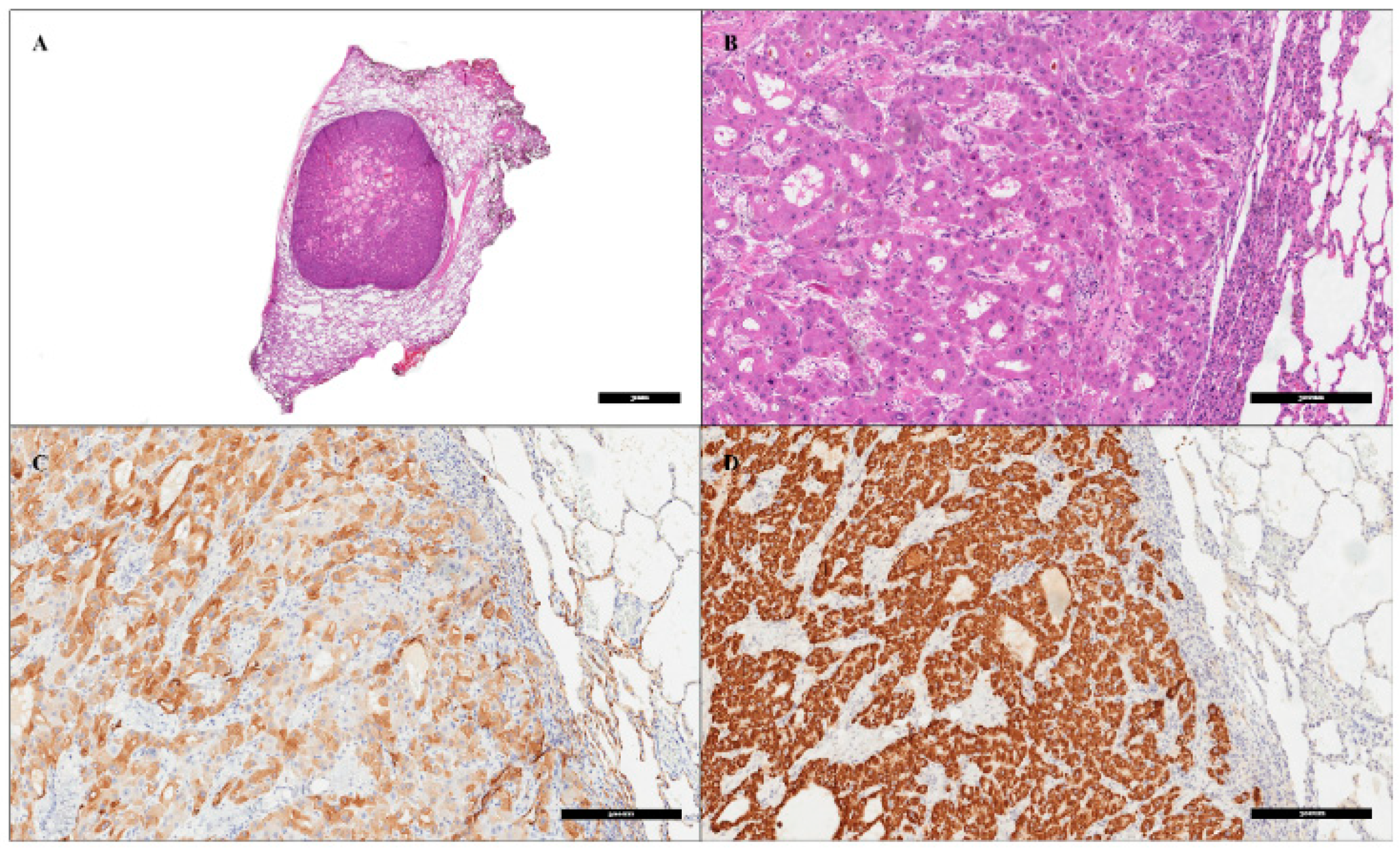
One month after the completion of chemotherapy, the patient underwent laser-assisted metastasectomy of three pulmonary nodules in the right lower lobe, wedge resection of a nodule in the middle lobe (Figure 1), and en bloc excision of a 20 mm paracaval mass with a small portion of the hemidiaphragm by posterolateral thoracotomy access. Open surgery was preferred to allow better palpation of the entire lung, as well as better control of the resection margins, particularly in laser-assisted metastasectomy. A laser-assisted technique was adopted in addition to the use of conventional wedge resections to mitigate against the risk of air leaks, for better lung sparing resection and a comparable R0 results [10]. All the nodules were confirmed to be FL-HCC metastases at histological examination (microscopical and immunohistochemical confirmations), with negative resection margins. No postoperative complications were registered. The patient was discharged after seven days, and follow-up was started. Nine months later, a total body CT-scan (TB-CTs) showed volumetric growth of an isolated pulmonary micronodule (10 mm vs. 4 mm; Figure 2) in the left lower lobe which had remained unchanged before that, with no further suspicious lesions. Multidisciplinary discussion confirmed the high probability of disease recurrence, and the patient underwent re-intervention. Video Assisted Thoracic Surgery (VATS) wedge resection of the left pulmonary nodule was performed. FL-HCC metastasis with wide negative margins was histologically confirmed (Figure 3). Two days after surgery, the patient was discharged from our department with no postoperative complications, the drainage was removed on the first postoperative day with no air-leak and no pain. TB-CTs performed one month and again three months later did not reveal the presence of other disease recurrence. In view of the long time to recurrence between the growth of the first and the second nodules, a deep surveillance was scheduled and is still ongoing with the aim of identifying further evidence of disease as earlier as possible. Survival time after initial the hepatectomy is 39 months, with no recurrence of the disease to date (Figure 4).

4. Discussion

FL-HCC is an extremely rare subtype of HCC first identified by Edmondson in 1956 [11], and recognized by the World Health Organization (WHO) as a distinct histological variety only in 2010, accounting for less than 1% of all primary hepatic cancers and with an age-adjusted prevalence of 0.02 per 100.000 persons in the US [2]. As we mentioned earlier, several improvements in terms of epidemiology and molecular aetiology have emerged in the literature in the last few years, although a conclusive flowchart for management of the disease is still lacking. FL-HCC case reports provide an overview of successful (or in some cases unsatisfactory) management on which to base further consensus.
As far as general features are concerned, no specific semeiotic sign is reported. This neoplasm frequently presents with non-specific symptoms such as malaise, stomach pain, weight loss and hepatomegaly. When liver pathology is suspected, a routine flow-chart is recommended [12]. FL-HCC lesions can initially be identified with ultrasounds, typically appearing as large masses with heterogeneous echogenicity [13]. However, contrast-enhanced CT scans and magnetic resonance imaging (MRI) are often recommended for better defining the tumour limits and its surroundings. Radiological CT features show in most FL-HCC heterogeneous hyperattenuation on arterial phase images due to the presence of large hypervascular tumour cells arranged around hypovascular fibrotic bands. Portal vein thrombosis and biliary obstruction are uncommon in FL-HCC. In MRI studies, FL-HCC is usually hypointense on T1-weighted images and hyperintense on T2-weighted images [14]. Anecdotal evidence has suggested nuclear medicine imaging for the study of this entity [13]. Differential diagnosis is most commonly performed with focal nodal hyperplasia (FNH), haemangioma, hepatic adenoma and conventional HCC [12].
At the time of diagnosis, this cancer is usually characterized by large size at the primary site, with a predilection for the left lobe of the liver, with nodal involvement in up to 50–65% of cases, and distant metastases in 20–30% of cases [5]. Definitive diagnosis typically requires a needle (or core) biopsy when indicated for guiding non-surgical treatment, but biopsy should be reserved for non-resectable cases [15].
Considering that FL-HCC is generally discovered in young adults, aggressive management is required to take cancer under control. When a metastatic neoplasm has escaped the primary site (liver), the recurrence free survival time becomes crucial and surgery is an affordable tool against cancer progression. For patients under 40 years of age (generally the top age cut-off for FL-HCC) surgical management would be a suitable first choice, always supposing that the FL-HCC presents commonplace biological behaviour in terms of aggressiveness due to the molecular mutation in chromosome 19. It is possible that in older patients this type of neoplasm would present a different biological behaviour, but reports are anecdotal, and we are far from being able to discuss this aspect in patients older than 40-years-old.
When feasible, surgical resection serves as the primary treatment option for FL-HCC, which typically shows limited responsiveness to chemotherapy and locoregional therapies. The recent literature suggests a better survival rate in patients with advanced FL-HCC who undergo combination neoadjuvant immunotherapy with Atezolizumab and Bevacizumab, instead of with sorafenib [14]. Five-year survival rates following surgical resection vary from 58% to 82%, whereas five-year survival rates in patients with unresectable FL-HCC range from 0% to 5%. Nonetheless, recurrence or metastases occur in over 50% of cases despite complete and radical (R0) surgical resection of the primary tumour (with rates ranging from 36% to 100%), with lungs being the most commonly affected site for metastases [15,16].
In a metastatic setting, little evidence is reported in the literature but a multimodal aggressive management seems to be strictly recommended [17,18,19].
Previous studies have recommended [19] the potential benefits of aggressive surgical intervention for FL-HCC cases with metastases. Additionally, favourable prognoses and promising long-term survival outcomes have been reported in cases of surgically manageable FL-HCC with recurrent resectable disease [9] emphasizing the pivotal role of surgery within a multidisciplinary treatment approach.
New therapies are expected in the future. Targetable DNA JB1-PRKACA gene fusion has been shown to be suitable (in preclinical evidence) for future personalized treatments in terms of immunotherapy or siRNAs derived drugs [16,17]. At the moment, target agents are unsatisfactory, failing to control FL-HCC by targeting the Akt/mTOR or EGFR pathway [18] despite their recognized involvement in Fl-HCC development.
To date there is no consensus regarding the optimal treatment approach for distant metastases arising from FL-HCC, which is inadequately characterized in the literature. This is probably the greatest limitation of this study. In fact, the lack of a shared consensus of the treatment of rare cancers results in different management in different centres of comparable cases in which different treatments are proposed with similar results [20,21,22]. Consequently, more case studies are urgently needed to reach a consensus. In our study, the time to relapse after the second resection was shorter than after the first, so we are not able to confirm that the patient will remain metastases-free after a further nine months. In this study, we have presented a case involving multiple metachronous bilateral lung metastases that were successfully managed through surgical intervention following adjuvant chemotherapy and multidisciplinary discussions. Multidisciplinary boards act as crucial elements in rare cases because different specialists are able to propose different options that, taken together, are able to delineate a joint strategy, that is lacking in FL-HCC. This is probably the main finding that emerges in the literature and that should always be kept in mind when managing lung metastatic FL-HCC.

5. Conclusions

Because of a limited understanding of its carcinogenesis, aetiology, and molecular biology, as well as the enigmatic nature of its natural history, FL-HCC poses a distinct challenge. Occurring predominantly in adolescents and young adults without underlying diseases, FL-HCC necessitates careful consideration of extended surgical resection and repeat surgery. This approach is warranted until more effective multimodal therapies are identified. Given the lack of conclusive knowledge surrounding FL-HCC, a proactive stance towards surgical intervention is suggested for optimizing patient outcomes while awaiting the introduction of new agents in the precision era management of rare cancers.

Author Contributions

Conceptualization, S.N., L.M. and A.B. (Alberto Bonis); methodology, A.B. (Alberto Busetto), A.D. and A.B. (Alberto Bonis); software, L.M.; validation, S.N., F.R., F.P. and F.C.; formal analysis, A.B. (Alberto Busetto); investigation, L.M., A.B. (Alberto Bonis), L.L., V.V. and A.B. (Alberto Busetto); resources, S.N.; data curation, A.B. (Alberto Busetto); writing—original draft preparation, L.M. and A.B. (Alberto Bonis); writing—review and editing, A.B. (Alberto Busetto), L.L., V.V. and F.P.; visualization, S.N.; supervision, A.D., F.R. and F.C. All authors have read and agreed to the published version of the manuscript.

Funding

This research received no external funding.

Institutional Review Board Statement

Ethical review and approval were waived for this study because our Institution does not require an Ethical Committee evaluation for single cases due to a general written statement that patients routinely sign what they are admitted to the hospital in which is expressly asked the agreement to contribute at the scientifical activity.

Informed Consent Statement

Written informed consent was obtained from the patient to publish this paper.

Data Availability Statement

The original contributions presented in the study are included in the article, further inquiries can be directed to the corresponding author.

Conflicts of Interest

The authors declare no conflict of interest.

References

  1. Lin, C.-C.; Yang, H.-M. Fibrolamellar Carcinoma: A Concise Review. Arch. Pathol. Lab. Med. 2018, 142, 1141–1145. [Google Scholar] [CrossRef] [PubMed]
  2. El-Serag, H.B.; Davila, J.A. Is Fibrolamellar Carcinoma Different from Hepatocellular Carcinoma? A US Population-Based Study. Hepatology 2004, 39, 798–803. [Google Scholar] [CrossRef] [PubMed]
  3. Cornella, H.; Alsinet, C.; Sayols, S.; Zhang, Z.; Hao, K.; Cabellos, L.; Hoshida, Y.; Villanueva, A.; Thung, S.; Ward, S.C.; et al. Unique Genomic Profile of Fibrolamellar Hepatocellular Carcinoma. Gastroenterology 2015, 148, 806–818.e10. [Google Scholar] [CrossRef] [PubMed]
  4. Kastenhuber, E.R.; Lalazar, G.; Houlihan, S.L.; Tschaharganeh, D.F.; Baslan, T.; Chen, C.-C.; Requena, D.; Tian, S.; Bosbach, B.; Wilkinson, J.E.; et al. DNAJB1-PRKACA Fusion Kinase Interacts with β-Catenin and the Liver Regenerative Response to Drive Fibrolamellar Hepatocellular Carcinoma. Proc. Natl. Acad. Sci. USA 2017, 114, 13076–13084. [Google Scholar] [CrossRef] [PubMed]
  5. Chaudhari, V.A.; Khobragade, K.; Bhandare, M.; Shrikhande, S.V. Management of Fibrolamellar Hepatocellular Carcinoma. Chin. Clin. Oncol. 2018, 7, 51. [Google Scholar] [CrossRef] [PubMed]
  6. Epstein, B.E.; Pajak, T.F.; Haulk, T.L.; Herpst, J.M.; Order, S.E.; Abrams, R.A. Metastatic Nonresectable Fibrolamellar Hepatoma: Prognostic Features and Natural History. Am. J. Clin. Oncol. 1999, 22, 22–28. [Google Scholar] [CrossRef]
  7. Weeda, V.B.; Murawski, M.; McCabe, A.J.; Maibach, R.; Brugières, L.; Roebuck, D.; Fabre, M.; Zimmermann, A.; Otte, J.B.; Sullivan, M.; et al. Fibrolamellar Variant of Hepatocellular Carcinoma Does Not Have a Better Survival than Conventional Hepatocellular Carcinoma--Results and Treatment Recommendations from the Childhood Liver Tumour Strategy Group (SIOPEL) Experience. Eur. J. Cancer 2013, 49, 2698–2704. [Google Scholar] [CrossRef] [PubMed]
  8. Kaseb, A.O.; Shama, M.; Sahin, I.H.; Nooka, A.; Hassabo, H.M.; Vauthey, J.-N.; Aloia, T.; Abbruzzese, J.L.; Subbiah, I.M.; Janku, F.; et al. Prognostic Indicators and Treatment Outcome in 94 Cases of Fibrolamellar Hepatocellular Carcinoma. Oncology 2013, 85, 197–203. [Google Scholar] [CrossRef]
  9. Groeschl, R.T.; Miura, J.T.; Wong, R.K.; Bloomston, M.; Lidsky, M.L.; Clary, B.M.; Martin, R.C.G.; Belli, G.; Buell, J.F.; Gamblin, T.C. Multi-Institutional Analysis of Recurrence and Survival after Hepatectomy for Fibrolamellar Carcinoma. J. Surg. Oncol. 2014, 110, 412–415. [Google Scholar] [CrossRef]
  10. Mammana, M.; Baldi, M.; Melan, L.; Dell’Amore, A.; Rea, F. Laser-Assisted Lung Metastasectomy: A Systematic Review. Updates Surg. 2023, 75, 1783–1793. [Google Scholar] [CrossRef]
  11. Edmondson, H.A. Differential Diagnosis of Tumors and Tumor-like Lesions of Liver in Infancy and Childhood. AMA J. Dis. Child. 1956, 91, 168–186. [Google Scholar] [CrossRef]
  12. Cannella, R.; Zins, M.; Brancatelli, G. ESR Essentials: Diagnosis of Hepatocellular Carcinoma-Practice Recommendations by ESGAR. Eur. Radiol. 2024, 34, 2127–2139. [Google Scholar] [CrossRef] [PubMed]
  13. Friedman, A.C.; Lichtenstein, J.E.; Goodman, Z.; Fishman, E.K.; Siegelman, S.S.; Dachman, A.H. Fibrolamellar Hepatocellular Carcinoma. Radiology 1985, 157, 583–587. [Google Scholar] [CrossRef]
  14. Ganeshan, D.; Szklaruk, J.; Kundra, V.; Kaseb, A.; Rashid, A.; Elsayes, K.M. Imaging Features of Fibrolamellar Hepatocellular Carcinoma. AJR Am. J. Roentgenol. 2014, 202, 544–552. [Google Scholar] [CrossRef]
  15. Alshareefy, Y.; Shen, C.Y.; Prekash, R.J. Exploring the Molecular Pathogenesis, Diagnosis and Treatment of Fibrolamellar Hepatocellular Carcinoma: A State of Art Review of the Current Literature. Pathol. Res. Pract. 2023, 248, 154655. [Google Scholar] [CrossRef]
  16. Patt, Y.Z.; Hassan, M.M.; Lozano, R.D.; Brown, T.D.; Vauthey, J.N.; Curley, S.A.; Ellis, L.M. Phase II Trial of Systemic Continuous Fluorouracil and Subcutaneous Recombinant Interferon Alfa-2b for Treatment of Hepatocellular Carcinoma. J. Clin. Oncol. 2003, 21, 421–427. [Google Scholar] [CrossRef] [PubMed]
  17. Tanaka, T.; Shimada, Y.; Furumoto, H.; Makino, Y.; Kudo, Y.; Maehara, S.; Hagiwara, M.; Kakihana, M.; Kajiwara, N.; Ohira, T.; et al. Comparative Analysis of the Results of Video-Assisted Thoracic Surgery Lobectomy Simulation Using the Three-Dimensional-Printed Biotexture Wet-Lung Model and Surgeons’ Experience. Interact. Cardiovasc. Thorac. Surg. 2021, 32, 284–290. [Google Scholar] [CrossRef]
  18. Kyoden, Y. Fibrolamellar Hepatocellular Carcinoma: A Case Report of 24-Year Postoperative Survival. HPB 2024, 26, S214–S215. [Google Scholar] [CrossRef]
  19. Ichiki, Y.; Sugio, K.; Baba, T.; Mizukami, M.; Oga, T.; Takenoyama, M.; Hanagiri, T.; Okamoto, K.; Yamaguchi, K.; Katagiri, S.; et al. Mediastinal Metastasis from a Fibrolamellar Hepatocellular Carcinoma: Report of a Case. Surg. Today 2010, 40, 360–364. [Google Scholar] [CrossRef]
  20. Bauer, J.; Köhler, N.; Maringer, Y.; Bucher, P.; Bilich, T.; Zwick, M.; Dicks, S.; Nelde, A.; Dubbelaar, M.; Scheid, J.; et al. The Oncogenic Fusion Protein DNAJB1-PRKACA Can Be Specifically Targeted by Peptide-Based Immunotherapy in Fibrolamellar Hepatocellular Carcinoma. Nat. Commun. 2022, 13, 6401. [Google Scholar] [CrossRef]
  21. Neumayer, C.; Ng, D.; Requena, D.; Jiang, C.S.; Qureshi, A.; Vaughan, R.; Prakash, T.P.; Revenko, A.; Simon, S.M. GalNAc-Conjugated siRNA Targeting the DNAJB1-PRKACA Fusion Junction in Fibrolamellar Hepatocellular Carcinoma. Mol. Ther. 2024, 32, 140–151. [Google Scholar] [CrossRef] [PubMed]
  22. Gummadi, J.; Wang, X.; Xie, C. Current Advances in the Treatment of Fibrolamellar Carcinoma of Liver. J. Hepatocell. Carcinoma 2023, 10, 745–752. [Google Scholar] [CrossRef] [PubMed]
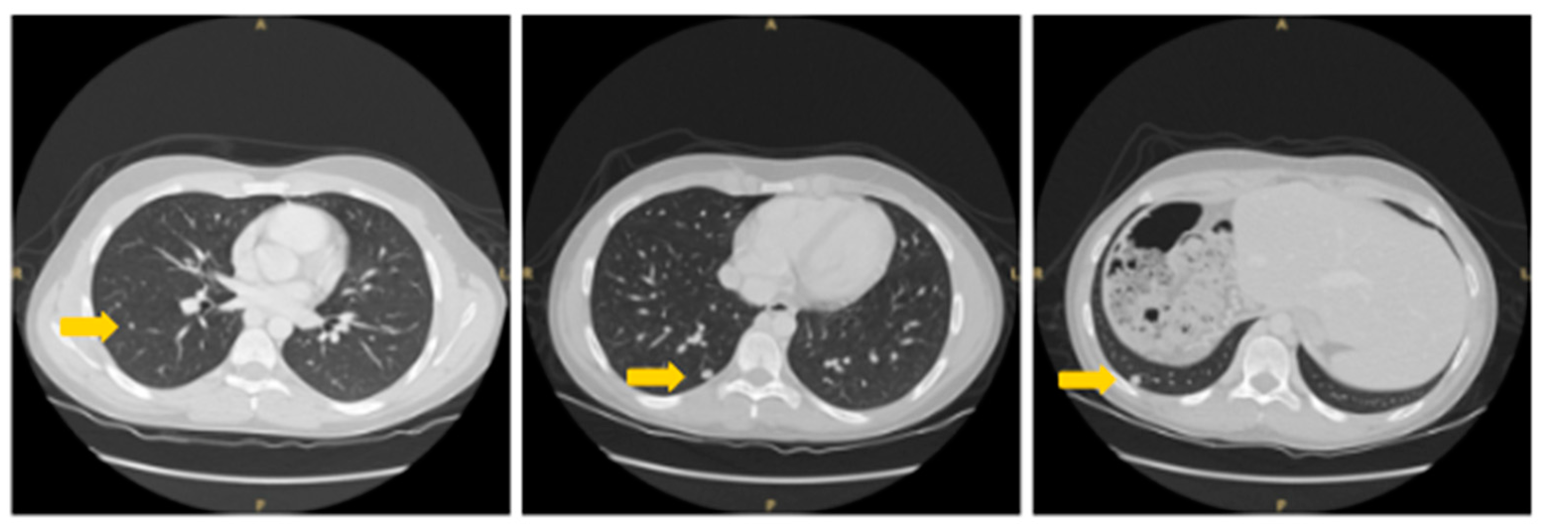
Figure 1. Three right pulmonary nodules highlighted by arrows.
Livers 04 00029 g001
Figure 2. Left pulmonary nodule highlighted by arrow. A: anterior; P: posterior; R: right; L: left.
Livers 04 00029 g002
Figure 3. Representative histopathological images of the metastasectomy specimen. At low magnification, the neoplasia was far from the surgical margins ((A), haematoxylin and eosin, scale bar: 3 mm). The neoplasia was composed of trabeculae and cords of neoplastic cells with prominent oncocytic and granular cytoplasm. The cells appeared large and polygonal, and exhibited well-defined cell borders ((B), haematoxylin and eosin, scale bar: 300 µm). Immunohistochemical analysis revealed positive staining for CK7 ((C), scale bar: 300 µm) and HepPar1 ((D), scale bar: 300 µm).
Livers 04 00029 g003
Figure 4. Chronological reconstruction of the aggressive management that was administered to our patient. Abbreviations: MW: microwave; XELOX: Chemotherapy protocol. Time is expressed as month/year.
Livers 04 00029 g004
Disclaimer/Publisher’s Note: The statements, opinions and data contained in all publications are solely those of the individual author(s) and contributor(s) and not of MDPI and/or the editor(s). MDPI and/or the editor(s) disclaim responsibility for any injury to people or property resulting from any ideas, methods, instructions or products referred to in the content.