Review

Deciphering the Gut–Liver Axis: A Comprehensive Scientific Review of Non-Alcoholic Fatty Liver Disease

1
ICMR-National Institute for Research in Environmental Health, Bhopal Bypass Road, Bhauri, Bhopal 462030, India
2
Internal Medicine Unit, Department of Clinical and Experimental Medicine, Gastroenterology and Hepatology Service, University Hospital Policlinico “G. Rodolico”, University of Catania, 95123 Catania, Italy
3
ReGenera Research Group for Aging-Intervention, 20144 Milano, Italy
4
Choithram Hospital and Research Centre Indore, Indore 452014, India
5
Stem Cell Research Centre, Department of Hematology, Sanjay Gandhi Post-Graduate Institute of Medical Sciences, Lucknow 226014, India
6
Department of Nutrition & Integrative Physiology, College of Health & Human Sciences, Florida State University, Tallahassee, FL 32306, USA
*
Author to whom correspondence should be addressed.
These authors contributed equally to this work.
Livers 2024, 4(3), 435-454; https://doi.org/10.3390/livers4030032
Submission received: 20 June 2024 / Revised: 27 August 2024 / Accepted: 29 August 2024 / Published: 12 September 2024

Abstract

:
Non-alcoholic fatty liver disease (NAFLD) has emerged as a significant global health issue. The condition is closely linked to metabolic dysfunctions such as obesity and type 2 diabetes. The gut–liver axis, a bidirectional communication pathway between the liver and the gut, plays a crucial role in the pathogenesis of NAFLD. This review delves into the mechanisms underlying the gut–liver axis, exploring the influence of gut microbiota, intestinal permeability, and inflammatory pathways. This review also explores the potential therapeutic strategies centered on modulating gut microbiota such as fecal microbiota transplantation; phage therapy; and the use of specific probiotics, prebiotics, and postbiotics in managing NAFLD. By understanding these interactions, we can better comprehend the development and advancement of NAFLD and identify potential therapeutic targets.

1. Introduction

Non-alcoholic fatty liver disease (NAFLD) is defined by the presence of at least 5% hepatic steatosis without common secondary causes like excessive alcohol consumption, chronic viral hepatitis, autoimmune hepatitis, congenital hepatic disorders, or long-term use of steatosis-inducing medications [1]. The prevalence of NAFLD is escalating globally, largely due to the obesity epidemic, and is anticipated to become the primary cause of liver transplantation by 2030, leading to rising healthcare costs [2]. Over the past few decades, the global prevalence of NAFLD has surged by more than 50%, from 25.3% between 1990 and 2006 to 38.0% between 2016 and 2019, reflecting the parallel rise in obesity and type 2 diabetes (T2D) [3]. In the majority of patients, NAFLD is associated with comorbidities like obesity, beta cell dysfunction, insulin resistance, T2D, and dyslipidemia [4]. The stringent associations between NAFLD and its mortality-driving comorbidities are not entirely understood but may involve continuous low-grade inflammation [2].
As our understanding of the metabolic underpinnings of this condition has evolved, new terminology and diagnostic criteria have been proposed, i.e., the term metabolic dysfunction-associated fatty liver disease (MAFLD) was introduced to better capture these metabolic associations. According to a 2020 consensus statement, MAFLD diagnosis requires the presence of hepatic steatosis in addition to one of the following criteria: overweight/obesity, type 2 diabetes, or evidence of metabolic dysregulation (elevated waist circumference, hypertension, hypertriglyceridemia, low HDL cholesterol, insulin resistance, or elevated C-reactive protein levels) [5,6]. More recently, in 2023, the term metabolic dysfunction-associated steatotic liver disease (MASLD) has been proposed to refine and simplify the diagnostic criteria. MASLD can be diagnosed if hepatic steatosis is present along with one of five cardiovascular risk factors: hypertension, type 2 diabetes, obesity, hypertriglyceridemia, or low HDL cholesterol, and without metabolic risk factors, the condition is termed cryptogenic steatotic liver disease (SLD). Additionally, a new category called metabolic dysfunction and alcoholic liver disease (MetALD) has been introduced for individuals with MASLD who consume more alcohol than the threshold for nonalcoholic status but less than the threshold for alcoholic liver disease (ALD) (average daily 20–50 g for women, 30–60 g for men). This shift from NAFLD to MAFLD and now MASLD reflects an ongoing effort to better characterize and diagnose liver diseases associated with metabolic dysfunction, ensuring a more targeted and precise approach to patient care and research [5,6].
Clinically, NAFLD is asymptomatic and is frequently diagnosed incidentally through abnormal liver enzyme findings or imaging studies by the presence of hepatic steatosis [7]. Additionally, NAFLD is driven by the excessive hepatic accumulation of lipids, particularly free fatty acids, fueled by the non-esterified fatty acid pool, dietary triglycerides, and de novo lipogenesis [8]. Lipotoxicity from non-esterified fatty acids and diacylglycerol promotes hepatic insulin resistance and endoplasmic reticulum stress, leading to chronic inflammation, liver fibrosis, cirrhosis, and ultimately hepatocarcinogenesis. As NAFLD progresses to non-alcoholic steatohepatitis (NASH), it is characterized by hepatic steatosis, inflammation, and cellular damage. Insulin resistance, oxidative stress, and pro-inflammatory cytokines such as TNF-α and IL-6 are key in this progression [9]. Persistent inflammation and liver damage in NASH can result in fibrosis, where extracellular matrix components are deposited in an attempt to repair the liver. Fibrosis advances through stages, ultimately leading to cirrhosis, where extensive scarring disrupts liver architecture and function, significantly increasing the risk of hepatocellular carcinoma (HCC). Mechanisms leading to HCC include genomic instability from chronic inflammation, epigenetic changes, and proliferative signaling due to ongoing liver cell damage and regeneration. Dysregulation in pathways such as insulin/IGF signaling, Wnt/β-catenin, and p53, as well as angiogenesis facilitated by factors like Vascular endothelial growth factor (VEGF), further contribute to carcinogenesis [9].
Theories on the pathogenesis of NAFLD have evolved from the two-hits hypothesis to the current multiple-hits hypothesis. The two-hits hypothesis posits that the first hit involves triglyceride accumulation in the liver (hepatic steatosis) due to sedentary lifestyles, high-fat diets, insulin resistance, and obesity. This accumulation increases the liver’s susceptibility to a second hit, characterized by lipotoxicity from free fatty acids, which then activates pro-inflammatory cytokines, promotes oxidative stress, and triggers fibrogenesis, leading to severe NAFLD phenotypes [10]. However, this hypothesis is now considered outdated as it fails to account for the roles of nutrition, gut microbiota, adipose tissue hormones, and concurrent insulin resistance in genetically predisposed individuals. The multiple-hits hypothesis suggests that genetic factors, diet, and environmental influences can cause gut microbiota dysbiosis, insulin resistance, and obesity, which together promote NAFLD development and are believed to interact in complex, interrelated ways [10]. Recent research has rapidly uncovered the link between gut microbiota and NAFLD, particularly in cases of obesity-related and high-fat-diet-induced NAFLD in both adults and children. Dysbiosis, or disruption of the typically beneficial gut microbiota, promotes the development of NAFLD by altering gut–liver homeostasis. This includes dysregulation of the gut barrier, transport of lipopolysaccharide (LPS) to the liver, altered bile acid profiles, and decreased short-chain fatty acids (SCFAs). The increased recognition of the importance of gut microbiota-mediated homeostasis in preventing NAFLD suggests that gut microbiota-targeted preventive and therapeutic strategies, such as probiotics, could be effective in combating NAFLD [10,11,12]. This review explores the intricate interactions and communication between gut microbiota and the liver, proposing potential therapeutic strategies focused on modulating gut microbiota for managing and treating NAFLD.

1.1. The Gut–Liver Axis: Key Insights and Interconnections

The gut–liver axis refers to the bidirectional network of signals (Figure 1) between the gut and the liver, connected via the hepatic portal vein, bile tract, and systemic circulation [13]. This axis is crucial for understanding the pathogenesis of various metabolic disorders including NAFLD and its progression to hepatocellular carcinoma (NAFLD-HCC).
The liver receives nearly two-thirds of its blood supply from the gastrointestinal tract via the portal vein, which transports nutrients, bacteria, and their components to the liver while bile and antibodies return to the small intestine to regulate the gut microbiome [14,15,16]. The gut–liver axis plays a crucial role in NAFLD pathogenesis due to its anatomical and functional connections. Dysbiosis, an imbalance in gut microbiota, can worsen the condition by increasing intestinal permeability, allowing harmful bacteria and their components to enter the liver. This stimulates hepatic immune cells and activates inflammatory pathways, potentially leading to NAFLD, which can progress to NASH, liver fibrosis, cirrhosis, and ultimately cancer [1,5,17,18]. This interaction is bidirectional, as gut dysfunction can impact liver health and vice versa, creating a cycle that promotes disease progression [19,20]. The gut–liver axis’s role in NAFLD is supported by the multiple-hit hypothesis, which states that different genetic, environmental, and lifestyle factors contribute to the disease development and progression. Other factors such as high caloric intake, physical inactivity, genetic predispositions [21], obesity, and gut dysbiosis-related damage to the intestinal integrity can lead to gut–liver axis malfunction, which might contribute to simple steatosis, thereby allowing bacterial components to enter circulation, a condition known as the “leaky” gut. This permeable gut permits hepatotoxic bacterial substances, including pathogen-associated molecular patterns (PAMPs) and damage-associated molecular patterns (DAMPs), to reach the liver through the hepatic portal circulation and activate toll-like receptors (TLRs) in hepatic cells. These TLRs sense bacterial products like lipoteichoic acid and lipopolysaccharides (LPS) influencing gut barrier function and permeability [22,23].
Subsequent triggers by increased lipotoxicity and enhanced intestinal permeability further worsen the metabolic and systemic profile of NAFLD patients [21]. Recent research emphasizes the key role of the gut–liver axis in liver diseases beyond NAFLD and states that the disruptions in the gut–liver axis, gut microbiome composition, and epithelial barrier function can increase microbial exposure, microbial translocation, infections, and a pro-inflammatory environment in the liver, thus causing disease progression [22].

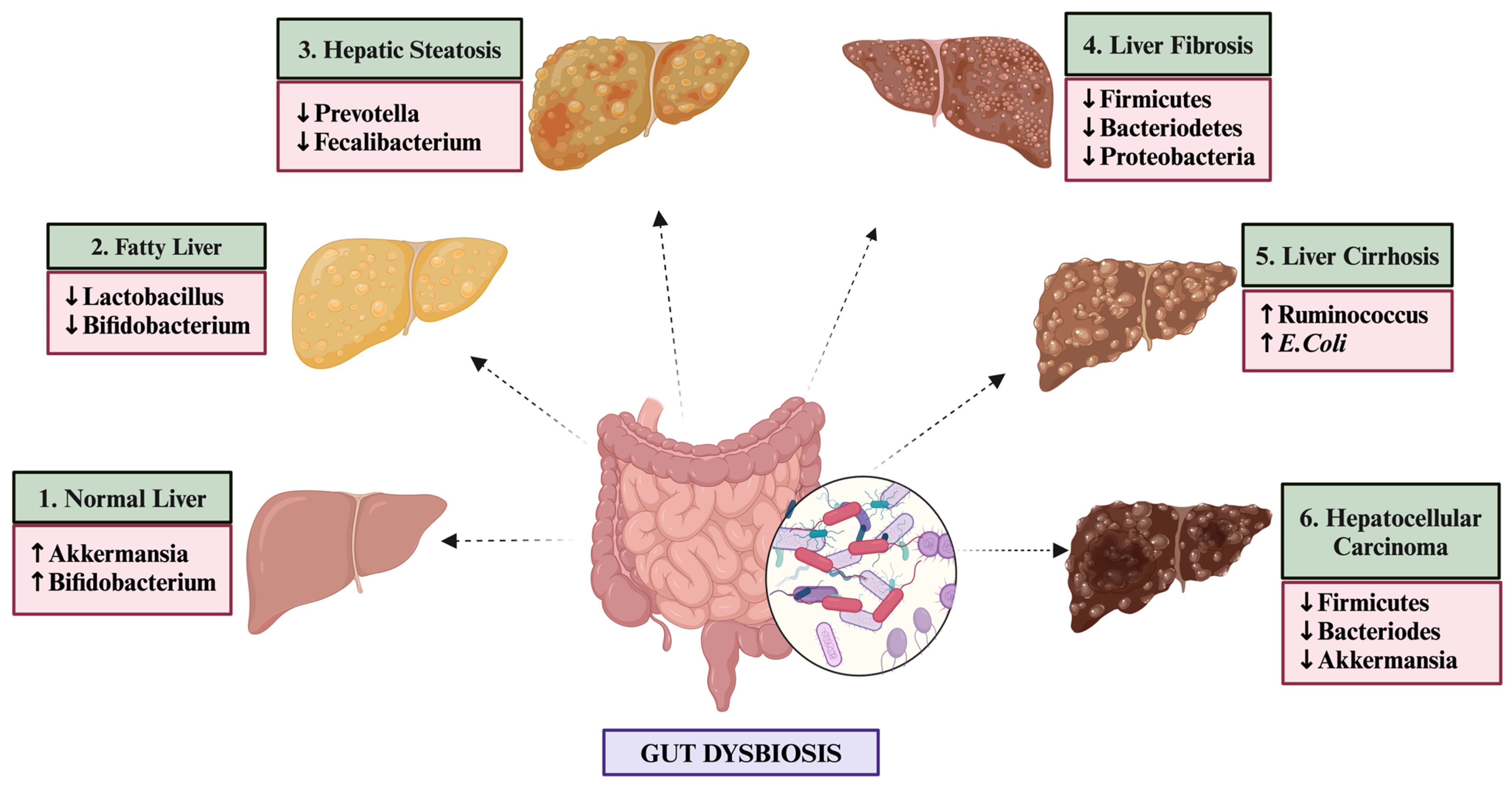
1.2. Distinct Gut Microbiome Signatures in NAFLD Patients

The intestinal tract harbors a vast number of microorganisms that play a vital role in maintaining metabolic homeostasis [10,24,25]. Among these, Firmicutes (primarily Gram-positive Bacilli, Clostridia, and Mollicutes) and Bacteroidetes (including Gram-negative Prevotella, Alistipes, and Parabacteroides) are the most dominant, constituting about 90% of the gut microbiota [19,24,26,27]. The epithelial mucosal and vascular barriers in the gut allow nutrient absorption while preventing the transmission of microbes and their toxins into the circulation. Disruption of gut barriers or dysbiosis, defined as a relative change in the composition and function of an individual’s commensal microbiota, can increase liver exposure to harmful substances and create a pro-inflammatory environment [10,26]. In NAFLD patients, gut microbiota imbalances manifest as reduced microbial diversity and an overgrowth of pathogenic bacteria like Escherichia coli, impairing the gut microbiota’s ability to maintain local homeostasis, whereas healthy individuals typically have higher microbial diversity and lower abundance of pathogenic bacteria [22,28,29,30] (Figure 2).
Among the extensive population of NAFLD patients, an observed increase in Proteobacteria, Lachnospiraceae, Escherichia, Enterobacteriaceae, Barnesiella intestinihominis, and Bacteroidetes is noted, though some studies reported a reduction or no change in Bacteroidetes [1,27,31], along with a decline in Prevotella and Firmicutes [28,29]. Da Silva et al. identified a reduction in levels of certain groups of gut microbiotas, including the Bacteroidetes and Firmicutes phyla, and an increase in the abundance of another group of microbiota in subjects with simple steatosis and NASH compared to controls suggesting that a particular gut microbiota may play a crucial role in the development and advancement of NAFLD [30]. In a similar study on the Korean population with NAFLD, a relatively lower alpha and beta diversity was reported with an abundance in the phylum Proteobacteria, family Enterobacteriaceae, genus Citrobacter and a significant decrease in the population levels of genus Faecalibacterium in subjects with NAFLD when compared to control. Additionally, they also reported a reduction in the abundance of butyrate-producing bacteria and a relative enhancement in the ethanol-producing bacteria in people with NAFLD [32]. In another study, it was revealed that the severity of NAFLD is directly linked to gut dysbiosis and alterations in the metabolic activity of the gastrointestinal microbiota. Here, Bacteroidetes was found to be associated with liver steatosis and NASH, while Ruminococcus was found to be significantly correlated with fibrosis [33]. A study on 171 Asians with NAFLD revealed significant variations in the microbiome diversity, with Ruminococcaceae and Veillonellaceae being the primary microbiota associated with the disease [34]. In a similar study on NAFLD patients, the correlation of gut metabolites with the abundance of specific genera was studied, and it was concluded that a decrease in Oscillospira is coupled to an up-regulation of 2-butanone, and an increase in Dorea and Ruminococcus was identified as the gut microbiome signatures linked with NAFLD onset [35].
Studies indicate that NAFLD is associated with a specific disbalance between two predominant phyla, namely Bacteroides and firmicutes. While some studies suggest increased Firmicutes are linked to NAFLD, others highlight the significant role of Bacteroide overgrowth in disease development [31,36,37,38]. A study on fecal microbiota of NAFLD cases showed an increased abundance of Enterobacteriaceae, Streptococcus, Blautia, Flavobacterium, Alkaliphilus, and a significantly reduced level of Akkermansia [39,40]. In another study, a substantial rise was found in the Bacteroidetes/Firmicutes ratio in NASH subjects independent of diabetes risk factors or drugs such as metformin use [38]. Additionally, NAFLD severity is also linked with increased fecal Bacteroides and decreased Prevotella levels [33]. Metagenome sequencing shows Bacteroides vulgatus and Eubacterium rectale are prevalent microbes in mild to moderate NAFLD, while Escherichia coli and Bacteroides vulgatus dominate in liver fibrosis [36,41].
High-throughput sequencing studies of NAFLD patients have revealed increased levels of Dysgonomonas, Escherichia coli, Veillonellaceae, and Bilophila, which promote endotoxin and endogenous ethanol production [19,42]. This leads to elevated systemic inflammation and insulin resistance. Conversely, beneficial bacteria like Akkermansia muciniphila, Faecalibacteriumprausnitzii (F. prausnitzii), Ruminococcaceae, Alistipes, and Bifidobacterium are reduced, impairing SCFA production and compromising the intestinal mucosal barrier [43,44]. Recent research indicates that dysbiosis of Alistipes in particular can have both beneficial and detrimental effects, with specific implications in liver fibrosis, colorectal cancer, and cardiovascular diseases. Its distinctive mechanism of fermenting amino acids, termed putrefaction, underscores its significant role in inflammation and liver-related conditions [45,46,47,48].

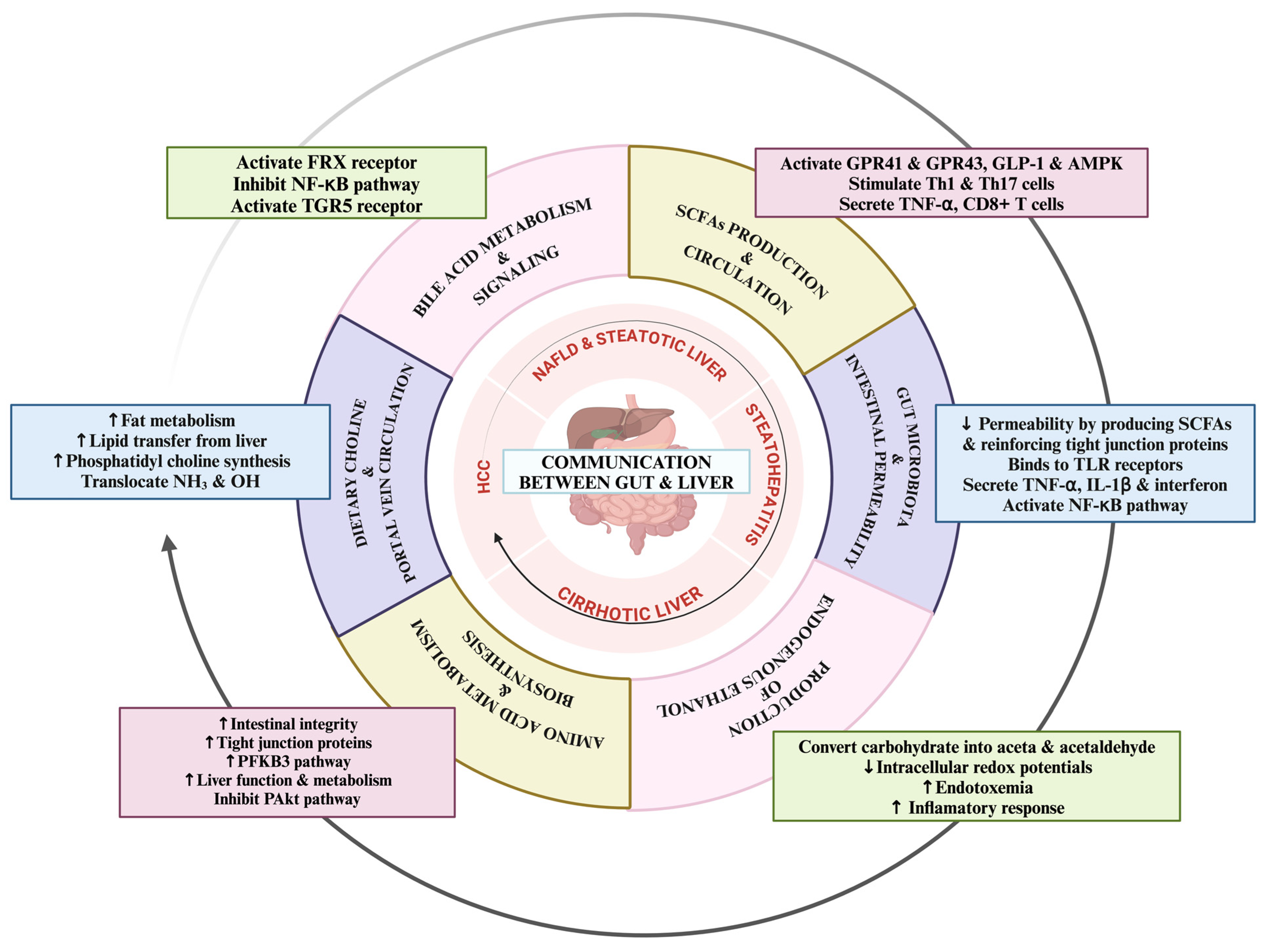
1.3. Dynamic Communication Mechanisms between Gut and Liver in NAFLD

The communication between the gut and liver in NAFLD is mediated via several mechanisms including the production and transport of live bacteria and its derived metabolites such as SCFAs, bile acids, trimethylamine oxide (TMAO), ethanol, choline, and amino acids (Figure 3) by the intestinal microbiota and the generation of proinflammatory effects during NAFLD progression [19].

2. Short-Chain Fatty Acids: Production, Circulation, and Effects on Liver Health

Short-chain fatty acids (SCFAs), particularly propionate, acetate, and butyrate, are produced through the fermentation of indigestible carbohydrates by gut bacteria [49,50,51,52]. While most SCFAs are utilized in the intestine as the source of energy, some are transported to the liver through the hepatic portal vein via the monocarboxylate transporter 1 (MCT-1) and the sodium-coupled monocarboxylate transporter 1 (SMCT-1) receptors [1]. The function of SCFAs in NAFLD is not entirely understood, but elevated fecal SCFA levels and a predominance of SCFA-producing bacteria are observed in NAFLD subjects compared to healthy individuals [53,54,55]. In general, butyrate primarily provides nourishment to colon epithelial cells, whereas propionate supports gluconeogenesis and cholesterol synthesis, and acetate acts as the key for lipogenesis and cholesterol biosynthesis in the liver [56,57,58,59].
Additionally, SCFAs influence liver metabolism, its metabolic substrates, and signaling molecules through various mechanisms, including appetite regulation, energy metabolism, insulin resistance, and adipose tissue metabolism. They manifest their metabolic effects by binding to G-protein-coupled receptors (GPRs) such as GPR41, GPR43, and GPR109A [51,59,60], with GPR43 being the most crucial for regulating insulin sensitivity and inflammatory responses, thereby influencing fat accumulation and metabolism in the liver cells and adipose tissue [59,60,61]. They activate GPR41 and GPR43, prompting enteroendocrine cells to release peptide YY (PYY), which slows intestinal transit and enhances nutrient absorption [62,63]. Furthermore, GPR activation stimulates glucagon-like peptide-1 (GLP-1), which promotes hepatic lipid β-oxidation and clinically reduces hepatic steatosis. SCFAs also activate AMP-activated protein kinase (AMPK), stimulating hepatic autophagy to facilitate triglyceride hydrolysis and free fatty acid β-oxidation [54,64,65].
Beyond metabolism, SCFAs play a role in immune modulation by stimulating pro-inflammatory T cells like Th1 and Th17 and influencing inflammatory cytokines, such as TNF-α, a key intermediator of liver inflammation [54,66]. Additionally, systemic acetate can enhance the recall response function of memory CD8+ T cells, thereby improving infection control [67]. Further research is needed to fully understand the characteristics and function of SCFAs and their role in NAFLD pathogenesis, particularly in relation to gut microbiome imbalance and the gut–liver axis.

2.1. Bile Acid Metabolism and Circulation Insights

Bile acids (BAs) play a significant role in enterohepatic circulation, particularly in the context of NAFLD. It is synthesized from cholesterol in the liver and secreted into the intestine to aid in lipid digestion and absorption [68,69]. Gut microbiota, especially Bacteroidetes, Lactobacillus, Bifidobacterium, and Clostridium XIVa with the help of enzyme bile salt hydrolases (BSH), converts primary BAs into secondary BAs, which are then reabsorbed in the ileum and carried back to the liver via the portal vein [1,49,70,71]. This enterohepatic circulation maintains BA homeostasis and is intricately linked to gut–liver communication [72]. In NAFLD, gut dysbiosis alters BA metabolism, causing an increase in BA synthesis, leading to higher levels of primary BAs and a disrupted primary-to-secondary BA ratio [73]. This imbalance affects the signaling of BA receptors, such as the farnesoid X receptor (FXR) [20,74] and Takeda G-protein receptor 5 (TGR5), which regulate lipid and glucose metabolism, insulin sensitivity, and inflammatory responses [70,75,76]. The FXR, which is primarily located in the liver and intestines, upon activation reduces hepatic lipogenesis and inflammation by inhibiting the NF-κB pathway [71]. Conversely, suppressed FXR activity, which is often observed in NAFLD, leads to increased lipid accumulation and inflammation [77]. For instance, germ-free mice, which lack gut microbiota, are resistant to liver steatosis, highlighting the role of microbial interactions in BA metabolism and NAFLD pathogenesis [78]. TGR5, another important bile acid receptor, upon activation, modulates glucose homeostasis and inflammatory cytokine production, which further improves liver steatosis and hepatocyte damage [79,80]. Dysbiosis-induced alterations in BA levels and signaling further exacerbate NAFLD by promoting hepatic lipid accumulation and reducing insulin sensitivity [80]. Clinical and preclinical studies demonstrate that targeting the intestinal FXR can influence BA metabolism and NAFLD outcomes, although differences between human and mouse models necessitate further research [54]. Maintaining healthy BA metabolism and gut microbiota is essential for preventing NAFLD progression, and understanding the gut–liver axis offers promising avenues for gut microbiota-targeted therapies.

2.2. Intestinal Barrier Integrity and Portal Vein Dynamics in the Gut–Liver Axis

Intestinal permeability, crucial for regulating gut–liver communication is determined by the integrity and robustness of the gut intestinal barrier, which comprises the mucus layer, intestinal epithelium, and the gut vascular barrier (GVB) and regulates the transport and circulation from the gut to the liver [81,82]. Enterocytes linked together by tight junction proteins, namely E-cadherins, occludins, and claudins, regulate the ingress into the hepatic portal vein and its way into the liver [81,82]. The portal vein provides a direct link between the intestine and the liver, delivering approximately 70% of blood, nutrients, and beneficial microbial products like SCFAs to the liver [31,53,83,84]. Gut dysbiosis disrupts the gut barrier integrity, allowing toxic factors like live bacteria, bacterial components (DAMPs, LPS), and proinflammatory metabolites such as ethanol and ammonia to enter the hepatic portal circulation and reach the liver. These toxins further activate immune cells and respective inflammatory pathways, thereby contributing to the NAFLD development and progression [85].
Impaired gut barrier function is a widely recognized characteristic of dysbiosis in subjects with NAFLD and NASH [1,86]. For instance, several studies have reported that the severity of NAFLD is associated with higher zonulin levels in patients compared to healthy controls [87,88,89]. Furthermore, in a meta-analysis recruiting 128 NAFLD patients, an increase of 39.1% was reported in intestinal gut permeability in NAFLD subjects as compared to 6.8% in healthy controls [61,90]. Clinically, it has been observed that patients with NAFLD exhibit higher portal LPS levels than those with simple steatosis. Additionally, metabolic alterations in the portal system, such as elevated L-tryptophan, DL-3-phenylacetic acid, and glycocholic acid, are observed in NAFLD patients [19,91].
Gut microbiota can enhance intestinal gut integrity by generating and synthesizing metabolites such as SCFAs, which reinforce and strengthen tight junctions [86]. Some bacteria like Akkermansia muciniphila can improve gut permeability and affect NAFLD development and progression by regulating tight junctions [83]. However, gut dysbiosis can disrupt these tight junctions, thereby allowing bacteria and their metabolites to translocate from the gut lumen to the liver where Kupffer cells release inflammatory cytokines, exacerbating liver inflammation [84]. This inflammatory response is generated via the stimulation of TLRs on Kupffer cells leading to the subsequent activation of the inflammatory cascade and production of proinflammatory cytokines namely interleukin (IL)-1β, tumor necrosis factor (TNF) α, and interferons [85]. Among the TLR family, TLR2, TRL4, TRL5, and TRL9 are best implicated in the pathogenesis of NAFLD [92]. TLR4 specifically binds with LPS, activates the NF-kB pathway, and further exacerbates the condition by contributing to chronic low-grade inflammation in obesity and NAFLD [20,59,66,93].

2.3. Dietary Choline: Implications for the Gut–Liver Axis

Choline, an important component of cell membrane phospholipid [94] is crucial for liver fat metabolism, particularly in forming very low-density lipoproteins (VLDL) necessary for lipid transfer from the liver via the phosphatidylcholine synthase (Pcs) pathway [1,95,96]. According to a study, the high choline intake is directly associated with reduced fatty liver risk in normal-weight women [97]. In animal models of hepatic steatosis, choline deficiency frequently results in liver fat accumulation and gets reversed in patients when choline supplementation is given [93]. The intestinal microbiota can influence choline metabolism by converting dietary choline into trimethylamines, which reduces circulating choline levels. This mimics choline-deficient diets, contributing to NAFLD development by impairing phosphatidylcholine synthesis necessary for VLDL assembly, thereby resulting in triglyceride buildup in hepatocytes [98,99].
Gut commensals such as E. coli and Desulfovibrio desulfuricans can convert choline to trimethylamine (TMA), which the liver further oxidizes to trimethylamine N-oxide (TMAO), and elevated TMAO levels impair glucose homeostasis, exacerbate insulin resistance, and contribute to atherosclerosis, obesity, and NAFLD [1,98]. A study found that higher plasma TMAO levels are linked to increased all-cause mortality in NAFLD patients [100,101]. Moreover, choline deficiency influences the gut microbiome composition and function, with higher levels of bacteria like Gammaproteobacteria and Erysipelotrichia, which convert choline to toxic methylamines, being directly connected to liver damage [1,102,103]. This alteration supports the role of gut microbes in NAFLD, with enhanced pathogenic bacterial growth potentially increasing choline demand and contributing to choline depletion and harmful metabolite production [54,103,104]. Thus, the gut microbiome significantly impacts NAFLD through altered choline metabolism and related mechanisms.

2.4. Endogenous Ethanol Production and Its Impact on the Gut–Liver Axis

Ethanol is typically produced in very small quantities in the intestine and is metabolized in the liver by the enzyme alcohol dehydrogenases [105]. Hepatic damage in alcoholic liver disease (ALD) and NAFLD is almost identical [106], suggesting a link between blood ethanol levels and changes in gut microbiota [107,108]. Certain gut microbiota can help in the fermentation of dietary carbohydrates and sugars into ethanol, which further gets converted into acetate and acetaldehyde via the upregulation of the CYP2E1 pathway, leading to fatty acid generation and fluctuations in intracellular redox potential, causing an increased intestinal permeability, endotoxemia, and inflammatory response, thus exacerbating liver damage [59,109,110,111]. Gut dysbiosis can enhance the level of ethanol-producing bacteria such as Escherichia, Klebsiella pneumonia, Bacteroides, Bifidobacterium, and Clostridium. A study reported a fivefold increase in alcohol-producing bacteria, namely Escherichia and Proteobacteria, with the quantity of ethanol synthesized to be directly linked to the abundance of Proteobacteria, particularly Klebsiella pneumoniae [105,112,113]. Additionally, a study identified Klebsiella pneumonia K14, a high alcohol producer, as a causative factor for NAFLD [113]. Another study found that mutations in Klebsiella pneumonia K14 and Collinsella aerofaciens influenced alcohol production and were directly correlated with the severity of NAFLD [114,115]. These findings highlight complex interactions between ethanol and NAFLD, warranting further research to clarify ethanol’s role in NAFLD development and advancement.

2.5. Amino Acid Metabolism in the Gut–Liver Axis

Amino acids and their derived metabolites, including phenylalanine, tryptophan, branched-chain amino acids (BCAAs), and microbiota-derived metabolites, play a crucial role in the development and pathogenesis of NAFLD by influencing liver function through mechanisms such as intestinal integrity, inflammation, lipogenesis, and insulin resistance [116].
Tryptophan, an example of an essential amino acid, is metabolized by gut bacteria through various pathways. The indole pathway converts tryptophan to indole, which enhances intestinal integrity and reduces the severity of NAFLD by promoting tight junction proteins and glycolysis via the PFKFB3 pathway [109,110]. In another pathway, butyrate is catalyzed by indoleamine 2,3-dioxygenase (IDO) to generate kynurenines (Kyn), which further over-activate the Kyn pathway in NAFLD, causing inflammation in various organs [109,110,111,112,113,114,115,116,117]. Indole-3-acetic acid, a derivative of indole, reduces hepatic lipogenesis and inflammation, thus improving NAFLD [118]. Serotonin, another tryptophan metabolite, inhibits energy expenditure in brown adipose tissue in NAFLD by inhibition of serotonin receptors, namely Thp1 and HTR2a, thereby reducing hepatic steatosis [105,119]. In NAFLD, there is disturbed tryptophan metabolism, and supplementation of tryptophan might increase the intestinal barrier integrity and improve liver NAFLD and function [110]. Phenylalanine and its derivatives, such as phenylacetic acid (PAA), are reported to contribute to the development and pathogenesis of NAFLD [120]. In a study, the transplantation of a liver steatosis-related microbiome into germ-free mice enhanced the PAA and hepatic triglyceride levels, thereby altering the gene expression related to lipogenesis and thus promoting triglyceride accumulation. PAA also inhibits the pAkt pathway, which increases hepatic steatosis by enhancing the use of BCAAs for lipid accumulation [105,121]. BCAAs like Valine, leucine, and isoleucine are linked to NAFLD via the impairment of the TCA cycle in NAFLD [122]. According to a study, elevated BCAA levels are associated with insulin resistance, liver inflammation, and ballooning, indicating severe NAFLD, while free dietary amino acid intake prevents unhealthy metabolic outcomes, as indicated in male mice [105,123,124].

2.6. Therapeutic Approaches Targeting NAFLD through Gut Microbiome-Centered Interventions

Gut microbiota interventions show promise in treating NAFLD by modifying microbial composition and function to enhance overall health. This can be achieved through dietary changes like increasing fiber intake or using probiotics, prebiotics, and postbiotics, as well as through methods such as phage therapy and fecal microbiota transplantation. These approaches aim to restore microbial balance, influence the gut–liver axis, and reduce liver inflammation and fat accumulation while minimizing side effects and promoting a safer, more sustainable management of liver conditions and metabolic health [125].
In contrast, current clinical drugs for NAFLD, such as pioglitazone and vitamin E, primarily target insulin resistance, oxidative stress, and inflammation. Pioglitazone, a thiazolidinedione, improves insulin sensitivity and reduces liver fat content but is associated with adverse effects like weight gain and an increased risk of heart failure. Vitamin E, an antioxidant, helps reduce oxidative stress and inflammation in the liver, showing beneficial effects in non-diabetic NAFLD patients but with potential long-term safety concerns [126].
Other pharmacological agents, such as glucagon-like peptide-1 (GLP-1) agonists and sodium-glucose co-transporter-2 (SGLT2) inhibitors, show promise for improving metabolic parameters and reducing liver fat [127,128]. However, these drugs can have side effects and do not address the underlying gut–liver axis, requiring long-term efficacy and safety evaluation [129,130]. For instance, pioglitazone can cause weight gain and increase the risk of congestive heart failure, metformin carries a risk of lactic acidosis, and vitamin E, while reducing oxidative stress, has been linked to a higher risk of prostate cancer with long-term use [131,132]. Moreover, these drugs often target specific pathways, which may limit their effectiveness in addressing the multifactorial nature of NAFLD [130].
Furthermore, gut microbiota therapies generally exhibit a favorable safety profile with minimal side effects. Most probiotics and prebiotics are well-tolerated, with occasional mild gastrointestinal symptoms such as bloating or gas being the most common adverse effects. Unlike clinical drugs, gut microbiota interventions do not carry risks of severe complications like congestive heart failure, lactic acidosis, or cancer. This makes them a safer option, especially for patients with NAFLD who are at increased risk of cardiovascular events or progressive liver disease [44,133]. Therefore, gut microbiota interventions offer a complementary approach by targeting the root cause of dysbiosis and providing a more holistic treatment option for NAFLD.

3. Probiotics

Probiotics are non-pathogenic live microorganisms that are generally considered beneficial for maintaining gut health and are mostly used in diarrhea [134,135,136] and malnutrition [137,138]. Recently, probiotic supplements have been reported to have worthwhile effects on both intro-intestinal and extro-intestinal diseases, including NAFLD [139,140].
Probiotics can restore gut microbiota balance [141], enhance lipid and glucose profiles [142,143], maintain intestinal gut barrier integrity [144], reduce inflammation [145], and inhibit oxidative stress [146]. These mechanisms theoretically contribute to the effective prevention of NAFLD.
Multiple animal studies have demonstrated that probiotics benefit NAFLD by reducing inflammation, hepatic triglyceride levels, overall body weight, and visceral adipose tissue weight, as well as by improving glucose homeostasis [147]. However, clinical evidence regarding the direct impact of probiotics on NAFLD is still insufficient, despite numerous animal studies exploring their therapeutic mechanisms. In a study aimed to evaluate the overall efficacy of probiotics in treating NASH using a hepatocyte-specific PTEN knockout mouse model which closely resembles human NAFLD indicated that probiotics significantly reduced serum transaminase levels, NAFLD activity scores, and the expression of pro-inflammatory cytokine genes. Additionally, probiotics alleviated oxidative stress, evidenced by anti-oxidative stress markers, and altered glutathione levels, suggesting a potential mechanism of action for their beneficial effects. Overall, probiotics demonstrated positive effects in mitigating NAFLD and preventing carcinogenesis in the PTEN knockout mice model [15]. Another study found that probiotic supplementation with Lactobacillus acidophilus in rabbits protected them against NAFLD. The treatment restored liver function, lipolytic gene expression, and antioxidant levels to normal [148].
Another meta-analysis aimed to summarize randomized controlled trials that examined the effects and efficacy of probiotics on NAFLD, which included the assessment of the impact of probiotics on liver function tests, specifically alanine aminotransferase (ALT), aspartate aminotransferase (AST), and gamma-glutamyl transferase (GGT). Overall, probiotics have shown beneficial effects and could be considered as an additional therapeutic approach for managing NAFLD [149]. A similar meta-analysis of 21 randomized clinical trials containing 1037 NAFLD subjects found that probiotic intervention significantly improved liver function tests, blood lipid levels, and blood glucose and insulin levels, thereby reducing hepatic steatosis. However, it did not affect the BMI index, level of inflammatory factors, or insulin resistance significantly. Subgroup analysis revealed that treatments lasting 12 weeks or longer resulted in better improvements in ALT, GGT, triglycerides, and blood sugar. Overall, the study indicates that probiotics effectively regulate liver function, liver steatosis, glucose homeostasis, and blood lipid levels in NAFLD subjects [150].
Over the past decade, clinical investigations into probiotic therapies for NAFLD have shown improvements, but the results and benefits remain contentious. The discrepancies in the trials mentioned above may be attributed to variations in study design, probiotic dosage, types of strains, and supplement duration, as well as the characteristics of the participants. Overall, probiotics may be beneficial in reducing transaminase levels and partially improving lipid profiles, particularly in the initial stages of NAFLD development [151]. However, long-term research is needed to establish more robust evidence and to determine whether probiotics can prevent the advancement and progression of NAFLD to liver cirrhosis and HCC. Currently, there are significant challenges in using probiotics to treat NAFLD. First, selecting the appropriate probiotics is complex due to the diverse range of strains, with different benefits and effects attributed to different strains; multi-strain probiotics have shown more effectiveness than single strains. Second, while initial-stage NAFLD patients have shown improvements with reduced transaminase levels following probiotics treatment, it is unclear whether probiotics can prevent advancement to liver fibrosis. Third, both the study duration and probiotic dosage impact efficacy outcomes. Despite these challenges, probiotics are considered a promising therapeutic strategy and are anticipated to become an effective, significant, and widely used treatment for NAFLD patients [151].

4. Prebiotics

Prebiotics are non-viable dietary components linked with gut microbiota modulation and can provide health-related benefits to the host. They primarily include polysaccharides like inulin, cellulose, hemicellulose, resistant starch, and pectins as well as oligosaccharides such as fructooligosaccharides, isomaltooligosaccharides, galactooligosaccharides, lactulose, xylooligosaccharides, and soy oligosaccharides, with fructooligosaccharides being widely researched in NAFLD [152]. These substances selectively stimulate the growth and activity of gut microbiota. Numerous animal studies have demonstrated that prebiotic supplementation can reduce fatty acid synthesis pathways, potentially lowering hepatic triglyceride accumulation caused by fructose. This effect is achieved through reduced expression of enzymes involved in hepatic lipogenesis, including fatty acid synthase and acetyl Co-A carboxylase. Additionally, oligofructose modifies the gut microbiome to promote Bifidobacterium, which enhances intestinal barrier function and lowers endotoxin levels [153].
Additionally, in another study on mice models, the administration of prebiotics decreased liver inflammation in obese mice via the glucagon-like peptide-2-dependent pathway, which also positively affected the gut barrier [154]. The study also evaluated the liver-protective effects of COS23, an enzymatically catalyzed byproduct of chitosan oligosaccharide (COS), in diet-induced obese mice and reported that it significantly reduced hepatic steatosis and improved liver injury by decreasing toxic lipids such as triglycerides and free fatty acids in the liver. It also regulated lipid-related pathways and inflammation while altering lipid profiles in plasma. Additionally, COS23 modulated gut microbiota, decreasing Mucispirillum and increasing Coprococcus abundance, and improved intestinal barrier function. These findings suggest that COS23 holds promise as a clinical treatment for NAFLD. However, human clinical trials are essential to corroborate this information [155].
Despite limitations in many studies, such as small sample sizes and insufficient data on patients’ diets and lifestyles, treatments with probiotics and prebiotics for NAFLD show promise. Strong evidence from short-term, high-quality human studies supports using dietary probiotics and prebiotics as a possible therapeutic approach for NAFLD. Nevertheless, additional research is required to establish a link between these results and alterations in gut microbiota [156].

5. Postbiotics

Postbiotics, which contain various bioactive substances, might exert promising effects by reducing hepatic lipid build-up [44,157]. A study explored the preventive role of postbiotics derived from Lactobacillus paracasei on NAFLD. The findings revealed that simultaneous ingestion of a high-fat diet (HFD) and postbiotics slowed weight gain, suppressed epididymal white fat hypertrophy and glycemic hike, improved serum biochemical markers associated with blood lipid metabolism, and reduced hepatic steatosis and low-grade liver inflammation in mice [158]. Bacterial sequencing demonstrated that postbiotics altered the gut microbiota in HFD mice, elevating the levels of Akkermansia and decreasing the relative abundance of the Lachnospiraceae NK4A136 group, Bilophila, and Ruminiclostridium [158].

6. Fecal Microbiota Transplantation (FMT)

FMT involves transferring fecal matter from a healthy donor to a patient, which can potentially correct dysbiosis and improve liver health. This procedure helps restore the balance of commensal gut microbiota and enhances the gut’s natural defenses, preventing the entry and translocation of potential pathogens [159]. As a result, FMT has been proposed to address other dysbiosis-related conditions in the gut microbiome, including those observed in NAFLD [48]. FMT restores the microbiome-mediated regulation of gut barrier integrity, preventing the entry and migration of potential pathogens, such as Clostridiales [160]. Initial animal studies provided the first evidence of FMT’s impact on NAFLD. Leroy et al. found that FMT from NAFLD mice triggered NAFLD development in most recipient mice, underscoring the substantial colonization of specific bacteria following FMT [161]. Additionally, a study investigated two different groups of mice fed a high-fat diet, with one group receiving FMT from healthy donors. This intervention led to a notable decrease in typical histological features associated with NAFLD in the treated group [162]. Interestingly, improvements in NASH-related histological features, including liver fibrosis and inflammatory infiltrates, were also observed post-FMT. These improvements correlated with reductions in body weight, fat content, and serum transaminase levels [163]. The clinical trials reported have demonstrated promising results of FMT in NAFLD, benefiting both diabetic and non-diabetic patients, with improvements noted in glycemic control and liver steatosis. Furthermore, initial reports suggest FMT may also be safe for use in patients with liver cirrhosis, a progression of NASH [48,164].
FMT seems to be a safe and effective treatment for NAFLD, though further randomized controlled trials (RCTs) and long-term follow-up studies are necessary to fully evaluate its effects and efficacy, especially in lean NAFLD patients who often do not benefit significantly from lifestyle and dietary changes, cholesterol-lowering agents, or probiotics [44].

7. Phage Therapy

Phage therapy, long overshadowed by antibiotics, is now being reconsidered as a potent tool against antibiotic-resistant bacteria [165]. Given the mechanistic links between bacterial microbiota, gastrointestinal, and liver disorders, the targeted manipulation of the gut microbiota through phages’ selective bactericidal action has garnered renewed attention. The human gut virome, dominated by phages, shows significant alterations in patients with liver diseases when compared to healthy controls [125]. Changes in the phageome have been observed in NAFLD patients, with lower viral diversity in advanced stages and higher abundance of Enterobacteria, Escherichia, and Lactobacillus phages [125].
Phages can target and reduce specific pathobionts, as evidenced by preclinical studies showing that selective elimination of pathobionts via phages improves inflammatory bowel disease, ethanol-induced liver disease, primary sclerosing cholangitis, and NAFLD in mouse models [166]. For example, phages targeting the cytolysin-producing Enterococcus faecalis reduced the circulation and presence of harmful protein cytolysin, which is linked to severe liver disease and mortality in NALD subjects [167]. In studies with humanized mice, oral administration of phages against cytolysin-positive E. faecalis reduced ethanol-induced liver injury, liver steatosis, and liver and gut inflammation. These findings highlight the role of harmful bacteria in fatty liver diseases and demonstrate that phage therapy can reduce liver disease and its associated complications in preclinical models. Phages typically have a narrow host range, infecting closely related strains within species, which minimizes collateral damage to the recipient’s microbiome but may also limit their ability to broadly modulate the gut microbiome [125]. The impact of the gut intestinal virome on bacterial microbiota and NAFLD progression remains largely unclear. However, phage therapy shows potential for treating NAFLD, as demonstrated by the prevention of NAFLD development in mice after eliminating ethanol-producing Klebsiella pneumoniae through phage therapy [113,168].

8. Conclusions and Future Prospects

The gut–liver axis plays a fundamental role in the development and pathogenesis of NAFLD. Gut dysbiosis, along with increased intestinal permeability, and altered bile acid signaling are the key factors responsible for these liver diseases. Advances in understanding this axis are driving the development of microbiota-based targeted therapeutic tools to prevent and treat NAFLD, ultimately improving patient outcomes. Despite the proven therapeutic potential of probiotics, fecal microbiota transplantation, and phage therapy in these conditions, comprehensive safety assessments and data on microbiota–host interactions are lacking. Most of the current knowledge is derived from animal studies, which face translational limitations due to physiological differences between species. Hence, large-scale controlled human studies with standardized and the most effective strains are needed to optimize dosages and treatment durations to individualize therapy for better disease management. The findings then need to be translated into clinical practice. Comprehensive studies on microbiota–host interactions will help pinpoint specific microbial strains or metabolites critical to NAFLD pathogenesis, aiding in the identification of reliable biomarkers for early diagnosis and monitoring. Additionally, integrating multi-omics technologies will offer a deeper, more comprehensive understanding of the molecular mechanisms at play, paving the way for personalized medicine and novel therapeutic targets. By advancing research in these areas, we can develop innovative strategies to mitigate the global burden of NAFLD and enhance patient outcomes.

Author Contributions

Conceptualization, M.K. (Manoj Kumar) and S.S.; writing and original draft preparation, M.K. (Mona Kriti) and S.S.; review and editing, R.C., F.M., M.M., A.J., V.V. and R.N.; visualization R.T. All authors have read and agreed to the published version of the manuscript.

Funding

This research received no external funding.

Institutional Review Board Statement

Not applicable.

Informed Consent Statement

Not applicable.

Data Availability Statement

No new data were created or analyzed in this study. Data sharing is not applicable to this article.

Conflicts of Interest

The authors declare no conflicts of interest.

References

  1. Kolodziejczyk, A.A.; Zheng, D.; Shibolet, O.; Elinav, E. The role of the microbiome in NAFLD and NASH. EMBO Mol. Med. 2019, 11, e9302. [Google Scholar] [CrossRef] [PubMed]
  2. Grander, C.; Grabherr, F.; Tilg, H. Non-alcoholic fatty liver disease: Pathophysiological concepts and treatment options. Cardiovasc. Res. 2023, 119, 1787–1798. [Google Scholar] [CrossRef] [PubMed]
  3. Wong, V.W.-S.; Ekstedt, M.; Wong, G.L.-H.; Hagström, H. Changing epidemiology, global trends and implications for outcomes of NAFLD. J. Hepatol. 2023, 79, 842–852. [Google Scholar] [CrossRef] [PubMed]
  4. Chalasani, N.; Younossi, Z.; Lavine, J.E.; Charlton, M.; Cusi, K.; Rinella, M.; Harrison, S.A.; Brunt, E.M.; Sanyal, A.J. The diagnosis and management of nonalcoholic fatty liver disease: Practice guidance from the American Association for the Study of Liver Diseases. Hepatology 2018, 67, 328–357. [Google Scholar] [CrossRef] [PubMed]
  5. Chen, L.; Tao, X.; Zeng, M.; Mi, Y.; Xu, L. Clinical and histological features under different nomenclatures of fatty liver disease: NAFLD, MAFLD, MASLD and MetALD. J. Hepatol. 2024, 80, e64–e66. [Google Scholar] [CrossRef]
  6. Rinella, M.E.; Sookoian, S. From NAFLD to MASLD: Updated naming and diagnosis criteria for fatty liver disease. J. Lipid Res. 2024, 65, 100485. [Google Scholar] [CrossRef]
  7. Lucas, C.; Lucas, G.; Lucas, N.; Krzowska-Firych, J.; Tomasiewicz, K. A systematic review of the present and future of non-alcoholic fatty liver disease. Clin. Exp. Hepatol. 2018, 4, 165–174. [Google Scholar] [CrossRef]
  8. Zhu, Z.; Zhang, X.; Pan, Q.; Zhang, L.; Chai, J. In-depth analysis of de novo lipogenesis in non-alcoholic fatty liver disease: Mechanism and pharmacological interventions. Liver Res. 2023, 7, 285–295. [Google Scholar] [CrossRef]
  9. Foerster, F.; Gairing, S.J.; Müller, L.; Galle, P.R. NAFLD-driven HCC: Safety and efficacy of current and emerging treatment options. J. Hepatol. 2022, 76, 446–457. [Google Scholar] [CrossRef]
  10. Jasirwan, C.O.M.; Lesmana, C.R.A.; Hasan, I.; Sulaiman, A.S.; Gani, R.A. The role of gut microbiota in non-alcoholic fatty liver disease: Pathways of mechanisms. Biosci. Microbiota Food Health 2019, 38, 81–88. [Google Scholar] [CrossRef]
  11. Tokuhara, D. Role of the gut microbiota in regulating non-alcoholic fatty liver disease in children and adolescents. Front. Nutr. 2021, 8, 700058. [Google Scholar] [CrossRef] [PubMed]
  12. Nagpal, R.; Kumar, M.; Yadav, A.; Hemalatha, R.; Yadav, H.; Marotta, F.; Yamashiro, Y. Gut microbiota in health and disease: An overview focused on metabolic inflammation. Benef. Microbes 2016, 7, 181–194. [Google Scholar] [CrossRef] [PubMed]
  13. Han, H.; Jiang, Y.; Wang, M.; Melaku, M.; Liu, L.; Zhao, Y.; Everaert, N.; Yi, B.; Zhang, H. Intestinal dysbiosis in nonalcoholic fatty liver disease (NAFLD): Focusing on the gut–liver axis. Crit. Rev. Food Sci. Nutr. 2023, 63, 1689–1706. [Google Scholar] [CrossRef] [PubMed]
  14. Barrow, F.; Khan, S.; Fredrickson, G.; Wang, H.; Dietsche, K.; Parthiban, P.; Robert, S.; Kaiser, T.; Winer, S.; Herman, A. Microbiota-driven activation of intrahepatic B cells aggravates NASH through innate and adaptive signaling. Hepatology 2021, 74, 704–722. [Google Scholar] [CrossRef] [PubMed]
  15. Arai, N.; Miura, K.; Aizawa, K.; Sekiya, M.; Nagayama, M.; Sakamoto, H.; Maeda, H.; Morimoto, N.; Iwamoto, S.; Yamamoto, H. Probiotics suppress nonalcoholic steatohepatitis and carcinogenesis progression in hepatocyte-specific PTEN knockout mice. Sci. Rep. 2022, 12, 16206. [Google Scholar] [CrossRef]
  16. Song, Q.; Zhang, X. The role of gut–liver axis in gut microbiome dysbiosis associated NAFLD and NAFLD-HCC. Biomedicines 2022, 10, 524. [Google Scholar] [CrossRef]
  17. Ji, Y.; Yin, Y.; Sun, L.; Zhang, W. The molecular and mechanistic insights based on gut–liver axis: Nutritional target for non-alcoholic fatty liver disease (NAFLD) improvement. Int. J. Mol. Sci. 2020, 21, 3066. [Google Scholar] [CrossRef]
  18. Compare, D.; Coccoli, P.; Rocco, A.; Nardone, O.; De Maria, S.; Cartenì, M.; Nardone, G. Gut–liver axis: The impact of gut microbiota on non alcoholic fatty liver disease. Nutr. Metab. Cardiovasc. Dis. 2012, 22, 471–476. [Google Scholar] [CrossRef]
  19. Liu, J.; Wu, A.; Cai, J.; She, Z.-G.; Li, H. The contribution of the gut-liver axis to the immune signaling pathway of NAFLD. Front. Immunol. 2022, 13, 968799. [Google Scholar] [CrossRef]
  20. Poeta, M.; Pierri, L.; Vajro, P. Gut–liver axis derangement in non-alcoholic fatty liver disease. Children 2017, 4, 66. [Google Scholar] [CrossRef]
  21. Frasinariu, O.E.; Ceccarelli, S.; Alisi, A.; Moraru, E.; Nobili, V. Gut-liver axis and fibrosis in nonalcoholic fatty liver disease: An input for novel therapies. Dig. Liver Dis. 2013, 45, 543–551. [Google Scholar] [CrossRef] [PubMed]
  22. Albillos, A.; De Gottardi, A.; Rescigno, M. The gut-liver axis in liver disease: Pathophysiological basis for therapy. J. Hepatol. 2020, 72, 558–577. [Google Scholar] [CrossRef]
  23. Paolella, G.; Mandato, C.; Pierri, L.; Poeta, M.; Di Stasi, M.; Vajro, P. Gut-liver axis and probiotics: Their role in non-alcoholic fatty liver disease. World J. Gastroenterol. 2014, 20, 15518. [Google Scholar] [CrossRef]
  24. Qin, J.; Li, R.; Raes, J.; Arumugam, M.; Burgdorf, K.S.; Manichanh, C.; Nielsen, T.; Pons, N.; Levenez, F.; Yamada, T. A human gut microbial gene catalogue established by metagenomic sequencing. Nature 2010, 464, 59–65. [Google Scholar] [CrossRef] [PubMed]
  25. Vaiserman, A.M.; Koliada, A.K.; Marotta, F. Gut microbiota: A player in aging and a target for anti-aging intervention. Ageing Res. Rev. 2017, 35, 36–45. [Google Scholar] [CrossRef] [PubMed]
  26. Kirpich, I.A.; Marsano, L.S.; McClain, C.J. Gut–liver axis, nutrition, and non-alcoholic fatty liver disease. Clin. Biochem. 2015, 48, 923–930. [Google Scholar] [CrossRef]
  27. Mouzaki, M.; Comelli, E.M.; Arendt, B.M.; Bonengel, J.; Fung, S.K.; Fischer, S.E.; McGilvray, I.D.; Allard, J.P. Intestinal microbiota in patients with nonalcoholic fatty liver disease. Hepatology 2013, 58, 120–127. [Google Scholar] [CrossRef]
  28. Woodhouse, C.; Patel, V.; Singanayagam, A.; Shawcross, D. the gut microbiome as a therapeutic target in the pathogenesis and treatment of chronic liver disease. Aliment. Pharmacol. Ther. 2018, 47, 192–202. [Google Scholar] [CrossRef]
  29. Lau, L.H.; Wong, S.H. Microbiota, obesity and NAFLD. In Obesity, Fatty Liver and Liver Cancer; Springer: Singapore, 2018; pp. 111–125. [Google Scholar]
  30. Da Silva, H.E.; Teterina, A.; Comelli, E.M.; Taibi, A.; Arendt, B.M.; Fischer, S.E.; Lou, W.; Allard, J.P. Nonalcoholic fatty liver disease is associated with dysbiosis independent of body mass index and insulin resistance. Sci. Rep. 2018, 8, 1466. [Google Scholar] [CrossRef]
  31. Wong, V.W.-S.; Tse, C.-H.; Lam, T.T.-Y.; Wong, G.L.-H.; Chim, A.M.-L.; Chu, W.C.-W.; Yeung, D.K.-W.; Law, P.T.-W.; Kwan, H.-S.; Yu, J. Molecular characterization of the fecal microbiota in patients with nonalcoholic steatohepatitis—A longitudinal study. PLoS ONE 2013, 8, e62885. [Google Scholar] [CrossRef]
  32. Oh, J.H.; Lee, J.H.; Cho, M.S.; Kim, H.; Chun, J.; Lee, J.H.; Yoon, Y.; Kang, W. Characterization of gut microbiome in Korean patients with metabolic associated fatty liver disease. Nutrients 2021, 13, 1013. [Google Scholar] [CrossRef] [PubMed]
  33. Boursier, J.; Mueller, O.; Barret, M.; Machado, M.; Fizanne, L.; Araujo-Perez, F.; Guy, C.D.; Seed, P.C.; Rawls, J.F.; David, L.A. The severity of nonalcoholic fatty liver disease is associated with gut dysbiosis and shift in the metabolic function of the gut microbiota. Hepatology 2016, 63, 764–775. [Google Scholar] [CrossRef] [PubMed]
  34. Lee, G.; You, H.J.; Bajaj, J.S.; Joo, S.K.; Yu, J.; Park, S.; Kang, H.; Park, J.H.; Kim, J.H.; Lee, D.H. Distinct signatures of gut microbiome and metabolites associated with significant fibrosis in non-obese NAFLD. Nat. Commun. 2020, 11, 4982. [Google Scholar] [CrossRef] [PubMed]
  35. Del Chierico, F.; Nobili, V.; Vernocchi, P.; Russo, A.; De Stefanis, C.; Gnani, D.; Furlanello, C.; Zandonà, A.; Paci, P.; Capuani, G. Gut microbiota profiling of pediatric nonalcoholic fatty liver disease and obese patients unveiled by an integrated meta-omics-based approach. Hepatology 2017, 65, 451–464. [Google Scholar] [CrossRef] [PubMed]
  36. Bastian, W.P.; Hasan, I.; Lesmana, C.R.A.; Rinaldi, I.; Gani, R.A. Gut microbiota profiles in nonalcoholic fatty liver disease and its possible impact on disease progression evaluated with transient elastography: Lesson learnt from 60 cases. Case Rep. Gastroenterol. 2019, 13, 125–133. [Google Scholar] [CrossRef]
  37. Rinninella, E.; Raoul, P.; Cintoni, M.; Franceschi, F.; Miggiano, G.A.D.; Gasbarrini, A.; Mele, M.C. What is the healthy gut microbiota composition? A changing ecosystem across age, environment, diet, and diseases. Microorganisms 2019, 7, 14. [Google Scholar] [CrossRef]
  38. Sobhonslidsuk, A.; Chanprasertyothin, S.; Pongrujikorn, T.; Kaewduang, P.; Promson, K.; Petraksa, S.; Ongphiphadhanakul, B. The association of gut microbiota with nonalcoholic steatohepatitis in Thais. BioMed Res. Int. 2018, 2018, 9340316. [Google Scholar] [CrossRef]
  39. Ponziani, F.R.; Bhoori, S.; Castelli, C.; Putignani, L.; Rivoltini, L.; Del Chierico, F.; Sanguinetti, M.; Morelli, D.; Paroni Sterbini, F.; Petito, V. Hepatocellular carcinoma is associated with gut microbiota profile and inflammation in nonalcoholic fatty liver disease. Hepatology 2019, 69, 107–120. [Google Scholar] [CrossRef]
  40. Nistal, E.; Saenz de Miera, L.E.; Ballesteros Pomar, M.; Sánchez-Campos, S.; García-Mediavilla, M.V.; Álvarez-Cuenllas, B.; Linares, P.; Olcoz, J.L.; Arias-Loste, M.T.; García-Lobo, J.M. An altered fecal microbiota profile in patients with non-alcoholic fatty liver disease (NAFLD) associated with obesity. Rev. Española Enferm. Dig. 2019, 111, 275–282. [Google Scholar] [CrossRef]
  41. Loomba, R.; Seguritan, V.; Li, W.; Long, T.; Klitgord, N.; Bhatt, A.; Dulai, P.S.; Caussy, C.; Bettencourt, R.; Highlander, S.K. Gut microbiome-based metagenomic signature for non-invasive detection of advanced fibrosis in human nonalcoholic fatty liver disease. Cell Metab. 2017, 25, 1054–1062.e5. [Google Scholar] [CrossRef]
  42. Shen, F.; Zheng, R.-D.; Sun, X.-Q.; Ding, W.-J.; Wang, X.-Y.; Fan, J.-G. Gut microbiota dysbiosis in patients with non-alcoholic fatty liver disease. Hepatobiliary Pancreat. Dis. Int. 2017, 16, 375–381. [Google Scholar]
  43. Watanabe, M.; Nakai, H.; Ohara, T.; Kawasaki, K.; Murosaki, S.; Hirose, Y. Beneficial effect of heat-killed Lactiplantibacillus plantarum L-137 on intestinal barrier function of rat small intestinal epithelial cells. Sci. Rep. 2024, 14, 12319. [Google Scholar] [CrossRef] [PubMed]
  44. Vallianou, N.G.; Kounatidis, D.; Psallida, S.; Vythoulkas-Biotis, N.; Adamou, A.; Zachariadou, T.; Kargioti, S.; Karampela, I.; Dalamaga, M. NAFLD/MASLD and the Gut–Liver Axis: From Pathogenesis to Treatment Options. Metabolites 2024, 14, 366. [Google Scholar] [CrossRef] [PubMed]
  45. Michail, S.; Lin, M.; Frey, M.R.; Fanter, R.; Paliy, O.; Hilbush, B.; Reo, N.V. Altered gut microbial energy and metabolism in children with non-alcoholic fatty liver disease. FEMS Microbiol. Ecol. 2015, 91, 1. [Google Scholar] [CrossRef] [PubMed]
  46. Stanislawski, M.A.; Lozupone, C.A.; Wagner, B.D.; Eggesbø, M.; Sontag, M.K.; Nusbacher, N.M.; Martinez, M.; Dabelea, D. Gut microbiota in adolescents and the association with fatty liver: The EPOCH study. Pediatr. Res. 2018, 84, 219–227. [Google Scholar] [CrossRef]
  47. Rattan, P.; Minacapelli, C.D.; Rustgi, V. The microbiome and hepatocellular carcinoma. Liver Transplant. 2020, 26, 1316–1327. [Google Scholar] [CrossRef]
  48. Abenavoli, L.; Maurizi, V.; Rinninella, E.; Tack, J.; Di Berardino, A.; Santori, P.; Rasetti, C.; Procopio, A.C.; Boccuto, L.; Scarpellini, E. Fecal Microbiota Transplantation in NAFLD Treatment. Medicina 2022, 58, 1559. [Google Scholar] [CrossRef]
  49. Tan, J.; McKenzie, C.; Potamitis, M.; Thorburn, A.N.; Mackay, C.R.; Macia, L. The role of short-chain fatty acids in health and disease. Adv. Immunol. 2014, 121, 91–119. [Google Scholar]
  50. Singh, S.; Kriti, M.; Anamika, K.; Sarma, D.K.; Verma, V.; Nagpal, R.; Mohania, D.; Tiwari, R.; Kumar, M. Deciphering the complex interplay of risk factors in type 2 diabetes mellitus: A comprehensive review. Metab. Open 2024, 22, 100287. [Google Scholar] [CrossRef]
  51. Rekha, K.; Venkidasamy, B.; Samynathan, R.; Nagella, P.; Rebezov, M.; Khayrullin, M.; Ponomarev, E.; Bouyahya, A.; Sarkar, T.; Shariati, M.A. Short-chain fatty acid: An updated review on signaling, metabolism, and therapeutic effects. Crit. Rev. Food Sci. Nutr. 2024, 64, 2461–2489. [Google Scholar] [CrossRef]
  52. Mann, E.R.; Lam, Y.K.; Uhlig, H.H. Short-chain fatty acids: Linking diet, the microbiome and immunity. Nat. Rev. Immunol. 2024, 24, 577–595. [Google Scholar] [CrossRef] [PubMed]
  53. Rau, M.; Rehman, A.; Dittrich, M.; Groen, A.K.; Hermanns, H.M.; Seyfried, F.; Beyersdorf, N.; Dandekar, T.; Rosenstiel, P.; Geier, A. Fecal SCFAs and SCFA-producing bacteria in gut microbiome of human NAFLD as a putative link to systemic T-cell activation and advanced disease. United Eur. Gastroenterol. J. 2018, 6, 1496–1507. [Google Scholar] [CrossRef]
  54. Pezzino, S.; Sofia, M.; Faletra, G.; Mazzone, C.; Litrico, G.; La Greca, G.; Latteri, S. Gut–Liver axis and non-alcoholic fatty liver disease: A vicious circle of dysfunctions orchestrated by the gut microbiome. Biology 2022, 11, 1622. [Google Scholar] [CrossRef] [PubMed]
  55. Yin, X.-Q.; An, Y.-X.; Yu, C.-G.; Ke, J.; Zhao, D.; Yu, K. The association between fecal short-chain fatty acids, gut microbiota, and visceral fat in monozygotic twin pairs. Diabetes Metab. Syndr. Obes. Targets Ther. 2022, 15, 359–368. [Google Scholar] [CrossRef] [PubMed]
  56. McNeil, N.I.; Cummings, J.; James, W. Short chain fatty acid absorption by the human large intestine. Gut 1978, 19, 819–822. [Google Scholar] [CrossRef] [PubMed]
  57. Spivak, I.; Fluhr, L.; Elinav, E. Local and systemic effects of microbiome-derived metabolites. EMBO Rep. 2022, 23, e55664. [Google Scholar] [CrossRef]
  58. Facchin, S.; Bertin, L.; Bonazzi, E.; Lorenzon, G.; De Barba, C.; Barberio, B.; Zingone, F.; Maniero, D.; Scarpa, M.; Ruffolo, C. Short-Chain Fatty Acids and Human Health: From Metabolic Pathways to Current Therapeutic Implications. Life 2024, 14, 559. [Google Scholar] [CrossRef]
  59. Thing, M.; Werge, M.P.; Kimer, N.; Hetland, L.E.; Rashu, E.B.; Nabilou, P.; Junker, A.E.; Galsgaard, E.D.; Bendtsen, F.; Laupsa-Borge, J. Targeted metabolomics reveals plasma short-chain fatty acids are associated with metabolic dysfunction-associated steatotic liver disease. BMC Gastroenterol. 2024, 24, 43. [Google Scholar] [CrossRef]
  60. Kimura, I.; Ozawa, K.; Inoue, D.; Imamura, T.; Kimura, K.; Maeda, T.; Terasawa, K.; Kashihara, D.; Hirano, K.; Tani, T. The gut microbiota suppresses insulin-mediated fat accumulation via the short-chain fatty acid receptor GPR43. Nat. Commun. 2013, 4, 1829. [Google Scholar] [CrossRef]
  61. Fianchi, F.; Liguori, A.; Gasbarrini, A.; Grieco, A.; Miele, L. Nonalcoholic fatty liver disease (NAFLD) as model of gut–liver axis interaction: From pathophysiology to potential target of treatment for personalized therapy. Int. J. Mol. Sci. 2021, 22, 6485. [Google Scholar] [CrossRef]
  62. Ang, Z.; Ding, J.L. GPR41 and GPR43 in obesity and inflammation–protective or causative? Front. Immunol. 2016, 7, 181337. [Google Scholar] [CrossRef] [PubMed]
  63. Samuel, B.S.; Shaito, A.; Motoike, T.; Rey, F.E.; Backhed, F.; Manchester, J.K.; Hammer, R.E.; Williams, S.C.; Crowley, J.; Yanagisawa, M. Effects of the gut microbiota on host adiposity are modulated by the short-chain fatty-acid binding G protein-coupled receptor, Gpr41. Proc. Natl. Acad. Sci. USA 2008, 105, 16767–16772. [Google Scholar] [CrossRef] [PubMed]
  64. Lee, J.; Hong, S.-W.; Rhee, E.-J.; Lee, W.-Y. GLP-1 receptor agonist and non-alcoholic fatty liver disease. Diabetes Metab. J. 2012, 36, 262–267. [Google Scholar] [CrossRef] [PubMed]
  65. Campos-Perez, W.; Martinez-Lopez, E. Effects of short chain fatty acids on metabolic and inflammatory processes in human health. Biochim. Biophys. Acta (BBA)-Mol. Cell Biol. Lipids 2021, 1866, 158900. [Google Scholar] [CrossRef] [PubMed]
  66. Xiong, J.; Chen, X.; Zhao, Z.; Liao, Y.; Zhou, T.; Xiang, Q. A potential link between plasma short-chain fatty acids, TNF-α level and disease progression in non-alcoholic fatty liver disease: A retrospective study. Exp. Ther. Med. 2022, 24, 598. [Google Scholar] [CrossRef]
  67. Rangan, P.; Mondino, A. Microbial short-chain fatty acids: A strategy to tune adoptive T cell therapy. J. Immunother. Cancer 2022, 10, e004147. [Google Scholar] [CrossRef]
  68. Lefebvre, P.; Cariou, B.; Lien, F.; Kuipers, F.; Staels, B. Role of bile acids and bile acid receptors in metabolic regulation. Physiol. Rev. 2009, 89, 147–191. [Google Scholar] [CrossRef]
  69. Cheng, Z.; Chen, Y.; Schnabl, B.; Chu, H.; Yang, L. Bile acid and nonalcoholic steatohepatitis: Molecular insights and therapeutic targets. J. Adv. Res. 2024, 59, 173–187. [Google Scholar] [CrossRef]
  70. Moreno-Castañeda, L.; Uribe, M. The Role of Bile Acids in Glucose Metabolism and Their Relation with Diabetes. Ann. Hepatol. 2017, 16, S15–S20. [Google Scholar]
  71. Armstrong, L.E.; Guo, G.L. Role of FXR in liver inflammation during nonalcoholic steatohepatitis. Curr. Pharmacol. Rep. 2017, 3, 92–100. [Google Scholar] [CrossRef]
  72. Joyce, S.A.; MacSharry, J.; Casey, P.G.; Kinsella, M.; Murphy, E.F.; Shanahan, F.; Hill, C.; Gahan, C.G. Regulation of host weight gain and lipid metabolism by bacterial bile acid modification in the gut. Proc. Natl. Acad. Sci. USA 2014, 111, 7421–7426. [Google Scholar] [CrossRef]
  73. Mouzaki, M.; Wang, A.Y.; Bandsma, R.; Comelli, E.M.; Arendt, B.M.; Zhang, L.; Fung, S.; Fischer, S.E.; McGilvray, I.G.; Allard, J.P. Bile acids and dysbiosis in non-alcoholic fatty liver disease. PLoS ONE 2016, 11, e0151829. [Google Scholar] [CrossRef] [PubMed]
  74. Parséus, A.; Sommer, N.; Sommer, F.; Caesar, R.; Molinaro, A.; Ståhlman, M.; Greiner, T.U.; Perkins, R.; Bäckhed, F. Microbiota-induced obesity requires farnesoid X receptor. Gut 2017, 66, 429–437. [Google Scholar] [CrossRef] [PubMed]
  75. Kim, H.; Fang, S. Crosstalk between FXR and TGR5 controls glucagon-like peptide 1 secretion to maintain glycemic homeostasis. Lab. Anim. Res. 2018, 34, 140–146. [Google Scholar] [CrossRef] [PubMed]
  76. Iracheta-Vellve, A.; Calenda, C.D.; Petrasek, J.; Ambade, A.; Kodys, K.; Adorini, L.; Szabo, G. FXR and TGR5 agonists ameliorate liver injury, steatosis, and inflammation after binge or prolonged alcohol feeding in mice. Hepatol. Commun. 2018, 2, 1379–1391. [Google Scholar] [CrossRef] [PubMed]
  77. Zhu, Y.; Liu, H.; Zhang, M.; Guo, G.L. Fatty liver diseases, bile acids, and FXR. Acta Pharm. Sin. B 2016, 6, 409–412. [Google Scholar] [CrossRef]
  78. Vallianou, N.; Christodoulatos, G.S.; Karampela, I.; Tsilingiris, D.; Magkos, F.; Stratigou, T.; Kounatidis, D.; Dalamaga, M. Understanding the role of the gut microbiome and microbial metabolites in non-alcoholic fatty liver disease: Current evidence and perspectives. Biomolecules 2021, 12, 56. [Google Scholar] [CrossRef]
  79. Finn, P.D.; Rodriguez, D.; Kohler, J.; Jiang, Z.; Wan, S.; Blanco, E.; King, A.J.; Chen, T.; Bell, N.; Dragoli, D. Intestinal TGR5 agonism improves hepatic steatosis and insulin sensitivity in Western diet-fed mice. Am. J. Physiol.-Gastrointest. Liver Physiol. 2019, 316, G412–G424. [Google Scholar] [CrossRef]
  80. Thomas, C.; Gioiello, A.; Noriega, L.; Strehle, A.; Oury, J.; Rizzo, G.; Macchiarulo, A.; Yamamoto, H.; Mataki, C.; Pruzanski, M. TGR5-mediated bile acid sensing controls glucose homeostasis. Cell Metab. 2009, 10, 167–177. [Google Scholar] [CrossRef]
  81. Berkan-Kawińska, A.; Piekarska, A. Hepatocellular carcinoma in non-alcohol fatty liver disease–changing trends and specific challenges. Curr. Med. Res. Opin. 2020, 36, 235–243. [Google Scholar] [CrossRef]
  82. Lombardi, M.; Troisi, J.; Motta, B.M.; Torre, P.; Masarone, M.; Persico, M. Gut–Liver Axis Dysregulation in Portal Hypertension: Emerging Frontiers. Nutrients 2024, 16, 1025. [Google Scholar] [CrossRef] [PubMed]
  83. Rao, Y.; Kuang, Z.; Li, C.; Guo, S.; Xu, Y.; Zhao, D.; Hu, Y.; Song, B.; Jiang, Z.; Ge, Z. Gut Akkermansia muciniphila ameliorates metabolic dysfunction-associated fatty liver disease by regulating the metabolism of L-aspartate via gut-liver axis. Gut Microbes 2021, 13, 1927633. [Google Scholar] [CrossRef] [PubMed]
  84. Cheng, C.; Tan, J.; Qian, W.; Zhang, L.; Hou, X. Gut inflammation exacerbates hepatic injury in the high-fat diet induced NAFLD mouse: Attention to the gut-vascular barrier dysfunction. Life Sci. 2018, 209, 157–166. [Google Scholar] [CrossRef] [PubMed]
  85. Reid, D.; Reyes, J.; McDonald, B.; Vo, T.; Reimer, R.; Eksteen, B. Kupffer cells undergo fundamental changes during the development of experimental NASH and are critical in initiating liver damage and inflammation. PLoS ONE 2016, 11, e0159524. [Google Scholar] [CrossRef]
  86. Miele, L.; Valenza, V.; La Torre, G.; Montalto, M.; Cammarota, G.; Ricci, R.; Masciana, R.; Forgione, A.; Gabrieli, M.L.; Perotti, G. Increased intestinal permeability and tight junction alterations in nonalcoholic fatty liver disease. Hepatology 2009, 49, 1877–1887. [Google Scholar] [CrossRef]
  87. Pacifico, L.; Bonci, E.; Marandola, L.; Romaggioli, S.; Bascetta, S.; Chiesa, C. Increased circulating zonulin in children with biopsy-proven nonalcoholic fatty liver disease. World J. Gastroenterol. 2014, 20, 17107. [Google Scholar] [CrossRef]
  88. Cakir, M.; Isbilen, A.; Eyupoglu, I.; Sag, E.; ÖREM, A.; Sen, T.; KAKLIKKAYA, N.; Kaya, G. Effects of long-term synbiotic supplementation in addition to lifestyle changes in children with obesity-related non-alcoholic fatty liver disease. Turk. J. Gastroenterol. 2017, 28, 377–383. [Google Scholar] [CrossRef]
  89. Loffredo, L.; Zicari, A.M.; Perri, L.; Carnevale, R.; Nocella, C.; Angelico, F.; Del Ben, M.; Mosca, A.; Zaffina, S.; Panera, N. Does Nox2 Overactivate in Children with Nonalcoholic Fatty Liver Disease? Mary Ann Liebert, Inc.: Larchmont, NY, USA, 2019. [Google Scholar]
  90. Giorgio, V.; Miele, L.; Principessa, L.; Ferretti, F.; Villa, M.P.; Negro, V.; Grieco, A.; Alisi, A.; Nobili, V. Intestinal permeability is increased in children with non-alcoholic fatty liver disease, and correlates with liver disease severity. Dig. Liver Dis. 2014, 46, 556–560. [Google Scholar] [CrossRef]
  91. Liu, J.; Geng, W.; Sun, H.; Liu, C.; Huang, F.; Cao, J.; Xia, L.; Zhao, H.; Zhai, J.; Li, Q. Integrative metabolomic characterisation identifies altered portal vein serum metabolome contributing to human hepatocellular carcinoma. Gut 2022, 71, 1203–1213. [Google Scholar] [CrossRef]
  92. Miura, K.; Ohnishi, H. Role of gut microbiota and Toll-like receptors in nonalcoholic fatty liver disease. World J. Gastroenterol. 2014, 20, 7381. [Google Scholar] [CrossRef]
  93. Van Herck, M.A.; Vonghia, L.; Francque, S.M. Animal models of nonalcoholic fatty liver disease—A starter’s guide. Nutrients 2017, 9, 1072. [Google Scholar] [CrossRef] [PubMed]
  94. Sanders, L.M.; Zeisel, S.H. Choline: Dietary requirements and role in brain development. Nutr. Today 2007, 42, 181–186. [Google Scholar] [CrossRef] [PubMed]
  95. Cole, L.K.; Vance, J.E.; Vance, D.E. Phosphatidylcholine biosynthesis and lipoprotein metabolism. Biochim. Biophys. Acta (BBA)-Mol. Cell Biol. Lipids 2012, 1821, 754–761. [Google Scholar] [CrossRef] [PubMed]
  96. van der Veen, J.N.; Kennelly, J.P.; Wan, S.; Vance, J.E.; Vance, D.E.; Jacobs, R.L. The critical role of phosphatidylcholine and phosphatidylethanolamine metabolism in health and disease. Biochim. Biophys. Acta (BBA)-Biomembr. 2017, 1859, 1558–1572. [Google Scholar] [CrossRef]
  97. Yu, D.; Shu, X.-O.; Xiang, Y.-B.; Li, H.; Yang, G.; Gao, Y.-T.; Zheng, W.; Zhang, X. Higher dietary choline intake is associated with lower risk of nonalcoholic fatty liver in normal-weight Chinese women. J. Nutr. 2014, 144, 2034–2040. [Google Scholar] [CrossRef]
  98. Liu, Y.; Dai, M. Trimethylamine N-oxide generated by the gut microbiota is associated with vascular inflammation: New insights into atherosclerosis. Mediat. Inflamm. 2020, 2020, 4634172. [Google Scholar] [CrossRef]
  99. Chen, X.; Qiu, W.; Ma, X.; Ren, L.; Feng, M.; Hu, S.; Xue, C.; Chen, R. Roles and Mechanisms of Choline Metabolism in Nonalcoholic Fatty Liver Disease and Cancers. Front. Biosci.-Landmark 2024, 29, 182. [Google Scholar] [CrossRef]
  100. Flores-Guerrero, J.L.; Post, A.; van Dijk, P.R.; Connelly, M.A.; Garcia, E.; Navis, G.; Bakker, S.J.; Dullaart, R.P. Circulating trimethylamine-N-oxide is associated with all-cause mortality in subjects with nonalcoholic fatty liver disease. Liver Int. 2021, 41, 2371–2382. [Google Scholar] [CrossRef]
  101. Ma, R.; Shi, G.; Li, Y.; Shi, H. Trimethylamine N-oxide, choline and its metabolites are associated with the risk of non-alcoholic fatty liver disease. Br. J. Nutr. 2024, 131, 1915–1923. [Google Scholar] [CrossRef]
  102. Spencer, M.D.; Hamp, T.J.; Reid, R.W.; Fischer, L.M.; Zeisel, S.H.; Fodor, A.A. Association between composition of the human gastrointestinal microbiome and development of fatty liver with choline deficiency. Gastroenterology 2011, 140, 976–986. [Google Scholar] [CrossRef]
  103. Dumas, M.-E.; Barton, R.H.; Toye, A.; Cloarec, O.; Blancher, C.; Rothwell, A.; Fearnside, J.; Tatoud, R.; Blanc, V.; Lindon, J.C. Metabolic profiling reveals a contribution of gut microbiota to fatty liver phenotype in insulin-resistant mice. Proc. Natl. Acad. Sci. USA 2006, 103, 12511–12516. [Google Scholar] [CrossRef] [PubMed]
  104. Jha, P.; Knopf, A.; Koefeler, H.; Mueller, M.; Lackner, C.; Hoefler, G.; Claudel, T.; Trauner, M. Role of adipose tissue in methionine–choline-deficient model of non-alcoholic steatohepatitis (NASH). Biochim. Biophys. Acta (BBA)-Mol. Basis Dis. 2014, 1842, 959–970. [Google Scholar] [CrossRef] [PubMed]
  105. Chen, J.; Vitetta, L. Gut microbiota metabolites in NAFLD pathogenesis and therapeutic implications. Int. J. Mol. Sci. 2020, 21, 5214. [Google Scholar] [CrossRef] [PubMed]
  106. Idalsoaga, F.; Kulkarni, A.V.; Mousa, O.Y.; Arrese, M.; Arab, J.P. Non-alcoholic fatty liver disease and alcohol-related liver disease: Two intertwined entities. Front. Med. 2020, 7, 448. [Google Scholar] [CrossRef]
  107. Dai, X.; Hou, H.; Zhang, W.; Liu, T.; Li, Y.; Wang, S.; Wang, B.; Cao, H. Microbial metabolites: Critical regulators in NAFLD. Front. Microbiol. 2020, 11, 567654. [Google Scholar] [CrossRef]
  108. Cope, K.; Risby, T.; Diehl, A.M. Increased gastrointestinal ethanol production in obese mice: Implications for fatty liver disease pathogenesis. Gastroenterology 2000, 119, 1340–1347. [Google Scholar] [CrossRef]
  109. Agus, A.; Planchais, J.; Sokol, H. Gut microbiota regulation of tryptophan metabolism in health and disease. Cell Host Microbe 2018, 23, 716–724. [Google Scholar] [CrossRef]
  110. Ritze, Y.; Bárdos, G.; Hubert, A.; Böhle, M.; Bischoff, S.C. Effect of tryptophan supplementation on diet-induced non-alcoholic fatty liver disease in mice. Br. J. Nutr. 2014, 112, 1–7. [Google Scholar] [CrossRef]
  111. Haroon, E.; Welle, J.R.; Woolwine, B.J.; Goldsmith, D.R.; Baer, W.; Patel, T.; Felger, J.C.; Miller, A.H. Associations among peripheral and central kynurenine pathway metabolites and inflammation in depression. Neuropsychopharmacology 2020, 45, 998–1007. [Google Scholar] [CrossRef]
  112. Zhu, L.; Baker, S.S.; Gill, C.; Liu, W.; Alkhouri, R.; Baker, R.D.; Gill, S.R. Characterization of gut microbiomes in nonalcoholic steatohepatitis (NASH) patients: A connection between endogenous alcohol and NASH. Hepatology 2013, 57, 601–609. [Google Scholar] [CrossRef]
  113. Yuan, J.; Chen, C.; Cui, J.; Lu, J.; Yan, C.; Wei, X.; Zhao, X.; Li, N.; Li, S.; Xue, G. Fatty liver disease caused by high-alcohol-producing Klebsiella pneumoniae. Cell Metab. 2019, 30, 675–688.e7. [Google Scholar] [CrossRef] [PubMed]
  114. Chen, X.; Zhang, Z.; Li, H.; Zhao, J.; Wei, X.; Lin, W.; Zhao, X.; Jiang, A.; Yuan, J. Endogenous ethanol produced by intestinal bacteria induces mitochondrial dysfunction in non-alcoholic fatty liver disease. J. Gastroenterol. Hepatol. 2020, 35, 2009–2019. [Google Scholar] [CrossRef] [PubMed]
  115. Purohit, A.; Kandiyal, B.; Kumar, S.; Pragasam, A.K.; Kamboj, P.; Talukdar, D.; Verma, J.; Sharma, V.; Sarkar, S.; Mahajan, D. Collinsella aerofaciens linked with increased ethanol production and liver inflammation contribute to the pathophysiology of NAFLD. Iscience 2024, 27, 108764. [Google Scholar] [CrossRef] [PubMed]
  116. Varshney, N.; Saini, V.; Jha, H.C. Significance of Microbial Biomolecules, Secondary Metabolites, and Their Impact on the Diverse Aspects of Human Health. In Industrial Microbiology and Biotechnology: An Insight into Current Trends; Springer: Berlin/Heidelberg, Germany, 2024; pp. 99–127. [Google Scholar]
  117. Cussotto, S.; Delgado, I.; Anesi, A.; Dexpert, S.; Aubert, A.; Beau, C.; Forestier, D.; Ledaguenel, P.; Magne, E.; Mattivi, F. Tryptophan metabolic pathways are altered in obesity and are associated with systemic inflammation. Front. Immunol. 2020, 11, 557. [Google Scholar] [CrossRef]
  118. Ji, Y.; Gao, Y.; Chen, H.; Yin, Y.; Zhang, W. Indole-3-acetic acid alleviates nonalcoholic fatty liver disease in mice via attenuation of hepatic lipogenesis, and oxidative and inflammatory stress. Nutrients 2019, 11, 2062. [Google Scholar] [CrossRef]
  119. Crane, J.D.; Palanivel, R.; Mottillo, E.P.; Bujak, A.L.; Wang, H.; Ford, R.J.; Collins, A.; Blümer, R.M.; Fullerton, M.D.; Yabut, J.M. Inhibiting peripheral serotonin synthesis reduces obesity and metabolic dysfunction by promoting brown adipose tissue thermogenesis. Nat. Med. 2015, 21, 166–172. [Google Scholar] [CrossRef]
  120. Hao, Y.; Hao, Z.; Zeng, X.; Lin, Y. Gut microbiota and metabolites of cirrhotic portal hypertension: A novel target on the therapeutic regulation. J. Gastroenterol. 2024, 59, 788–797. [Google Scholar] [CrossRef]
  121. Hoyles, L.; Fernández-Real, J.-M.; Federici, M.; Serino, M.; Abbott, J.; Charpentier, J.; Heymes, C.; Luque, J.L.; Anthony, E.; Barton, R.H. Molecular phenomics and metagenomics of hepatic steatosis in non-diabetic obese women. Nat. Med. 2018, 24, 1070–1080. [Google Scholar] [CrossRef]
  122. Sunny, N.E.; Kalavalapalli, S.; Bril, F.; Garrett, T.J.; Nautiyal, M.; Mathew, J.T.; Williams, C.M.; Cusi, K. Cross-talk between branched-chain amino acids and hepatic mitochondria is compromised in nonalcoholic fatty liver disease. Am. J. Physiol.-Endocrinol. Metab. 2015, 309, E311–E319. [Google Scholar] [CrossRef]
  123. Gaggini, M.; Carli, F.; Rosso, C.; Buzzigoli, E.; Marietti, M.; Della Latta, V.; Ciociaro, D.; Abate, M.L.; Gambino, R.; Cassader, M. Altered amino acid concentrations in NAFLD: Impact of obesity and insulin resistance. Hepatology 2018, 67, 145–158. [Google Scholar] [CrossRef]
  124. Rives, C.; Martin, C.M.P.; Evariste, L.; Polizzi, A.; Huillet, M.; Lasserre, F.; Alquier-Bacquie, V.; Perrier, P.; Gomez, J.; Lippi, Y. Dietary Amino Acid Source Elicits Sex-Specific Metabolic Response to Diet-Induced NAFLD in Mice. Mol. Nutr. Food Res. 2024, 68, 2300491. [Google Scholar] [CrossRef] [PubMed]
  125. Koning, M.; Herrema, H.; Nieuwdorp, M.; Meijnikman, A.S. Targeting nonalcoholic fatty liver disease via gut microbiome-centered therapies. Gut Microbes 2023, 15, 2226922. [Google Scholar] [CrossRef] [PubMed]
  126. David, D.; Eapen, C.E. What are the current pharmacological therapies for nonalcoholic fatty liver disease? J. Clin. Exp. Hepatol. 2021, 11, 232–238. [Google Scholar] [CrossRef] [PubMed]
  127. Lavine, J.E.; Schwimmer, J.B.; Van Natta, M.L.; Molleston, J.P.; Murray, K.F.; Rosenthal, P.; Abrams, S.H.; Scheimann, A.O.; Sanyal, A.J.; Chalasani, N. Effect of vitamin E or metformin for treatment of nonalcoholic fatty liver disease in children and adolescents: The TONIC randomized controlled trial. JAMA 2011, 305, 1659–1668. [Google Scholar] [CrossRef] [PubMed]
  128. Lombardi, R.; Mantovani, A.; Cespiati, A.; Francione, P.; Maffi, G.; Del Zanna, E.; Maffeis, C.; Colecchia, A.; Passigato, N.; Ferrarese, A. Evolution of liver fibrosis in diabetic patients with NAFLD in a follow-up study: Hepatoprotective effects of sodium-glucose co-transporter-2 inhibitors. Dig. Liver Dis. 2024, 56, 551–558. [Google Scholar] [CrossRef]
  129. Nassir, F. NAFLD: Mechanisms, treatments, and biomarkers. Biomolecules 2022, 12, 824. [Google Scholar] [CrossRef]
  130. Zachou, M.; Flevari, P.; Nasiri-Ansari, N.; Varytimiadis, C.; Kalaitzakis, E.; Kassi, E.; Androutsakos, T. The role of anti-diabetic drugs in NAFLD. Have we found the Holy Grail? A narrative review. Eur. J. Clin. Pharmacol. 2024, 80, 127–150. [Google Scholar]
  131. Gharabagh, L.H.; Shargh, A.; Azar, M.R.M.H.; Esmaeili, A. Comparison between the effect of Empagliflozin and Pioglitazone added to metformin in patients with type 2 diabetes and nonalcoholic fatty liver disease. Clin. Res. Hepatol. Gastroenterol. 2024, 48, 102279. [Google Scholar] [CrossRef]
  132. Chang, M.L.; Tai, J.; Cheng, J.S.; Chen, W.T.; Yang, S.S.; Chiu, C.H.; Chien, R.N. Factors associated with treatment responses to pioglitazone in patients with steatotic liver disease: A 3-year prospective cohort study. Diabetes Obes. Metab. 2024, 26, 2969–2978. [Google Scholar] [CrossRef]
  133. Zyoud, S.e.H.; Alalalmeh, S.O.; Hegazi, O.E.; Shakhshir, M.; Abushamma, F.; Al-Jabi, S.W. An examination of global research trends for exploring the associations between the gut microbiota and nonalcoholic fatty liver disease through bibliometric and visualization analysis. Gut Pathog. 2024, 16, 31. [Google Scholar] [CrossRef]
  134. Hempel, S.; Newberry, S.J.; Maher, A.R.; Wang, Z.; Miles, J.N.; Shanman, R.; Johnsen, B.; Shekelle, P.G.J.J. Probiotics for the prevention and treatment of antibiotic-associated diarrhea: A systematic review and meta-analysis. JAMA 2012, 307, 1959–1969. [Google Scholar] [PubMed]
  135. Mantegazza, C.; Molinari, P.; D’Auria, E.; Sonnino, M.; Morelli, L.; Zuccotti, G.V. Probiotics and antibiotic-associated diarrhea in children: A review and new evidence on Lactobacillus rhamnosus GG during and after antibiotic treatment. Pharmacol. Res. 2018, 128, 63–72. [Google Scholar] [CrossRef] [PubMed]
  136. Kumar, M.; Nagpal, R.; Kumar, R.; Hemalatha, R.; Verma, V.; Kumar, A.; Chakraborty, C.; Singh, B.; Marotta, F.; Jain, S. Cholesterol-lowering probiotics as potential biotherapeutics for metabolic diseases. J. Diabetes Res. 2012, 2012, 902917. [Google Scholar] [CrossRef]
  137. Pan, Y.; Yang, L.; Dai, B.; Lin, B.; Lin, S.; Lin, E.J. Effects of probiotics on malnutrition and health-related quality of life in patients undergoing peritoneal dialysis: A randomized controlled trial. J. Renal Nutr. 2021, 31, 199–205. [Google Scholar] [CrossRef] [PubMed]
  138. Liu, X.; Zhang, Y.; Chu, J.; Zheng, J.; Cheng, X.; Li, X.; Long, J.J.M. Effect of probiotics on the nutritional status of severe stroke patients with nasal feeding that receive enteral nutrition: A protocol for systematic review and meta-analysis of randomized controlled trials. Medicine 2021, 100, e25657. [Google Scholar] [CrossRef] [PubMed]
  139. Arellano-García, L.; Portillo, M.P.; Martínez, J.A.; Milton-Laskibar, I. Usefulness of probiotics in the management of NAFLD: Evidence and involved mechanisms of action from preclinical and human models. Int. J. Mol. Sci. 2022, 23, 3167. [Google Scholar] [CrossRef]
  140. Carpi, R.Z.; Barbalho, S.M.; Sloan, K.P.; Laurindo, L.F.; Gonzaga, H.F.; Grippa, P.C.; Zutin, T.L.M.; Girio, R.J.; Repetti, C.S.F.; Detregiachi, C. The effects of probiotics, prebiotics and synbiotics in non-alcoholic fat liver disease (NAFLD) and non-alcoholic steatohepatitis (NASH): A systematic review. Int. J. Mol. Sci. 2022, 23, 8805. [Google Scholar] [CrossRef]
  141. Kim, S.-K.; Guevarra, R.B.; Kim, Y.-T.; Kwon, J.; Kim, H.; Cho, J.H.; Kim, H.B.; Lee, J.-H. Role of probiotics in human gut microbiome-associated diseases. J. Microbiol. Biotechnol. 2019, 29, 1335–1340. [Google Scholar] [CrossRef]
  142. Rodríguez-Pastén, A.; Fernández-Martínez, E.; Pérez-Hernández, N.; Soria-Jasso, L.E.; Cario-Cortés, R. Prebiotics and probiotics: Effects on dyslipidemia and NAFLD/NASH and the associated mechanisms of action. Curr. Pharm. Biotechnol. 2023, 24, 633–646. [Google Scholar]
  143. Dai, Y.; Quan, J.; Xiong, L.; Luo, Y.; Yi, B. Probiotics improve renal function, glucose, lipids, inflammation and oxidative stress in diabetic kidney disease: A systematic review and meta-analysis. Ren. Fail. 2022, 44, 862–880. [Google Scholar] [CrossRef]
  144. Liu, Q.; Yu, Z.; Tian, F.; Zhao, J.; Zhang, H.; Zhai, Q.; Chen, W. Surface components and metabolites of probiotics for regulation of intestinal epithelial barrier. Microb. Cell Factories 2020, 19, 23. [Google Scholar] [CrossRef] [PubMed]
  145. Torres, S.; Fabersani, E.; Marquez, A.; Gauffin-Cano, P. Adipose tissue inflammation and metabolic syndrome. The proactive role of probiotics. Eur. J. Nutr. 2019, 58, 27–43. [Google Scholar] [PubMed]
  146. Den, H.; Dong, X.; Chen, M.; Zou, Z. Efficacy of probiotics on cognition, and biomarkers of inflammation and oxidative stress in adults with Alzheimer’s disease or mild cognitive impairment—A meta-analysis of randomized controlled trials. Aging 2020, 12, 4010. [Google Scholar] [CrossRef] [PubMed]
  147. Kobyliak, N.; Falalyeyeva, T.; Beregova, T.; Spivak, M. Probiotics for experimental obesity prevention: Focus on strain dependence and viability of composition. Endokrynol. Pol. 2017, 68, 659–667. [Google Scholar] [CrossRef] [PubMed]
  148. Aziz, M.; Hemeda, S.A.; Albadrani, G.M.; Fadl, S.E.; Elgendey, F. Ameliorating effect of probiotic on nonalcoholic fatty liver disease and lipolytic gene expression in rabbits. Sci. Rep. 2023, 13, 6312. [Google Scholar] [CrossRef]
  149. Musazadeh, V.; Roshanravan, N.; Dehghan, P.; Ahrabi, S.S. Effect of Probiotics on Liver Enzymes in Patients With Non-alcoholic Fatty Liver Disease: An Umbrella of Systematic Review and Meta-Analysis. Front. Nutr. 2022, 9, 844242. [Google Scholar] [CrossRef]
  150. Zhou, X.; Wang, J.; Zhou, S.; Liao, J.; Ye, Z.; Mao, L. Efficacy of probiotics on nonalcoholic fatty liver disease: A meta-analysis. Medicine 2023, 102, e32734. [Google Scholar] [CrossRef]
  151. Cao, C.; Shi, M.; Wang, X.; Yao, Y.; Zeng, R. Effects of probiotics on non-alcoholic fatty liver disease: A review of human clinical trials. Front. Nutr. 2023, 10, 1155306. [Google Scholar] [CrossRef]
  152. Reshef, N.; Gophna, U.; Reshef, L.; Konikoff, F.; Gabay, G.; Zornitzki, T.; Knobler, H.; Maor, Y. Prebiotic Treatment in Patients with Nonalcoholic Fatty Liver Disease (NAFLD)—A Randomized Pilot Trial. Nutrients 2024, 16, 1571. [Google Scholar] [CrossRef]
  153. Mijangos-Trejo, A.; Nuño-Lambarri, N.; Barbero-Becerra, V.; Uribe-Esquivel, M.; Vidal-Cevallos, P.; Chávez-Tapia, N. Prebiotics and Probiotics: Therapeutic Tools for Nonalcoholic Fatty Liver Disease. Int. J. Mol. Sci. 2023, 24, 14918. [Google Scholar] [CrossRef]
  154. Cani, P.D.; Possemiers, S.; Van de Wiele, T.; Guiot, Y.; Everard, A.; Rottier, O.; Geurts, L.; Naslain, D.; Neyrinck, A.; Lambert, D.M.; et al. Changes in gut microbiota control inflammation in obese mice through a mechanism involving GLP-2-driven improvement of gut permeability. Gut 2009, 58, 1091–1103. [Google Scholar] [CrossRef] [PubMed]
  155. Qian, M.; Lyu, Q.; Liu, Y.; Hu, H.; Wang, S.; Pan, C.; Duan, X.; Gao, Y.; Qi, L.W.; Liu, W.; et al. Chitosan Oligosaccharide Ameliorates Nonalcoholic Fatty Liver Disease (NAFLD) in Diet-Induced Obese Mice. Mar. Drugs 2019, 17, 391. [Google Scholar] [CrossRef] [PubMed]
  156. Tarantino, G.; Finelli, C. Systematic review on Intervention with Prebiotics/Probiotics in Patients with Obesity-Related Nonalcoholic Fatty liver Disease. Future Microbiol. 2015, 10, 889–902. [Google Scholar] [CrossRef] [PubMed]
  157. Song, W.; Wen, R.; Liu, T.; Zhou, L.; Wang, G.; Dai, X.; Shi, L. Oat-based postbiotics ameliorate high-sucrose induced liver injury and colitis susceptibility by modulating fatty acids metabolism and gut microbiota. J. Nutr. Biochem. 2024, 125, 109553. [Google Scholar] [CrossRef] [PubMed]
  158. Pan, Z.; Mao, B.; Zhang, Q.; Tang, X.; Yang, B.; Zhao, J.; Cui, S.; Zhang, H. Postbiotics prepared using Lactobacillus paracasei CCFM1224 prevent nonalcoholic fatty liver disease by modulating the gut microbiota and liver metabolism. Int. J. Mol. Sci. 2022, 23, 13522. [Google Scholar] [CrossRef]
  159. Singh, S.; Sharma, P.; Sarma, D.K.; Kumawat, M.; Tiwari, R.; Verma, V.; Nagpal, R.; Kumar, M. Implication of obesity and gut microbiome dysbiosis in the etiology of colorectal cancer. Cancers 2023, 15, 1913. [Google Scholar] [CrossRef]
  160. Suk, K.T.; Koh, H. New perspective on fecal microbiota transplantation in liver diseases. J. Gastroenterol. Hepatol. 2022, 37, 24–33. [Google Scholar] [CrossRef]
  161. Le Roy, T.; Llopis, M.; Lepage, P.; Bruneau, A.; Rabot, S.; Bevilacqua, C.; Martin, P.; Philippe, C.; Walker, F.; Bado, A.; et al. Intestinal microbiota determines development of non-alcoholic fatty liver disease in mice. Gut 2013, 62, 1787–1794. [Google Scholar] [CrossRef]
  162. Kelly, C.J.; Zheng, L.; Campbell, E.L.; Saeedi, B.; Scholz, C.C.; Bayless, A.J.; Wilson, K.E.; Glover, L.E.; Kominsky, D.J.; Magnuson, A.; et al. Crosstalk between Microbiota-Derived Short-Chain Fatty Acids and Intestinal Epithelial HIF Augments Tissue Barrier Function. Cell Host Microbe 2015, 17, 662–671. [Google Scholar] [CrossRef]
  163. Zhou, D.; Pan, Q.; Shen, F.; Cao, H.X.; Ding, W.J.; Chen, Y.W.; Fan, J.G. Total fecal microbiota transplantation alleviates high-fat diet-induced steatohepatitis in mice via beneficial regulation of gut microbiota. Sci. Rep. 2017, 7, 1529. [Google Scholar] [CrossRef]
  164. Qiu, X.-X.; Cheng, S.-L.; Liu, Y.-H.; Li, Y.; Zhang, R.; Li, N.-N.; Li, Z. Fecal microbiota transplantation for treatment of non-alcoholic fatty liver disease: Mechanism, clinical evidence, and prospect. World J. Gastroenterol. 2024, 30, 833. [Google Scholar] [CrossRef] [PubMed]
  165. Pal, N.; Sharma, P.; Kumawat, M.; Singh, S.; Verma, V.; Tiwari, R.R.; Sarma, D.K.; Nagpal, R.; Kumar, M. Phage therapy: An alternative treatment modality for MDR bacterial infections. Infect. Dis. 2024, 1–33. [Google Scholar] [CrossRef] [PubMed]
  166. Fujiki, J.; Schnabl, B. Phage therapy: Targeting intestinal bacterial microbiota for the treatment of liver diseases. JHEP Rep. 2023, 5, 100909. [Google Scholar] [CrossRef] [PubMed]
  167. Duan, Y.; Llorente, C.; Lang, S.; Brandl, K.; Chu, H.; Jiang, L.; White, R.C.; Clarke, T.H.; Nguyen, K.; Torralba, M.; et al. Bacteriophage targeting of gut bacterium attenuates alcoholic liver disease. Nature 2019, 575, 505–511. [Google Scholar] [CrossRef]
  168. Gan, L.; Feng, Y.; Du, B.; Fu, H.; Tian, Z.; Xue, G.; Yan, C.; Cui, X.; Zhang, R.; Cui, J.; et al. Bacteriophage targeting microbiota alleviates non-alcoholic fatty liver disease induced by high alcohol-producing Klebsiella pneumoniae. Nat. Commun. 2023, 14, 3215. [Google Scholar] [CrossRef]
Figure 1. This schematic illustrates the complex bidirectional communication between the liver and gut, known as the gut–liver axis. The diagram also highlights the flow of various substances and complex signals through this axis, which plays a crucial role in maintaining homeostasis and influencing disease states. SCFA: Short Chain Fatty Acids; BA: Bile Acids; ROS: Reactive Oxygen Species; PAMPs: Pathogen-Associated Molecular Patterns; DAMPs: Damage-Associated Molecular Patterns; CD4: Cluster of Differentiation.
Livers 04 00032 g001
Figure 2. This diagram illustrates the progression of Non-Alcoholic Fatty Liver Disease (NAFLD) from a normal liver to hepatocellular carcinoma (HCC), highlighting the critical role of gut dysbiosis which advances through stages like fatty liver, hepatic steatosis, liver fibrosis, and liver cirrhosis. Gut dysbiosis, marked by decreased beneficial bacteria, namely Firmicutes and Bifidobacterium, and increased harmful bacteria such as E. coli and Proteobacteria, exacerbates these conditions by promoting inflammation and liver damage.
Livers 04 00032 g002
Figure 3. The figure illustrates the dynamic interplay and communication mechanisms between the gut microbiota and the liver, highlighting various physiological processes and pathways involved. It also emphasizes how alterations in microbial metabolism and signaling pathways can influence the advancement of NAFLD and its associated complications. FRX: Farnesoid X Receptor; TGR5: Takeda G protein-coupled receptor 5; GPR: G protein Receptors; TNFα: Tumor Necrosis Factor-alpha; Th Cells: T helper cells; AMPK: Adenosine monophosphate-activated protein kinase; CD: Cluster of Differentiation; NH3: Ammonia; OH: Ethanol; PFKB: Phosphofructokinase B; IL1β: Interleukin 1β; NFκB: Nuclear Factor kappa-light-chain-enhancer of activated B cells.
Livers 04 00032 g003
Disclaimer/Publisher’s Note: The statements, opinions and data contained in all publications are solely those of the individual author(s) and contributor(s) and not of MDPI and/or the editor(s). MDPI and/or the editor(s) disclaim responsibility for any injury to people or property resulting from any ideas, methods, instructions or products referred to in the content.

