Article

Green Synthesis of Silver Nanoclusters for Sensitive and Selective Detection of Toxic Metal Ions

Department of Chemistry, College of Science, Qassim University, Buraidah 51452, Saudi Arabia
*
Authors to whom correspondence should be addressed.
Analytica 2025, 6(2), 15; https://doi.org/10.3390/analytica6020015
Submission received: 15 March 2025 / Revised: 20 April 2025 / Accepted: 21 April 2025 / Published: 24 April 2025

Abstract

:
This research introduces a novel synthetic method for introducing highly luminescent silver nanoclusters (AgNCs). The technique relies on coffee Arabica seed extraction (CSE), which is the focus of this study. Our developed and manufactured ecologically friendly approach has enhanced the selectivity of AgNCs for Hg(II) ions. The coffee extract was employed in the synthesis process to stabilize and enhance the quantity of AgNCs generated. Various advanced techniques were used to characterize the AgNCs precisely in their prepared condition concerning size, surface modification, and composition. The fluorescence quenching of the AgNCs was the mechanism via which the CSE-AgNCs reacted to the principal metal ions in the experiment. Using this sensing methodology, a very accurate and selective sensing method is provided for Hg(II) in the dynamic range of 0.117 µM to 1.4 µM, with a limit of detection (LOD) equal to 35.21 nM. Comparative research was conducted to determine how selective CSE-AgNCs are for Hg(II) ions compared to other ions. Consequently, a notable degree of selectivity of AgNCs towards these Hg(II) metal ions was achieved, allowing the sensitive detection of Hg(II) metal ions, even their interfering metal ions, in the environment. AgNCs can detect Hg(II) at acceptable values within the nanomolar range. Based on their characteristics, Hg(II) ions were detected in real samples using CSE-AgNCs.

1. Introduction

Mercury(II) ions are a substantial environmental contaminant due to their permanence and high toxicity. Mercury ions are released into the environment, which poses a significant hazard to human health and ecosystems. It has resulted from its pervasive use in various industrial processes, including mining, manufacturing, and agriculture [1,2]. High levels of mercury can result in population declines and ecosystem imbalances, which can impair reproduction. Furthermore, consuming seafood contaminated with mercury can result in severe health hazards for humans. Mercury is a highly potent neurotoxin that can affect the central nervous system and cause symptoms such as tremors, muscle paralysis, memory loss, and cognitive impairment [3,4]. It can also cause cardiovascular system injury, which can result in cardiac conditions such as arrhythmias. Additionally, it has the potential to cause kidney dysfunction or failure by damaging the kidneys [5]. The presence of mercury ions in water significantly endangers the environment and human health. A multifaceted approach, including public awareness campaigns, remediation efforts, and pollution prevention, is necessary to address mercury pollution [6].
Fluorescent nanomaterials, distinguished by their capacity to emit light upon excitation, have emerged as potent instruments for optical sensing applications [7,8,9,10]. These materials, which include a diverse array of nanostructures, including quantum dots [11], carbon nanotubes [12], and metal nanoclusters [13,14,15], provide distinctive advantages in selectivity, sensitivity, and adaptability. The exceptional sensitivity of fluorescent nanomaterials is one of their core advantages [16,17,18,19]. Their intense fluorescence emission, which can detect analytes at trace levels, results from their small size and quantum confinement effects. This high sensitivity is advantageous in environmental monitoring, biomedical diagnostics, and food safety applications [20,21]. Selectivity is an additional critical attribute of fluorescent nanomaterials. Selective recognition of analytes can be accomplished by accurately adjusting these materials’ surface chemistries and properties. This selectivity is crucial for preventing interference from other components in complex samples and guaranteeing precise and dependable measurements [22,23]. Fluorescent nanomaterials provide diverse optical properties that can be adjusted in addition to their sensitivity and selectivity. These materials’ sizes, shapes, composition, and surface modifications can be adjusted to control their emission wavelength, intensity, and lifetime. As a result of this tunability, sensors can be developed that are tailored to specific applications and detection targets [24,25].
Silver nanoclusters (AgNCs) have attracted considerable attention in recent years due to their distinctive optical properties, making them promising candidates for various applications, such as sensing, bioimaging, and catalysis [26,27,28]. They have been synthesized and characterized in recent years. Toxic chemicals are frequently employed in conventional methods of AgNCs synthesis, which can have adverse health and environmental consequences. As a result, there has been an increasing interest in developing sustainable and environmentally friendly methods for producing AgNCs. These nanoclusters have been synthesized using several methods reported in the literature, including traditional chemical reduction procedures and contemporary green synthesis technologies. Chemical approaches frequently utilize reducing agents like sodium borohydride in conjunction with stabilizing ligands to inhibit aggregation and regulate cluster size [29]. Methods such as electrochemical reduction, photochemical processes, and thermal degradation have been utilized for accurate structural modification [30,31]. Recently, green synthesis techniques employing natural extracts, including plant-derived biomolecules, have been popular due to their environmentally benign, economical, and biocompatible characteristics [32,33]. Moreover, templated synthesis employing biomimetic materials like DNA, proteins, or dendrimers has facilitated the precise manufacture of AgNCs with customized characteristics [34,35]. The methodologies establish a robust basis for the innovative synthesis technique presented in this study, whereby coffee extract is employed to produce Ag NCs, highlighting the ecologically sustainable fluorescence-based sensing mechanism exclusive to this research.
Coffee seed extract (CSE) is a nutrient-dense source of bioactive compounds, such as phenolic acids, alkaloids, and flavonoids, which exhibit antioxidant and reducing properties [36]. The roasted seeds were substituted with green coffee Arabica seeds. Because of the roasting process of green seeds, the fractional transformation of chlorogenic acids is generated, which leads to the beginning of several different chemical events [37,38]. These reactions include lactonization, dehydration, and molecular degradation. Furthermore, the lactonization process is responsible for inducing the synthesis of chlorogenic acid lactones [39]. It should be noted that the components of CSE comprise a range of active groups. These active groups are a substantial chelating agent that absorbs on metal surfaces through the carbonyl and carboxylate groups present in the chlorogenic acid molecules [40]. These compounds have the potential to function as natural reducing and capping agents in the synthesis of metal nanoparticles, such as AgNCs. CSE offers several advantages as a green and sustainable alternative to conventional chemical reagents, including enhanced biocompatibility, reduced toxicity, and lower cost [41]. This investigation suggests a novel method for synthesizing AgNCs that employs CSE as a stabilizing and reducing agent. The green synthesis of AgNCs using coffee Arabica seeds extract (CSE) provides several benefits, such as the potential for sensing applications as fluorescent probes for various analytes, improved biocompatibility because of the presence of natural biomolecules, and reduced toxicity and environmental impact due to the natural origin of CSE. Additionally, the tunable optical properties can be adapted by adjusting the synthesis conditions [42,43,44].
This study examines the potential applications, characterization, and synthesis of AgNCs using CSE. The properties of CSE-AgNCs, AgNCs synthesized based on CSE, will be investigated in various synthesis parameters, including pH, CSE concentration, and reaction time. UV–visible spectroscopy, fluorescence spectroscopy, transmission electron microscopy (TEM), and dynamic light scattering (DLS) will characterize the AgNCs. Additionally, we will examine the potential of CSE-capped AgNCs as optical sensors for detecting specific analytes, including Hg(II) metal ions. The interaction between AgNCs and analytes can result in variations in their fluorescence intensity, which can serve as a foundation for quantitative detection. The AgNC-based sensors’ dynamic range, selectivity, and sensitivity will be assessed. In summary, this investigation introduces a novel method for the environmentally friendly synthesis of AgNCs using CSE. The synthesized AgNCs have the potential to be employed as fluorescent probes in a variety of applications, such as sensing and bioimaging. We aim to contribute to developing sustainable and innovative nanomaterials by investigating the synthesis, characterization, and applications of CSE-capped AgNCs.

2. Materials and Methods

2.1. Chemicals

The seeds of the Arabica coffee plant were purchased at a local market. To ensure the greatest possible level of purity, all of the chemicals and reagents were purchased from Sigma Aldrich Company. Spectrum Company purchased a dialysis membrane with a 12–14 kD molecular weight limit. The instruments are mentioned in Supplementary Information S1.

2.2. Coffee Extraction

A total of 20 g of finely ground green seeds were mixed with 80 mL of bi-distilled water in a 100 mL volumetric flask and incubated at 90 °C for 5 h. A clean extract was obtained by filtering the resultant solution with the Whatman No. 1 filter paper. Extra steps included a 10 min centrifugation run at 4400 rpm for the filtrate. To cap and reduce the silver nanocluster formation process, the transparent solution was refrigerated at 4 °C. To prepare for extraction, we followed the extraction technique.

2.3. CSE-AgNCs Synthesis

The glassware was washed with aqua regia and then rinsed thrice with bi-distilled water. The cleaning process was quite thorough. To make CSE-AgNCs, an aqueous solution was utilized, and coffee Arabica molecules were also included in the mixture as a stabilizing and reducing agent. In a round flask with a capacity of 25 mL at a temperature of 37 °C, 5 mL of 5 mM AgNO3 was added to 5 mL of CSE solution. After rapidly agitating the solution that was produced for five minutes, 0.3 mL of 1 M NaOH was inserted concurrently, and the resulting solution was agitated for 24 h. The solution changed color: first, it was yellow, then it turned dark yellow, and ultimately, it became brown. The resulting solution was dialyzed in water that was extremely clean, and the water that had been polluted was replaced every 10 h for a period of 48 h to eliminate any and all contaminants. Dialysis was used in the CSE-AgNCs synthesis process primarily as a purification step to remove residual impurities and unreacted precursor materials. The AgNCs that were produced were kept in a dark environment at a temperature of 4 C. The quantum yield of the nanoclusters was measured by measuring the integrated emission intensity of the nanoclusters and the quinine sulfate probe in a sulfuric acid solution. As a reference, the excitation wavelength was 362 nm [45].

2.4. Sensing Procedure

The stock solution of nanoclusters was produced by diluting the CSE-AgNCs solution in its previously prepared state twenty times with an adequate quantity of phosphate buffer with a pH of seven and a half. Following the generation of the treatments, the subsequent measures were carried out to ensure their effectiveness. After that, the fluorescence measurements were carried out by incorporating 100 μL CSE-AgNCs and 10 μL 0.25 mM stock solutions of the metal ions being investigated. The total volume of the mixture is 2.1 mL, complete with water. The excitation wavelength was 362 nm. To conduct more research on selectivity and sensitivity, the same circumstances were utilized to analyze a wide range of ions, including metal ion varieties.

3. Results

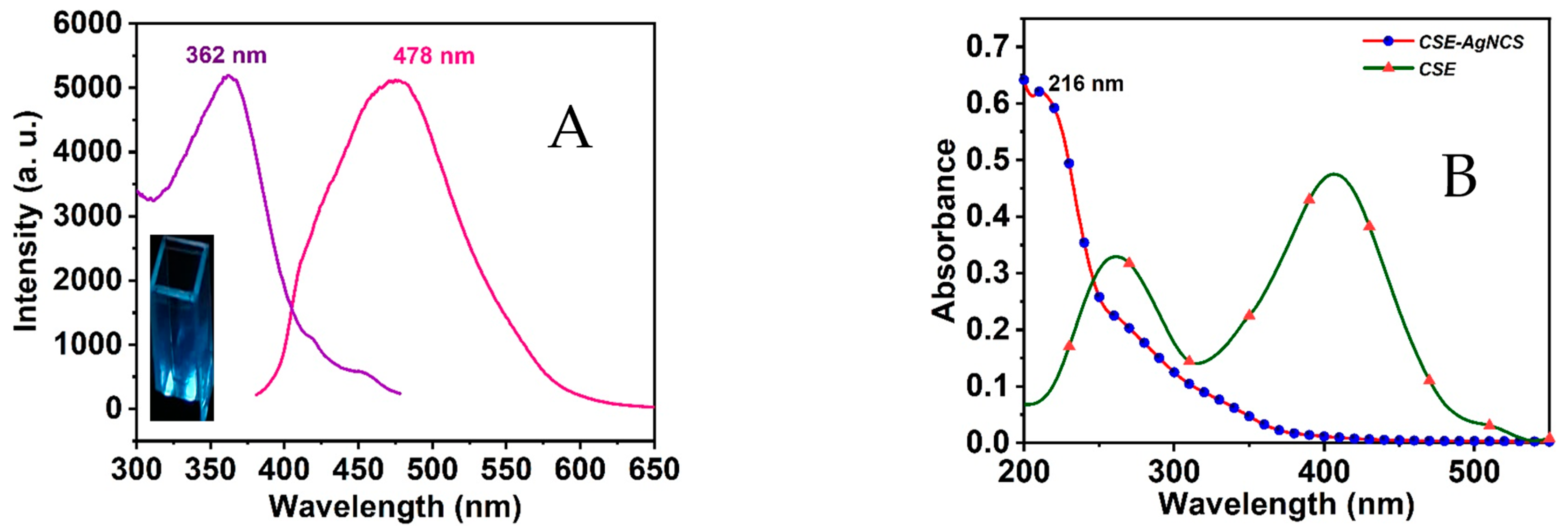
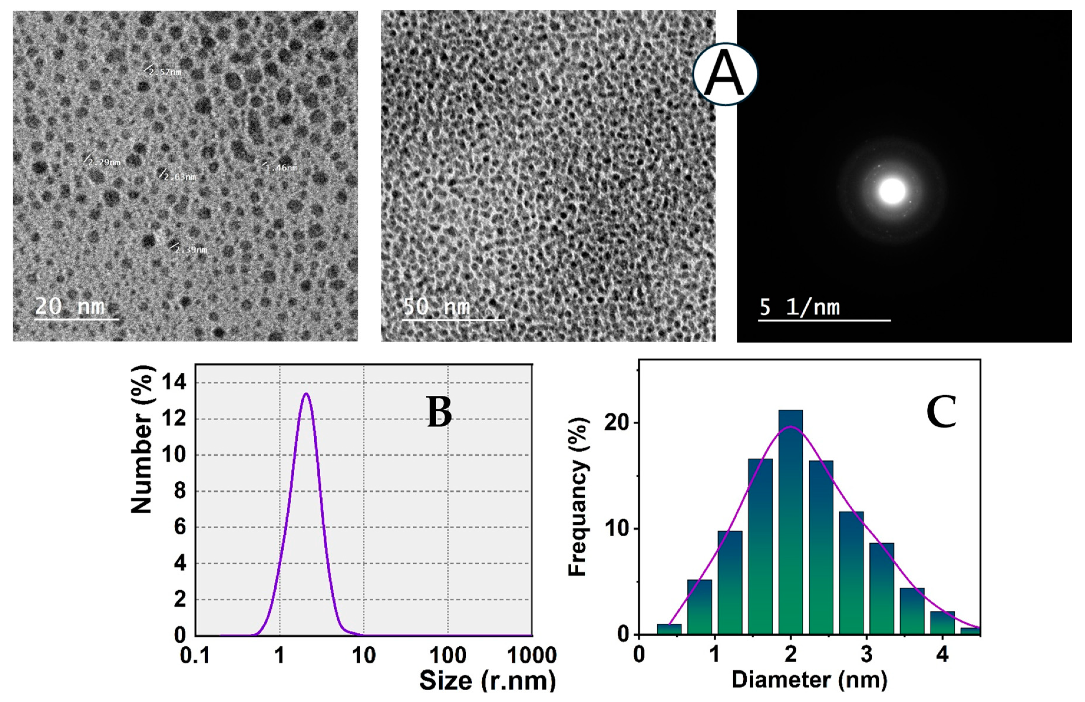
3.1. CSE-AgNCs Preparation

We devised a simple, single-pot, environmentally friendly synthetic process for synthesizing AgNCs at a temperature of 37 °C. The natural seed extraction of coffee Arabica that is accessible for commercial use serves as the foundation for this technique. We brought the pH of the reaction medium down to roughly 12.5 to maximize the ability of the coffee molecules to reduce. In addition, using coffee Arabica as a capping agent makes it possible for the AgNCs to have biocompatibility and environmental characteristics. Furthermore, the surface modifications and chelation of substantial metal ions are induced by the CSE coating layer on AgNCs. Moreover, the metal-binding properties of chlorogenic acids, the primary component of coffee Arabica, are essential for eliminating the detrimental effects of heavy metals [46].
The synthesized CSE-AgNCs solution appears brown in visible light and displays an intense blue luminescence under UV lighting, with a quantum yield of around 5.2%. The remarkable optical properties of the CSE-AgNCs are validated by the luminescence spectrum depicted in Figure 1A. Additionally, the UV–vis spectra of the coffee extraction and the synthesized AgNCs were recorded (Figure 1B). Figure 1A demonstrates that the fluorescence maxima of the CSE-AgNCs have a principal peak at 478 nm when excited at a wavelength of 362 nm. The CSE-AgNCs exhibited a substantial Stokes shift of around 112 nm [47,48], attributed to the innovative coffee extraction as a reducing agent. If the Stokes shift is adequate, the self-quenching of the chemical probe can be inhibited [49,50]. When silver nanoclusters are smaller than 5 nm, their emission peak may be seen [51]. An absorption peak is also observed within the wavelength range of 200 to 550 nm. It is centered approximately at 216 nm, as seen in Figure 1B. The absorbance spectra of the synthesized CSE-AgNCs markedly differs from that of the coffee extraction spectra. This demonstrates the creation of nanoclusters in the reaction media. Furthermore, there is an absence of an absorbance peak at 430 nm, indicative of surface plasmon resonance (SPR) for bigger nanoparticles (>10 nm) [52].
Furthermore, the TEM pictures, DLS (dynamic light scattering), and SD (size distribution) studies of CSE-AgNCs are shown to describe the synthesized nanoclusters in terms of size and form. Figure 2A illustrates that the TEM picture verifies that the mono-dispersed NCs mostly exhibit spherical morphology with an average diameter of around 2 nm. Consequently, the findings indicate that the green CSE reduction method is a significant approach for producing silver nanoclusters with exceptional efficacy. The DLS analysis and size distribution (SD) histogram (Figure 2B,C) demonstrate that the CSE-AgNCs consist of monodispersed particles and are virtually uniformly distributed. The results approximated the mean particle size of AgNCs to be around 2 nm. The diffraction pattern provides valuable insight into the microstructure of Ag nanoclusters. The concentric rings observed in the electron diffraction pattern suggest a crystalline structure and periodic atomic arrangement. The presence of high-order reflections in the image indicates a well-defined crystallographic order, which is essential for understanding the phase properties of the Ag nanoclusters.
To investigate the chemical composition of CSE-AgNCs, XPS analysis was utilized. The results showed two unique peaks: 367.45 eV (Ag 3d5/2) and 373.45 eV (Ag 3d3/2). In addition, the binding energy of Ag 3d5/2 is between that of Ag0 and Ag+, which is between 366.36 eV and 373.74 eV, according to [53]. This suggests that Ag0 is present here. There was a change in the chemical environment that surrounded the Ag atoms, as shown by the fact that the peaks moved to lower binding energies (Figure 3A). The presence of both Ag0 and Ag+ in the mixture may be responsible for this phenomenon. CSE is made up of an amino acid that is composed of functional groups like carboxyl and amino groups. As a result, this phenomenon may suggest that silver atoms are involved in interactions with either the N or O members of the CSE function groups. In addition, the presence of carbon, nitrogen, and oxygen in the capping agent of the coffee seeds extraction may be identified using the survey of our XPS analysis (the survey graph is shown in Figure 3B). Furthermore, this provides evidence that the CSE ligand protects the surfaces of the AgNCs that were obtained by synthesis.

3.2. FT-IR Analyses

Figure 4 represents the FT-IR data collected for the natural CSE and the CSE-AgNCs in the 4000–400 cm1 wavenumber range. Various chemical components are present in green coffee, as seen by the FT-IR of the crude green coffee extract [54]. The Fourier transform infrared spectroscopy (FTIR) analysis reveals several distinctive peaks. One of these peaks, which has a frequency range of 805 to 868 cm1, is linked to the β-type glycosidic connections resulting from carbohydrate materials. Within the 1026 to 1154 cm1 range, an additional peak is connected to the peak of carbohydrate components based on the II-arabinogalactan compound. A stretching mode that can be observed between 1247 and 1452 cm1 is shown by the caffeine component, which is another important fact to consider. There is also the identification of phenolic groups at a frequency of 1247 cm1 [55], which is the consequence of the hydrolysis of chlorogenic acids. They are detected at this frequency. More specifically, the peaks that appear at 1452 and 1645 cm1 are what distinguish the deprotonated carboxylic groups of the chlorogenic acids from other types of carboxylic groups. The peak at 1751 cm−1 corresponds to the stretching vibration of carbonyl (C=O) groups, which are characteristic of esters or aldehydes. This feature is particularly present in the CSE spectrum. Among the peaks that fall in 2857 to 3294 cm1, it has been shown that hydroxyl groups of phenol components and other aliphatic alkanes, such as methylene and methyl groups, are responsible for their appearance [56]. A further illustration of the FTIR measurement of the CSE-AgNCs can be seen in Figure 4. The spectrum indicates the changes that have occurred in the chemistry of the CSE solution. Within the range of 720–1620 cm1, the CSE solution exhibits a variety of peak shifts and peak forms. The decrease in Ag+ that occurs due to the action of phenolic components could be responsible for this consequence.

3.3. Optimum Conditions

In a distinct series of tests, we investigated the impact of the reaction temperature on the emission intensities of the CSE-AgNCs that was prepared. Increasing the temperature of the reaction medium from 22 °C to 37 °C increases the luminescence intensities. Subsequently, the luminescence intensities are substantially reduced by increasing the temperature to 50, 60, and 70 °C. This behavior can be accounted for by the growth of larger nanoclusters at high temperatures, which results in the aggregation-induced quenching of AgNCs fluorescence [57]. Consequently, the optimal temperature is determined to be 37 °C, as illustrated in Figure 5A. It was noted that the luminescence intensities of CSE-AgNCs depend upon CSE concentrations. It is noteworthy that the luminescence intensity of CSE-AgNCs exhibits a disproportionate enrichment in the presence of 5 mL of CSE using a 2 to 7 mL range of CSE (Figure 5B). The luminescence intensity of CSE-AgNCs experiences an undesired decrease as the CSE concentration is increased to 7 mL. Consequently, the optimized concentration of CSE is 5 mL, as illustrated in the inset of Figure 5B. In this study, we observed that the fluorescence properties of the synthesized Ag NCs remained stable over a three-month storage period, with no significant changes in emission intensity or extinction profiles. This stability can be attributed to the effective capping and stabilizing properties of the Arabica coffee seed extract (CSE), which prevents the aggregation and degradation of the nanoclusters.

3.4. CSE-AgNCs Reactivity

Firstly, we evaluated the CSE-AgNCs fluorescence in the presence of Hg(II) and different metal ions, including alkali, alkaline earth, and transition metal ions. The CSE-AgNCs exhibited a great response to mercury ions: about 86.9% of the fluorescence signal was suppressed by 1.4 µM Hg(II). Moreover, the other metal ions did not induce any notable quenching at the same concentration of 1.4 µM (Figure 6). The quenching of CSE-AgNCs fluorescence by Hg(II) alone offers an efficient approach for their measurement. The detection limits for Hg(II) are at the nanomolar range of 35.21 nM. Consequently, further processing is necessary to distinguish the quenching phenomena.

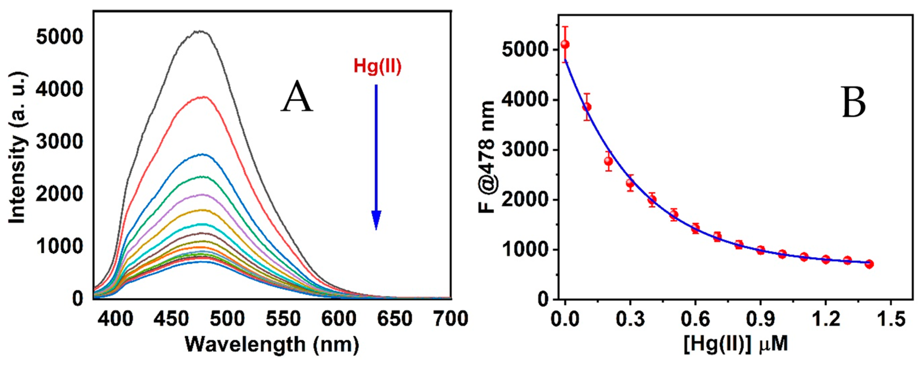
3.5. Sensing Mechanism

In this context, the mechanism is predicated on the hypothesis that Hg(II) is bound with the surface of the CSE-AgNCs by inducing metal ions and the chemical groups of the CSE surface molecule in the CSE-AgNCs. Thus, this action resulted in a significant quenching of the CSE-AgNCs fluorescence emission through an electron transfer mechanism. The intense blue luminescence of CSE-AgNCs is plainly visible in Figure 7 following the titrimetric reaction of CSE-AgNCs in the presence of Hg(II) solution, which can be quenched. The emission spectra that result from the increase in Hg(II) ions indicate that the emission intensity of the CSE-AgNCs is reduced. Still, the wavelength of the AgNCs maximal peak is unaffected. Consequently, resonance energy transmission cannot be validated as the proposed mechanism. AgNCs that were stabilized with various scaffold molecules were reported in various research studies. These nanoclusters were employed to identify substantial metal ions through the aggregation of the nanoclusters [57]. This performance results from the metal complex induction between the active Hg(II) ions and the surface scaffold molecules.
The spontaneous interaction of Hg(II) and Ag+ on the nanoclusters’ structure is the cause of the quenching phenomenon of the CSE-AgNCs. Consequently, a mixture solution was generated during the titration of Hg(II) by combining a 20 mM phosphate-buffer solution at pH 7.4 with a CSE-AgNCs solution. The solution was subsequently applied to the titration procedure, as illustrated in Figure 7A. The fluorescence intensity of CSE-AgNCs decreases due to the excessive addition of Hg(II) concentrations. The fluorescence intensity begins to stabilize at 1.4 µM. The detection of Hg(II) ions based on the eco-friendly sensor nanoclusters was established via a quenching mechanism within the range of 0 to 1.4 µM, as evidenced by the relationship between the fluorescence intensities of CSE-AgNCs and various concentrations of Hg(II) ions, as illustrated in Figure 7B.
In order to introduce the plot of relative intensities (F0/F) vs. [Hg(II)], a Stern–Volmer equation was applied [58]. The relation that was produced is shown in Figure 8. A linear association was established between the concentrations ranging from 0 to 14 µM, with a value of regression factor R2 equal to 0.991. Based on the assumption that the fluorescence intensities could be measured with an accuracy of ±1%, the limit of detection (LOD) was predicted to be 35.21 nM [59].
F0/F = 1 + Ksv [Q]
In this equation, F0 and F are the emission of CSE-AgNCs and CSE-AgNCs in the presence of Hg(II) metal ions, Ksv is the Stern–Volmer constant, and Q is the concentration of the quencher molecules or ions (Hg(II) metal ions).
Figure 8. Stern–Volmer based on CSE-AgNCs system using different Hg(II) solution concentrations.
Analytica 06 00015 g008

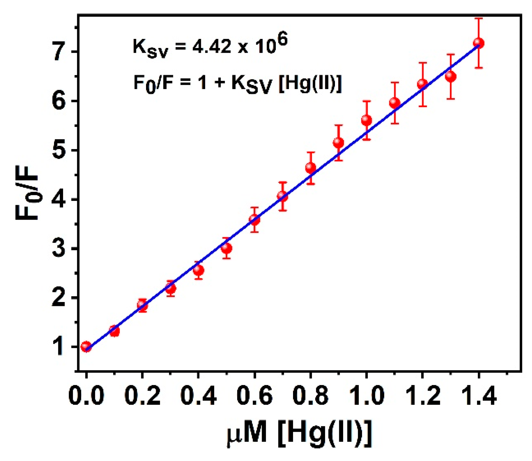
3.6. Binding Constant

To further understand the metal nanocluster binding process, the fluorescence spectra changes in the CSE-AgNCs probe were analyzed in the presence of different concentrations of Hg(II) to determine the binding constant. The binding constant was determined with the modified Stern–Volmer equation.
F0/F0 − F = 1/A + 1/A. Kb [Q]
In this equation, F0 represents the fluorescence intensity of the unbound ligand AgNCs, F denotes the fluorescence intensity of the Hg(II)-AgNCs complex, Q signifies the concentration of [Hg(II)], A is a constant, and Kb indicates the binding constant [60,61]. A linear connection was seen when F0/(F0/F) was graphed against the concentration of 1/[Q]: (y = α + βx), where y = F0/(F0 − F), the intercept equals 1/A, the slope equals 1/A.Kb, x equals 1/[Q], and Kb was derived from α/β (Figure 9). Inferred from the fluorescence titration curves of the AgNCs probe with Hg(II), Kb, the binding association constant was determined to be 3.88 × 10⁶ M1. The quantum yield (QY) was calculated to be 0.32, and the utilized equations were inserted in Supporting Information S2.

3.7. CSE-AgNCs Selectivity

To examine the selectivity of CSE-AgNCs, we conducted a study whereby we examined the impact of several cations, each with a concentration of 1.4 µM, on CSE-AgNCs under optimal circumstances, as seen in Figure 10A. It was observed that the fluorescence intensities of the Hg(II)-CSE-AgNCs system were not affected by the presence of other cations, which demonstrates that CSE-AgNCs are capable of selectively sensing Hg(II). Additionally, the mercury ion was transferred to reduce the luminescence intensity of AgNCs by the transfer of electrons or sources of energy. Because CSE components have the ability to chelate Hg(II) ions and produce luminescence quenching, it is possible that the quenching effect of Hg(II) might be attributable to these constituents. In addition, chlorogenic acid is one of the compounds that is found in the highest concentration in Arabica green coffee. Coffee contains a substantial amount of this important component, which is also useful from a biological standpoint [62]. The luminescence of the Ag nanocluster in the Hg(II)-CSE-AgNCs system is also significantly improved by the additional presence of the EDTA solution. Because Hg(II) ions can attach to the chelation groups of EDTA molecules, it is possible to remove Hg(II) ions from the surface of CSE-AgNCs in a considerable manner, which results in the luminescence intensity of AgNCs being restored. As seen in Figure 10B, the remarkable recovery of the fluorescence of AgNCs was accomplished by about 90.7% restoration of its initial value in the presence of EDTA solution. This significant achievement was accomplished after five cycles. There is no discernible change in the luminescence intensity of CSE-AgNCs as the number of cycles rises; the intensity remains roughly the same after five cycles. In addition, the kinetic differences that occur during the restoration of the luminescence of AgNCs make it possible for Hg(II) ions to remain very selective. In light of this, the repeatability of the CSE-AgNCs approach for identifying Hg(II) ions was sufficient throughout the course of five cycles.

3.8. Environmental Application

Using a CSE-AgNCs sensor, this investigation aimed to identify the presence of Hg(II) metal ions in mineral and tap water samples. In the absence of any additional processing, the water samples that were examined were utilized immediately. After the samples were adjusted in a phosphate-buffer solution with a buffer concentration of 20 mM and a pH of 7.4, the detection was carried out. The actual samples were also subjected to the introduction of Hg(II) in amounts that were particular to each individual sample. The findings that were achieved are dependent on the standard curve that was produced from the number of experiments that were performed. It was determined how much mercury ion (Hg(II)) is present in mineral and tap water samples. Based on the data shown in Table 1, it can be concluded that the average recovery range for Hg(II) detection was between 97.00% and 99.07%. The obtained findings were examined and compared to the data acquired from the ICP-MS, which indicated that this approach is essential for identifying Hg(II) in environmental samples.

4. Conclusions

This study illustrates the successful synthesis of silver nanoclusters (AgNCs) using coffee Arabica seeds extract (CSE) in a green, one-pot, and straightforward manner. CSE is employed as a novel reducing and capping agent in the synthesis of AgNCs. Our research introduces a novel sensing mechanism enabling AgNCs to function as green optical sensors. This mechanism capitalizes on the fluorescence intensity of AgNCs to detect mercury (Hg(II)) metal cations. The coffee constituents’ high chelation ability on AgNCs toward Hg(II) led to substantial fluorescence quenching, facilitating the quantitative determination of Hg(II) concentrations. Furthermore, the fluorescence quenching effect is further enhanced by the reduction in Hg(II) on the surface of the AgNCs in the presence of Ag(I), which serves as an effective method for detecting Hg(II). Additionally, our investigation investigated the impact of other metal ions on the detection of Hg(II). The results suggest that the CSE-AgNCs demonstrate high selectivity and sensitivity toward Hg(II) in the presence of other metal ions, thereby minimizing potential interference and assuring accurate detection. This innovative approach emphasizes the potential of green chemistry and provides a sustainable and eco-friendly method for developing advanced sensing technologies. CSE-AgNCs are a promising instrument for real sample analysis, notably in detecting Hg(II) in environmental samples, due to their robust fluorescence quenching properties and significant chelation ability. Our research represents a substantial advancement in developing green, efficient, highly selective sensors for detecting hazardous metal ions, particularly mercury. It offers a valuable contribution to the field of optical sensing. It also opens new avenues for applying green-synthesized nanoclusters in environmental monitoring.

Supplementary Materials

The following supporting information can be downloaded at: https://www.mdpi.com/article/10.3390/analytica6020015/s1, Supplementary Information S1: Experimental; S2: Results and Discussions.

Author Contributions

Conceptualization, S.M.S. and R.A.; methodology, R.A., S.A. and S.M.S.; formal analysis, S.M.S., S.A. and R.A.; investigation, S.M.S., S.A. and R.A.; resources, S.M.S., R.A. and S.A.; data curation, S.M.S. and R.A.; writing—original draft preparation, S.M.S., R.A. and S.A.; writing—review and editing, S.M.S., R.A. and S.A.; supervision, S.M.S. and R.A. All authors have read and agreed to the published version of the manuscript.

Funding

The authors gratefully acknowledge Qassim University, represented by the Deanship of Graduate Studies and Scientific Research, on the financial support for this research under the number (QU-J-PG-2-2025-56078) during the academic year 1446 AH/2024 AD.

Data Availability Statement

All data will be made available upon reasonable request.

Conflicts of Interest

The authors declare no conflicts of interest.

References

  1. Baimenov, A.Z.; Berillo, D.A.; Moustakas, K.; Inglezakis, V.J. Efficient removal of mercury (II) from water using cryogels and comparison to commercial adsorbents under environmentally relevant conditions. J. Hazard. Mater. 2020, 399, 123056. [Google Scholar] [CrossRef] [PubMed]
  2. Gworek, B.; Dmuchowski, W.; Baczewska-Dąbrowska, A.H. Mercury in the terrestrial environment: A review. Environ. Sci. Eur. 2020, 32, 128. [Google Scholar] [CrossRef]
  3. Cusset, F.; Reynolds, S.J.; Carravieri, A.; Amouroux, D.; Asensio, O.; Dickey, R.C.; Fort, J.; Hughes, B.J.; Paiva, V.H.; Ramos, J.A.; et al. A century of mercury: Ecosystem-wide changes drive increasing contamination of a tropical seabird species in the South Atlantic Ocean. Environ. Pollut. 2023, 323, 121187. [Google Scholar] [CrossRef] [PubMed]
  4. Al-Sulaiti, M.M.; Soubra, L.; Al-Ghouti, M.A. The causes and effects of mercury and methylmercury contamination in the marine environment: A review. Curr. Pollut. Rep. 2022, 8, 249–272. [Google Scholar] [CrossRef]
  5. Rana, M.N.; Tangpong, J.; Rahman, M.M. Toxicodynamics of lead, cadmium, mercury and arsenic-induced kidney toxicity and treatment strategy: A mini review. Toxicol. Rep. 2018, 5, 704–713. [Google Scholar] [CrossRef]
  6. Zulaikhah, S.T.; Wahyuwibowo, J.; Pratama, A.A. Mercury and its effect on human health: A review of the literature. Int. J. Public Health Sci. 2020, 9, 103–114. [Google Scholar] [CrossRef]
  7. Ali, R.; Alminderej, F.M.; Messaoudi, S.; Saleh, S.M. Ratiometric ultrasensitive optical chemisensor film based antibiotic drug for Al(III) and Cu(II) detection. Talanta 2021, 221, 121412. [Google Scholar] [CrossRef]
  8. Saleh, S.M.; Ali, R.; Hegazy, M.E.F.; Alminderej, F.M.; Mohamed, T.A. The natural compound chrysosplenol-D is a novel, ultrasensitive optical sensor for detection of Cu(II). J. Mol. Liq. 2020, 302, 112558. [Google Scholar] [CrossRef]
  9. Saleh, S.M.; Ali, R.; Alminderej, F.; Ali, I.A. Ultrasensitive optical chemosensor for Cu(II) detection. Int. J. Anal. Chem. 2019, 2019, 7381046. [Google Scholar] [CrossRef]
  10. Aroua, L.M.; Ali, R.; Albadri, A.E.; Messaoudi, S.; Alminderej, F.M.; Saleh, S.M. A new, extremely sensitive, turn-off optical sensor utilizing Schiff base for fast detection of Cu(II). Biosensors 2023, 13, 359. [Google Scholar] [CrossRef]
  11. Nazri, N.A.A.; Azeman, N.H.; Luo, Y.; Bakar, A.A.A. Carbon quantum dots for optical sensor applications: A review. Opt. Laser Technol. 2021, 139, 106928. [Google Scholar] [CrossRef]
  12. Hendler-Neumark, A.; Bisker, G. Fluorescent single-walled carbon nanotubes for protein detection. Sensors 2019, 19, 5403. [Google Scholar] [CrossRef] [PubMed]
  13. Ali, R.; Alminderej, F.M.; Saleh, S.M. A simple, quantitative method for spectroscopic detection of metformin using gold nanoclusters. Spectrochim. Acta Part A Mol. Biomol. Spectrosc. 2020, 241, 118744. [Google Scholar] [CrossRef] [PubMed]
  14. Ali, R.; Ali, I.A.; Messaoudi, S.; Alminderej, F.M.; Saleh, S.M. An effective optical chemosensor film for selective detection of mercury ions. J. Mol. Liq. 2021, 336, 116122. [Google Scholar] [CrossRef]
  15. Saleh, S.M.; El-Sayed, W.A.; El-Manawaty, M.A.; Gassoumi, M.; Ali, R. An eco-friendly synthetic approach for copper nanoclusters and their potential in lead ions sensing and biological applications. Biosensors 2022, 12, 197. [Google Scholar] [CrossRef]
  16. Saleh, S.M.; Ali, R.; Wolfbeis, O.S. New silica and polystyrene nanoparticles labeled with longwave absorbing and fluorescent chameleon dyes. Microchim. Acta 2011, 174, 429–434. [Google Scholar] [CrossRef]
  17. Saleh, S.M.; Elkady, E.M.; Ali, R.; Alminderej, F.; Mohamed, T.A. Novel chemical sensor for detection Ca(II) ions based on ferutinin. Spectrochim. Acta Part A Mol. Biomol. Spectrosc. 2018, 205, 264–268. [Google Scholar] [CrossRef]
  18. Ali, R.; Ghannay, S.; Messaoudi, S.; Alminderej, F.M.; Aouadi, K.; Saleh, S.M. A reversible optical sensor film for mercury ions discrimination based on isoxazolidine derivative and exhibiting pH sensing. Biosensors 2022, 12, 1028. [Google Scholar] [CrossRef] [PubMed]
  19. Zhang, W.; Ma, M.; Zhang, X.A.; Zhang, Z.Y.; Saleh, S.M.; Wang, X.D. Fluorescent proteins as efficient tools for evaluating the surface PEGylation of silica nanoparticles. Methods Appl. Fluoresc. 2017, 5, 024003. [Google Scholar] [CrossRef]
  20. Saleh, S.M.; Ali, R.; Algreiby, A.; Alfeneekh, B.; Ali, I.A. A novel organic chromo-fluorogenic optical sensor for detecting chromium ions. Heliyon 2024, 10, e37480. [Google Scholar] [CrossRef]
  21. Ali, R.; Saleh, S.M. Design a Friendly Nanoscale Chemical Sensor Based on Gold Nanoclusters for Detecting Thiocyanate Ions in Food Industry applications. Biosensors 2024, 14, 223. [Google Scholar] [CrossRef] [PubMed]
  22. Saleem, M.; Rafiq, M.; Hanif, M. Organic material based fluorescent sensor for Hg2+: A brief review on recent development. J. Fluoresc. 2017, 27, 31–58. [Google Scholar] [CrossRef]
  23. Wu, D.; Sedgwick, A.C.; Gunnlaugsson, T.; Akkaya, E.U.; Yoon, J.; James, T.D. Fluorescent chemosensors: The past, present and future. Chem. Soc. Rev. 2017, 46, 7105–7123. [Google Scholar] [CrossRef]
  24. Ali, R.; Alfeneekh, B.; Chigurupati, S.; Saleh, S.M. Green synthesis of pregabalin-stabilized gold nanoclusters and their applications in sensing and drug release. Arch. Der Pharm. 2022, 355, 2100426. [Google Scholar] [CrossRef]
  25. Ali, R.; Saleh, S.M.; Aly, S.M. Fluorescent gold nanoclusters as pH sensors for the pH 5 to 9 range and for imaging of blood cell pH values. Microchim. Acta 2017, 184, 3309–3315. [Google Scholar] [CrossRef]
  26. Shang, Y.; Gao, H.; Li, L.; Ma, C.; Gu, J.; Zhu, C.; Yang, Z.; Wang, C.; Zhang, Y.; Chen, G. Green Synthesis of Fluorescent Ag Nanoclusters for Detecting Cu2+ Ions and Its “Switch-On” Sensing Application for GSH. J. Spectrosc. 2021, 2021, 8829654. [Google Scholar] [CrossRef]
  27. Yourston, L.E.; Krasnoslobodtsev, A.V. Micro RNA sensing with green emitting silver nanoclusters. Molecules 2020, 25, 3026. [Google Scholar] [CrossRef] [PubMed]
  28. Zhao, K.; Shen, W.; Cui, H. Highly chemiluminescent silver nanoclusters with a dual catalytic center. J. Mater. Chem. C 2018, 6, 6549–6555. [Google Scholar] [CrossRef]
  29. Xu, J.; Bi, Y.; Zhao, H.; Shi, L.; Zhang, N.; Xin, X. In situ synthesis of well-dispersed silver nanoparticles from silver nanoclusters hydrogel for catalytic reduction of 4-Nitrophenol. Appl. Surf. Sci. 2025, 683, 161759. [Google Scholar] [CrossRef]
  30. Zhang, Y.; Lv, M.; Gao, P.; Zhang, G.; Shi, L.; Yuan, M.; Shuang, S. The synthesis of high bright silver nanoclusters with aggregation-induced emission for detection of tetracycline. Sens. Actuators B Chem. 2021, 326, 129009. [Google Scholar] [CrossRef]
  31. Han, F.; Wang, W.; Li, D.; Xu, S.; Sun, Y.; Lin, L.; Ma, L.; Li, J.; Li, L. Green preparation of silver nanocluster composite AgNCs@CF-g-PAA and its application: 4-NP catalytic reduction and hydrogen production. RSC Adv. 2023, 13, 11807–11816. [Google Scholar] [CrossRef] [PubMed]
  32. Khatoon, U.T.; Velidandi, A.; Rao, G.N. Sodium borohydride mediated synthesis of nano-sized silver particles: Their characterization, anti-microbial and cytotoxicity studies. Mater. Chem. Phys. 2023, 294, 126997. [Google Scholar] [CrossRef]
  33. Guo, J.; Yang, H.; Liu, Y.; Liu, W.; Zhao, R.; Li, H.; Long, W.; Xu, W.; Guo, M.; Zhang, X. Atomically precise silver clusterzymes protect mice from radiation damages. J. Nanobiotechnol. 2021, 19, 377. [Google Scholar] [CrossRef]
  34. Tao, Y.; Aparicio, T.; Li, M.; Leong, K.W.; Zha, S.; Gautier, J. Inhibition of DNA replication initiation by silver nanoclusters. Nucleic Acids Res. 2021, 49, 5074–5083. [Google Scholar] [CrossRef]
  35. Liu, S.; Yan, Q.; Cao, S.; Wang, L.; Luo, S.H.; Lv, M. Inhibition of bacteria in vitro and in vivo by self-assembled DNA-silver nanocluster structures. ACS Appl. Mater. Interfaces 2022, 14, 41809–41818. [Google Scholar] [CrossRef]
  36. Mediani, A.; Kamal, N.; Lee, S.Y.; Abas, F.; Farag, M.A. Green extraction methods for isolation of bioactive substances from coffee seed and spent. Sep. Purif. Rev. 2023, 52, 24–42. [Google Scholar] [CrossRef]
  37. Clifford, M.N. Chlorogenic acids. In Coffee, 1 (Chemistry); Clarke, R.J., Macrae, R., Eds.; Elsevier Applied Science Publ.: London, UK, 1985; pp. 153–202. [Google Scholar]
  38. Schrader, K.; Kiehne, A.; Engelhardt, U.H.; Maier, H.G. Determination of chlorogenic acids with lactones in roasted coffee. J. Sci. Food Agric. 1996, 71, 392–398. [Google Scholar] [CrossRef]
  39. de Paulis, T.; Schmidt, D.E.; Bruchey, A.K.; Kirby, M.T.; McDonald, M.P.; Commers, P.; Lovinger, D.M.; Martin, P.R. Dicinnamoylquinides in roasted coffee inhibit the human adenosine transporter. Eur. J. Pharmacol. 2002, 442, 215–223. [Google Scholar] [CrossRef] [PubMed]
  40. Yao, C.; Ding, Y.; Li, P.; Song, Q.; Wang, G.; Cheng, D. Effects of chlorogenic acid on the binding process of cadmium with bovine serum albumin: Amulti-spectroscopic docking study. J. Mol. Struct. 2020, 1204, 127531. [Google Scholar] [CrossRef]
  41. Kalinowska, M.; Sienkiewicz-Gromiuk, J.; Świderski, G.; Pietryczuk, A.; Cudowski, A.; Lewandowski, W. Zn(II) complex of plant phenolic chlorogenic acid: Antioxidant, antimicrobial and structural studies. Materials 2020, 13, 3745. [Google Scholar] [CrossRef]
  42. Jaiswal, R.; Matei, M.F.; Subedi, P.; Kuhnert, N. Does roasted coffee contain chlorogenic acid lactones or/and cinnamoylshikimate esters? Food Res. Int. 2014, 61, 214–227. [Google Scholar] [CrossRef]
  43. Belay, A.; Gholap, A.V. Characterization and determination of chlorogenic acids (CGA) in coffee beans by UV-Vis spectroscopy. Afr. J. Pure Appl. Chem. 2009, 3, 234–240. [Google Scholar]
  44. Moores, R.G.; McDermott, D.L.; Wood, T.R. Determination of chlorogenic acid in coffee. Anal. Chem. 1948, 20, 620–624. [Google Scholar] [CrossRef]
  45. Saleh, S.M.; Ali, R.; nd Elshaarawy, R.F. A ratiometric and selective fluorescent chemosensor for Ca(II) ions based on a novel water-soluble ionic Schiff-base. RSC Adv. 2016, 6, 68709–68718. [Google Scholar] [CrossRef]
  46. Yan, L.; Li, J.; Cai, H.; Shao, Y.; Zhang, G.; Chen, L.; Wang, Y.; Zong, H.; Yin, Y. Carbon dots/Ag nanoclusters-based fluorescent probe for ratiometric and visual detection of Cu2+. J. Alloys Compd. 2023, 945, 169227. [Google Scholar] [CrossRef]
  47. Dong, Y.Y.; Deng, F.; Zhao, J.J.; He, J.; Ma, M.G.; Xu, F.; Sun, R.C. Environmentally friendly ultrosound synthesis and antibacterial activity of cellulose/Ag/AgCl hybrids. Carbohydr. Polym. 2014, 99, 166–172. [Google Scholar] [CrossRef]
  48. Capek, P.; Paulovičová, E.; Matulová, M.; Mislovičová, D.; Navarini, L.; Liverani, F.S. Coffea arabica instant coffee-chemical view and immunomodulating properties. Carbohydr. Polym. 2014, 103, 418–426. [Google Scholar] [CrossRef]
  49. Alvarez, N.M.M.; Pastrana, J.M.; Lagos, Y.; Lozada, J.J. Evaluation of mercury (Hg2+) adsorption capacity using exhausted coffee waste. Sustain. Chem. Pharm. 2018, 10, 60–70. [Google Scholar] [CrossRef]
  50. Liu, C.; Pujol, D.; Olivella, M.À.; De la Torre, F.; Fiol, N.; Poch, J.; Villaescusa, I. The role of exhausted coffee compounds on metal ions sorption. Water Air Soil Pollut. 2015, 226, 1–10. [Google Scholar] [CrossRef]
  51. Chen, Z.; Lu, D.; Zhang, G.; Yang, J.; Dong, C.; Shuang, S. Glutathione capped silver nanoclusters-based fluorescent probe for highly sensitive detection of Fe3+. Sens. Actuators B Chem. 2014, 202, 631–637. [Google Scholar] [CrossRef]
  52. Anandalakshmi, K.; Venugobal, J.; Ramasamy, V.J.A.N. Characterization of silver nanoparticles by green synthesis method using Pedalium murex leaf extract and their antibacterial activity. Appl. Nanosci. 2016, 6, 399–408. [Google Scholar] [CrossRef]
  53. Liu, C.; Yang, D.; Wang, Y.; Shi, J.; Jiang, Z. Fabrication of antimicrobial bacterial cellulose–Ag/AgCl nanocomposite using bacteria as versatile biofactory. J. Nanopart. Res. 2012, 14, 1084. [Google Scholar] [CrossRef]
  54. Saleh, S.M.; Ali, R.; Hirsch, T.; Wolfbeis, O.S. Detection of biotin-avidin affinity binding by exploiting a self-referenced system composed of upconverting luminescent nanoparticles gold nanoparticles. J. Nanopart. Res. 2011, 13, 4603–4611. [Google Scholar] [CrossRef]
  55. Farah, A. Coffee constituents. In Coffee: Emerging Health Effects and Disease Prevention; Chu, Y.-F., Ed.; John Wiley & Sons Inc.: Hoboken, NJ, USA; Blackwell Publishing Ltd.: Hoboken, NJ, USA, 2012; Chapter 2. [Google Scholar]
  56. Pujol, D.; Liu, C.; Gominho, J.; Fiol, N.; Villaescusa, I.; Pereira, H. The chemical composition of exhausted coffee waste. Ind. Crop. Prod. 2013, 50, 423–429. [Google Scholar] [CrossRef]
  57. Panthi, G.; Park, M. Synthesis of metal nanoclusters and their application in Hg2+ ions detection: A review. J. Hazard. Mater. 2022, 424, 127565. [Google Scholar] [CrossRef]
  58. Saleh, S.M.; Almotiri, M.K.; Ali, R. Green synthesis of highly luminescent gold nanoclusters and their application in sensing Cu(II) and Hg(II). J. Photochem. Photobiol. A Chem. 2022, 426, 113719. [Google Scholar] [CrossRef]
  59. Ali, R.; Lang, T.; Saleh, S.M.; Meier, R.J.; Wolfbeis, O.S. Optical sensing scheme for carbon dioxide using a solvatochromic probe. Anal. Chem. 2011, 83, 2846–2851. [Google Scholar] [CrossRef]
  60. Karak, D.; Banerjee, A.; Sahana, A.; Guha, S.; Lohar, S.; Adhikari, S.S.; Das, D. 9-Acridone-4-carboxylic acid as an efficient Cr(III) fluorescent sensor: Trace level detection, estimation and speciation studies. J. Hazard. Mater. 2011, 188, 274–280. [Google Scholar] [CrossRef]
  61. Elshaarawy, R.F.; Ali, R.; Saleh, S.M.; Janiak, C. A novel water-soluble highly selective “switch-on” ionic liquid-based fluorescent chemi-sensor for Ca(II). J. Mol. Liq. 2017, 241, 308–315. [Google Scholar] [CrossRef]
  62. Tajik, N.; Tajik, M.; Mack, I.; Enck, P. The potential effects of chlorogenic acid the main phenolic components in coffee on health: Acomprehensive review of the literature. Eur. J. Nutr. 2017, 56, 2215–2244. [Google Scholar] [CrossRef]
Figure 1. (A) Optical characteristics of CSE-AgNCs; and (B) solution absorbances of CSE, and CSE-AgNCs; 100 μL CSE-AgNCs in 2 mL phosphate-buffer solution.
Analytica 06 00015 g001
Figure 2. (A) TEM images and diffraction pattern, (B) DLS, and (C) SD histogram of as-prepared CSE-AgNCs; CSE-AgNCs solution was diluted twenty-fold using phosphate buffer.
Analytica 06 00015 g002
Figure 3. (A) XPS spectra of Ag 3d; and (B) survey spectra of CSE-AgNCs.
Analytica 06 00015 g003
Figure 4. FT-IR data of CSE and CSE-AgNCs. CSE-AgNCs samples were utilized as-prepared for measurements without any further dilution.
Analytica 06 00015 g004
Figure 5. (A) Influences of temperature on AgNCs synthesis process and (B) CSE initial concentration effect.
Analytica 06 00015 g005
Figure 6. Metal ions sensing based on CSE-AgNCs.
Analytica 06 00015 g006
Figure 7. (A) Luminescence titration of CSE-AgNCs versus Hg(II) ions; (B) luminescence intensity of CSE-AgNCs fluorescence intensity at 478 nm versus Hg(II) ions.
Analytica 06 00015 g007
Figure 9. Modified Stern-Volmer equation for CSE-AgNCs system using different Hg(II) metal ion solutions.
Analytica 06 00015 g009
Figure 10. (A) Emissions of CSE-AgNCs: (I) effect of different significant cations and (II) effect of Hg(II) and other cations; and (B) introduction of EDTA restores emissions of CSE-AgNCs.
Analytica 06 00015 g010
Table 1. Detection of Hg(II) in environmental samples was accomplished using CSE-AgNCs as substantial chemical sensors (n = 3).
Added
Hg(II) µM
ICP-MS
µM
Found
µM
RSD *
(%)
Recovery
(%)
Mineral water0.100.1030.09871.1198.70
0.700.7050.6920.8998.85
1.401.4291.3860.7999.00
Tap water0.100.1160.0980.09797.00
0.700.7070.6830.8897.57
1.401.4181.3870.8599.07
* RSD: relative standard deviation.
Disclaimer/Publisher’s Note: The statements, opinions and data contained in all publications are solely those of the individual author(s) and contributor(s) and not of MDPI and/or the editor(s). MDPI and/or the editor(s) disclaim responsibility for any injury to people or property resulting from any ideas, methods, instructions or products referred to in the content.

