Abstract
Over the last few decades, nanomaterials have been synthesized using one-step green approaches, which are rapid, simple, cost-effective, eco-friendly, and nonhazardous. Silver nanomaterials (PS-Ag/AgCl) were synthesized using Pistia stratiotes aqueous leaf extract. The leaf extract acts as a reducing, stabilizing, and capping agent. The compounds responsible for the reduction of silver salt and the functional groups present in leaf extract were explored by FT-IR (Fourier-transform infrared spectroscopy) as well as other characterization techniques. The synthesized nanomaterials had a spherical surface morphology and were 15–50 nm in size, according to HR-TEM (high-resolution transmission electron microscopy). UV–visible spectroscopy showed an absorbance peak in the range of 420 nm. Surface morphology was explored by SEM (scanning electron microscopy), and elemental composition was analyzed by EDX (energy-dispersive X-ray spectroscopy). The crystallinity and crystal lattice of Ag/AgCl nanomaterials were explored by XRD (X-ray powder diffraction). The silver nanomaterials showed antibacterial activity against Gram-negative (Escherichia coli) bacteria. As a result, the nanomaterials (PS-Ag/AgCl) are safe and nontoxic.
1. Introduction
In the last few decades, the green synthesis of nanomaterials has achieved an alternative approach for the synthesis of metallic nanomaterials with negligible formation of toxic waste products [1]. Noble-metal nanomaterials (e.g., gold, platinum, silver, palladium, etc.) [2,3,4,5] have been synthesized by the green method, which gives tremendous results regarding the product [6]. Nanomaterial synthesis approaches are mainly divided into three types: the chemical approach, the physical approach, and the green approach. The chemical and physical approaches use high pressure and temperature and hazardous chemicals, which affect nature and increase the cost of the process [7,8,9]. To overcome these negative impacts, researchers adopted the green approach method and used plant extract for the synthesis of metal nanomaterials. The green synthetic approach of metal nanomaterials utilizing micro-organisms bacteria, yeast, and fungi has been known, but there are many demerits, including more purification steps and an intricate process of maintaining microbial growth [4,10,11]. To overcome these shortcomings, we used the Pistia stratiotes extract for the synthesis of AgNPs.
Metal nanomaterials have wide applications in different biological fields and are used in biomedicine, pharmaceutical industries, cosmetics industries, electronics, biosensors, catalysis, and optics. Much of the research has especially been reported in biomedical applications [12], like anticancer [13], antibacterial [14], and antioxidant [15,16,17]. These properties are due to the size (1–100 nm) and shape of metallic nanomaterials [18]. The shape and size of green synthesis metallic nanoparticles depend on the amount of salt, pH value, temperature, process of timing, and contents of the plant extract, and they are highly sensitive to the environment under which the process is being undertaken.
In the view of chemistry, the plant extract has functional groups such as hydroxyl (-OH), amine (-NH), etc., and many plant derivatives such as tannins, terpenoid saponins, starches, polypeptides [19], and other heterocyclic compounds [20,21]. These functional groups of plant derivatives bind with metals and act as reducing and capping/stabilizing agents for the metal atom [6]. Capping agents also prevent the agglomeration of the nanomaterials and reduce their toxicity. Green synthetic PS-AgNPs are environmentally friendly, cost-effective, negligibly toxic, and negligibly maintained in-process, and they enhance the application of the synthesis of metallic nanomaterials by using plant extract.
Many plants, including Azadirachta indica [22], Berberis vulgaris [23], Jatropha curcas [24], Hypericum perforatum [25], Eriobotrya japonica [26], and Fumarica parviflora [27], have been used to synthesize silver nanoparticles for different pharmacological properties. The leaf and root of Pistia stratiotes can be used as a source of nutrients with antibacterial, antifungal, antidermatophytic, and toxicological properties [28]. It belongs to the Araceae family, commonly known as jalkumbhi, water cabbage, and water lettuce, which is a free-floating aquatic plant [29] and widespread in lakes and ponds in tropical and subtropical regions. In this work, we synthesized PS-Ag/AgCl NPs by utilizing the plant extract of Pistia stratiotes. The capping enhances biochemical activity such as antibacterial, antioxidant, anticancer, and anti-inflammatory activity. Herein, X-ray powder diffraction and UV–vis were used to confirm the synthesis of PS-Ag/AgCl NPs; their shape and size were determined using HR-TEM and SEM; and their functional groups were confirmed using FT-IR.
2. Experimental
2.1. Material
Silver salt (AgNO3) of analytical grade was purchased from Sigma-Aldrich. Pistia stratiotes plant was collected from the university campus pond during February. During the synthesis and other studies, we used deionized water and ethanol for washing.
Escherichia coli strain AKS-1 16S ribosomal RNA gene, partial sequence GenBank: MK478816 was obtained from the Department of Environmental Microbiology, Babasaheb Bhimrao Ambedkar University, Lucknow. The culture was maintained in their appropriate agar slants at 4 °C and used as stock culture.
2.2. Characterization of PS-AgNPs
The growth of PS-Ag/AgCl nanoparticles was synthesized by Pistia stratiotes leaf extract at room temperature and in normal conditions. High-resolution transmission electron microscopy (HR-TEM) and scanning electron microscopy (SEM) were used to measure and characterize the surface morphology, size, and crystalline nature of PS-Ag/AgCl NPs. Ultraviolet–visible spectroscopy (UV–vis) and X-ray diffraction (XRD) data were used to evaluate the crystalline nature of PS-Ag/AgCl NPs. Fourier-transform infrared spectroscopy (FTIR) was used to explore the preparation of PS-Ag/AgCl NPs by considering the possible presence of functional groups in the plant extract for the reduction, stabilization, and capping of silver nanomaterials. EDS was used to confirm the contamination of Ag and other elements present in PS-AgNPs.
2.3. Anti-Bacterial Activity of PS-AgNPs
To test the toxic effect of PS-Ag/AgCl NPs prepared by Pistia stratiotes leaf extract on Gram-negative (E. coli) bacteria, growth inhibition analysis was conducted in Luria–Bertani (LB) broth media. The experiments were performed using the last reported method, with a few changes (M. Sathishkumar et al., 2009) [30]. For growth inhibition analysis, three well-plates, each containing 50 mL of LB broth media and a sufficient amount of PS-Ag/AgCl NPs, were inoculated with 50 mL of the freshly grown bacterial suspension to maintain the bacterial concentration in the same range in the well. The three-well plates were then incubated in a rotary shaker at 165 rpm at 37 °C. The growth of the pathogen was monitored every hour for 24 h by the absorbance at 600 nm. A control experiment containing only media and bacteria bare of PS-Ag/AgCl NPs was also included.
3. Results and Discussion
3.1. Green Synthesis Parameter
The leaves of the plant Pistia stratiotes were collected and washed three times with tap water and deionized water. The washed plant leaves were dried in sunlight for 2 h to prepare the extract. A solution was made by boiling 20 g of dried leaves in 300 mL of deionized water for 2 h, which was then filtered using Whatman filter paper. The synthesis of PS-Ag/AgCl NPs started with 150 mL of plant extract and 1.7 g of silver nitrate salt. The plant extract solution was mixed drop-by-drop with silver salt in an Erlenmeyer flask and kept at room temperature until the color changed from colorless to black at 1200 rpm. The change in color within one hour indicated the reduction of Ag+ to Ag. The solution was sonicated for half an hour and rinsed with ethanol. The collected nanomaterials were placed on a petri plate and dried in a vacuum oven at 60 °C for 24 h. The dried silver nanomaterial (Ag/AgCl) was utilized for further study.
3.2. Characterization of PS-Ag/AgCl NPs
3.2.1. FTIR Study
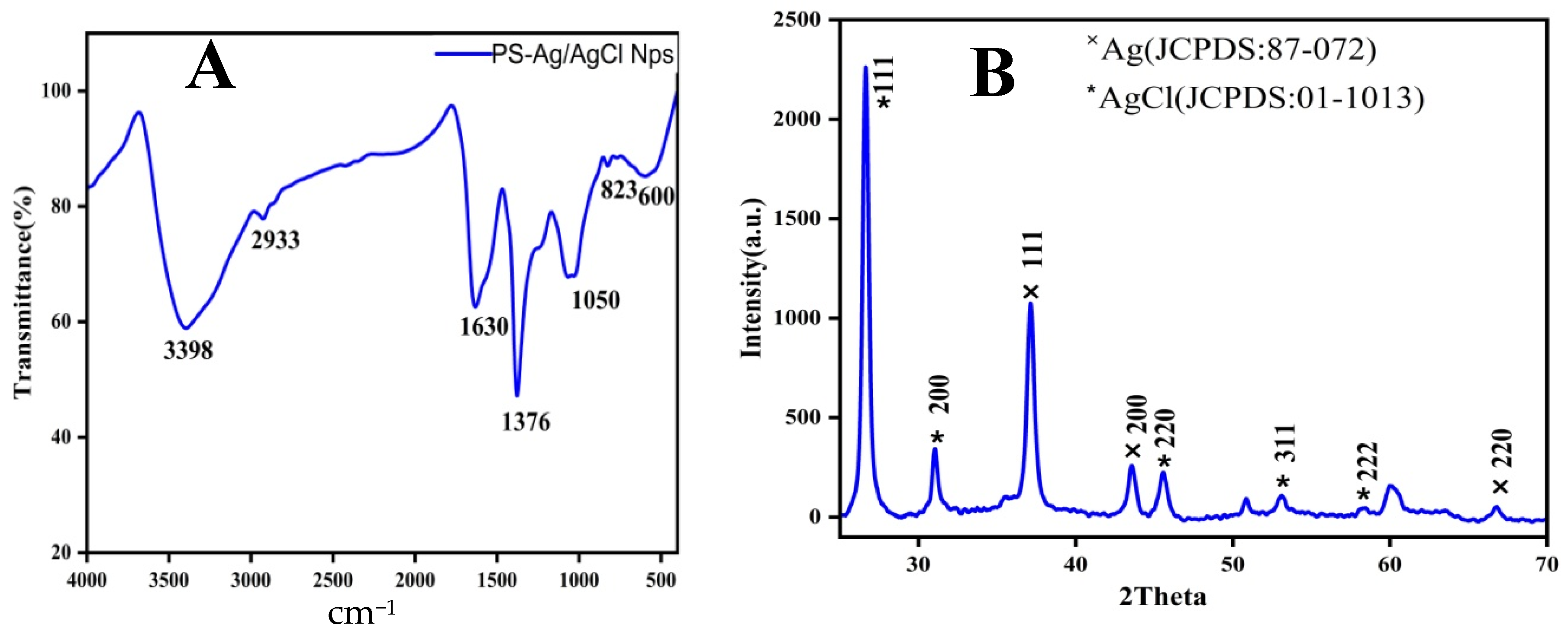
Plant extracts of Pistia stratiotes play a role in the reduction and capping of silver metal, and different functional groups of phytoconstituent compounds play an important role in the synthesis of silver nanomaterials, which were characterized by FTIR spectroscopy. A sharp peak at 3398 cm−1 is owing to the N-H stretching vibration of functional groups -NH2 and O-H and the overlapping of the stretching vibration of attribution for water and Pistia stratiotes leaf extract [22]. The peak at 2933 cm−1 shows the primary and secondary amines of the C-H stretching vibration [30]. The peak at 1630 cm−1 agrees with amide C=O stretching [22]. The peaks at 1066 cm−1 and 600 cm−1 are due to the stretching of the phenol group [31]. Furthermore, the peaks at 1376 cm−1 and 823 cm−1 agree with the -C-O- stretching of phenol or tertiary alcohol. The mentioned peaks are mainly attributed to flavonoids, terpenoids, and polyphenols. The phytoconstituents of plant extract have a strong affinity for metal ions and a protective coat-like shell, which prevents their further aggregation and stabilization of silver nanomaterials (Figure 1A).
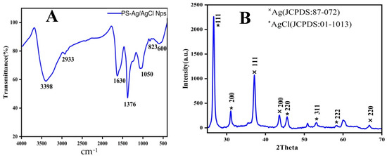
Figure 1.
(A) FTIR and (B) XRD data of nanomaterials (PS-Ag/AgCl NPs).
3.2.2. XRD Study
XRD patterns of PS-Ag/AgCl NPs synthesized from the leaf extract of Pistia stratiotes. Several Bragg reflections with 2θ values of 26.63, 37.04, 37.11, 43.57, 45.57, 53.10, 58.36, 66.79, angle [32] corresponding to (111), (200), (111), (200), (220), (311), (222), and (220) planes, respectively, for PS-Ag/AgCl NPs at 25 °C. Ag and AgCl nanomaterial peaks are present in Figure 1B. It means Ag is present on the surface of the AgCl and also shows a face-centered cubic structure. The sharp peaks revealed that the crystalline nature of PS-Ag/AgCl NPs was formed by leaf extract.
3.2.3. UV–Visible Study
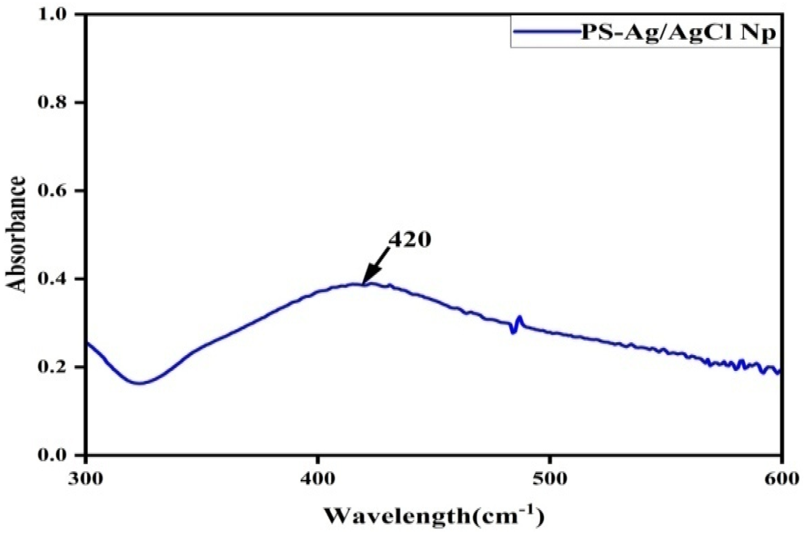
The formation of silver nanoparticles in colloidal solution has been confirmed by UV–visible analysis. The color change of silver nanoparticles from a colorless to a reddish-brown aqueous solution with bioreduction. The change in color is due to surface plasmon vibration in PS-Ag/AgCl NPs. The UV–visible spectra show the appearance of different absorption maxima between 392 and 460 nm [25]. Figure 2 shows the peak at approx. 420 nm; this intense peak is due to the formation of uniform spherical PS-Ag/AgCl NPs [25].
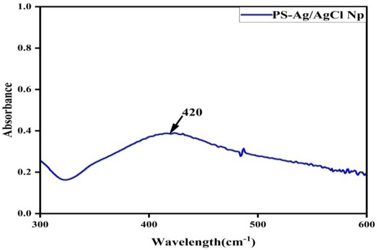
Figure 2.
Electronic microscopy study (HR-TEM and EDX).
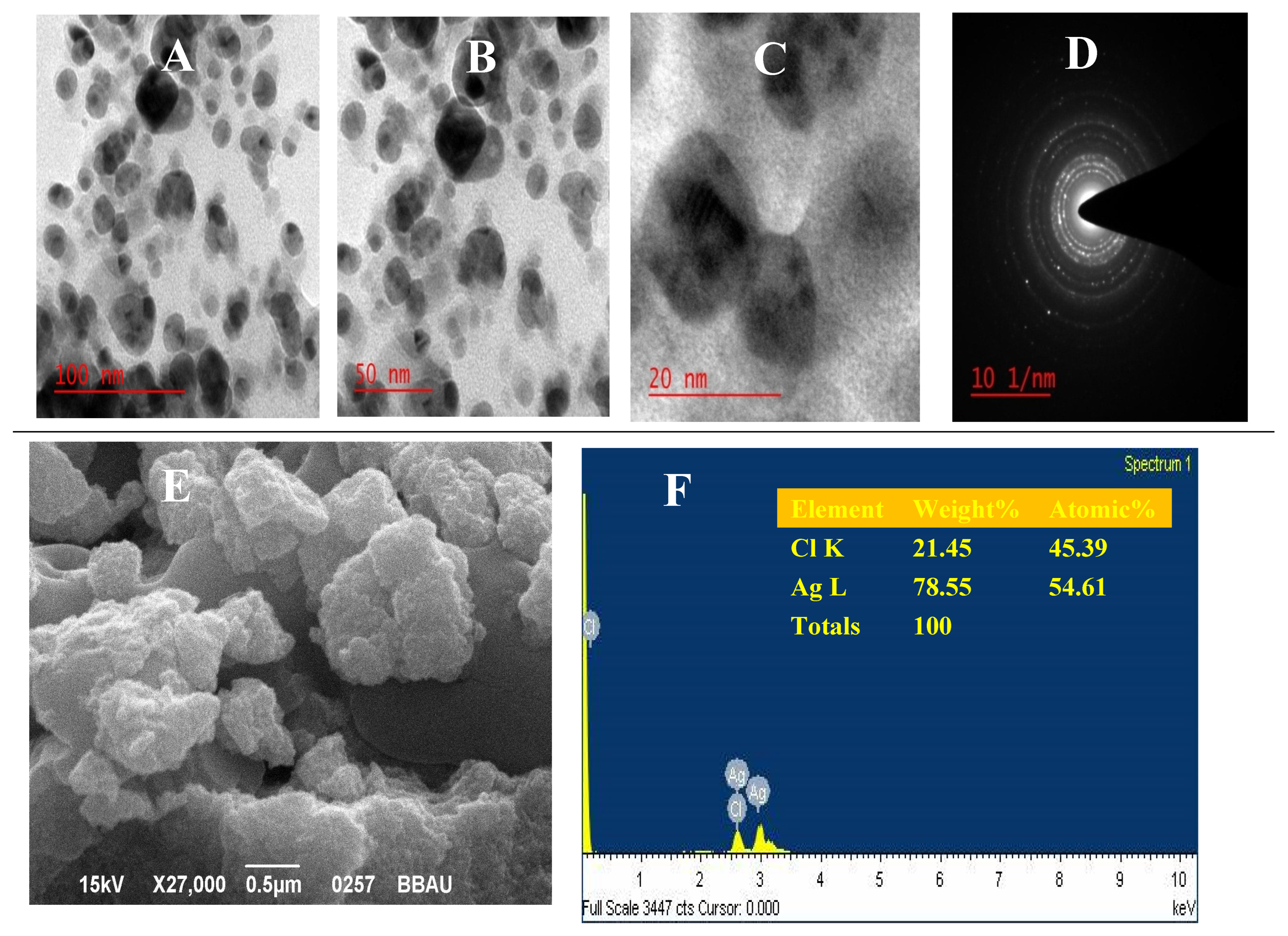
The HRTEM analysis has been used to explore the size, shape, and surface morphology of silver nanomaterials [22]. The images are present in Figure 3A–C. The image of biochemically synthesized nanomaterials (PS-Ag/AgCl NPs) shows nanoscale and uniform particles. The nanoparticles are spherical and have a size range of 15 nm to 50 nm. These findings are also confirmed by other researchers [33,34,35]. Crystallinity and lattice pattern were explored by the SAED image in Figure 3D. The SEM image also explains the surface morphology of nanomaterials, and EDX analysis explored the composition, purity, and percentage of nanomaterials in Figure 3E,F, respectively. There is more silver metal compared to chlorine. It is also seen that the AgCl surface is covered by the silver metals.
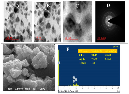
Figure 3.
(A–C) HR-TEM images and (D) SAED pattern of PS-Ag/AgCl NPs; (E) SEM image; (F) EDX data.
4. Anti-Bacterial Activities of Green Synthesized PS-Ag/AgCl NPs
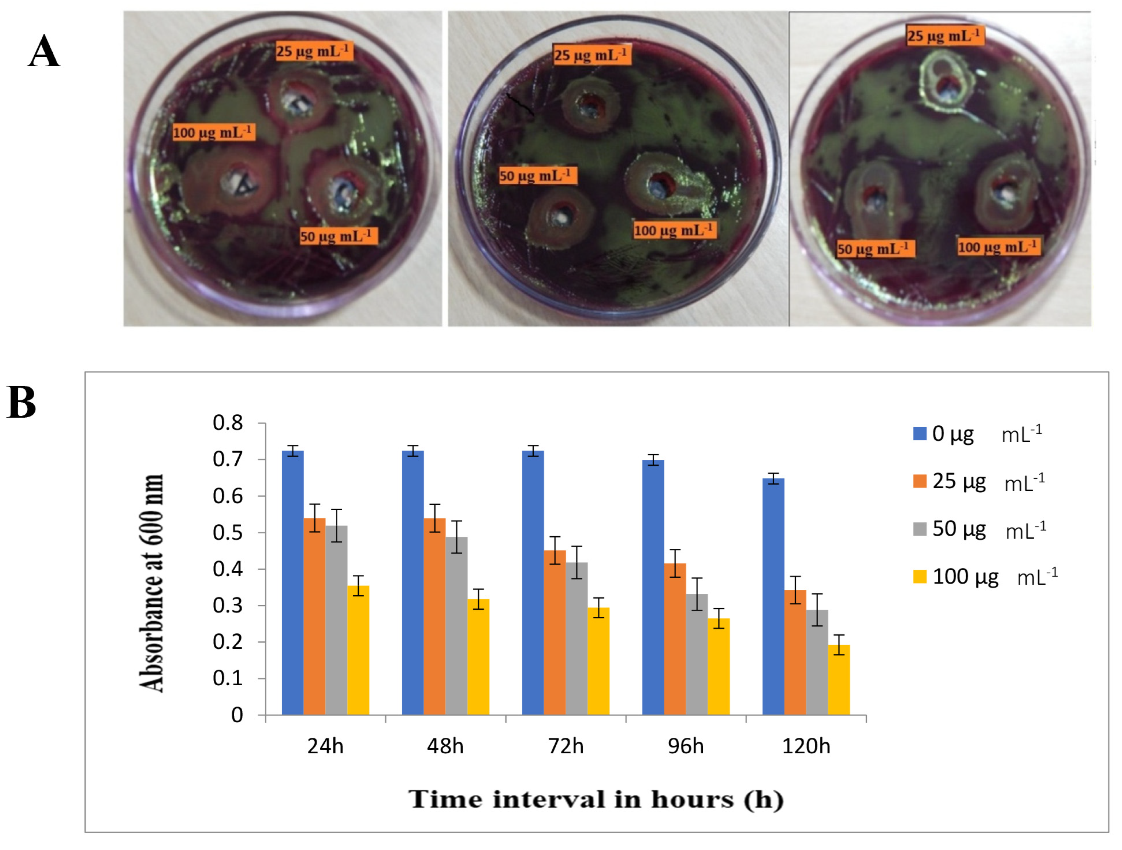
The gram-negative bacteria E. coli were finalized for evaluating the antibacterial activity of green synthesized silver nanomaterials (PS-AgNPs). As shown in Figure 4A, the absorbance at 600 nm for the pathogen with different doses of silver nanomaterials was plotted over 120 h. The anti-bacterial activity of silver nanomaterials against bacteria was explored in a potential dose-dependent manner. The growth inhibition was observed even in the minimum concentration range of 25 µg/mL−1 of silver nanomaterials. However, the maximum inhibition concentration (MIC) of as-prepared silver nanomaterials was 100 µg/mL−1 for E. coli bacteria that could be effectively inhibited within 120 h. After 50 µg/mL−1 concentrations at 72 h periods, the stationary phase was observed, and after 120 h, bacterial cultures declined, so spectrophotometer readings were taken up to 120 h. This has occurred due to cell wall damage or unfavourable conditions due to the anti-bacterial activity of the nanomaterials of Pistia stratiotes leaf extracts. However, this may be due to the silver nanomaterials being spherical, with an average particle size of 15 nm to 50 nm. In addition, as per the report [36,37], the silver nanomaterials synthesized by KÜP and his coworkers using the leaf extract of Aesculuship pocastanum had a bigger average size of 50.0–5.0 nm but showed stronger anti-bacterial effects for a bacteria with an MIC of 1.56 µg/mL−1 (Wei et al., 2020) [38]. It seems that the toxic effects of green synthesized silver nanomaterials for bacterial strains have an ambiguous relationship with the average size.
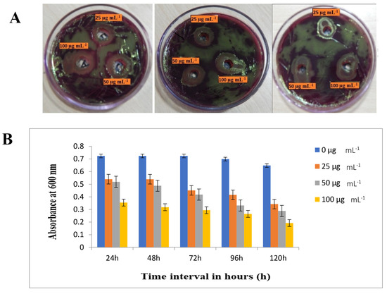
Figure 4.
(A) Antibacterial activity test of green synthesized PS-AgNPs for E. coli. (B) Graph representing the growth inhibition of the E. coli bacteria at 600 nm absorbance during a period of 24 h to 120 h.
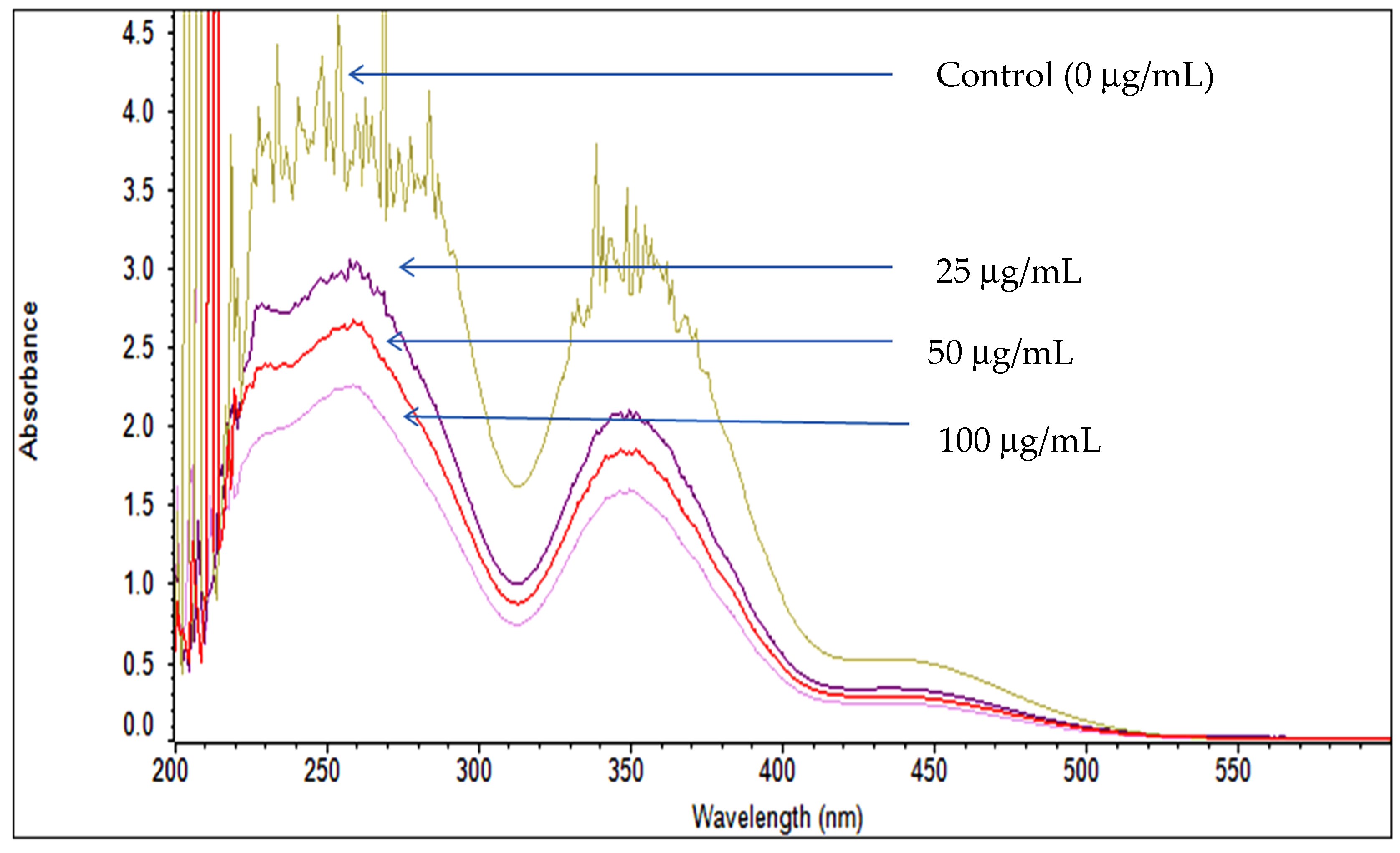
We have to observe different antibacterial activity, which may be due to the capped constituents of the biosynthesized silver nanomaterials, which may lead to various mechanisms for cell wall damage and toxic effects of the bacterial cells. It has been represented in Figure 4B with absorbance at 600 nm at different time intervals. The densities of the cell culture after the 120 h time interval were also measured via scanning spectrophotometry analysis at 600 nm. As per the scanning results, the control (0 µg mL−1) flask had a higher absorbance and decreased continuously when increasing the concentration of the silver nanomaterials in the flask (Figure 5). It has represented the viability of the bacterial cultures at different stress conditions due to silver nanomaterials. However, the anti-bacterial activity of silver nanomaterials was also measured by Eosin methylene blue (EMB) agar petri plates via a well diffusion assay, as represented in Figure 4A. The clear zone of inhibition has been noted in triplicate experiments with different concentrations of silver nanomaterials.
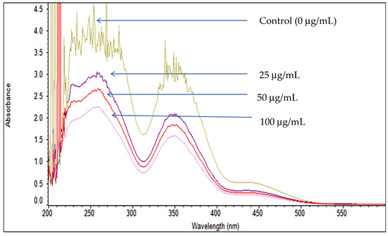
Figure 5.
Spectrophotometer scanning graph representing the decrease in absorbance by increasing wavelength after the treatment of PS-Ag/AgCl NPs at different concentrations after 120 h of 600 nm wavelength exposure.
5. Conclusions
Eco-friendly, cost-effective, and comparatively less toxic PS-Ag/AgCl NPs were synthesized using Pistia stratiotes plant leaves via a green approach. Furthermore, the biological process is simpler and easier for downstream processing. The nanomaterials are spherical. No chemical reagents or surfactants were used in this method to stabilize nanoparticles. PS-Ag/AgCl NPs were confirmed using FT-IR, UV–vis, HRTEM, SEM, EDX, and XRD analysis. The PS-Ag/AgCl NPs expressed effective antibacterial activity and may be useful as a silver dressing for wounds or as an alternative material. PS-Ag/AgCl NPs were further studied to fully characterize the cytotoxicity.
Author Contributions
A.K.G.: Investigation, methodology, Data correction, original draft, editing, communication. G.P.: Supervision, review and editing. All authors have read and agreed to the published version of the manuscript.
Funding
This research received no external funding.
Institutional Review Board Statement
Not applicable.
Informed Consent Statement
Not applicable.
Data Availability Statement
Data are contained within the article.
Conflicts of Interest
The authors declare no conflict of interest.
References
- Gul, A.R.; Shaheen, F.; Rafique, R.; Bal, J.; Waseem, S.; Park, T.J. Grass-mediated biogenic synthesis of silver nanoparticles and their drug delivery evaluation: A biocompatible anti-cancer therapy. J. Chem. Eng. 2021, 407, 127202. [Google Scholar] [CrossRef]
- Al-Radadi, N.S. Facile one-step green synthesis of gold nanoparticles (AuNp) using licorice root extract: Antimicrobial and anticancer study against HepG2 cell line. Arab. J. Chem. 2021, 14, 102956. [Google Scholar] [CrossRef]
- Yang, W.; Ma, Y.; Tang, J.; Yang, X. “Green synthesis” of monodisperse Pt nanoparticles and their catalytic properties. Colloids Surf. A Physicochem. Eng. Asp. 2007, 302, 628–633. [Google Scholar] [CrossRef]
- He, Y.; Wei, F.; Ma, Z.; Zhang, H.; Yang, Q.; Yao, B.; Huang, Z.; Li, J.; Zeng, C.; Zhang, Q. Green synthesis of silver nanoparticles using seed extract of Alpinia katsumadai, and their antioxidant, cytotoxicity, and antibacterial activities. RSC Adv. 2017, 7, 39842–39851. [Google Scholar] [CrossRef]
- Abbas, G.; Kumar, N.; Kumar, D.; Pandey, G. Effect of Reaction Temperature on Shape Evolution of Palladium Nanoparticles and Their Cytotoxicity against A-549 Lung Cancer Cells. ACS Omega 2019, 4, 21839–21847. [Google Scholar] [CrossRef] [PubMed]
- Wei, S.; Wang, Y.; Tang, Z.; Xu, H.; Wang, Z.; Yang, T.; Zou, T. A novel green synthesis of silver nanoparticles by the residues of Chinese herbal medicine and their biological activities. RSC Adv. 2021, 11, 1411–1419. [Google Scholar] [CrossRef] [PubMed]
- Rosenthal, S.J.; Chang, J.C.; Kovtun, O.; McBride, J.R.; Tomlinson, I.D.J.C. Biocompatible quantum dots for biological applications. Chem. Biol. 2011, 18, 10–24. [Google Scholar] [CrossRef] [PubMed]
- Berenguel-Alonso, M.; Ortiz-Gómez, I.; Fernández, B.; Couceiro, P.; Alonso-Chamarro, J.; Capitán-Vallvey, L.; Salinas-Castillo, A.; Puyol, M.J.S. An LTCC monolithic microreactor for the synthesis of carbon dots with photoluminescence imaging of the reaction progress. Sens. Actuators B-Chem. 2019, 296, 126613. [Google Scholar] [CrossRef]
- Harada, M.; Kuwa, M.; Sato, R.; Teranishi, T.; Takahashi, M.; Maenosono, S. Cation distribution in monodispersed MFe2O4 (M = Mn, Fe, Co, Ni, and Zn) nanoparticles investigated by X-ray absorption fine structure spectroscopy: Implications for magnetic data storage, Catalysts, Sensors, and Ferrofluids. ACS Appl. Nano Mater. 2020, 3, 8389–8402. [Google Scholar] [CrossRef]
- Saifuddin, N.; Wong, C.W.; Yasumira, A.A. Rapid biosynthesis of silver nanoparticles using culture supernatant of bacteria with microwave irradiation. E-J. Chem. 2009, 6, 61–70. [Google Scholar] [CrossRef]
- Jain, D.; Daima, H.K.; Kachhwaha, S.; Kothari, S.L. Synthesis of plant-mediated silver nanoparticles using papaya fruit extract and evaluation of their anti microbial activities. Dig. J. Nanomater. Biostruct. 2009, 4, 557–563. [Google Scholar]
- Wang, Y.; Wei, S.; Wang, K.; Wang, Z.; Duan, J.; Cui, L.; Zheng, H.; Wang, Y.; Wang, S. Evaluation of biosynthesis parameters, stability and biological activities of silver nanoparticles synthesized by Cornus Officinalis extract under 365 nm UV radiation. RSC Adv. 2020, 10, 27173–27182. [Google Scholar] [CrossRef]
- Dai, Y.; Xu, C.; Sun, X.; Chen, X. Nanoparticle design strategies for enhanced anticancer therapy by exploiting the tumour microenvironment. Chem. Soc. Rev. 2017, 46, 3830–3852. [Google Scholar] [CrossRef] [PubMed]
- Frattini, A.; Pellegri, N.; Nicastro, D.; de Sanctis, O. Effect of amine groups in the synthesis of Ag nanoparticles using aminosilanes. Mater. Chem. Phys. 2005, 94, 148–152. [Google Scholar] [CrossRef]
- Lee, S.H.; Jun, B.H. Silver Nanoparticles: Synthesis and Application for Nanomedicine. Int. J. Mol. Sci. 2019, 20, 865. [Google Scholar] [CrossRef] [PubMed]
- Jeyaraj, M.; Sathishkumar, G.; Sivanandhan, G.; MubarakAli, D.; Rajesh, M.; Arun, R.; Kapildev, G.; Manickavasagam, M.; Thajuddin, N.; Premkumar, K.; et al. Biogenic silver nanoparticles for cancer treatment: An experimental report. Colloids Surf. B 2013, 106, 86–92. [Google Scholar] [CrossRef]
- Rajoka, M.S.R.; Mehwish, H.M.; Zhang, H.; Ashraf, M.; Fang, H.; Zeng, X.; Wu, Y.; Khurshid, M.; Zhao, L.; He, Z. Antibacterial and antioxidant activity of exopolysaccharide mediated silver nanoparticle synthesized by Lactobacillus brevis isolated from Chinese koumiss. Colloids Surf. B 2020, 186, 110734. [Google Scholar] [CrossRef] [PubMed]
- Nahar, K.N.; Rahaman, M.; Khan, G.M.; Islam, M.; Al-Reza, S.M. Green synthesis of silver nanoparticles from Citrus sinensis peel extract and its antibacterial potential. Asian J. Green Chem. 2021, 5, 135–150. [Google Scholar]
- Mohamad, N.A.N.; Arham, N.A.; Jai, J.; Hadi, A. Plant extract as reducing agent in synthesis of metallic nanoparticles: A review. Adv. Mater. Res. 2014, 832, 350–355. [Google Scholar] [CrossRef]
- Faheem, M.; Rathaur, A.; Pandey, A.; Kumar Singh, V.; Tiwari, A.K. A review on the modern synthetic approach of benzimidazole candidate. ChemistrySelect 2020, 5, 3981–3994. [Google Scholar] [CrossRef]
- Faheem, M.; Tiwari, A.K.; Singh, V.K. A Review on Modern Synthetic Route for the Construction of 1, 3-Diazanaphthalene Moiety. Curr. Org. Chem. 2020, 24, 1108–1138. [Google Scholar] [CrossRef]
- Ahmed, S.; Saifullah Ahmad, M.; Swami, B.L.; Ikram, S. Green synthesis of silver nanoparticles using Azadirachta indica aqueous leaf extract. J. Radiat. Res. Appl. Sci. 2016, 9, 1–7. [Google Scholar] [CrossRef]
- Behravan, M.; Panahi, A.H.; Naghizadeh, A.; Ziaee, M.; Mahdavi, R.; Mirzapour, A. Facile green synthesis of silver nanoparticles using Berberis vulgaris leaf and root aqueous extract and its antibacterial activity. Int. J. Boil. Macromol. 2019, 124, 148–154. [Google Scholar] [CrossRef] [PubMed]
- Bar, H.; Bhui, D.K.; Sahoo, G.P.; Sarkar, P.; Pyne, S.; Misra, A. Green synthesis of silver nanoparticles using seed extract of Jatropha curcas. Colloids Surf. A Physicochem. Eng. Asp. 2009, 348, 212–216. [Google Scholar] [CrossRef]
- Alahmad, A.; Feldhoff, A.; Bigall, N.C.; Rusch, P.; Scheper, T.; Walter, J.G. Hypericum perforatum L.-mediated green synthesis of silver nanoparticles exhibiting antioxidant and anticancer activities. Nanomaterials 2021, 11, 487. [Google Scholar] [CrossRef]
- Jabir, M.S.; Hussien, A.A.; Sulaiman, G.M.; Yaseen, N.Y.; Dewir, Y.H.; Alwahibi, M.S.; Soliman, D.A.; Rizwana, H. Green synthesis of silver nanoparticles from Eriobotrya japonica extract: A promising approach against cancer cells proliferation, inflammation, allergic disorders and phagocytosis induction. Artif. Cells Nanomed. Biotechnol. 2021, 49, 48–60. [Google Scholar] [CrossRef]
- Patil, M.P.; Piad, L.L.A.; Bayaraa, E.; Subedi, P.; Tarte, N.H.; Kim, G.-D. Doxycycline hyclate mediated silver–silver chloride nanoparticles and their antibacterial activity. J. Nanostruct. Chem. 2019, 9, 53–60. [Google Scholar] [CrossRef]
- Wasagu, R.S.; Lawal, M.; Shehu, S.; Alfa, H.H.; Muhammad, C. Nutritive values, mineral and antioxidant properties of Pistia stratiotes (water lettuce). Niger. J. Basic Appl. Sci. 2013, 21, 253–257. [Google Scholar] [CrossRef][Green Version]
- Khan, M.A.; Marwat, K.B.; Gul, B.; Wahid, F.; Khan, H.; Hashim, S. Pistia stratiotes L.(Araceae): Phytochemistry, use in medicines, phytoremediation, biogas and management options. Pak. J. Bot. 2014, 46, 851–860. [Google Scholar]
- Alfuraydi, A.A.; Devanesan, S.; Al-Ansari, M.; AlSalhi, M.S.; Ranjitsingh, A.J. Eco-friendly green synthesis of silver nanoparticles from the sesame oil cake and its potential anticancer and antimicrobial activities. J. Photochem. Photobiol. B Biol. 2019, 192, 83–89. [Google Scholar] [CrossRef]
- Hemlata; Meena, P.R.; Singh, A.P.; Tejavath, K.K. Biosynthesis of silver nanoparticles using cucumis prophetarum aqueous leaf extract and their antibacterial and antiproliferative activity against cancer cell lines. ACS Omega 2020, 5, 5520–5528. [Google Scholar] [CrossRef] [PubMed]
- Shahbandeh, M.; Eghdami, A.; Moghaddam, M.M.; Nadoushan, M.J.; Salimi, A.; Fasihi-Ramandi, M.; Mohammadi, S.; Mirzaei, M.; Mirnejad, R. Conjugation of imipenem to silver nanoparticles for enhancement of its antibacterial activity against multidrug-resistant isolates of Pseudomonas aeruginosa. J. Biosci. 2021, 46, 26. [Google Scholar] [CrossRef]
- Mittal, A.K.; Chisti, Y.; Banerjee, U.C. Synthesis of metallic nanoparticles using plant extracts. Biotechnol. Adv. 2013, 31, 346–356. [Google Scholar] [CrossRef]
- Singh, P.K.; Bhardwaj, K.; Dubey, P.; Prabhune, A. UV-assisted size sampling and antibacterial screening of Lantana camara leaf extract synthesized silver nanoparticles. RSC Adv. 2015, 5, 24513–24520. [Google Scholar] [CrossRef]
- Krishnaraj, C.; Jagan, E.; Rajasekar, S.; Selvakumar, P.; Kalaichelvan, P.; Mohan, N. Synthesis of silver nanoparticles using Acalypha indica leaf extracts and its antibacterial activity against water borne pathogens. Colloids Surf. B Biointerfaces 2010, 76, 50–56. [Google Scholar] [CrossRef] [PubMed]
- Sathishkumar, M.; Sneha, K.; Won, S.W.; Cho, C.W.; Kim, S.; Yun, Y.S. Cinnamon Zeylanicum Bark Extract and Powder Mediated Green Synthesis of Nano-Crystalline Silver Particles and Its Bactericidal Activity. J. Photochem. Photobiol. B 2009, 73, 332–338. [Google Scholar] [CrossRef]
- Kup, F.O.; Coskuncay, S.; Duman, F. Biosynthesis of silver nanoparticles using leaf extract of Aesculus hippocastanum (horse chestnut): Evaluation of their antibacterial, antioxidant and drug release system activities. Mater. Sci. Eng. C 2020, 107, 110207. [Google Scholar] [CrossRef]
- Wei, S.M.; Wang, Y.H.; Tang, Z.S.; Hu, J.H.; Su, R.; Lin, J.J.; Zhou, T.; Guo, H.; Wang, N.; Xu, R.R. A size-controlled green synthesis of silver nanoparticles by using the berry extract of Sea Buckthorn and their biological activities. New J. Chem. 2020, 44, 9304–9312. [Google Scholar] [CrossRef]
Disclaimer/Publisher’s Note: The statements, opinions and data contained in all publications are solely those of the individual author(s) and contributor(s) and not of MDPI and/or the editor(s). MDPI and/or the editor(s) disclaim responsibility for any injury to people or property resulting from any ideas, methods, instructions or products referred to in the content. |
© 2023 by the authors. Licensee MDPI, Basel, Switzerland. This article is an open access article distributed under the terms and conditions of the Creative Commons Attribution (CC BY) license (https://creativecommons.org/licenses/by/4.0/).

