Proceeding Paper

A Computational and Spectroscopic Approach to Elucidate the Coordination Structures in Iron–Catechol Polymers †

Instituto de Química del Sur (INQUISUR-CONICET), Departamento de Química, Universidad Nacional del Sur, Av. Alem 1253, Bahía Blanca B8000CPB, Argentina
*
Author to whom correspondence should be addressed.
Presented at the 28th International Electronic Conference on Synthetic Organic Chemistry (ECSOC-28), 15–30 November 2024; Available online: https://sciforum.net/event/ecsoc-28.
Chem. Proc. 2024, 16(1), 7; https://doi.org/10.3390/ecsoc-28-20208
Published: 14 November 2024

Abstract

:
Coordination polymer particles (CPPs) with catechol functionalities present significant potential for various applications. To elucidate their structural features, we synthesized CPPs using bidentate bis-catechol and catechol–pyridine ligands and employed both computational and spectroscopic techniques. The results reveal distinct coordination environments: a four-center coordination for catechol–catechol and an octahedral coordination for catechol–pyridine. Experimental UV-Vis, IR, and Raman spectroscopic analyses confirmed these findings, further supporting the accuracy of the computational model.

1. Introduction

The use of catechol derivatives has garnered significant interest in the development of functional materials [1]. Catechols exhibit a remarkable ability to chelate various metals [2], making them ideal ligands for the generation of coordination polymer particles (CPPs) [3]. These particles form through the self-assembly of metal ions and polydentate organic ligands in a poor solvent, which induces rapid precipitation (Figure 1). However, characterizing their final polymeric structure remains a significant challenge, partly due to their amorphous nature.
In this work, we present the use of two synthesized bidentate ligands bearing bis-catechol (ligand A) and catechol–pyridine (ligand B) functional groups in the preparation of CPPs through mixing with an Fe(III) salt (Figure 1). DFT calculations were performed using a simplified computational model to gain further insights into the structure of the coordination polymers and to support the characterization conducted with UV-Vis, IR, and Raman spectroscopies.

2. Methods

2.1. Polymer Preparation

The synthesis of CPPs involved dissolving the organic ligand in ethanol under magnetic stirring, followed by the dropwise addition of an aqueous FeCl3.6H2O solution. Subsequently, a 0.1 M aqueous NaOH solution was added until a deep violet-blue color was achieved. The reaction mixture was stirred overnight, then subjected to centrifugation, washing with water and ethanol, and finally dried.
Experimental details for the synthesis of the ligands are reported in our previous work [4].

2.2. Spectra

UV-Vis spectra of CPPs were recorded on a Thermo Scientific™ ISA-220 with an Integrating Sphere Accessory in reflectance configuration. Spectra were collected over a wavelength range from 200 to 800 nm.
IR spectra were obtained using a Nicolet Nexus FT spectrophotometer in the range of 4000–400 cm−1 using an ATR module or KBr pellets.
For Raman spectra, a confocal Raman microscope was utilized with a 785 nm laser at 1% of the nominal potency (500 mW).

2.3. Calculations

All calculations were performed using the Gaussian 09 program [5]. Geometry optimizations of simplified CPP models were carried out using the BP-D3BJ [6,7] functional and the def2-TZVPP(Fe), TZVP(O, N), and SVP(C, H) basis sets [8], which have proven to be a reliable method for obtaining optimized geometries of iron-based complexes [9]. Frequency calculations were subsequently performed using the B3LYP-D3BJ [10,11] functional and the same basis set [8].

3. Results and Discussion

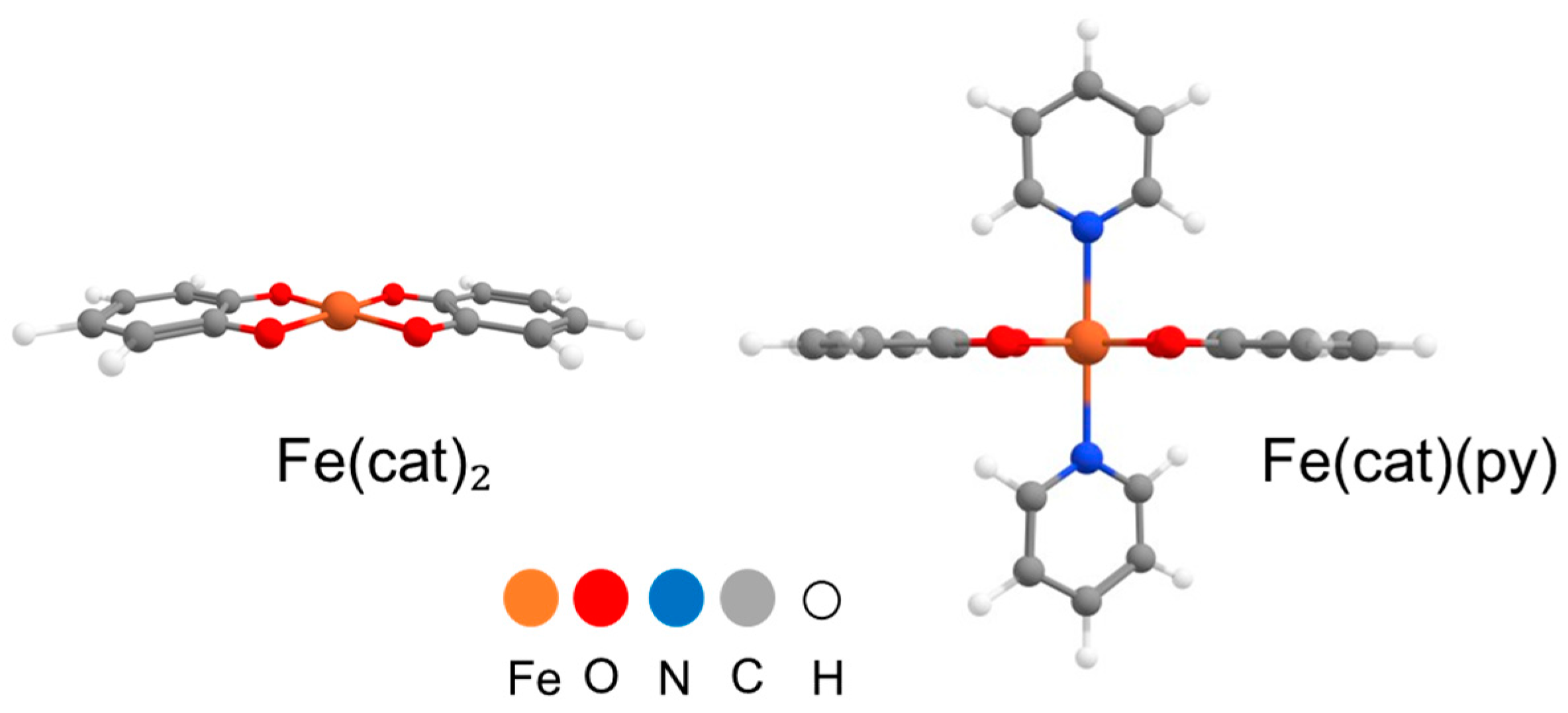
Computational modeling was carried out based on the ultraviolet–visible (UV-Vis) spectra of the polymers, which show an absorption maximum at 550 nm. This band corresponds to the metal–ligand charge transfer (MLCT) transition, characteristic of the coordination of two catecholate ligands to an iron atom. Simplified molecular models were proposed to represent the structures of the coordination polymers, which we named Fe(cat)₂ and Fe(cat)(py) (Figure 2). These structures were used for theoretical calculations of the respective infrared (IR) and Raman spectra. To assign the bands in the experimentally recorded spectra, we utilized the spectra of the free ligands A and B, as well as those simulated by DFT methods.

3.1. IR Spectral Assignment

  • Region from 4000 to 2000 cm−1
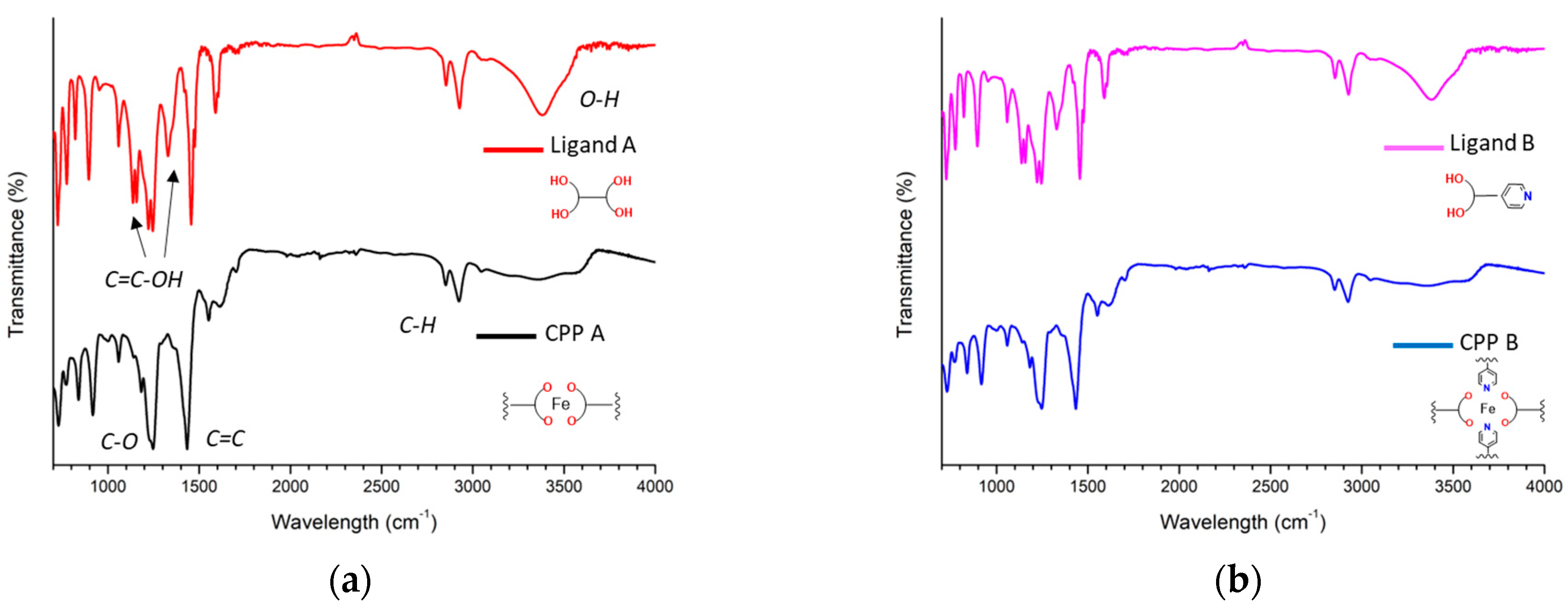
In this region, the O-H stretching band of the free catechol (A and B ligands), located around 3400 cm−1, is no longer present in the spectra of the polymers. This observation corroborates the deprotonation of the hydroxyl group, which is necessary for coordination with the metal center. Furthermore, bands corresponding to the aromatic (~2950 cm−1) and aliphatic (~2850 cm−1) C-H stretching vibrations were identified, remaining practically unchanged upon polymer formation.
  • Region from 2000 to 1000 cm1
This region exhibits bands characteristic of aromatic ring vibrations and C-O bonds. Specifically, the bands at 1440 cm1 and 1250 cm1 correspond to C=C and C-O stretching modes, respectively, and are present in both the free ligands and the polymer samples. Conversely, the bands at 1330 cm1 and 1150 cm1, associated with in-plane bending vibrations of the free catechol C-OH group, completely disappear in the polymer spectra, as expected.
  • Region from 1000 to 700 cm1
Three characteristic bands associated with C-H bending vibrations of the aromatic ring and aliphatic chain (C4 or longer) are observed. When comparing the spectra of the free ligands with those of the polymers, a slight shift of these bands to higher wave numbers is noted, from 890, 820, and 725 cm−1 to 920, 840, and 730 cm1, respectively. This shift is possibly due to the electronic effect of the metal on the electron density in the aromatic ring and the hydrocarbon chain.
  • Region from 700 to 400 cm1
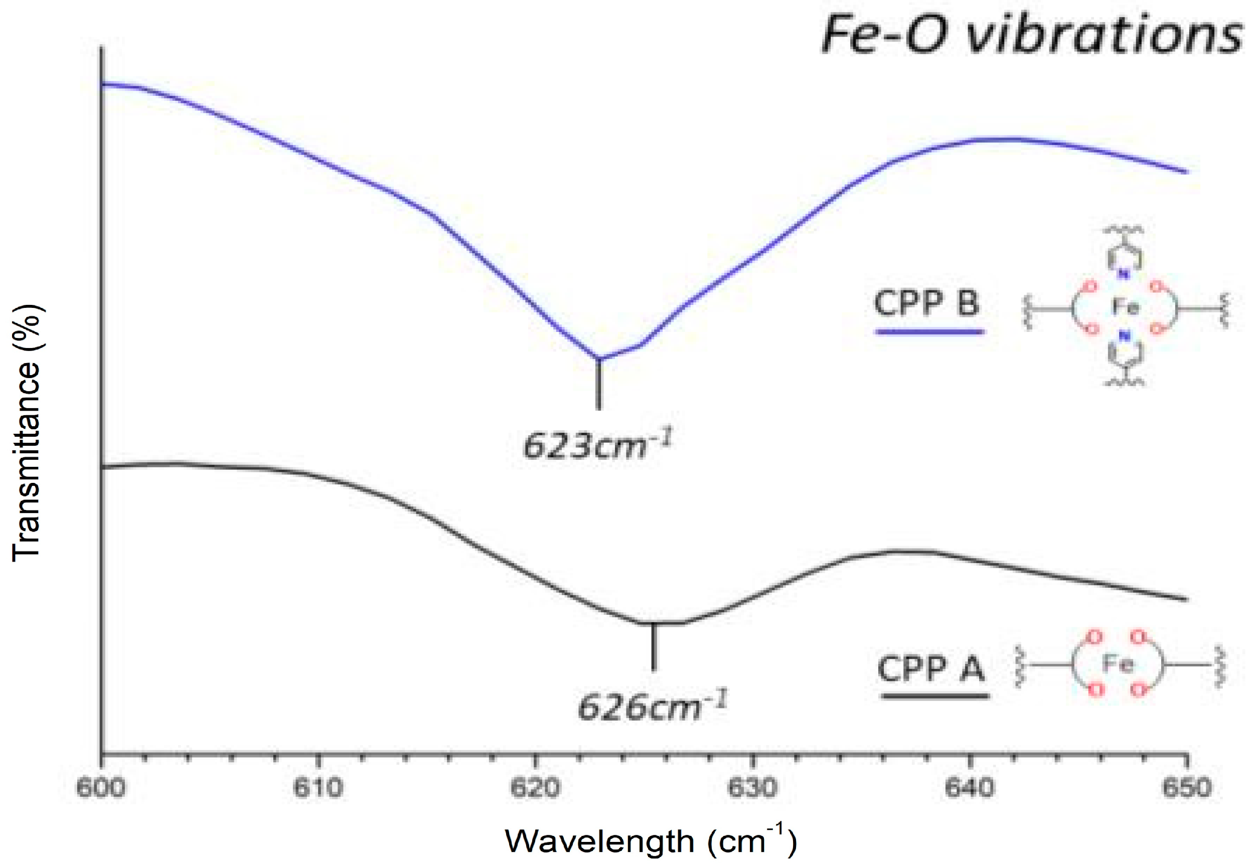
Since the ATR crystal did not allow for spectral analysis in this low region of the IR spectra (Figure 3), it was necessary to prepare the samples using KBr pellets (Figure 4). The most relevant signal in this region corresponds to the Fe-O bond and is located at ~620 cm1. This signal is present in the polymers but absent in the free ligands, evidencing the coordination of the catecholate to the iron.
The nature of the vibration modes corresponding to CCP A and CPP B were in agreement with the calculated. The experimental [Exp] and calculated [DFT] wavenumbers for some selected normal vibration modes are shown in Table 1. Probably the differences between the experimental and theoretical results can be attributed to the interaction packing forces.

3.2. Raman Spectral Assignment

Raman spectroscopy was employed to enhance the structural characterization of the polymers and, in particular, to identify the Fe-N coordination, which is essential for the formation of the polymeric Fe-(cat)(py) structure.
  • Region from 2000 to 1200 cm−1
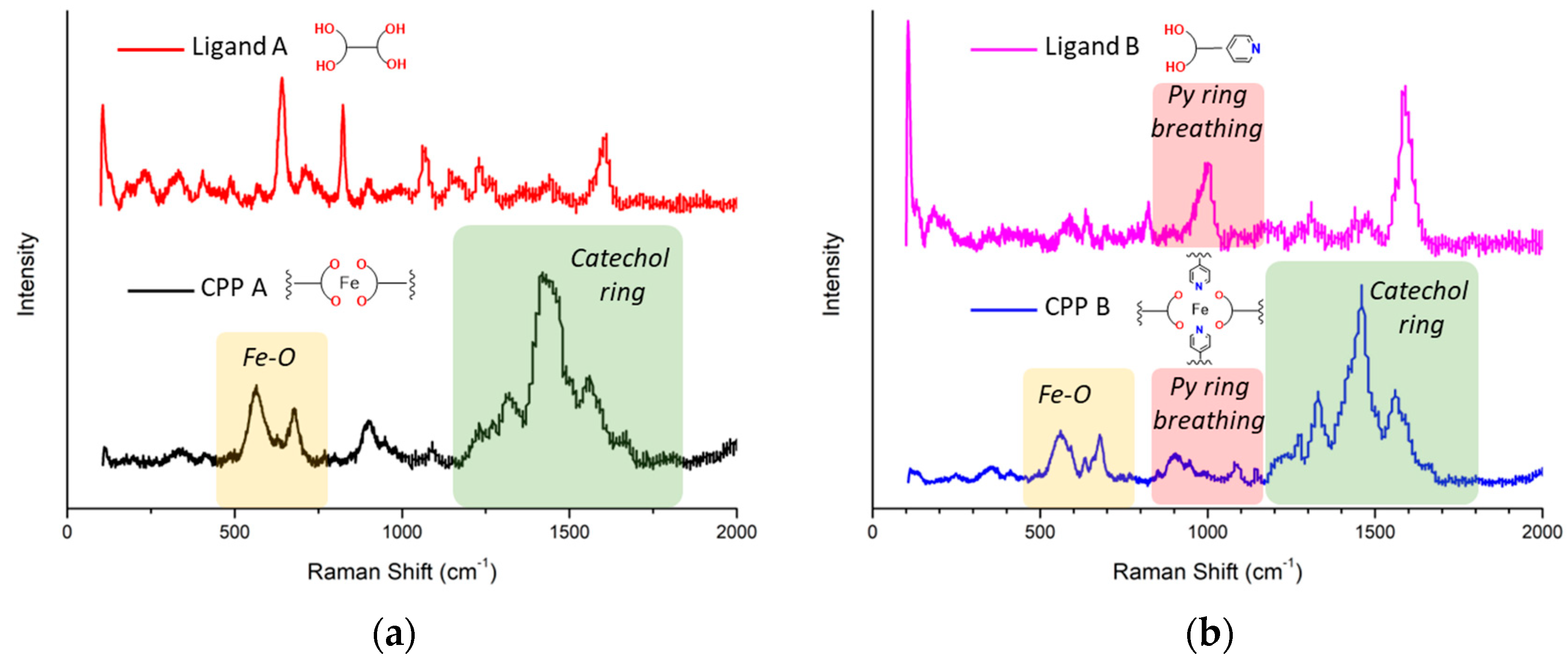
In this region, the characteristic bands of the vibrations of the aromatic ring and the =C-O bonds are found. In the case of the polymers, four signals are observed at 1560, 1450, 1330, and 1265 cm−1. For the ligands, only one signal is observed around 1600 cm−1. This could be due to the fact that the polymers are amorphous, which means they do not follow a symmetry pattern, leading to an increased number of observed bands.
  • Region from 1200 to 800 cm−1
This region is of particular interest for the polymer containing the pyridine ring Fe(cat)(py), as the significant decrease in the intensity of the band at 1000 cm−1 (Figure 5), corresponding to the pyridine ring breathing mode, upon polymer formation serves as evidence of the coordination of the pyridine nitrogen to the metal.
  • Region from 800 to 400 cm−1
In this region of the Raman spectra, two distinct bands appear at 690 and 555 cm−1, assigned to the Fe-O stretching vibration (marked with a yellow rectangle in Figure 5). These bands exhibit significantly higher intensity compared to the infrared spectra, providing compelling evidence for the formation of the Fe-O bond in the polymers. In contrast, the free ligands display a single band at 650 cm−1, attributed to an OH bending mode of the catechol moiety, which disappears upon polymer formation.

4. Conclusions

The UV-Vis spectra revealed a characteristic absorption maximum at 550 nm, indicative of a bidentate coordination of two catecholate ligands to the iron center. DFT calculations were instrumental in interpreting the IR and Raman spectra, confirming the deprotonation of the catechol ligands, the formation of Fe-O bonds, and the coordination of the pyridine nitrogen. The agreement between the experimental and calculated spectra validates the proposed model, providing a foundation for future studies on analogous coordination polymer systems using fast and simple methods for their characterization.

Author Contributions

V.D. carried out the conceptualization, investigation, methodology, and writing; F.N. participated in the investigation, methodology, and writing; M.C. participated in the methodology; G.R. carried out the funding acquisition, project administration, investigation, and writing. All authors have read and agreed to the published version of the manuscript.

Funding

This work was generously supported by the Consejo Nacional de Investigaciones Científicas y Técnicas (CONICET, PIP N° 11220200101665CO), Agencia Nacional de Promoción Científica y Tecnológica (ANPCyT, PICT 2018-2471), and Universidad Nacional del Sur (UNS, PGI 24/Q142) from Argentina.

Institutional Review Board Statement

Not applicable.

Informed Consent Statement

Not applicable.

Data Availability Statement

Data are available upon request.

Conflicts of Interest

The authors declare no conflicts of interest.

References

  1. Zhang, W.; Wang, R.; Sun, Z.; Zhu, X.; Zhao, Q.; Zhang, T.; Cholewinski, A.; Yang, F.; Zhao, B.; Pinnaratip, R.; et al. Catechol Functionalized Hydrogels: Biomimetic Design, Adhesion Mechanism, and Biomedical Applications. Chem. Soc. Rev. 2020, 49, 433–464. [Google Scholar] [CrossRef] [PubMed]
  2. Saiz-Poseu, J.; Mancebo-Aracil, J.; Nador, F.; Busqué, F.; Ruiz-Molina, D. The Chemistry behind Catechol-Based Adhesion. Angew. Chem. Int. Ed. 2019, 58, 696–714. [Google Scholar] [CrossRef] [PubMed]
  3. Suárez-García, S.; Solórzano, R.; Novio, F.; Alibés, R.; Busqué, F.; Ruiz-Molina, D. Coordination Polymers Nanoparticles for Bioimaging. Coord. Chem. Rev. 2021, 432, 213716. [Google Scholar] [CrossRef]
  4. Nador, F.; Mancebo-Aracil, J. Thiol-yne click reaction: An interesting way to derive thiol-provided catechols. RSC Adv. 2021, 11, 2074–2082. [Google Scholar] [CrossRef] [PubMed]
  5. Frisch, M.J.; Trucks, G.W.; Schlegel, H.B.; Scuseria, G.E.; Robb, M.A.; Cheeseman, J.R.; Scalmani, G.; Barone, V.; Mennucci, B.; Petersson, G.A.; et al. Gaussian 09, Revision C.01; Gaussian, Inc.: Wallingford, CT, USA, 2010. [Google Scholar]
  6. Perdew, J. Density-functional approximation for the correlation energy of the inhomogeneous electron gas. Phys. Rev. B 1986, 33, 8822–8824. [Google Scholar] [CrossRef] [PubMed]
  7. Grimme, S.; Antony, J.; Ehrlich, S.; Krieg, H. A Consistent and Accurate Ab Initio Parametrization of Density Functional Dispersion Correction (DFT-D) for the 94 Elements H-Pu. J. Chem. Phys. 2010, 132, 154104. [Google Scholar] [CrossRef] [PubMed]
  8. Weigend, F.; Ahlrichs, R. Balanced Basis Sets of Split Valence, Triple Zeta Valence and Quadruple Zeta Valence Quality for H to Rn: Design and Assessment of Accuracy. Phys. Chem. Chem. Phys. 2005, 7, 3297. [Google Scholar] [CrossRef] [PubMed]
  9. Ye, S.; Neese, F. Accurate Modeling of Spin-State Energetics in Spin-Crossover Systems with Modern Density Functional Theory. Inorg. Chem. 2010, 49, 772–774. [Google Scholar] [CrossRef] [PubMed]
  10. Becke, D. Density-functional thermochemistry. III. The role of exact exchange. J. Chem. Phys. 1993, 98, 5648–5652. [Google Scholar] [CrossRef]
  11. Lee, C.; Yang, W.; Parr, R.G. Development of the Colle-Salvetti correlation-energy formula into a functional of the electron density. Phys. Rev. B 1988, 37, 785. [Google Scholar] [CrossRef] [PubMed]
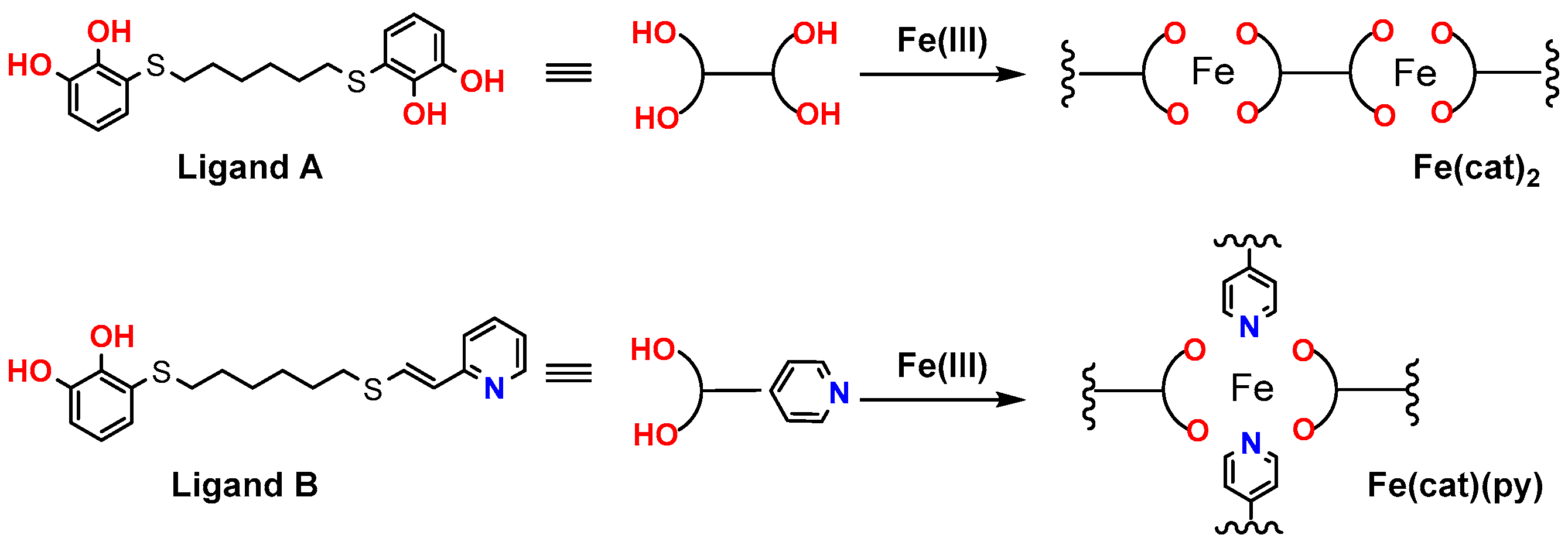
Figure 1. Formation of coordination polymer particles (CPPs).
Chemproc 16 00007 g001
Figure 2. Geometries of the structures used for theoretical IR and Raman calculations.
Chemproc 16 00007 g002
Figure 3. ATR Infrared spectra of the ligands (a) for Ligand A and (b) for Ligand B, and below their corresponding polymers (a) for CPP A and (b) for CPP B.
Chemproc 16 00007 g003
Figure 4. Fe-O vibrations on the KBr pellets infrared spectrum for both polymers.
Chemproc 16 00007 g004
Figure 5. Raman spectra of the ligands and their corresponding polymers. (a) For ligand A and (b) for ligand B.
Chemproc 16 00007 g005
Table 1. Main infrared frequencies (cm−1) and assignments for the two systems.
Vibrationsυ =C-Hυ C-H *υ C=Cυ C-OPy Ring breathingγ C-Hγ C-Hδ C-H *Fe-O
Fe(cat)2 [DFT]3101-14851254-956871-608
CPP A [Exp]2930285514401250-920840730626
Fe(cat)(py) [DFT]3095-147512671042945869-643
CPP B [Exp]29302855143512501000920840730623
* These vibrations are not found in DFT modeling due to simplification of the structures.
Disclaimer/Publisher’s Note: The statements, opinions and data contained in all publications are solely those of the individual author(s) and contributor(s) and not of MDPI and/or the editor(s). MDPI and/or the editor(s) disclaim responsibility for any injury to people or property resulting from any ideas, methods, instructions or products referred to in the content.