Proceeding Paper

Enzyme Inhibition and Antibiotics Properties of Six (6) Weeks Stable Chrysophyllum albidum Leaf Silver Nano-Particles †

by
Kingsley Ozioma Omeje
*,
Florence N. Nworah
,
Benjamin O. Ezema
and
Sabinus O. O. Eze
Department of Biochemistry, Faculty of Biological Sciences, University of Nigeria, Nsukka 410001, Nigeria
*
Author to whom correspondence should be addressed.
Presented at the 4th International Electronic Conference on Applied Sciences, 27 October–10 November 2023; Available online: https://asec2023.sciforum.net/.
Eng. Proc. 2023, 56(1), 196; https://doi.org/10.3390/ASEC2023-16578
Published: 4 December 2023
(This article belongs to the Proceedings of The 4th International Electronic Conference on Applied Sciences)

Abstract

:
Antibiotic resistance has posed a major public health challenge because of antibiotics misuse, overdose, underdose, ignorance of antibiotics usage and substandard production from producers, thus entailing the need for an alternative antibiotic agent production. Here, a commonly used antibiotic plant, Chrysophyllum albidum leaf, was used to produce silver nanoparticles (AgNPs) and was characterized using XRD, FTIR, DSC and DLS. The characterization data showed the production of six (6)-week stable AgNPs, with high antioxidant properties and amylase, glucosidase and cholinesterases inhibition properties. Similarly, the product exhibited stable antibiotics properties on Escherichia coli, Klebsiella pneumoniae and Staphylococcus aureus after six (6) weeks of storage at 4 °C.

1. Introduction

The synthesis and characterization of nanoparticles have been receiving great attention in recent times due to their wide array of applications in different areas of chemistry, medicine biology [1] and drug delivery [2]. The size of nanomaterials usually range between 1 to 100 nm [3].
Antibiotics are substances that inhibit the growth of microorganisms. Antibiotics resistance (AMR) is a major problem for public health, particularly now, and has led to the prevention and treatment of different infections [4], with S. aureus and K. pneumoniae being the most prevalent pathogenic antibiotic-resistant organisms of global concern [4].
Chrysophyllum albidum is a perennial plant also known as African star apple, with pharmacological or medicinal potentials [5], used for the treatment of stomach-ache and diarrhea [6] and inhibiting microbial growth of known wound contaminants [7,8], malaria and yellow fever.
Some advantages associated with compounding medicinal plants into nanoparticles include an increase in the component’s concentration, solubility, and half-life, thus enhancing drug delivery to its target [2,9]. Despite the advantages attributed to compounding medicinal plants into nanoparticles, there is a dearth of literature on the silver nitrate nanoparticle of C. albidum, which could increase the potency of this common and generally acclaimed antibiotic medicinal plant. One of the challenges affecting the efficacies of antibiotics could be the reduction of the intended concentration due to factors in the biological system, which could be overcome by compounding this medicinal plant into silver nanoparticles and increasing its concentration at the target site, which forms the aim of this study. Here, the biological activities of C. albidum leaf silver nanoparticles (Ag-C. albidum-NPs) were reported.

2. Materials and Methods

Reagents used include 1,1-diphenyl-2-picryl hydrazyl (Sigma-Aldrich, St. Louis, MO, USA), AgNO3, Acetylcholine iodide (Sigma-Aldrich), Butrylcholine iodide (Sigma-Aldrich). All the reagents used were analytical-grade.

2.1. Sample Collection and Preparation

The taxonomic classification of Chrysophyllum albidum was carried out by Mr. Felix Nwafor of the Pharmacognosy Department, University of Nigeria, Nsukka as Chrysophyllum albidum G. Don. (Sapotaceae) with the voucher number PCG/UNN/0359. The plant sample (Leaves) was dried by airdrying for 72 h, ground and macerated in 1000 mL of 80% ethanol for 72 h. At the end of maceration, the mixture was filtered and concentrated for analysis.

2.2. Synthesis of Silver Nanoparticles

The synthesis of C. albidum silver nanoparticles AgNPs was carried out as described by Osibe et al. [10] using aqueous C. albidum extract to an equal volume of 1 mM AgNO3 and was incubated at 25 °C for 72 h.

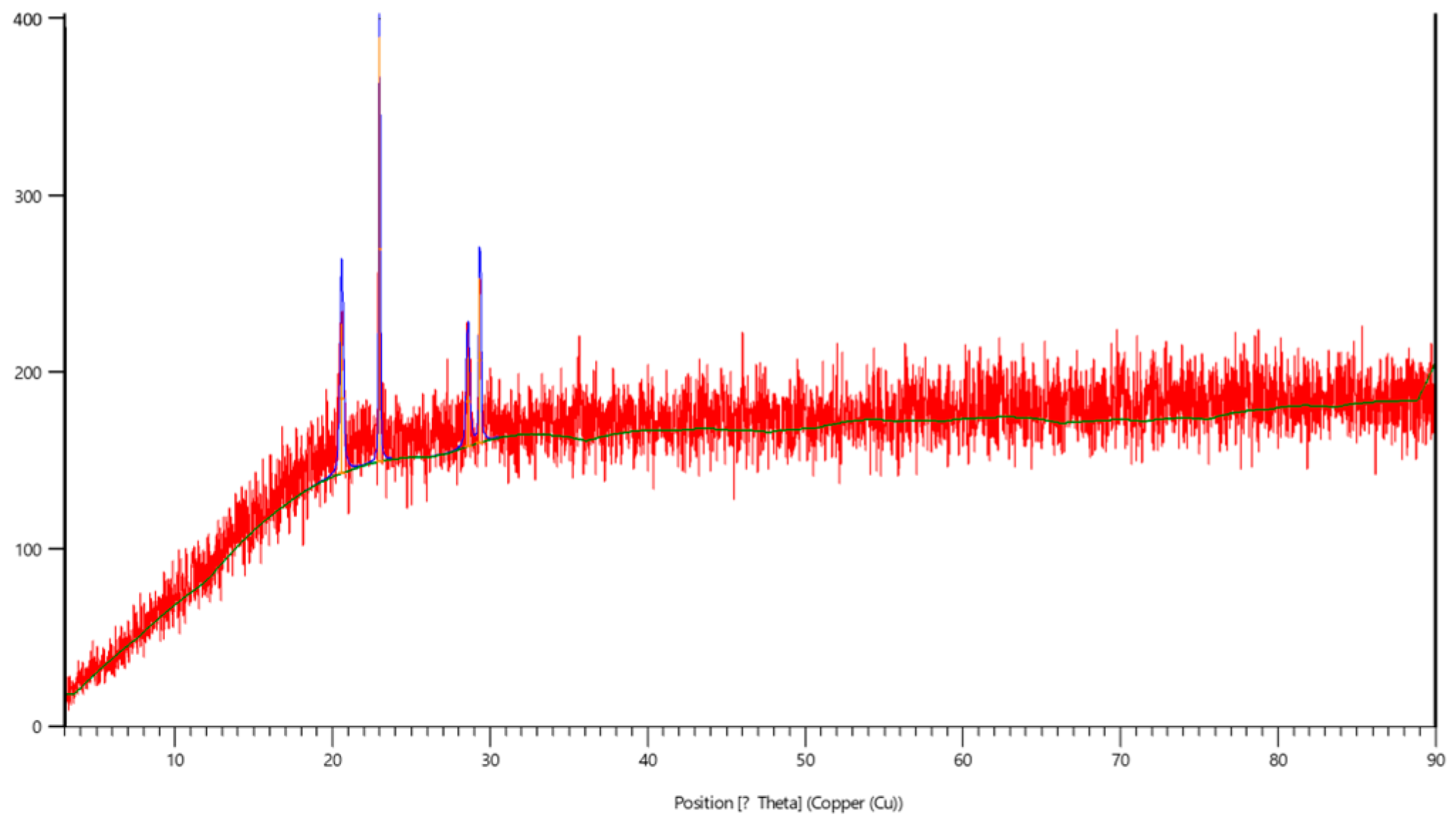
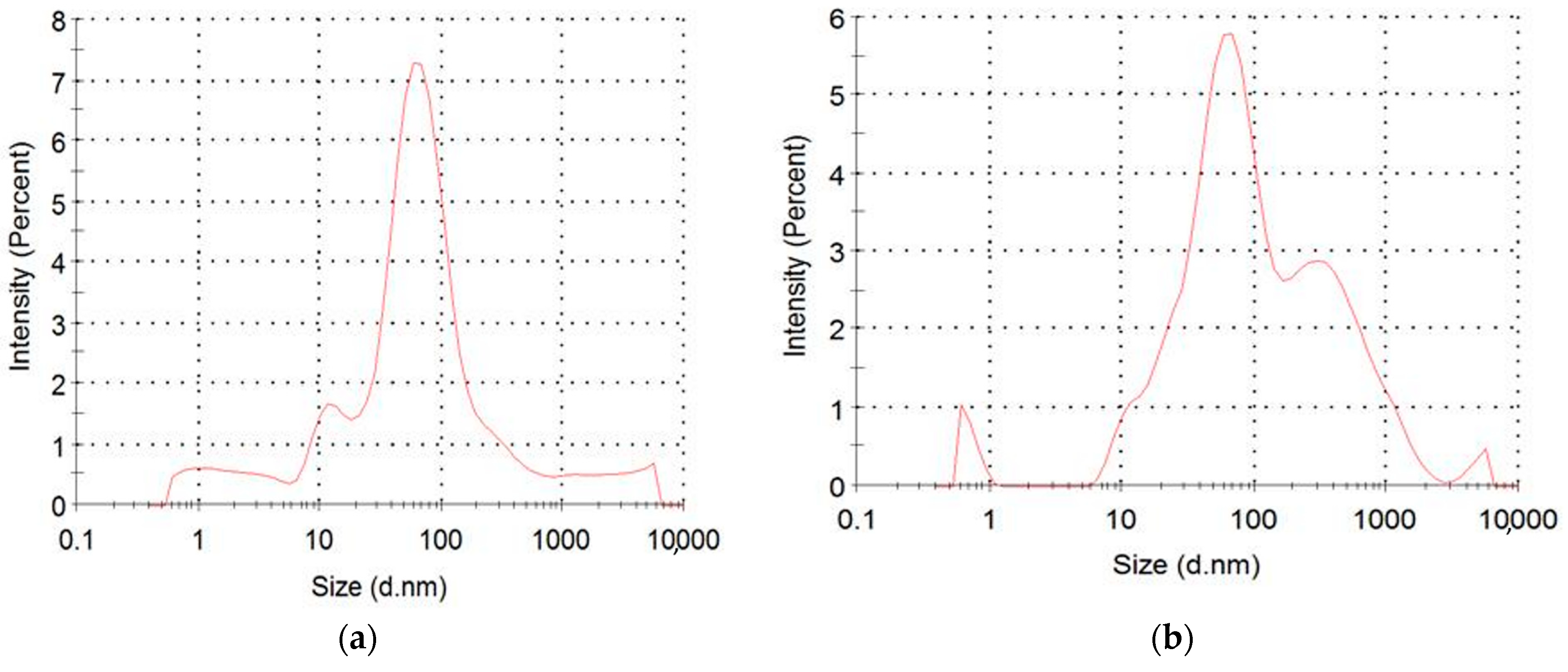
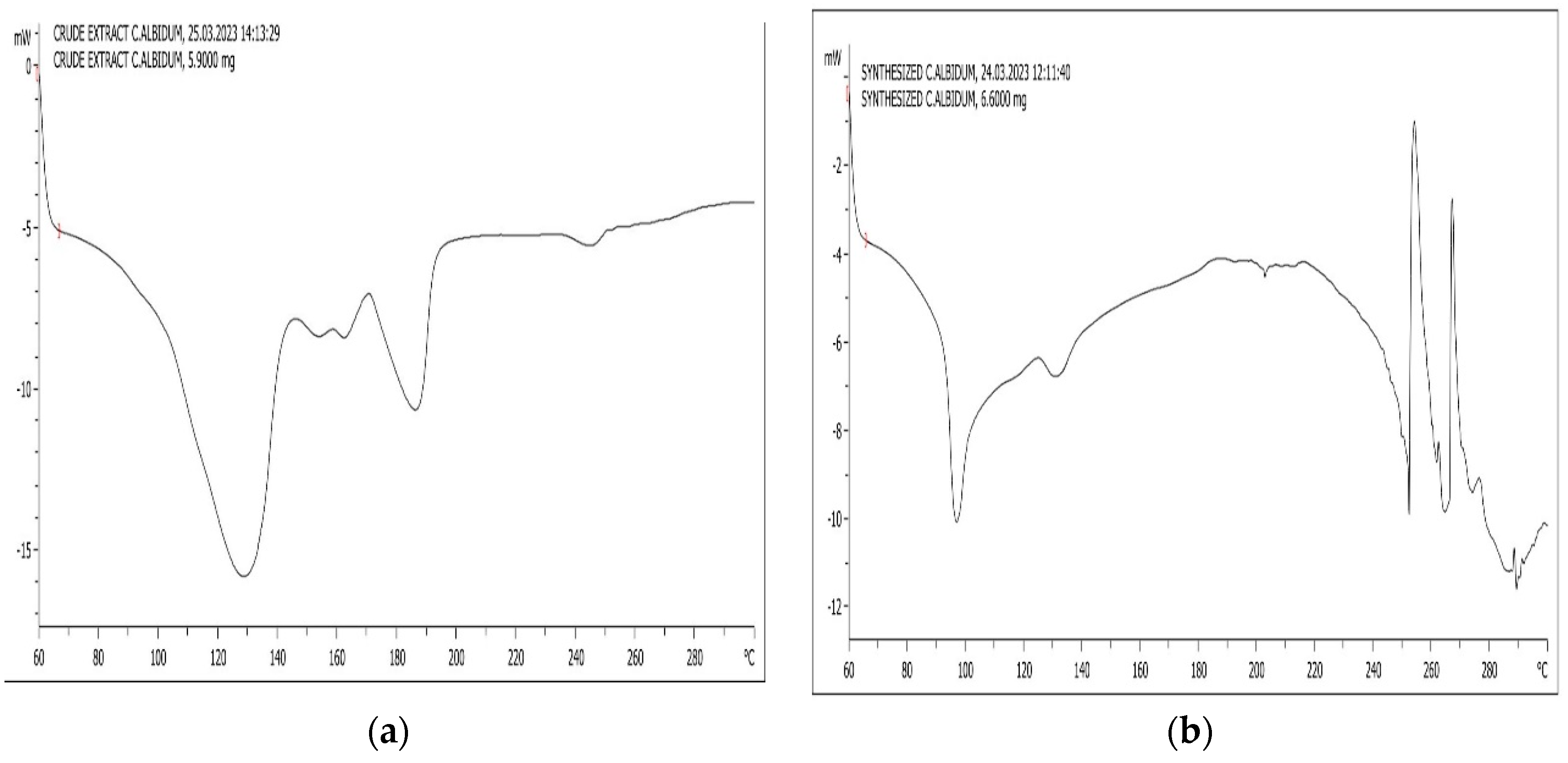
2.3. Characterization of Nanoparticles

The thermostability of Ag-C. albidum-NPs was determined using differential scanning calorimetry (Shimadzu, Kyoto, Japan). The particle size was determined using dynamic light scattering (Malvern Zeta sizer, Worcestershire, UK). X-ray diffraction and phase identification of the formulated nanoparticles (AgNPs) were recorded in the X-ray diffraction (Philips Diffractometer, Eindhoven, The Netherlands). Functional groups of the synthesized AgNPs were identified using Fourier transform infrared spectroscopy (Shimadzu, Kyoto, Japan).

2.4. In Vitro Antioxidant Activity

2.4.1. 1,1-Diphenyl-2-picryl Hydrazyl (DPPH) Scavenging Potential

The DPPH scavenging potential of Ag-C. albidum-NPs was assessed as described by Shen et al. [11].
DPPH   radical   scavenging   ( % )   = C o n t r o l   a b s o r b a n c e S a m p l e   a b s o r b a n c e C o n t r o l   a b s o r b a n c e × 100

2.4.2. Determination of Hydroxyl Radical Scavenging Activity

The hydroxyl radical (-OH) scavenging activity was measured by the method of Jin et al. [12], and the absorbance was read at 536 nm. Scavenging activity (%) = (Abs. sample − Abs. blank)/(Abs0 − Abs. blank) × 100. Ascorbic acid was used as the reference standard.

2.4.3. Nitric Oxide Radical Scavenging

The nitric oxide radical scavenging capacity of the fractions was measured by Griess reaction, following Sangermeswaran et al. [13]. Ascorbic acid was used as the reference standard.
Percentage of inhibition = [(Ao − A1)/Ao] × 100.

2.4.4. Ferric Cyanide (Fe3+) Reducing Antioxidant Power Assay

The reducing power of the extracts was measured by the direct reduction of Fe3+(CN−)6 to Fe2+(CN−)6 by the absorbance measurement of the formation of the Perl’s Prussian Blue complex following the addition of excess Fe3+ [14]. Finally, the reaction mixture absorbance was measured spectrophotometrically at 700 nm.

2.4.5. Acetylcholinesterase and Butrylcholinesterase Inhibitory Activity Assay

Cholinesterases (AchE) and (BuchE) inhibitions were studied using Ellman et al. [15].
A C h E   a c t i v i t y   % = A o A 1 A o × 100 ;   B C h E   a c t i v i t y   % = A o A 1 A o × 100 ;

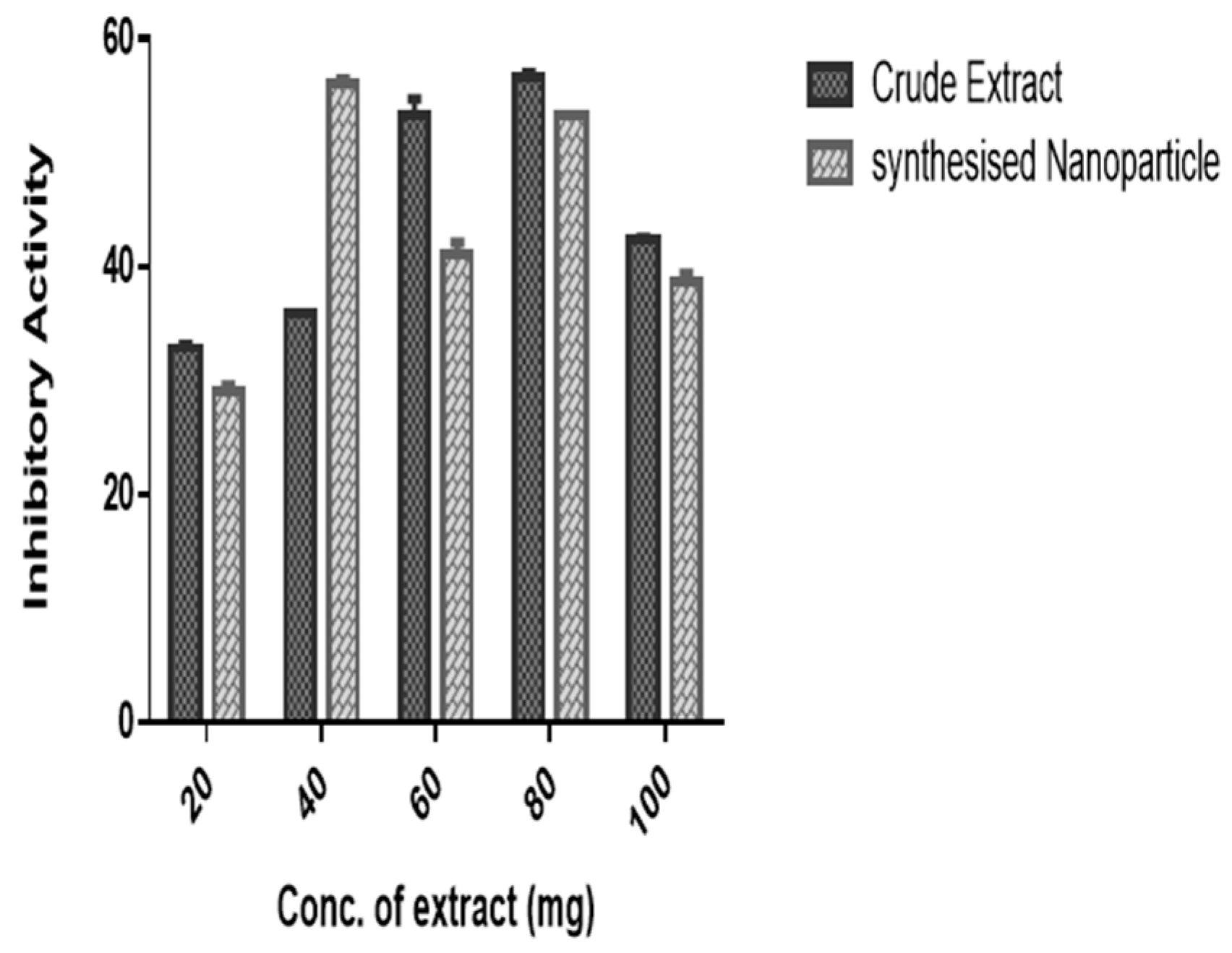
2.4.6. α-Amylase and α-Glucosidase Inhibition Assay

The α-amylase activity inhibition assay was conducted as described by Gulati et al.’s method [16]. The α-glucosidase activity inhibition assay was determined according to the procedure of Liu et al. [17].
Percentage   ( % )   amylase   and   glucosidase   inhibition   =   A b s . C o n t r o l A b s . S a m p l e A b s . C o n t r o l × 100

3. Results and Discussion

X-ray diffraction is an appropriate technique for determining the phases of materials (Ag-C. albidum nanoparticles): four intense diffraction peaks were apparent at the 2 h values of 20.56, 22.95, 28.56 and 29.33, corresponding to the Ag-planes, respectively (Figure 1). The generated data are in tandem with the report of [10,18], with the high intensity of the peaks corresponding to Ag.NPs [19].
One of the tools used in analyzing the functional groups present in a compound is Fourier transform infrared spectroscopy. Thus, the identification of the functional groups in both the aqueous extract of C. albidum and synthesized Ag C. albidum NPs is shown in Figure 2a,b. The spectra of the C. albidum extract showed absorbance bands at 3280, 2918, 2851 and 2117. The broad band 3280 cm−1 represents the stretching vibrations of N–H, and the 2918 and 2851 cm−1 bands correspond to C–H stretching vibrations. Additionally, FT-IR spectra of the Ag-C. albidum-NPs show an absorbance band at 3250 cm representing stretching vibrations of a N–H group, 2914 cm bands corresponding to C–H stretching vibrations and 2117 cm bands representing N=N=N stretching. Furthermore, peaks at 1997, 1871 and 1718 cm represent amino acids, N-H+ charged amine and C=O stretching, respectively. There was a shift in absorbance from 3280 to 3250 cm−1, with an increase in band intensity (Figure 2b), suggesting a possible involvement of this functional group in the reduction of silver ions to nanoparticles. Several studies reported the use of FT-IR in evaluating the functional groups present in the plant material that aided in the biosynthesis of Ag-C. albidum-NPs and in identifying possible biomolecules responsible for some of the biological activities obtained [20,21]. According to Tavan et al. [22], FTIR was used in identifying the functional groups of P. frutescens extract and Pf-AgNPs.
Dynamic light scattering (DLS) is used to measure the size of the synthesized particle. The AgNPs had a peak diameter of 54.15 nm, and Ag-C. albidum-NPs had modal peaks at 64.75 nm, respectively, as shown in Figure 3a,b. DLS technique was described as a means of measuring the diameter of nanoparticles in suspension [19,21]. An average diameter of 28.3 to 161 nm, with a Polydispersity Index of 0.72, was reported [22]. The results of DLS agreed with the reports of Omeje et al. [21] and Tavan et al. [22].
The thermostability of the AgNPs and Ag-C. albidum-NPs was also evaluated using differential scanning calorimetry (DSC). It was observed that the Ag-nanoparticle was releasing energy, with a melting temperature of 129.8 and 188.6 °C, while Ag-C. albidum-NPs underwent an exothermic reaction at 97.6 °C, with subsequent heating giving rise to endothermic activity at 257.4 and 270.1 °C, respectively (Figure 4a,b).
There was an in vitro inhibitory assay of α-amylase with both crude and Ag-C. albidum-NPs, as shown in Figure 5. Crude extract providing more than 50% inhibition of alpha amylase was obtained at high concentrations of 60 (53.22 ± 1.5%) and 80 mg/mL (56.62 ± 0.4%), respectively. Additionally, Ag-C. albidum-NPs provided 56.03 ± 0.45% inhibition of alpha amylase at 40 mg/mL and 53.24 ± 0.06% inhibition of alpha amylase at 80 mg/mL concentration of Ag-C. albidum-NPs (Figure 5). At 20 and 100 mg/mL of both products, less than 50% inhibition of the enzyme resulted, respectively. The potential of crude and Ag-C. albidum-NPs to inhibit α-glucosidase was also studied and reported in Figure 6. The rate of inhibition increased proportionally to the increase in concentration of the extracts. At 60 mg/mL of the crude extract, more than 50% inhibition of the enzyme was reported, with a steady decline as the concentration increased (80–100 mg/mL). Similarly, the highest percentage inhibition (47.22 ± 0.4%) was obtained at 60 mg/mL for Ag-C. albidum-NPs. The Ag-C. albidum-NPs could not achieve 50% inhibition of the enzyme at all concentrations studied (Figure 6).
Additionally, the in vitro bioactivity of crude and Ag-NPs of C. albidum products was evaluated using acetylcholinesterase and butyrylcholinesterase. Various degrees of enzyme inhibition were obtained, as shown in Figure 7 and Figure 8. The percentage inhibition of acetylcholinesterase of the crude leaf extract was 39.58 ± 0.9, 55.15 ± 0.03 and 79.82 ± 0.83% at 20, 40 and 60 mg/mL. A subsequent increase of concentration (80–100 mg/mL) provided an AchE percentage inhibition of 66.56 ± 0.22 and 63.32 ± 0.70, respectively. Furthermore, the inhibitory capacity of the crude and Ag-C. albidum-NPs was evaluated on butyrylcholinesterase, one of the enzymes responsible for stabilizing the nervous system, and the results are shown in Figure 8. There was >50% of butylrylcholinesterase activity inhibition at all the concentrations studied for both crude and complex NPs produced. However, a concentration-dependent increase in inhibition was observed from 20–60 mg/mL. A high concentration (80–100 mg/mL) showed a reduction in inhibition of the enzyme (Figure 8). On the other hand, Ag-C. albidum-NPs provided a consistent increase in inhibition with a concomitant increase in extract concentrations. According to Omeje et al. [23] the inhibition of some physiological enzymes involved in the etiology of some chronic diseases (diabetes and Alzheimer’s disease) is effective for their management.
The antibacterial potential of Ag-C. albidum-NPs was evaluated on some common bacteria pathogens, such as K. pneumoniae, E. coli, and S. aureus, using graded concentrations of 6.5, 12.5, 25 and 50 mg, and their zone of inhibitions were reported. At 6.5 mg, there was a 0.83 and 1.09 inhibition of K. pneumoniae and S. aureus, respectively. A subsequent increase of the extract concentrations to 12.5 mg provided a 1.21, 3.06 and 3.27 mm zone of inhibition on K. pneumonia, E. coli, and S. aureus, respectively. Additionally, a progressive increase in the zone of inhibition was obtained as the concentration of the extract increased from 25 to 50 mg/mL across all organisms studied, as shown in Table 1. A concentration of 50 mg/mL provided the highest zone of inhibition among all studied organisms (K. pneumonia, E. coli, and S. aureus), with 9.88, 11.73 and 19.03 mm, respectively. However, a significant difference in the zone of inhibition was obtained for the extract when compared to a standard antibiotic (Streptomycin) (Table 1).
The power of medicinal material to scavenge free radicals in cells is an essential pharmacological property. Thus, the in vitro ability of Ag-C. albidum-NPs to mop DPPH generated free radicals was evaluated. The radical scavenging potential of Ag-C. albidum-NPs increased concomitantly with an increase in concentration (20–80 mg/mL), except at 100 mg when a significant decrease in the scavenging power was obtained. The report of this study is comparable to the study of NPs on S. aureus and E. coli [24], and Pf-AgNPs were reported to exert antimicrobial power against S. aureus, Candida albicans and E. coli [21].
There was an increase in the scavenging potential of Ag-C. albidum-NPs and the standard antioxidant molecule (Vit. C.) at all concentrations studied (Table 2). Furthermore, the hydroxyl radical (OH*) scavenging ability of Ag-C. albidum-NPs was shown to increase with an increase in concentration, with 60 mg providing the highest desirable activity. Higher concentrations of 80 and 100 mg yielded a lower inhibition when compared to the standard (Vit. C) and 60 mg concentration. The inhibition of OH* by the compound was significantly low when compared to ascorbic acid inhibition (81.53%). Similarly, the NO* scavenging power of Ag-C. albidum-NPs was studied at varying (20–100 mg/mL) concentrations, respectively. None of the concentrations provided 50% inhibition of the radicals produced by nitroprusside, as shown in Table 2. However, 60 mg/mL also provided the highest inhibition of the radical (48.22 ± 1.57%). The varying concentrations’ scavenging power is significantly lower when compared to the 86.82 ± 0.41% inhibition obtained for Vit C. Table 2 shows the ferric reducing power of the Ag-C. albidum-NPs studied; a concentration-dependent increase in electron transfer rate inhibition was obtained for the compound (20–60 mg), and a further increase in the concentration of the compound (80–100 mg) led to a decrease in the FRAP scavenging power of the compound (Table 2). The antioxidant abilities of medicinal plants are attributed to plants being rich in phytochemicals, such as polyphenols and flavonoids [25]. Several plants have been reported to have radical scavenging powers [22,23]. The radical scavenging power of NPs could be attributed to the presence of some compounds in C. albidum being used for the synthesis. Similarly, Omeje et al. [23] also attributed the antioxidant power of plant materials to the presence of tannins. This supports the earlier assertion of Deng et al. [26] on tannins and of Patel et al. [27] on alkaloids, with flavonoids being described as a potent antioxidant agent [28].

4. Conclusions

In conclusion, the green synthesis method was used to develop a commonly used antibiotic medicinal plant (Chrysophyllum albidum) into silver nanoparticles (AgNPs) and was characterized. From the results obtained in this study, the produced C. albidum-Ag-NPs had a good stability under ambient conditions for six (6) weeks, and the compound possessed free radical scavenging power, antibacterial properties and showed the ability to inhibit alpha amylase and glucosidase, which have been implicated in diabetes etiology. Additionally, C. albidum-Ag-NPs exhibited anti-cholinesterases activities, suggesting that they could be appropriate in managing some neurological diseases, such as Alzheimer’s disease. Overall, the study contributes to improving the concentration of active C. albidum-Ag-NPs from antibiotic, antioxidant and anticholinesterase medicinal plants, which could be delivered to the site of action at higher concentrations with a prolonged shelf life of the material.

Author Contributions

Conceptualization, K.O.O., S.O.O.E. and F.N.N.; methodology, K.O.O., B.O.E. and F.N.N.; software, K.O.O. and B.O.E.; formal analysis, K.O.O. and B.O.E.; investigation, K.O.O., B.O.E., F.N.N. and S.O.O.E.; resources, K.O.O. and B.O.E.; data curation, K.O.O., B.O.E., F.N.N. and S.O.O.E.; validation, K.O.O., B.O.E., F.N.N. and S.O.O.E.; visualization, K.O.O., B.O.E. and F.N.N.; supervision, K.O.O., B.O.E., F.N.N. and S.O.O.E.; writing—original draft preparation, K.O.O., B.O.E., F.N.N. and S.O.O.E.; writing—review and editing, K.O.O., B.O.E., F.N.N. and S.O.O.E. All authors have read and agreed to the published version of the manuscript.

Funding

This research received no external funding.

Institutional Review Board Statement

Not applicable.

Informed Consent Statement

Not applicable.

Data Availability Statement

Data are contained within the article.

Acknowledgments

The authors K.O.O., B.O.E., F.N.N. and S.O.O.E acknowledge the assistance and technical support from the Biochemistry Department, University of Nigeria, Nsukka, Nigeria.

Conflicts of Interest

The authors declare no conflict of interest.

References

  1. Vishwasrao, C.; Momin, B.; Ananthanarayan, L. Green synthesis of silver nanoparticles using sapota fruit waste and evaluation of their antimicrobial activity. Waste Biomass Valor. 2019, 10, 2353–2363. [Google Scholar] [CrossRef]
  2. Bharathi, D.; Josebin, M.D.; Vasantharaj, S.; Bhuvaneshwari, V. Biosynthesis of silver nanoparticles using stem bark extracts of Diospyros montana and their antioxidant and antibacterial activities. J. Nanostruct. Chem. 2018, 8, 83–92. [Google Scholar] [CrossRef]
  3. Nayagam, V.; Gabriel, M.; Palanisamy, K. Green synthesis of silver nanoparticles mediated by Coccinia grandis and Phyllanthus emblica: A comparative comprehension. Appl. Nanosci. 2018, 8, 205–219. [Google Scholar] [CrossRef]
  4. Prestinaci, F.; Pezzotti, P.; Pantosti, A. Antimicrobial resistance: A global multifaceted phenomenon. Pathog. Glob. Health 2015, 109, 309–318. [Google Scholar] [CrossRef] [PubMed]
  5. Ezema, B.O.; Omeje, K.O.; Ozioko, J.N.; Urama, D.C.; Omeje, H.C.; Nnawelezi, A.; Ejim, A. Cholinesterase inhibition, biological activity, and characterization of Chrysophyllum albidum leaf and stem-bark chloroform extract using GC-MS: An in vitro study. Trop. J. Nat. Prod. Res. 2021, 5, 130–134. [Google Scholar] [CrossRef]
  6. Ajayi, A.M.; Chidebe, E.O.; Ben-Azu, B. Chrysophyllum albidum (African star apple) fruit-supplemented diet enhances cognitive functions and attenuates lipopolysaccharide-induced memory impairment, oxidative stress, and release of proinflammatory cytokines. Nutrire 2020, 45, 20. [Google Scholar] [CrossRef]
  7. Droepenu, E.K.; Jackson, N.A.; Kyene, M.O.; Ayertey, F.; Brew-Daniels, H.; Mensah, T.; Adomako, C.; Nartey, A.P.; Gyampoh, A.O.; Boamah, L.A. Synthesis and characterization of Chrysophyllum albidum (African Star Apple) plant-mediated Zinc oxide nanostructures and evaluation of their antioxidant, anti-inflammatory, and antimicrobial activity. J. Nanomater. 2022, 5508224, 14. [Google Scholar] [CrossRef]
  8. Adebayo, A.H.; Abolaji, A.O.; Kela, R.; Ayepola, O.O.; Olorunfemi, T.B.; Taiwo, O.S. Antioxidant activities of the leaves of Chrysophyllum albidum G. Pak. J. Pharm. Sci. 2011, 24, 545–551. [Google Scholar]
  9. Puja, A.M.; Rupa, E.J.; Kim, Y.J.; Yang, D.-C. Medicinal Plant Enriched Metal Nanoparticles and Nanoemulsion for Inflammation Treatment: A Narrative Review on Current Status and Future Perspective. Immuno 2023, 3, 182–194. [Google Scholar] [CrossRef]
  10. Osibe, D.A.; Chiejina, N.V.; Ogawaa, K.; Aoyagi, H. Stable antibacterial silver nanoparticles produced with seed-derived callus extract of Catharanthus roseus. Artif. Cells Nanomed. Biotechnol. 2018, 46, 1266–1273. [Google Scholar] [CrossRef]
  11. Shen, X.; Chen, W.; Zheng, Y.; Lei, X.; Tang, M.; Wang, H.; Song, F. Chemical composition, antibacterial and antioxidant activities of hydrosols from different parts of Areca catechu L. and Cocos nucifera L. Ind. Crops Prod. 2017, 96, 110–119. [Google Scholar] [CrossRef]
  12. Jin, H.; Cia, M.; Li, Y.; Zhou, J. 1,10-phenanthroline-Fe2+ oxidative assay of hydroxyl radical produced by H2O2/Fe. Prog. Biochem. Biophys. 1996, 23, 553–555. [Google Scholar]
  13. Sangameswaran, B.B.; Balakrishnan, B.; Deshraj, B.R.; Jayakar, C. In vitro antioxidant activity of roots of Thespesia lampas Dalz and Gibs. Pak. J. Pharm. Sci. 2009, 22, 368–372. [Google Scholar] [PubMed]
  14. Oyaizu, M. Studies on products of browning reaction. Antioxidative activities of products of browning reaction prepared from glucosamine. Jpn. J. Nutr. Dietet. 1986, 44, 307–315. [Google Scholar] [CrossRef]
  15. Ellman, G.L.; Courtney, K.D.; Andres, V.; Featherstone, R.M. A new and rapid colorimetric determination of AchE activity. Biochem. Pharmacol. 1961, 7, 88–95. [Google Scholar] [CrossRef] [PubMed]
  16. Gulati, V.; Harding, I.H.; Palombo, E.A. Enzyme inhibitory and antioxidant activities of traditional medicinal plants: Potential application in the management of hyperglycemia. BMC Complement. Altern. Med. 2012, 12, 77. [Google Scholar] [CrossRef] [PubMed]
  17. Liu, S.; Yu, Z.; Zhu, H.; Zhang, W.; Chen, Y. In vitro α-glucosidase inhibitory activity of isolated fractions from water extract of Qingzhuan dark tea. BMC Complement. Altern. Med. 2016, 16, 378. [Google Scholar] [CrossRef] [PubMed]
  18. Venkatesan, J.; Kim, S.-K.; Shim, M.S. Antimicrobial, antioxidant, and anticancer activities of biosynthesized silver nanoparticles using marine algae Ecklonia cava. Nanomaterials 2016, 6, 235. [Google Scholar] [CrossRef]
  19. Srikar, S.K.; Giri, D.D.; Pal, D.B.; Mishra, P.K.; Upadhyay, S.N. Green synthesis of silver nanoparticles: A review. Green Sustain. Chem. 2016, 6, 34. [Google Scholar] [CrossRef]
  20. González-Pedroza, M.G.; Benítez, A.R.T.; Navarro-Marchal, S.A.; Martínez-Martínez, E.; Juan, A.; Marchal, J.A.; Boulaiz, H.; Morales-Luckie, R.A. Biogeneration of silver nanoparticles from Cuphea procumbens for biomedical and environmental applications. Sci. Rep. 2023, 13, 790. [Google Scholar] [CrossRef]
  21. Omeje, K.O.; Magbo, C.; Ossai, E.C.; Ozioko, J.N.; Ezema, B.O.; Nnolim, N.E.; Eze, S.O.O. Immobilization of fungal peroxidase on paramagnetic nanoparticles for synthetic dye decolorization. Mater. Proc. 2022, 9, 24. [Google Scholar] [CrossRef]
  22. Tavan, M.; Hanachi, P.; Mirjalili, M.H.; Dashtbani-Roozbehani, A. Comparative assessment of the biological activity of the green synthesized silver nanoparticles and aqueous leaf extract of Perilla frutescens (L.). Sci. Rep. 2023, 13, 6391. [Google Scholar] [CrossRef] [PubMed]
  23. Omeje, K.O.; Ezema, B.O.; Onaebi, C.N.; Onoyima, S.C.; Ezeorba, T.P.C.; Eze, S.O.O. HPLC fngerprint of favonoids, enzyme inhibition and antioxidant activity of Newbouldia laevis stem-bark: An in vitro and in silico study. Fut. J. Pharma. Sci. 2023, 9, 36–52. [Google Scholar] [CrossRef]
  24. Shah, M.Z.; Guan, Z.H.; Din, A.U.; Ali, A.; Rehman, A.U.; Jan, K.; Faisal, S.; Saud, S.; Adnan, M.; Wahid, F.; et al. Synthesis of silver nanoparticles using Plantago lanceolata extract and assessing their antibacterial and antioxidant activities. Sci. Rep. 2021, 11, 20754. [Google Scholar] [CrossRef] [PubMed]
  25. Kumar, S.; Kumar, V.; Rana, M.; Kumar, D. Enzymes inhibitors from plants: An alternate approach to treat diabetes. Pharmacogn. Commun. 2012, 2, 18–33. [Google Scholar] [CrossRef]
  26. Deng, L.; Qi, Y.; Liu, Z.; Xi, Y.; Xue, W. Effect of tannic acid on blood components and functions. Colloids Surf. B Biointerfaces 2019, 184, 110505. [Google Scholar] [CrossRef]
  27. Patel, K.; Gadewar, M.; Tripathi, R.; Prasad, S.K.; Patel, D.K. A review on medicinal importance, pharmacological activity and bioanalytical aspects of beta-carboline alkaloid Harmine. Asian Pac. J. Trop. Biomed. 2012, 2, 660. [Google Scholar] [CrossRef]
  28. Srivastava, J.; Kumar, S.; Vankar, P.S. Correlation of antioxidant activity and phytochemical profile in native plants. Nutr. Food Sci. 2012, 42, 71–79. [Google Scholar] [CrossRef]
Figure 1. X-ray diffraction spectra of Ag-C. albidum-NPs synthesized from the leaf of C. albidum.
Engproc 56 00196 g001
Figure 2. (a) FT-IR spectra of AgNPs and (b) FT-IR spectra of Ag-C. albidum-NPs.
Engproc 56 00196 g002
Figure 3. (a) DLS of spectra of AgNPs and (b) DLS spectra of synthesized Ag-C. albidum-NPs.
Engproc 56 00196 g003
Figure 4. (a) DSC thermogram of the AgNPs and (b) DSC thermogram of Ag-C. albidum-NPs.
Engproc 56 00196 g004
Figure 5. Amylase inhibition by crude extract and Ag-C. albidum-NPs.
Engproc 56 00196 g005
Figure 6. Glucosidase inhibition by crude extract and Ag-C. albidum-NPs.
Engproc 56 00196 g006
Figure 7. Inhibition of AchE by crude extract and Ag-C. albidum-NPs.
Engproc 56 00196 g007
Figure 8. Inhibition of BuchE by crude extract and Ag-C. albidum-NPs.
Engproc 56 00196 g008
Table 1. Antibacterial Activity of Ag-C. albidum-NPs.
Conc. (mg)Klebsiella pneumoniaeEscherichia coliStaphylococcus aureus
6.50.83-1.09
12.51.213.063.27
256.9219.4121.75
509.8811.7319.03
Streptomycin27.9737.3038.58
Table 2. Antioxidant potentials of silver C. albidum-Nanoparticles.
Antioxidant
Potentials
20 mg/mL40 mg/mL60 mg/mL80 mg/mL100 mg/mLVit. C mg/mL)
DPPH (%)20.11 * a ± 0.0130.10 * b ± 1.0042.71 * c ± 1.9942.06 * c ± 2.6039.88 * c ± 0.1073.20 * ± 0.93
OH* Scavenging ability (%)9.84 * a ± 0.2022.97 * b ± 0.0955.83 * c ± 0.1950.91 * c ± 0.2646.15 * d ± 0.2181.53 * ± 0.86
NO* Scavenging ability (%)19.93 * a ± 1.3027.08 * b ± 0.7748.22 * c ± 1.5747.80 * c ± 0.9043.83 * c ± 0.1186.82 * ± 0.41
FRAP (%)22.84 * a ± 1.4935.20 * b ± 1.0750.48 * c ± 0.3239.10 * b ± 0.1947.94 * d ± 0.5078.90 * ± 0.88
Key: Data are triplicate results (mean ± standard error); data with (*) are statistically significant (p < 0.05) to Vit. C (Standard); values with different lowercases are statistically significant (p < 0.05) across concentrations.
Disclaimer/Publisher’s Note: The statements, opinions and data contained in all publications are solely those of the individual author(s) and contributor(s) and not of MDPI and/or the editor(s). MDPI and/or the editor(s) disclaim responsibility for any injury to people or property resulting from any ideas, methods, instructions or products referred to in the content.

