Proceeding Paper

Mucoadhesive Pentoxifylline Microsphere for Non-Invasive Nasal Drug Delivery †

Department of Pharmaceutics, P. S. G. V. P. Mandal’s College of Pharmacy, Shahada Dist. Nandurbar, Jalgaon 425409, Maharashtra, India
*
Author to whom correspondence should be addressed.
Presented at the 4th International Electronic Conference on Applied Sciences, 27 October–10 November 2023; Available online: https://asec2023.sciforum.net/.
Eng. Proc. 2023, 56(1), 319; https://doi.org/10.3390/ASEC2023-15957
Published: 9 November 2023
(This article belongs to the Proceedings of The 4th International Electronic Conference on Applied Sciences)

Abstract

:
The aim of this study was to formulate and evaluate mucoadhesive sodium alginate microspheres for the nasal administration of Pentoxifylline to avoid first-pass metabolism. Microspheres were prepared using an ionic gelation process using a 23-factorial design. We investigated the effects of several factors on particle size and in–vitro mucoadhesion, including the drug-to-polymer weight ratio, calcium chloride (CaCl2) concentration, and cross-linking time. The particle size of the mucoadhesive microsphere was found in the 27.01 to 33.78 µm range, while the in vitro mucoadhesive result showed in the range 76.14 to 87.58%. The microspheres were characterized by SEM to study the shape and distribution of drugs within the microspheres. The surface morphology studied by SEM showed a spherical shape and the smooth surface of pentoxifylline-sodium alginate-loaded microspheres containing 2% w/v of Carbopol prepared by the ionotropic gelation method. The PM6 formulation shows highest percentage of in vitro diffusion (84.78%). In vitro dissolution tests were performed in a pH 6.2 phosphate buffer and indicated a non–Fickenian type of transport for the diffusion of drug from the Pentoxifylline mucoadhesive microsphere. It has been shown that the Hixson–Crowell model best describes the release of Pentoxifylline from Carbopol. The PM6 formulation utilized use of the Hixson–Crowell diffusion model of drug release, which was determined to be the model that best fit the data (r2 = 0.9697). The formulation showed that the Fickian mechanism of drug release was acting when the n value was less than 0.5.

1. Introduction

Conventional formulations of hemorheological agents are well absorbed from the gastrointestinal tract but undergo substantial first-pass hepatic metabolism. Its absolute oral bioavailability is approximately 25%. Therefore, multiple doses are recommended to maintain effective plasma concentrations. However, conventional dosage forms have shown disadvantages due to the inability to retain and localize the system within the gastrointestinal tract. Therefore, an alternative route of administration was decided. The nasal route has attracted the attention of many researchers and developers due to its high potential for drug delivery. The nasal cavity offers many advantages as a drug delivery site because it has a large surface area for absorption and highly vascularized epithelial tissue [1,2].
Mucoadhesive microcarriers systems are an interesting topic in the development of drug delivery systems to increase residence time at the site of application or absorption. Microspheres have excellent bioadhesive properties and readily swell when in contact with the nasal mucosa, thus increasing drug bioavailability and residence time after intranasal administration and thus can be used for long-term drug localization [3]. The use of suitable mucoadhesive polymers on the surface of the microcarriers has other advantages due to more intimate contact with the nasal mucosa. The result is efficient absorption, increased drug bioavailability, improved patient compliance, and targeting to the site of absorption [3,4].

2. Material and Method

Pentoxifylline was obtained as a gift sample from Zydus, Ankleshwar, and sodium alginate was procured from Yarrow Chemical, Mumbai. All other chemicals and reagents used in this investigation were of research grade.

2.1. Preparation of Pentoxifylline–Sodium Alginate Microspheres

Experimental designs were employed to prepare Pentoxifylline microsphere. The details of factorial designs are shown in Table 1. Microspheres were prepared using the ionotropic gelation method. The required amount of sodium alginate was accurately weighed and dissolved in distilled water using a mechanical stirrer. Drugs were added after a while. A mechanical stirrer was used to thoroughly mix the above solutions.
The solution was then sonicated for about 30 min to remove air bubbles. After sonication, the solution was left for 30 min. Using a 23-gauge syringe needle; the resulting solution was added dropwise to 50 mL of an 8% calcium chloride (CaCl2) solution containing 10% v/v acetic acid. The microspheres were washed three times with distilled water [5,6,7].

2.2. Experimental Design

The experimental design was applied to the prepared pentoxifylline microspheres shown in Table 1. All formulations contained 1% pentoxifylline.

3. Result and Discussion

3.1. Particle Size

Particle size determinations of the microspheres from all the batches were performed, and the results, shown in Table 2, were found to be in the range of 27.01 to 33.78 µm, which is suitable for intranasal absorption. Figure 1 (a) 3D and (b) contour graph show the particle size of the microsphere increased as the polymer concentration increased, owing to an increase in polymer concentration, which increased the viscosity of the polymeric solution, and thus, microspheres with a larger particle size were formed. On this basis, it was decided that the concentration of polymer was to be optimized prior to preparing the microspheres. As the polymer concentration increases, so does the concentration of CaCl2, and increasing the time of cross-linking results in the formation of larger microspheres [8,9].

3.2. Surface Morphology

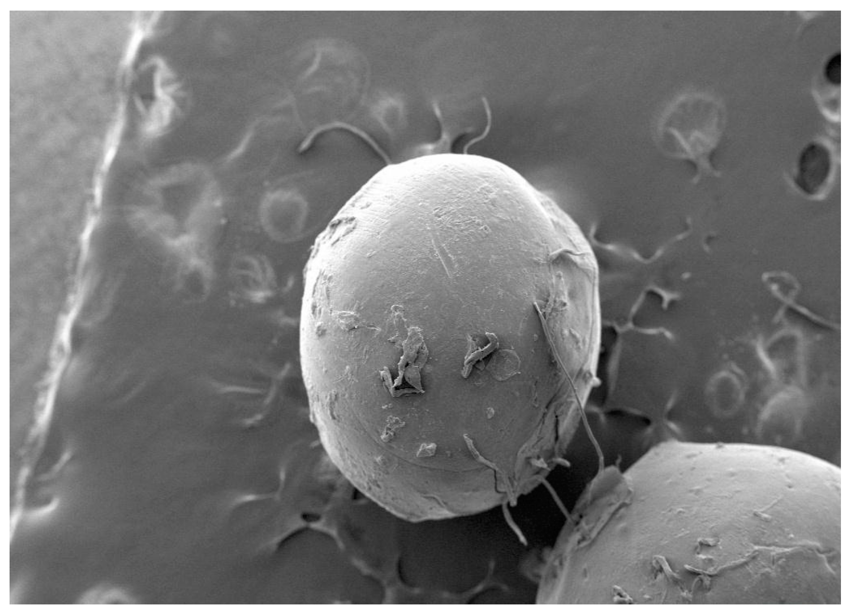
Figure 2, shows SEM photographs of a pentoxifylline-loaded sodium alginate microsphere. The surface morphology studied by SEM showed the spherical shape and smooth surface of a pentoxifylline–sodium alginate loaded microsphere containing 2% w/v of Carbopol prepared by the ionotropic gelation method. On the other hand, further increases in Carbopol concentration above 2% w/v leads to the formation of aggregates, smaller and discrete particles [10].

3.3. Encapsulation Efficacy

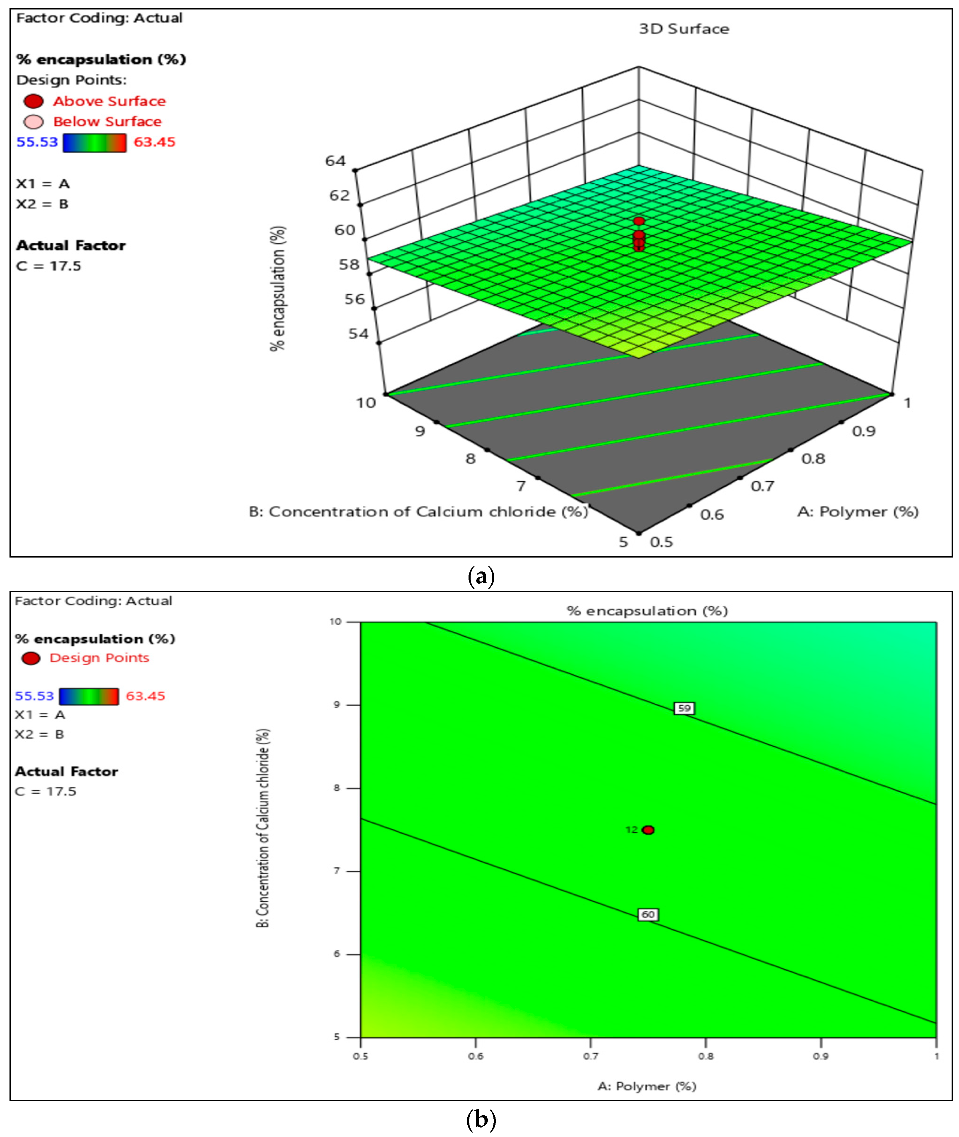
The encapsulation efficacy was found to be in the range of 56.24 to 63.45%, which is shown in Table 3. Figure 3 (a) 3D and (b) contour graph reveal that, the encapsulation efficacy was dependent on drug loading, the concentration of the polymer used, and cross-linking time. The formulation loaded with a high amount of drug showed higher encapsulations. The encapsulation efficacy decreases with an increase in the concentration of CaCl2 and cross-linking time [11,12].

3.4. In-Vitro Mucoadhesion

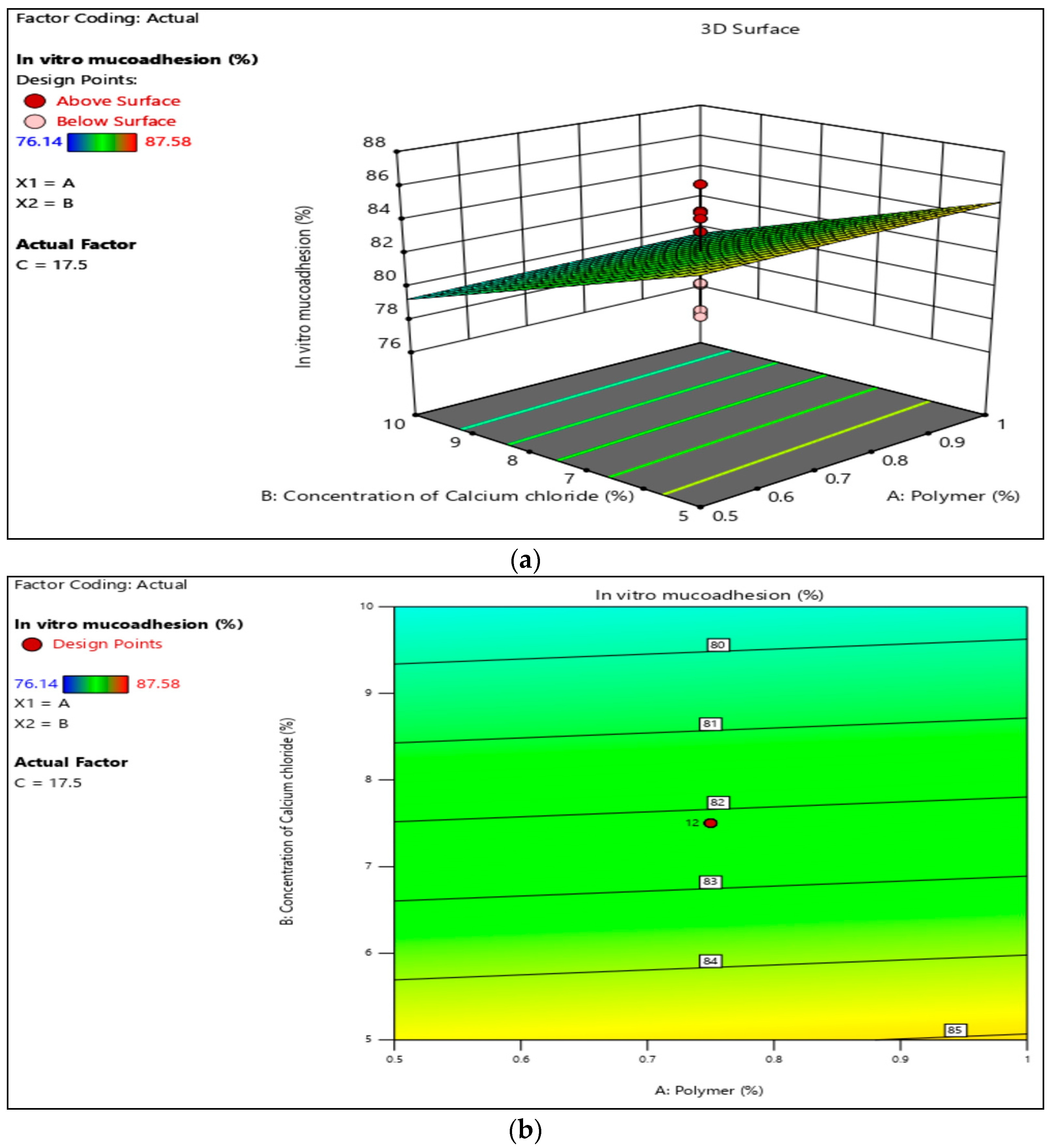
The in vitro mucoadhesion of all the batches is shown on Table 4. It was found that all the formulation batches were in the range of 76.14 to 87.58%. Figure 4 (a) 3D and (b) contour graph shows that increasing the polymer concentration ratio increases mucoadhesion due to a higher percentage of the polymer interacting with the mucosal surface [13].

3.5. In-Vitro Diffusion Study

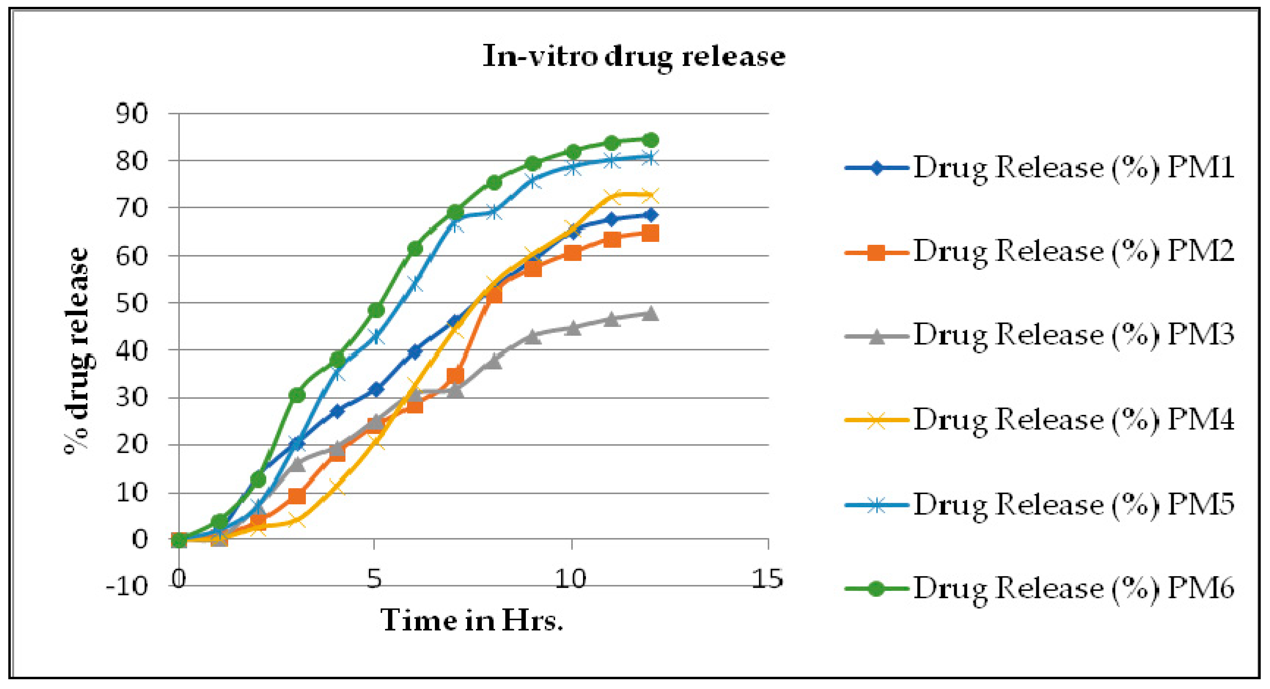
A drug release study was conducted using Franz diffusion cells, which have donor and receptor compartments separated by a dialysis membrane. Before dispersing the sample equivalent to 20 mg of drug onto the donor compartment, the dialysis membrane was carefully equilibrated with phosphate buffer at 6.6 pH. The donor compartment is filled with simulated nasal fluid, while the receptor compartment is filled with phosphate buffer at 6.6 pH. The pH of the nasal cavity is within the pH range, and the solution temperature is kept at 37 ± 0.5 °C. To maintain the sink condition, 1 mL of the sample was withdrawn and replaced with a fresh sample after a predetermined interval, and samples were spectrophotometrically measured at 274 nm using a UV spectrophotometer [14,15]. The % drug release values are shown in Table 5. Figure 5 shows drug release at different time intervals.

3.6. Kinetics of Drug Release

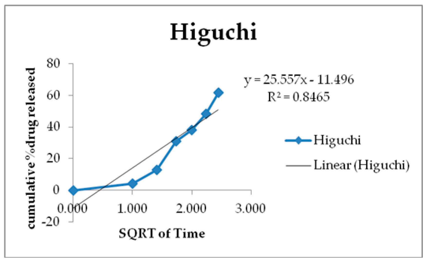
We investigated the drug release mechanism by applying multiple kinetic models to study the drug release of the optimized formulations, which are expressed in Figure 6, Figure 7, Figure 8, Figure 9 and Figure 10. It has been established that the Hixson-Crowell model is suitable for explaining the mechanism by which sodium alginate and 2% Carbopol release of Pentoxifylline microsphere.
The PM6formulation followed the Hixson–Crowell diffusion model of drug release (r2 = 0.9697), and it was best-fitted to the Hixson–Crowell diffusion model. The kinetics of formulation indicated the Fickian mechanism of drug release when the n value was less than 0.5. Details of the kinetic study are shown in Table 6 [16].

4. Conclusions

Mucoadhesive Pentoxifylline microspheres created by the ion induced gelation method were successfully prepared. Carbopol was used as a mucoadhesive polymer. A 23-Factorial experimental design was employed to identify optimal formulation parameters for a microsphere preparation with the minimum value of particle size and maximum value of in-vitro mucoadhesion. From the mathematical models generated, an optimal formulation comprising the drug, a polymer ratio (1:2), a CaCl2 concentration of (5–10%), and a cross-linking time (10–15 min) was identified to provide desired values for a particle size of 27.01 to 33.78 µm and in vitro mucoadhesion of 76.14 to 87.58%. The surface morphology studied by SEM showed the spherical shape and smooth surface of pentoxifylline–sodium alginate-loaded microsphere containing 2% w/v of Carbopol. In- vitro dissolution tests were performed in pH 6.2 phosphate buffer and indicated a Fickenian-type of transport for the diffusion of the drug from the Pentoxifylline mucoadhesive microspheres.

Author Contributions

Conceptualization, S.T.; methodology, S.T.; validation, S.T.; investigation, S.T.; writing–original draft preparation, S.T.; writing–review and editing, S.T. and S.P.; supervision, S.P. All authors have read and agreed to the published version of the manuscript.

Funding

This research received no external funding.

Institutional Review Board Statement

Not applicable.

Informed Consent Statement

Not applicable.

Data Availability Statement

Data are contained within the article.

Acknowledgments

We would like to convey our obligation to the principal, P. S. G. V. P. Mandal’s College of Pharmacy, Shahada, District Nandurbar, for furnishing all the essential facilities for the completion of this research.

Conflicts of Interest

The authors declare no conflicts of interest.

References

  1. Annamaraju, P.; Baradhi, K.M. Pentoxifylline. Pentoxifylline—[Updated 19 September 2022]. In StatPearls [Internet]; StatPearls Publishing: Treasure Island, FL, USA, 2022. [Google Scholar]
  2. Gudziol, V.; Hummel, T. Effects of Pentoxifylline on Olfactory Sensitivity: A Postmarketing Surveillance Study. Arch. Otolaryngol.–Head Neck Surg. 2009, 135, 291. [Google Scholar] [CrossRef] [PubMed]
  3. Belgamwar, V.S.; Patel, H.S.; Joshi, A.S.; Agrawal, A.; Surana, S.J.; Tekade, A.R. Design and Development of Nasal Mucoadhesive Microspheres Containing Tramadol HCl for CNS Targeting. Drug Deliv. 2011, 18, 353–360. [Google Scholar] [CrossRef] [PubMed]
  4. Jiang, L.; Gao, L.; Wang, X.; Tang, L.; Ma, J. The Application of Mucoadhesive Polymers in Nasal Drug Delivery. Drug Dev. Ind. Phar. 2010, 36, 323–336. [Google Scholar] [CrossRef]
  5. Chaudhari, H.C.; Chaudhari, L.Y.; Chaudhari, P.A.; Bhavsar, S.P.; Tadavi, S.A. Formulation and Evaluation of Microsphere Containing Telmisartan Drug by Ionotropic Gelation Method. Int. J. Res. Trends Innov. 2022, 7, 1489–1500. [Google Scholar]
  6. Patil, N.N. Formulations and Evaluations of Metformin Microspheres by Ionotropic Gelation Technique. World J. Pharm. Pharm. Sci. 2017, 6, 1473–1486. [Google Scholar] [CrossRef]
  7. Patil, S.B.; Sawant, K.K. Development, Optimization and in Vitro Evaluation of Alginate Mucoadhesive Microspheres of Carvedilol for Nasal Delivery. J. Microencapsul. 2009, 26, 432–443. [Google Scholar] [CrossRef] [PubMed]
  8. Patil, S.; Babbar, A.; Mathur, R.; Mishra, A.; Sawant, K. Mucoadhesive Chitosan Microspheres of Carvedilol for Nasal Administration. J. Drug Target. 2010, 18, 321–331. [Google Scholar] [CrossRef] [PubMed]
  9. Rathnanadh, M.; Kumar, D.S.; Shirwaikar, A.; Kumar Ravi Sampath Kumar, D.; Prasad, R.S. Preparation of Mucoadhesive Micrspheres for Nasal Delivery by Spray Drying. Indian J. Pharm. Sci. 2007, 69, 651–657. [Google Scholar] [CrossRef]
  10. Tamizharasi, S.; Rathi, J.C.; Rathi, V. Formulation, and Evaluation of Pentoxifylline-Loaded Poly(?-Caprolactone) Microspheres. Indian J. Pharm. Sci. 2008, 70, 333. [Google Scholar] [CrossRef] [PubMed]
  11. Kashyap, N.; Mishra, A.; Pathak, A.K. Preparation and Evaluation of Mucoadhasive Microspheres of Propranolol HCl for Nasal Delivery. Int. J. Adv. Pharm. 2015, 4, 49–54. [Google Scholar]
  12. Arefin, P.; Hasan, I.; Islam, M.S.; Reza, M.S. Formulation and In Vitro Evaluation of Eudragit RL 100 Loaded Fexofenadine HCl Microspheres. Bangladesh Pharm. J. 2016, 19, 58–67. [Google Scholar] [CrossRef]
  13. Jain, S.; Jain, N.; Gupta, Y.; Jain, A.; Jain, D.; Chaurasia, M. Mucoadhesive Chitosan Microspheres for Non-Invasive and Improved Nasal Delivery of Insulin. Indian J. Pharm. Sci. 2007, 69, 498. [Google Scholar] [CrossRef]
  14. Gavini, E.; Rassu, G.; Sanna, V.; Cossu, M.; Giunchedi, P. Mucoadhesive Microspheres for Nasal Administration of an Antiemetic Drug, Metoclopramide: In-Vitro/Ex-Vivo Studies. J. Pharm. Pharmacol. 2010, 57, 287–294. [Google Scholar] [CrossRef] [PubMed]
  15. Taksande, J.B.; Umekar, M.J. Preparation of Intranasal Pregabalin Microspheres: In Vitro, Ex Vivo and In Vivo Pharmacodynamic Evaluation. J. Pharm. Res. 2018, 12, 10. [Google Scholar]
  16. Sonje, A.G.; Mahajan, H.S. Nasal inserts containing ondansetron hydrochloride based on Chitosan-gellan gum polyelectrolyte complex: In vitro–in vivo studies. Mater. Sci. Eng. C 2016, 64, 329–335. [Google Scholar] [CrossRef] [PubMed]
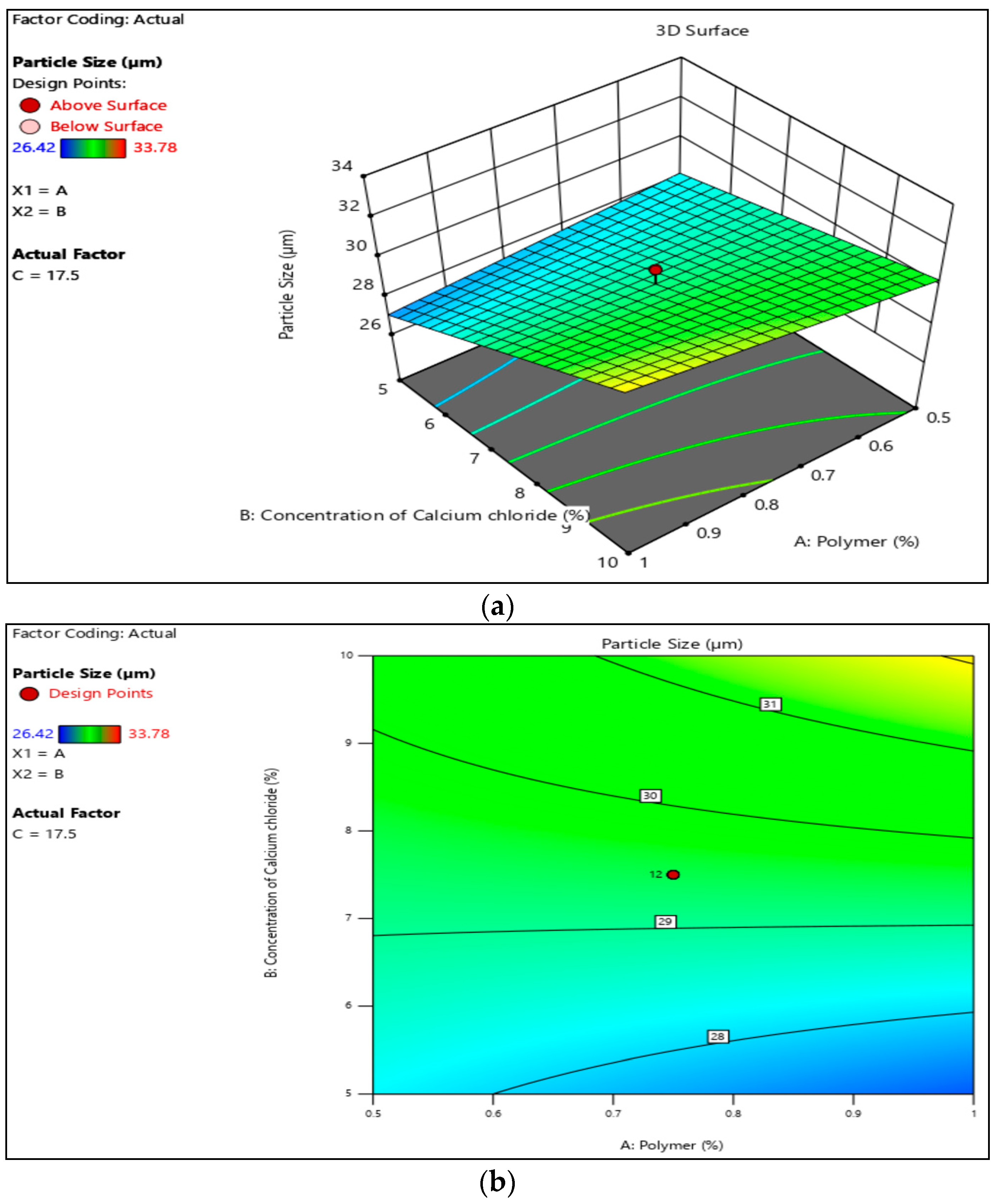
Figure 1. Particle size of Pentoxifylline microsphere: (a) 3D graph of particle size; (b) contour graph of particle size.
Engproc 56 00319 g001
Figure 2. Scanning electron microscopy of optimized formulation of Pentoxifylline microsphere.
Engproc 56 00319 g002
Figure 3. % Drug encapsulation efficacy of Pentoxifylline microspheres: (a) 3 D graph of % encapsulation; (b) contour graph of % encapsulation.
Engproc 56 00319 g003
Figure 4. In-vitro mucoadhesion study of Pentoxifylline microspheres: (a) 3D graph of in-vitro mucoadhesion; (b) contour graph of in-vitro mucoadhesion.
Engproc 56 00319 g004
Figure 5. % In-vitro drug release profile.
Engproc 56 00319 g005
Figure 6. Korsmeyer-Peppas drug release kinetics.
Engproc 56 00319 g006
Figure 7. Zero-order drug release kinetics study of optimized formulation.
Engproc 56 00319 g007
Figure 8. Higuchi drug release kinetics study of optimized formulation.
Engproc 56 00319 g008
Figure 9. First-order drug release kinetics study of optimized formulation.
Engproc 56 00319 g009
Figure 10. Hixson–Crowell drug release kinetics study of optimized formulation.
Engproc 56 00319 g010
Table 1. 23 Factorial design of Pentoxifylline microsphere.
NameUnitsLowHigh–Alpha+Alpha
Sodium AlginateGm1.952.051.929292.07071
CarbopolMg450550429.289570.711
Table 2. Particle size of Pentoxifylline microspheres.
Formulation CodeParticle Size, µm
PM127.01 ± 0.08
PM230.48 ± 0.02
PM329.11 ± 0.05
PM433.78 ± 0.03
PM532.74 ± 0.07
PM631.45 ± 0.03
Table 3. % drug encapsulation of Pentoxifylline microspheres.
Formulation Code% Drug Encapsulation
PM158.63 ± 0.10
PM256.24 ± 0.04
PM359.75 ± 0.08
PM463.45 ± 0.06
PM559.27 ± 0.07
PM660.45 ± 0.02
Table 4. In-vitro mucoadhesion study of Pentoxifylline microspheres.
Formulation Code% In-Vitro Mucoadhesion
PM178.15 ± 0.07
PM276.14 ± 0.08
PM380.85 ± 0.04
PM487.58 ± 0.06
PM585.63 ± 0.07
PM684.12 ± 0.01
Table 5. % Drug release study of Pentoxifylline microspheres.
Drug Release (%)
Time in Hrs.PM1PM2PM3PM4PM5PM6
0000000
11.89 ± 0.050.71 ± 0.040.23 ± 0.010.23 ± 0.071.89 ± 0.054.02 ± 0.023
213.27 ± 0.093.79 ± 0.037.1 ± 0.052.6 ± 0.057.11 ± 0.0712.81 ± 0.05
320.44 ± 0.089.49 ± 0.0715.9 ± 0.044.27 ± 0.0620.4 ± 0.0530.86 ± 0.09
427.11 ± 0.0418.05 ± 0.0519.5 ± 0.0111.39 ± 0.0535.4 ± 0.0738.3 ± 0.08
531.88 ± 0.0324.02 ± 0.0825.21 ± 0.0520.66 ± 0.0342.82 ± 0.0548.53 ± 0.02
639.96 ± 0.0528.55 ± 0.0930.92 ± 0.0532.79 ± 0.0554.23 ± 0.0861.84 ± 0.07
746.16 ± 0.0734.73 ± 0.0531.9 ± 0.0444.23 ± 0.0566.85 ± 0.0769.26 ± 0.06
853.3 ± 0.0851.82 ± 0.0738.07 ± 0.0354.24 ± 0.0469.29 ± 0.0475.69 ± 0.04
959.26 ± 0.0357.36 ± 0.0543.07 ± 0.0260.45 ± 0.0575.93 ± 0.0679.53 ± 0.03
1065.22 ± 0.0260.71 ± 0.0444.76 ± 0.0565.7 ± 0.0778.81 ± 0.0482.15 ± 0.05
1167.62 ± 0.0763.8 ± 0.0546.66 ± 0.0472.36 ± 0.0680.25 ± 0.0784.05 ± 0.03
1268.58 ± 0.0265.01 ± 0.0747.86 ± 0.0772.87 ± 0.0580.96 ± 0.0884.78 ± 0.01
Table 6. Kinetics model.
Zero OrderFirst OrderHiguchiKorsmeyer-PeppasHixson-Crowell
R20.79250.95660.84650.9520.9697
K61.1562.04425.5571.536-
N----0.2152
Disclaimer/Publisher’s Note: The statements, opinions and data contained in all publications are solely those of the individual author(s) and contributor(s) and not of MDPI and/or the editor(s). MDPI and/or the editor(s) disclaim responsibility for any injury to people or property resulting from any ideas, methods, instructions or products referred to in the content.