Article

Preliminary Evidence of Enhanced Immunogenicity of Hepatitis B Virus Vaccines When Co-Administered with Calcium Phosphate, Aluminum Hydroxide, and Cytosine Phospho-Guanine Oligodeoxynucleotides Combined Adjuvant in BALB/c Mice

by
Oumou Ouattara
1,*,
Josephine W. Kimani
2 and
James H. Kimotho
3
1
PAU—Institute for Basic Sciences, Technology, and Innovation (PAUSTI), Pan African University, Nairobi P.O. Box 62000-00200, Kenya
2
Department of Biochemistry, Jomo Kenyatta University of Agriculture and Technology (JKAUT), Nairobi P.O. Box 62000-00200, Kenya
3
Innovation and Technology Transfer Department (ITTD), Kenya Medical Research Institute (KEMRI), Nairobi P.O. Box 54840-00200, Kenya
*
Author to whom correspondence should be addressed.
Immuno 2025, 5(1), 12; https://doi.org/10.3390/immuno5010012
Submission received: 22 January 2025 / Revised: 4 March 2025 / Accepted: 7 March 2025 / Published: 14 March 2025

Abstract

:
Hepatitis B virus (HBV) infection is a major public health risk. Despite the introduction of successful vaccines, which are normally single adjuvanted, there are still some drawbacks, including non-responsiveness in certain groups, short durability of immunity, inadequate protection, and the need for additional doses to be addressed. This study aimed to develop an optimized combination of Cytosine-phosphate-Guanine Oligonucleotides (CPG-ODN2395, CPG-ODN-18281-2 23 mer) and calcium phosphate, and to assess its immunogenicity and toxicity when co-administrated with the commercial HBV vaccine (BEVAC, containing aluminum hydroxide) and an in-house aluminum hydroxide-adjuvanted HBs purified antigen in Balb/c mice. Tail blood was collected from vaccinated Balb/c mice on days 14 and 28 post-immunization to determine the antibody secretion level using an enzyme-linked immunosorbent assay (ELISA). The Tumor Necrosis Factor (TNF-a) and interleukin-6 (IL-6) cytokine expression levels were assessed through real-time PCR, and the safety profile was checked through biochemical and hematological analysis. Our results showed that the combination of CPG-ODN2395, CPG-ODN 18281-2 23 mer, and CAP significantly enhanced the IgG antibody secretion level (p < 0.0001), which also showed a significant increase in IL-6 expression (p < 0.0001). The safety evaluations revealed no adverse impact on liver and kidney function, with normal ALT, AST, urea, and creatinine levels (p < 0.55). Hematological assessments revealed stable parameters across all groups. This study concludes that combining CpG ODNs and calcium phosphate adjuvants with hepatitis B vaccinations has the potential to enhance a stronger immunological response to hepatitis B infection than single adjuvants. These results highlight the promise of this innovative adjuvant system, necessitating more research in clinical environments to increase vaccine effectiveness and sustained protection against HBV.

1. Introduction

Hepatitis B virus (HBV) infections are a main global health concern causing significant human suffering and economic loss worldwide [1]. The infection, caused by a partly double-stranded hepatotoxic DNA virus, can cause chronic and acute hepatitis leading to life-threatening conditions including cirrhosis or liver cancer [2,3]. According to the WHO, 254 million people were living with chronic HBV infection in 2022, with 1.2 million new infections occurring each year [4].
Vaccination remains the most efficient and cost-effective method of public health prevention, even with advancements in antiviral medication [5].
Several licensed single adjuvanted HBV vaccines for human use exist. For instance, the recombinant HBV vaccines are made of recombinant hepatitis B surface antigen (HBsAg) with aluminum hydroxide or synthetic oligonucleotide with CpG patterns as an adjuvant. These stimulate Toll-like receptor 9 (TLR9), enhancing innate immune activation and extended immune response [1]. However, several drawbacks need to be addressed. For instance, adults require three doses spread out over six months for optimal protection, increasing the overall cost of vaccination. There is hypo responsiveness in older persons and a longer time for seroprotection [6]. There is also limited effectiveness in people with compromised immune systems, diabetics, obesity, smokers, and adults [1]. The vaccine’s duration of immunity decreases over time [7].
Adjuvants are added to vaccines to enhance the immune response [8]. They offer several advantages including improved immunogenicity, reduced vaccine dose, improved vaccine efficacy, broadened protection, and reduced vaccine reactogenicity [8]. However, using a single adjuvant has limitations, such as the inability to affect the immune response qualitatively and limited adjuvanticity [9,10]. Therefore, adjuvant formulations that are currently successful often consist of multiple compounds that collaborate to stimulate different immune pathways, unlike previously approved adjuvants. Several combined adjuvants were previously authorized for use in vaccines for both humans and animals, and more are currently being developed. They are uniquely designed to enhance and guide the appropriate immune responses to favor Th1, Th2, or Th17 [11]. For instance, the work of Thakur et al. assessed the efficacy of four adjuvants, alum, saponin, cationic liposomes, and monophosphoryl lipid-A, in conjunction with the autoclaved Leishmania donovani (ALD) antigen against mouse visceral leishmaniasis (VL) in BALB/c mice, which demonstrated strong protective effectiveness compared to the infected controls [12]. Additionally, mice immunized with HBsAg and a combination of Cytosine Phosphate Guanine Oligodeoxynucleotides (CpG ODNs) and one of the two polyphosphazene (PCEP or PCPP) showed significantly increased HBsAg-specific antibody responses [13]. Combining TLR-3 agonist poly I: C and TLR-9 agonist CpG ODN with a DNA-encoded HIV Gag vaccination improved CD8+ T cell response, enhancing IFN-γ- and IL-2-producing cells [14].
This study investigated the immunogenicity and toxicity of adjuvant combinations generated from CpG Oligodeoxynucleotides (ODN-18281-2 23-mer, CPG-ODN2395), and calcium phosphate co-administrated with HBV vaccine (BEVAC, which contains aluminum hydroxide) and an in-house aluminum hydroxide-adjuvanted HBV purified antigen. CpG-ODNs are known to improve antigen presentation, resulting in vaccine-specific responses and activating the innate immune system, releasing Th1 and pro-inflammatory cytokines [15]. Recent studies have shown the effect of CpGs-ODNs on humoral immune responses as well as cytokine expression such as IL6, TNF-a, and INF-γ [16,17]. Calcium phosphate absorbs the vaccine antigen on its surface and creates a deposit on the injection site, slowing the antigen’s release, and improving vaccine immunogenicity [18]. Therefore, combining CPG-ODNs and calcium phosphate could increase the immune response while providing safe and long-term seroprotection against HBV infection.

2. Materials and Methods

2.1. Study Approval

The licenses to conduct this research were obtained from the National Commission for Science, Technology & Innovation, Kenya (License No: NACOSTI/P/24/35409, approval number 123477), the Mount Kenya University Ethics Review Committee (Approval number 2710), and the KEMRI-Animal Care and Use Committee (KEMRI ACUC/1 June 2024).

2.2. Animal Model

Female mice of the BALB/c strain (20 ± 2 g, 7–8 weeks old) were acquired from the Kenya Institute of Primate Research (KPRE), Kenya, and acclimatized for 14 days at a temperature of 21 ± 3 °C and humidity of 40–70%. The subjects were sustained on a 12 h light/dark/light cycle and provided sufficient mouse food and water. All measures were undertaken to lessen injury and distress to the mice by adhering to the principles outlined in the 3Rs of ethical animal research: replacement, reduction, and refinement.

2.3. Study Design and Sample Size Determination

Using a laboratory-based experimental design, the experiment was carried out at KEMRI, particularly at the Innovation & Technology Transfer Division (ITTD). The sample size was calculated using the ‘Resource Equation (E)’ to determine the minimum and maximum number of animals necessary for the study. In all, 57 female Balb/C mice were split into 19 groups comprising 3 mice each based on the treatment, as shown in Table 1.

2.4. Adjuvant Combination and Immunization Protocol

Three adjuvants, namely CPG-ODN 2395 [CPG1, 5′-TC*GT*CG TTT TC*G GCG C*GC G*CC G-3], CPG-ODN 18281-2 (23 mer) [CPG2 5′-T*GA*CTGT*GAACGTTCGGATGAT*T-3′], and calcium phosphate, were combined with the hepatitis B commercial vaccine (BEVAC™, which is an alumni hydroxide-adjuvanted vaccine) and an in-house aluminum hydroxide-adjuvanted HBV purified antigen (HBs Antigen). All the adjuvants were used at their optimal dosage determined from a published study [17,18,19] The total number of combinations was determined according to the combination law C (n, k) = n!/k! (N − k)! [20]. Seven (7) possible adjuvant combinations were generated. The combinations and administered vaccine dosages are shown in Table 1 below.
Every mouse was immunized intramuscularly (IM) into their left or right muscle on day 0 and day 14.

2.5. Samples Collection

For ELISA and biochemical assays, 50 μL of blood samples were obtained by tail puncture, mixed with 150 μL of heparin, and centrifuged at 2000 rpm for 10 min on days 14 and 28. The plasma was harvested and stored at −20 °C for later investigation. Using 1.5 mL EDTA-coated collection tubes, 300 μL of whole blood was collected for hematological analysis based on the cardiac puncture technique and kept at 8 °C. Further, in sterile 1.5 mL Eppendorf tubes, spleen tissues were collected on day 28 and place on ice. The tissues were stored at −80 °C for subsequent investigations. The mice were sacrificed by euthanization using CO2.

2.6. Assessment of Humoral Immune Response

The HBV anti-spike IgG levels were determined using an ELISA Kit (Solarbio Science & Technology Co., Ltd., Beijing, China), according to the manufacturer’s protocol. The ELISA plates were examined at 450 nm using the VersaMax™ ELISA Microplate Reader (Sunnyvale, CA, USA). The Optical Density (OD) values were calculated, and an OD450 nm of ≥0.1 signified the existence of mouse HBV anti-spike (S) IgG antibodies.

2.7. Extraction of Total RNA from Spleen Tissue

Total RNA was extracted from the mice’s spleen tissues using the Total RNA Extraction Kit (Solarbio Science & Technology Co., Ltd., Beijing, China), according to the manufacturer’s protocol. The concentration and purity were measured with the NanoDrop™2000/2000c spectrophotometer (Thermo Fisher Scientific, Waltham, MA, USA) at 260/280 nm absorbance. The samples were preserved at −80 °C for cDNA synthesis and subsequent gene expression analysis.

2.8. cDNA Synthesis and Relative Quantification of IL-6 and TNF Alpha mRNA Levels

cDNA synthesis and amplification for each sample were performed in a single tube using the TaqMan One-Step RT-qPCR Kit (Solarbio Science & Technology Co., Ltd., Beijing, China). The reaction volume used was 12.5 µL including 2.5 μL of the PCR buffer, 0.75 μL of the Rtase mix, 0.5 μL of forward primer (10 μM), 0.5 μL of the reverse primer (10 μM), 0.625 μL (10X) of Syber green dye, 2.5 μL of the RNA template, and 5.125 μL of nuclease-free water. The experiment proceeded using the Applied Biosystems’ Quant Studio 5 platform (PE Applied Biosystems, Waltham, MA, USA), with the following thermocycler profile: one cycle of Reverse Transcription in 10 min at 50 °C, one cycle of initial denaturation for 2 min at 95 °C followed by 40 cycles of denaturation at 95 °C for 20 s and annealing/extension at 60 °C for 1 min. The primers used to amplify TNF Alpha, IL6, and the housekeeping genes are presented in Table 2. The relative quantification of gene expression was determined using the deltadelta threshold cycle (ΔΔCt) formula, ΔCt = Ct (gene of interest) − Ct (housekeeping gene), and the PBS control group was used to calibrate [21].

2.9. Assessment of Hematological and Biochemical Analysis

The hematological analysis was conducted with the Huma Count 30TS hematology analyzer (Human Diagnostics Worldwide, Wiesbaden, Germany). The levels of serum in Aspartate Transaminase (AST), Alanine Transferase (ALT), Gamma-Glutamyltransfearse (GGT), urea, and creatinine were assessed using the Reflotron colorimetric test kit (Woodley Equipment Company, Lancashire, UK), following the kit’s guideline.

2.10. Data Analysis

The ELISA optical densities and the gene expression Ct values were primarily recorded in Microsoft Excel (2013). Gene expression was determined using the delta–delta threshold cycle 2 (ΔΔCt) method. The normality tests were performed to assess whether the data followed a normal distribution using the Shapiro–Wilk test, and the statistical analysis was performed using Graph Pad Prism version 8.0.2, normal one-way analysis of variance (ANOVA) for three or more groups, and t-tests for two groups to determine statistically significant differences. Additionally, two-way ANOVA was performed on the hematological data. The values were considered significant at 95% confidence intervals (p < 0.05).

3. Results

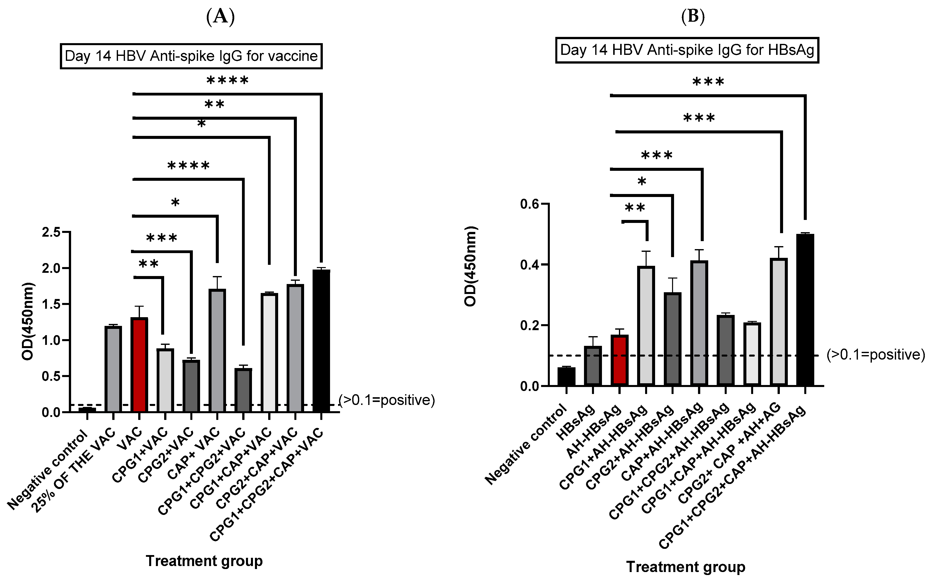
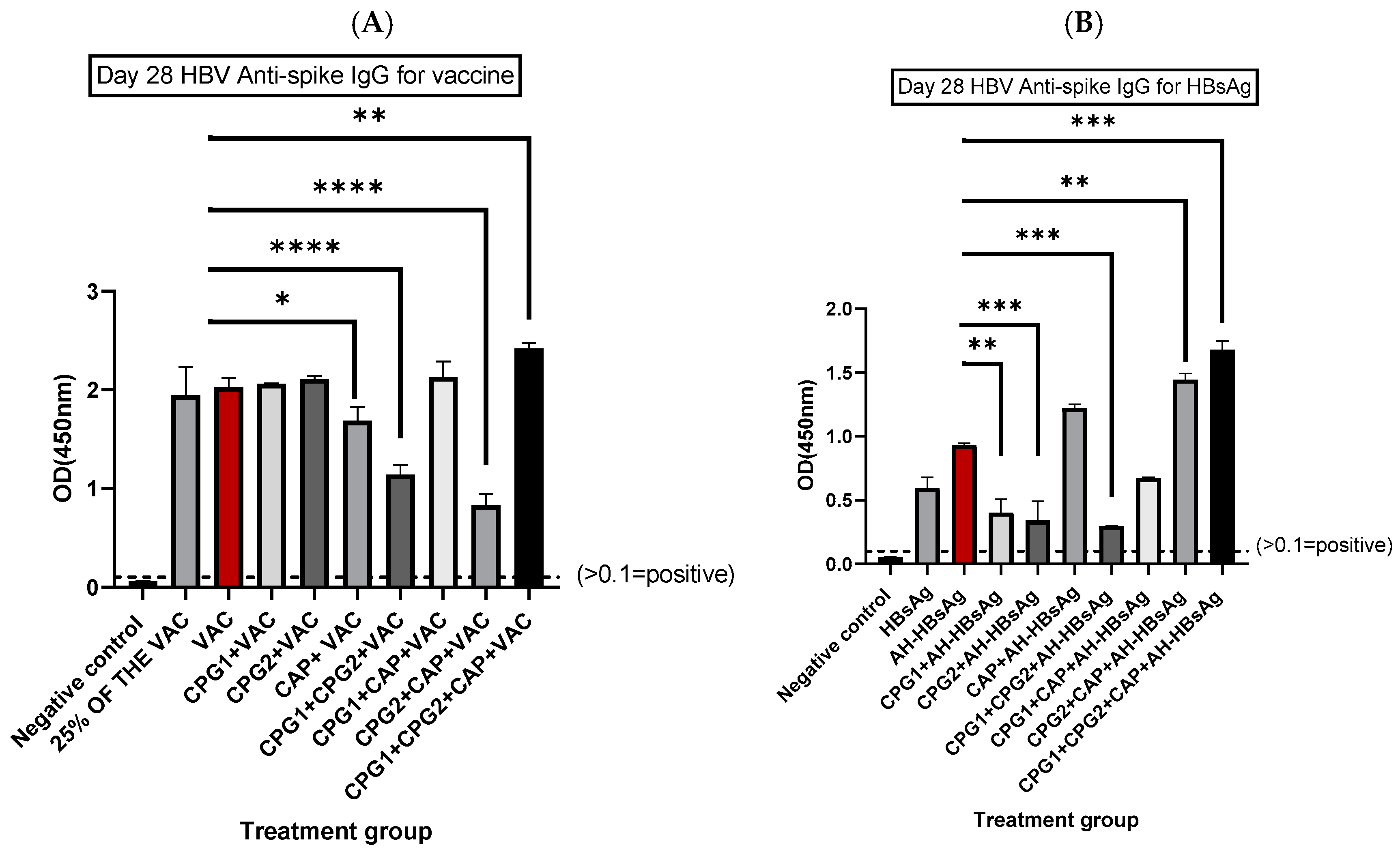
3.1. Determination of HBV Spike-Specific IgG Antibodies at Days 14 and 28 Post-Immunization

The OD 450 nm measurements indicated that antibodies were present in the plasma of all immunized mice on both days 14 and 28 following immunization (OD > 0.1). On day 14, a combination of three adjuvants CPG1 (CpG-ODN 2395) + CPG2 (CpG-ODN18281-2) +CAP co-administrated with either AH-HBsAg or the BEVAC vaccine exhibited the highest activity, leading to a significant fold increase in OD of 1.5 and 2.9 (p < 0.0001 Figure 1). The combination of CPG2 + CAP showed significant fold increases of 1.34 and 2.5 when co-administrated with AH-HBsAg and the BEVAC vaccine in comparison to the individual vaccine (p < 0.004; Figure 1). At the same time, mice immunized with the CAP, CPG1, and CPG2 co-administrated with AH-HBsAg showed fold increases of 2.4, 2.3, and 1.8 (p < 0.04; Figure 1B). Furthermore, the AH-HBsAg supplemented with the CPG1 + CPG2 and CPG1 + CAP groups also increased the antibody IgG level (Fold increase of 1.3 and 1.2); nonetheless, there was no statistical significance when compared to the AH-HBsAg (p < 0.55; Figure 1B). On the other hand, CAP appeared to better enhance the activity of the BEVAC vaccine, unlike the ODN-based adjuvants. CPG1 + CAP and CAP when co-administrated with the BEVAC vaccine showed a significant (p < 0.040) increase in IgG secretion level of a 1.29- and 1.25-fold increase, respectively. Conversely, the group comprising CPG1, CPG2, and CPG1 + CPG2 co-administrated with the BEVAC vaccine gave a significantly decreased IgG level compared to the alone vaccine group (p < 0.0001; Figure 1A). The presence of the CAP adjuvant increased the adjuvant combination potency, hence, all the groups immunized with the combinations incorporating CAP adjuvant showed increased IgG levels compared to the CpG-ODNs (Figure 1).
On day 28 post-immunization, the ODs showed a marked increase in both the HBsAg and BEVAC vaccine groups relative to day 14 (p < 0.0001). The groups that received the combination of the triple adjuvants combination together with either BEVAC or AH-HBsAg demonstrated a statistically significant increase in IgG levels when compared to the results observed in the other combination groups (p < 0.0001; Figure 2). In addition, a statistically significant increase was noted in CPG2 + CAP co-administrated with AH-HBsAg compared to the individual AH-HBsAg (p < 0.002; Figure 2B). Conversely, the AH-HBsAg supplemented with the CPG1, CPG2, and CPG1 +CPG2 groups showed a statistically significant decrease in the antibody IgG level (p < 0.0001, Figure 2B). On the other hand, an elevation in the IgG level was observed in the BEVAC vaccine groups when supplemented with the CPG1, CPG2, and CPG1 + CAP groups compared to the BEVAC vaccine alone group. Nonetheless, the same groups revealed no statistically significant differences in the antibody titers (p < 0.969; Figure 2A). Furthermore, a statistically significant decrease was shown in the BEVAC vaccine groups when supplemented with CPG2 + CAP, CPG1 + CPG2, and CAP (p < 0.0001; Figure 2A). In summary, the data suggest that the combination comprising CPG1, CPG2, and CAP yields the greatest efficacy on both days 14 and 28 when co-administered with either the aluminum hydroxide-HBsAg or the commercial BEVAC vaccine (p < 0.0001; Figure 2).
The unpaired t-test analysis of IgG levels on days 14 and 28 of the most effective combination (CPG1 + CPG2 + CAP) co-administrated with the BEVAC vaccine showed a statistically significant difference compared to groups immunized with the aluminum hydroxide-adjuvanted-HBsAg on both days 14 and 28 (p < 0.0001 Figure 1 and Figure 2). This finding confirms that the CPG1 + CPG2 + CAP adjuvant combination was able to improve the immunogenicity with both the commercial vaccine, which is the rDNA vaccine and the purified surface antigen (Figure 1 and Figure 2).

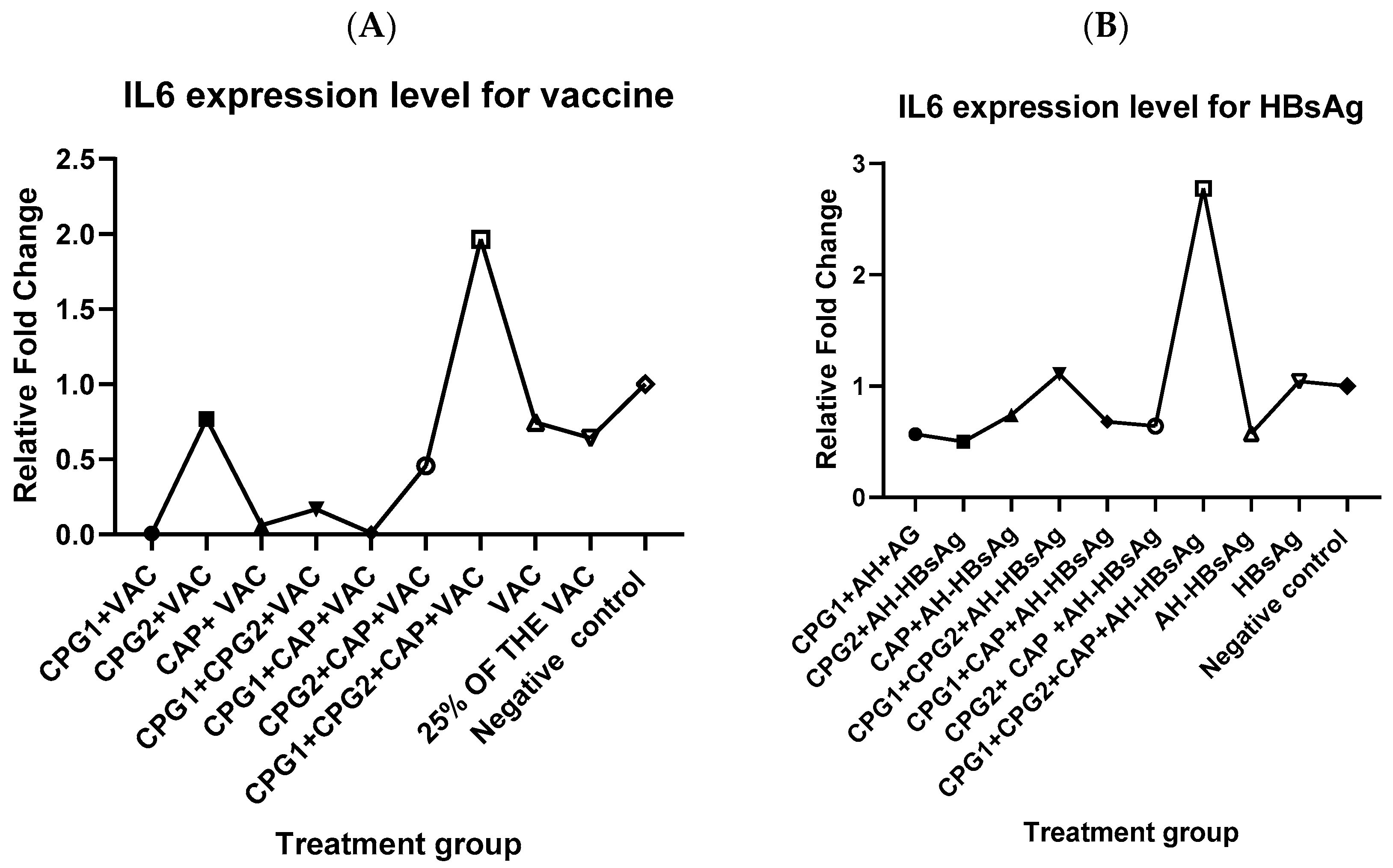
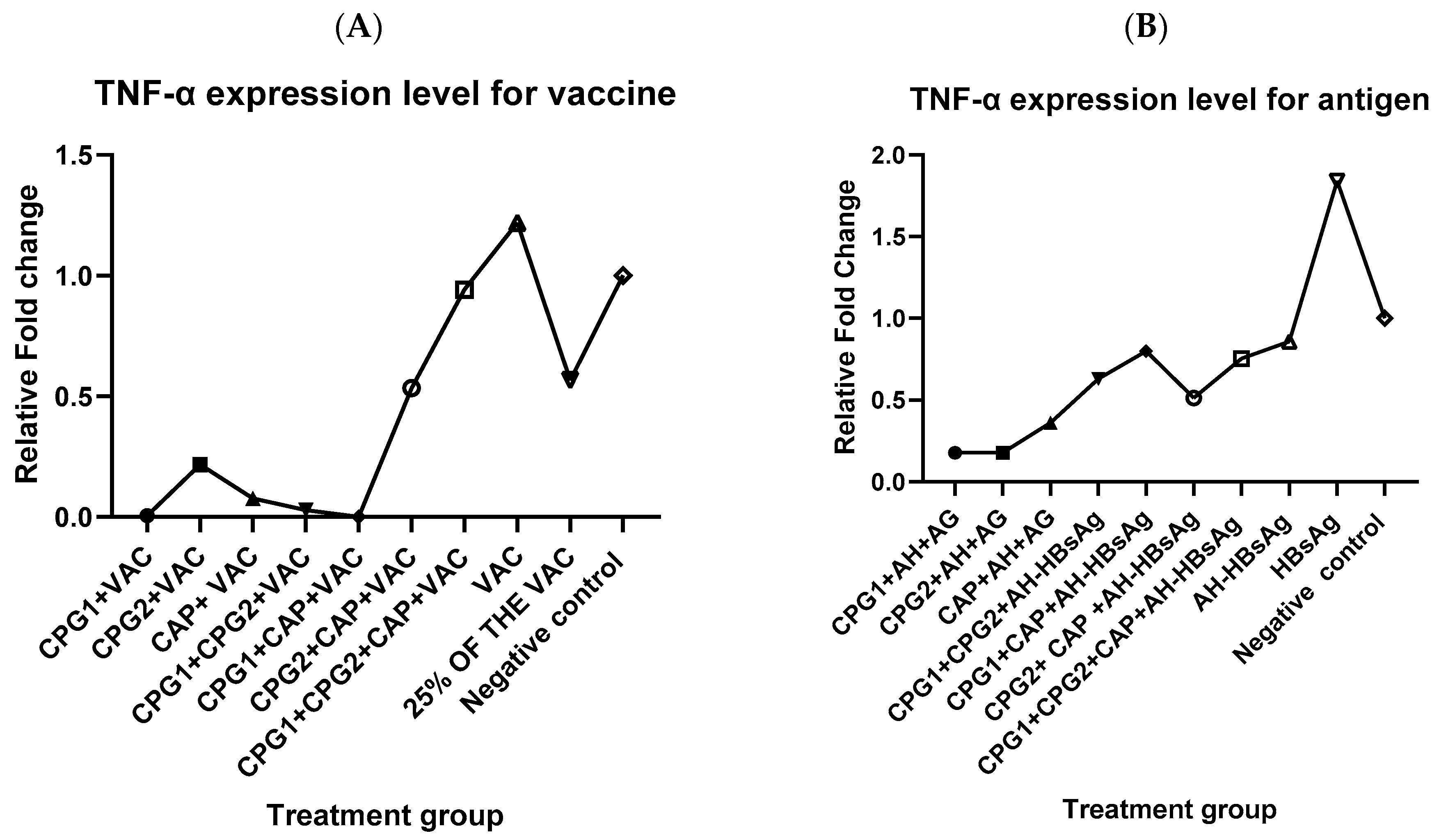
3.2. Effect of Most Potent Adjuvant Combination on IL-6 and TNF-α Expression

The aluminum hydroxide-adjuvanted–HBV antigen-supplemented groups showed a significantly higher expression level of IL6 and TNF-α genes relative to the BEVAC vaccine-supplemented groups (p < 0.001; Figure 3). Both the BEVAC vaccine and AH-HBsAg groups supplemented with triple-combined adjuvant (CPG1 + CPG2 + CAP) demonstrated the highest expression level of IL6 compared to their BEVAC vaccine and AH-HBsAg alone groups. In contrast, the groups that included single adjuvant (CPG1, CPG2, and CAP), as well as CPG1 + CPG2 and CPG1 + CAP, showed a decrease in the IL6 cytokine level when co-administrated with either BEVAC vaccine or AH-HBsAg (Figure 3). Furthermore, the CPG1 + CPG2 group showed an increase in IL6 expression compared to the untreated group (1.109 against 1) (Figure 3B). Therefore, CPG1 +CPG2 +CAP was the best combination since it increased IL6 expression when combined with either the BEVAC vaccine or the aluminum hydroxide-adjuvanted HBs-purified antigen.
A reduced expression level of TNF-α was observed in all the treatment groups compared to the untreated group (fold change < 1), except the HBsAg and BEVAC vaccine alone groups, which exhibited the highest levels of TNF-α expression (Figure 4).
The relationship between the IgG secretion levels and the cytokine expression of various adjuvant combinations administrated with the commercial vaccine (BEVAC) and the aluminum hydroxide-adjuvanted HBsAg were determined using Pearson correlation analysis (Figure 5). A positive correlation was found between the IgG secretion level and IL6 cytokine expression (r = 0.280, Figure 5A), suggesting that increased IL-6 expression may be associated with higher IgG antibody production. IL-6 is known to promote B cell differentiation and enhance antibody production. A negative correlation existed between the IgG secretion level and TNF-α (r = −0.048, p = 0.909), suggesting that TNF-α may be downregulated by the immune system as a regulatory mechanism to maintain the immune system. Excessive TNF-α production is generally associated with chronic inflammation and tissue damage, which was not observed in our study as evidenced by biochemical and hematological parameters. However, there was no statistically significant correlation between IgG levels and either IL-6 or TNF-α, as indicated by the results presented in Figure 5B (p > 0.05).

3.3. Assessment of Hematological and Biochemical Parameters

The hematological parameters of the Balb/c mice immunized using the most effective adjuvant combination alongside the different treatments are detailed in Table 3. The two-wayANOVA analysis for all the treatments including CPG1 + CPG2 + CAP co-administrated with the BEVAC vaccine and the aluminum hydroxide-adjuvanted HBsAg, BEVAC vaccine without tri-adjuvant, and AH-adjuvanted-HBsAg without tri-adjuvant revealed a statistically significant (p < 0.001) lower count for platelets (438 ± 0.1, 571 ± 0.3, 514 ± 0.03, and 494 ± 0.1) compared to the untreated groups (759.5 ± 0.2). Furthermore, a lower count for lymphocytes (46 ± 8.4, 52 ± 5.6, 54.5 ± 3.53, 43 ± 7.7, and 24.5 ± 0.7) and a higher count for neutrophils (38.5± 7.7, 35 ± 7.0, 31.5 ± 2.1, 39 ± 8.4, and 59 ± 21.2) was observed compared to the normal range. However, the analysis revealed no statistically significant differences (p > 0.55). The other hematological parameters were not significantly different (p > 0.05) between the treatment and negative control groups. Despite a minor reduction in platelet and lymphocytes counts and a slightly higher count for neutrophils, all values remained within the normal physiological range for the BALB/c mice, suggesting no hematological toxicity (Table 3).
The biochemical analysis of the blood from the female BALB /C mice immunized with the most effective adjuvant combination (CPG1 + CPG2 + CAP) co-administrated with the BEVAC vaccine and aluminum hydroxide-adjuvanted HBsAg as well as the untreated group are presented in Figure 6. There was no statistically significant change in the ALT (32.35 and 21.45 U/L against 26.35 U/L), AST (39.35 and 28.15 U/L against 32.15 U/L), UREA (15.85 and 8.45 against 9.90 mg/dL), and creatinine (0.70 and 1.05 mg/dL against 0.60) levels between the adjuvant combination supplemented and non-treated group, respectively (p > 0.05) (Figure 6).

4. Discussion

Hepatitis B infection is a significant public health risk. Despite the availability of several vaccines, certain populations still have lower immune response rates. Improving HBV vaccines with adjuvants may induce better immune responses. Most of the licensed HBV vaccines use a single adjuvant, which has limitations in generating all necessary protective immune responses [13]. Therefore, better adjuvants are still required to elicit a robust immune response. The outcome of this study indicates that the novel combination of CpG 1 + CpG 2 + CAP was the best among several other combinations since it elicited significantly higher levels of specific IgG antibodies (p < 0.0001) compared to the BEVAC vaccine alone and aluminumhydroxide-adjuvanted HBsAg groups. Furthermore, it demonstrated long-term stability of high titers of IgG antibodies (day 28 post-immunization), suggesting that the novel combination possesses the potential for extended immune protection.
The significant increase in the IgG level could be attributed to the synergetic effect of the different adjuvants [11], indicating that the tri-component adjuvant formed complexes that may have enhanced the transport of both antigen and adjuvants to antigen-presenting cells (APCs) [24]. Dendritic cells are powerful antigen-presenting cells crucial for the control of both innate and adaptive immune responses. In reaction to antigenic stimuli, immature dendritic cells activate and mature, releasing cytokines and chemokines, and migrating from peripheral organs to lymph nodes to deliver antigens to naïve T cells [25]. CpG ODNs improve antigen presentation to APCs, resulting in vaccine-specific responses and activating the innate immune system, releasing Th1 and pro-inflammatory cytokines [15]. Kemi et al [17] recently demonstrated that CPG-ODNs could increase the antibody secretion level when administrated with the HBV vaccine; here in this study, we observed that the combination of CPG-ODNs with CAP markedly enhanced the humoral immune response. Calcium phosphate (CAP) augments the immune response by serving as a transporter for antigens and activating both humoral and cellular immunological responses. This dual mechanism is essential for efficient immunization against diverse diseases [26]. Several studies validate our findings, for instance, a study indicated that the combination of Monophosphoryl Lipid A (MPL) and Polyinosinic(poly I: C) as adjuvants in an influenza vaccine enhanced the generation of antigen-specific antibodies, reduced cytokine levels and cellular infiltrates at infection sites, and elicited substantial memory T and B cell responses in mice [27]. Additionally the combination of Toll-like receptor 3 (TLR-3) agonist poly I: C and Toll-like receptor9 (TLR-9) agonist CpG ODN with a DNA-encoded HIV Gag vaccination improved CD8+ T cell response, leading to an increase in IFN-γ- and IL-2-producing cells [14]. The outcome of this study was in agreement with that of Haseda et al., who developed a novel adjuvant combination consisting of 1,2-dioleoyl-3-trimethylammonium propane (DOTAP), D35, and aluminum salts, showing strong T cell and antibody responses in BALB/c mice post-immunization with model protein antigens [28]. Our findings corroborate the report of Wang et al. on Balbc/mice which showed a significant increase in the humoral immune response upon co-administration of MF59 and CpG5 with the porcine epidemic virus vaccine [29]. Ebensen et al. combined the c-di-AMP with alum in the administration of β-Gal protein as an antigen and achieved a more robust humoral immune response [30]. Their findings were in agreement with our findings. Furthermore, the immunization of mice using a co-formulation of the HH2–CpG complex alongside pertussis toxoid resulted in a notable increase in the induction of toxoid-specific antibody titers in comparison to the use of toxoid alone, leading to elevated levels of IgG1 and IgG2a, indicative of a balanced Th1/Th2 response, which also resulted in elevated IgA titers [24]. Combining CpG with nanoemulsion was shown to more successfully promote antibody production and cytokine release (IL-17 and IFN-γ) than using each component alone [31]. Mice immunized with a single subcutaneous injection of HBsAg and a combination of CpG ODN and one of the two polyphosphazenes (PCEP or PCPP) showed significantly increased HBsAg-specific antibody responses, surpassing those achieved with either adjuvant alone [13]. Furthermore, the combination of the Dimethyldioctadecylammonium (DDA) adjuvant with Emulsigen revealed strong immunological responses in cattle [11]. Therefore, the efficacy of the optimized adjuvant combination was demonstrated with the commercial aluminum hydroxide-adjuvanted vaccine (BEVAC) and aluminumhydroxide-adjuvanted-HBsAg in enhancing the antibody IgG secretion level in BalB/c mice.
To select the most effective adjuvant combination based on the cellular immune response, we conducted an IL6 and Tumor Necrosis Factor (TNF-α) cytokine expression analysis. The findings showed that the combination of CPG1 + CPG1 + CAP significantly increased the IL6 gene expression level (p < 0.0001) when co-administrated with both the commercial BEVAC vaccine and the aluminum hydroxide-adjuvanted-HBsAg compared to the other treatment groups. The three adjuvants may have acted synergistically to activate the immune cell implicated in the secretion of the IL6 cytokines. Since CpG-ODNs enhance the expression of MHC II and other co-stimulatory molecules, they stimulate the release of cytokines such as TNF-α and IL-6, as well as the synthesis of immunoglobulins by B cells [17]. IL-6 is a pleiotropic cytokine that is mainly expressed in activated monocytes or macrophages, fibroblasts, or activated endothelium cells of inflamed tissues [17]. IL-6 is crucial in B cell differentiation into antibody-secreting plasma cells. The process enhances the synthesis of immunoglobulins, such as IgM and IgG, which are important in neutralizing the hepatitis B virus (HBV) and ensuring sustained immunity following vaccination. Other studies indicate that IL-6 plays a significant role in augmenting antibody production following vaccination, highlighting its critical function in fostering a strong humoral response [30,31]. IL-6 affects T cell responses by facilitating Th2 polarization, potentially increasing antibody production while also possibly suppressing Th1 responses that are essential for viral clearance. It also facilitates the activation and proliferation of CD8+ T cells, which have a crucial role in cellular immunity against HBV. This dual role facilitates a balanced immune response, effectively managing the virus while enhancing immune memory [32,33]. This finding is not unexpected as studies have confirmed the potential effect of the adjuvant combination on the cytokine expression levels. For instance, Ebensen et al. showed that the combination of c-di-AMP/alum with β-Gal exhibited increased production of Th1 cytokines, specifically IFN-γ and IL-2, as well as Th2 cytokines including IL-4, IL-5, IL-10, and IL-13, along with the Th17 cytokine [30]. This supports our results on the synergic effect of the optimized adjuvant combination on the IgG antibody level as well as the IL6 cytokine expression level. Our study contradicts the finding of Le et al., which showed that the combination of MPL and poly I: C adjuvanted with influenza vaccine decreased the levels of cytokines [27]. The reliability of this result was supported by Karaba et al., who reported that an increased existence of inflammatory cytokines served as a sign of elevated antibody titers [34]. Therefore, the combination of CPG-ODNs and CAP boosted the cellular immune response through the secretion of IL6 cytokine expression level.
Tumor Necrosis Factor-alpha (TNF-α) is an inflammatory cytokine produced by macrophages and monocytes in response to acute inflammation. It plays a crucial role in various cellular signaling processes, which can result in either necrosis or apoptosis [35]. In this study, the TNF-α expression level of the CPG1 + CPG2 + CAP combination co-administrated with the BEVAC vaccine and AH-HBsAg was lower compared to the negative control group (FCT = 1.000). However, the difference in outcome was not statistically significant (p > 0.05). This result agrees with the study of Kemi et al., which revealed that mice immunized with HBV supplemented with CPG1 expressed reduced levels of TNF-α relative to the untreated group [17]. Furthermore, the findings of [27] support this result, which demonstrated a reduced level of TNF-α expression relative to the control group after a combination of MPL and poly I: C co-administered with influenza vaccine. The downregulation of TNF-α could be caused by the capacity of the adjuvant to influence the immune response through modifications in cytokine production. For instance, specific adjuvants have demonstrated the capability to enhance the secretion of IL-6 and IL-10, which support adaptive immunity, while possibly decreasing the levels of TNF-α, a cytokine linked to various inflammatory responses [34,35].
To further confirm the applicability of our optimized adjuvant combination, a hematological analysis was performed 28 days post-immunization to assess the potential toxicity effect of the adjuvant combination on blood parameters. The hematological parameters serve as biomarkers for detecting organ or tissue damage, identifying infections, parasitosis, and other illnesses [36]. Therefore, the hematological analysis results in this study did not reveal any significant (p > 0.5) toxicity effect on the immunized Balb/c mice since all the parameters were normal compared to the untreated group except for the platelets, which had a lower count compared to the untreated group (438 ± 0.1, and 571 ± 0.3 against 759.5 ± 0.2) (p < 0.001). This reduction may indicate an adaptive immune response designed to avoid excessive inflammation or coagulation activation, which might arise following robust immunological stimulation such as vaccination [37]. However, the platelets were within the range value according to Silva et al. [36]. Furthermore, elevated neutrophils was also observed compared to the normal range, which may signify a vigorous innate immune response, essential for combating infections and eliminating germs [38]. Neutrophils are known to be essential in resolving inflammation by phagocytosing foreign particles and cellular debris, thereby averting tissue damage [38]. The lymphocyte level was also lower compared to the normal range. Decreased lymphocyte numbers occur due to their transfer to tissues requiring immune activation [39]. Therefore, the reduced lymphocyte counts may indicate a regulatory mechanism aimed to balance various elements of the immune system under strong stimulation [38]. This balance guarantees that while one aspect of immunity augmented innate responses via neutrophils, for instance, other elements adapt correspondingly to avert excessive inflammation or autoimmunity [39]. Nevertheless, the changes in neutrophils and lymphocytes were not statistically significant (p > 0.99). Additionally, no negative effect on the physical features was observed, including changes in the body weight and behavior groups in both groups of mice where the BEVAC vaccine and HBsAg were co-administrated with the optimized adjuvant combination.
Blood biochemical profiles function as a crucial diagnostic instrument by indicating the physiological condition of the animal [16]. Therefore, the renal biochemical profile was evaluated by the analysis of urea and creatinine levels to assess the potential damage of the adjuvant combination on kidney function, while ALT and AST were evaluated to check any potential damage to the liver. The result showed no statistically significant differences between the CPG1 + CPG2 + CAP-supplemented and the untreated groups (p > 0.05). So, the result reveals no toxicity profile of the optimized combination on the liver and kidney function of the Balb/c mice.

5. Conclusions

This work effectively generated and assessed an optimized vaccine adjuvant combination utilizing CPG-ODNs and calcium phosphate (CpG-ODN2395, CpG-ODN18281-2, and CAP) delivered alongside either a commercial hepatitis B vaccine (BEVAC) or an aluminum hydroxide-adjuvanted HBs-purified antigen in Balb/c mice. To the best of our understanding, this is among the few studies that have explored the potential of several adjuvant combinations in enhancing the activities of hepatitis B vaccines both commercial and in-house. The findings indicated that the CpG1 + CpG2 + CAP adjuvant combination was the most effective by producing a strong humoral immune response, as seen by increased antibody titers and markedly higher IL-6 gene expression levels. No indications of toxicity were detected based on the hematological and biochemical parameters, indicating that the adjuvant combination is both immunogenic and safe. This study y’ key contribution lies in demonstrating the synergistic potential of combining CpG-ODNs and CAP to significantly boost the immune response to hepatitis B vaccines. Considering that HBV infection remains a global health challenge, this enhanced adjuvant system could lead to more effective vaccination strategies, potentially reducing the number of doses required and improving long-term protection. The study presented noteworthy findings; however, the influence of sample size on scientific conclusions requires the repetition of the study with an increased sample size. In addition to that limitation, the study exclusively utilized BALB/c mice, raising questions about the potential for different outcomes if alternative mouse species or animal models were employed. Therefore, additional exploration is necessitated for clinical use.

Author Contributions

Conceptualization, O.O. and J.H.K.; methodology, O.O. and J.H.K. validation, O.O., J.W.K. and J.H.K.; formal analysis, O.O., J.W.K. and J.H.K.; investigation, O.O.; data curation, O.O; resources, O.O, J.W.K. and J.H.K.; writing—original draft, O.O.; writing—review and editing, J.W.K. and J.H.K.; visualization, O.O.; supervision, J.W.K. and J.H.K.; project administration, O.O.; funding acquisition, O.O. All authors have read and agreed to the published version of the manuscript.

Funding

The African Union Commission supported this work through the Pan African University Institute for Basic Sciences, Technology, and Innovation (PAUSTI).

Institutional Review Board Statement

All mice studies complied with the Guide for the Care and Use of Laboratory Animals at Kenya Medical Research Institute (KEMRI). The animal study protocols were reviewed and approved by the KEMRI Animal Care and Use Committee and Mount Kenya University (MKU) Animal Care and Use Ethics Review Committee with reference numbers KEMRI ACUC/1 June 2024 and REF: MKU/ISERC/3666 (approval number 2710), respectively.

Informed Consent Statement

Not applicable.

Data Availability Statement

All data are provided in this manuscript.

Acknowledgments

The authors extend their gratitude to the Pan African University Institute for Basic Sciences, Technology and Innovation (PAUSTI), the Kenya Medical Research Institute KEMRI, and Jomo Kenyatta University of Agriculture and Technology (JKUAT). We also acknowledge the valuable assistance of Reuben Ayivor-Djanie in optimizing the PCR process.

Conflicts of Interest

The authors declare no conflicts of interest.

References

  1. Leroux-Roels, G. Old and new adjuvants for hepatitis B vaccines. Med. Microbiol. Immunol. 2015, 204, 69–78. [Google Scholar] [CrossRef] [PubMed]
  2. Liang, Z.; Qiu, J.; Xiang, Q.; Yi, J.; Zhu, J.; Zhao, Q. Epidemiology of hepatitis B virus infection among preconception couples in South China: A cross-sectional study. BMJ Open 2023, 13, e061165. [Google Scholar] [CrossRef]
  3. Yuen, M.-F.; Chen, D.-S.; Dusheiko, G.M.; Janssen, H.L.A.; Lau, D.T.Y.; Locarnini, S.A.; Peters, M.G.; Lai, C.-L. Hepatitis B virus infection. Nat. Rev. Dis. Prim. 2018, 4, 18036. [Google Scholar] [CrossRef] [PubMed]
  4. Bishop, N. Hepatitis B. Contin. Pract. 1992, 19, 35–38. [Google Scholar]
  5. Meireles, L.C. Three decades of hepatitis B control with vaccination. World J. Hepatol. 2015, 7, 2127–2132. [Google Scholar] [CrossRef]
  6. Ndung’U, F.; Nyanjom, S.; Omari, S.; Wainaina, E.; Mugasiali, R.; Kimotho, J. Preliminary assessment of adjuvant activities of Glycine max (L.) Merr saponin extract in BALB/c mice immunized with hepatitis B virus vaccine. F1000Research 2023, 12, 1145. [Google Scholar] [CrossRef]
  7. Di Lello, F.A.; Martínez, A.P.; Flichman, D.M. Insights into induction of the immune response by the hepatitis B vaccine. World J. Gastroenterol. 2022, 28, 4249–4262. [Google Scholar] [CrossRef]
  8. Mount, A.; Koernig, S.; Silva, A.; Drane, D.; Maraskovsky, E.; Morelli, A.B. Combination of adjuvants: The future of vaccine design. Expert Rev. Vaccines 2013, 12, 733–746. [Google Scholar] [CrossRef]
  9. Coffman, R.L.; Sher, A.; Seder, R.A. Vaccine adjuvants: Putting innate immunity to work. Immunity 2010, 33, 492–503. [Google Scholar] [CrossRef]
  10. Jangra, S.; Landers, J.J.; Rathnasinghe, R.; O’konek, J.J.; Janczak, K.W.; Cascalho, M.; Kennedy, A.A.; Tai, A.W.; Baker, J.R.; Schotsaert, M.; et al. A Combination Adjuvant for the Induction of Potent Antiviral Immune Responses for a Recombinant SARS-CoV-2 Protein Vaccine. Front. Immunol. 2021, 12, 729189. [Google Scholar] [CrossRef]
  11. Levast, B.; Awate, S.; Babiuk, L.; Mutwiri, G.; Gerdts, V.; Hurk, S.V.D.L.-V.D. Vaccine potentiation by combination adjuvants. Vaccines 2014, 2, 297–322. [Google Scholar] [CrossRef] [PubMed]
  12. Thakur, A.; Kaur, H.; Kaur, S. Evaluation of the immunoprophylactic potential of a killed vaccine candidate in combination with different adjuvants against murine visceral leishmaniasis. Parasitol. Int. 2015, 64, 70–78. [Google Scholar] [CrossRef]
  13. Mutwiri, G.; Gerdts, V.; Hurk, S.v.D.L.-V.D.; Auray, G.; Eng, N.; Garlapati, S.; Babiuk, L.A.; Potter, A. Combination adjuvants: The next generation of adjuvants? Expert Rev. Vaccines 2011, 10, 95–107. [Google Scholar] [CrossRef] [PubMed]
  14. Zhu, Q.; Egelston, C.; Gagnon, S.; Sui, Y.; Belyakov, I.M.; Klinman, D.M.; Berzofsky, J.A. Using 3 TLR ligands as a combination adjuvant induces qualitative changes in T cell responses needed for antiviral protection in mice. J. Clin. Investig. 2010, 120, 607–616. [Google Scholar] [CrossRef] [PubMed]
  15. Shirota, H.; Klinman, D. CpG Oligodeoxynucleotides as Adjuvants for Clinical Use. In Immunopotentiators in Modern Vaccines; Elsevier: Amsterdam, The Netherlands, 2017; pp. 163–198. [Google Scholar]
  16. Celise, D.A.; Kimotho, J.; Kimani, J.W.; Muriithi, A.K.; Odari, E.O. Increase in the Immune Response in Balb/c Mice after the Co-Administration of a Vector-Based COVID-19 Vaccine with Cytosine Phosphoguanine Oligodeoxynucleotide. Vaccines 2023, 11, 53. [Google Scholar] [CrossRef]
  17. Kemi, T.; Nelly, S.; Kariuki, D.; Kimotho, J. Co-administration of a Hepatitis B vaccine with CpG-ODN 2395 induces stronger immune response in BALB/c mice. F1000Research 2024, 13, 404. [Google Scholar] [CrossRef]
  18. Masson, J.-D.; Thibaudon, M.; Bélec, L.; Crépeaux, G. Calcium phosphate: A substitute for aluminum adjuvants? Expert Rev. Vaccines 2017, 16, 289–299. [Google Scholar] [CrossRef]
  19. Moni, S.S.; Abdelwahab, S.I.; Jabeen, A.; Elmobark, M.E.; Aqaili, D.; Gohal, G.; Oraibi, B.; Farasani, A.M.; Jerah, A.A.; Alnajai, M.M.A.; et al. Advancements in Vaccine Adjuvants: The Journey from Alum to Nano Formulations. Vaccines 2023, 11, 1704. [Google Scholar] [CrossRef]
  20. Math: Permutation and Combination. 2010. Available online: https://sathee.prutor.ai/article/maths/permutation-and-combination/# (accessed on 11 August 2024).
  21. Atmaca, H.T. Expression of serotonin 2A, 2C, 6 and 7 receptor and IL-6 mRNA in experimental toxoplasmic encephalitis in mice. Heliyon 2019, 5, e02890. [Google Scholar] [CrossRef]
  22. Sudo, K.; Yamada, Y.; Saito, K.; Shimizu, S.; Ohashi, H.; Kato, T.; Moriwaki, H.; Ito, H.; Seishima, M. TNF-α and IL-6 signals from the bone marrow derived cells are necessary for normal murine liver regeneration. Biochim. Biophys. Acta (BBA) Mol. Basis Dis. 2008, 1782, 671–679. [Google Scholar] [CrossRef]
  23. Medrano, G.; Guan, P.; Barlow-Anacker, A.J.; Gosain, A. Comprehensive selection of reference genes for quantitative RT-PCR analysis of murine extramedullary hematopoiesis during development. PLoS ONE 2017, 12, e0181881. [Google Scholar] [CrossRef] [PubMed]
  24. Kindrachuk, J.; Jenssen, H.; Elliott, M.; Townsend, R.; Nijnik, A.; Lee, S.F.; Gerdts, V.; Babiuk, L.A.; Halperin, S.A.; Hancock, R.E. A novel vaccine adjuvant comprised of a synthetic innate defence regulator peptide and CpG oligonucleotide links innate and adaptive immunity. Vaccine 2009, 27, 4662–4671. [Google Scholar] [CrossRef] [PubMed]
  25. Banchereau, J.; Steinman, R.M. Dendritic cells and the control of immunity. Nature 1998, 392, 245–252. [Google Scholar] [CrossRef] [PubMed]
  26. Sun, Z.; Li, W.; Lenzo, J.C.; Holden, J.A.; McCullough, M.J.; O’connor, A.J.; O’brien-Simpson, N.M. The Potential of Calcium Phosphate Nanoparticles as Adjuvants and Vaccine Delivery Vehicles. Front. Mater. 2021, 8, 788373. [Google Scholar] [CrossRef]
  27. Le, C.T.T.; Ahn, S.Y.; Ho, T.L.; Lee, J.; Lee, D.-H.; Hwang, H.S.; Kang, S.-M.; Ko, E.-J. Adjuvant effects of combination monophosphoryl lipid A and poly I:C on antigen-specific immune responses and protective efficacy of influenza vaccines. Sci. Rep. 2023, 13, 12231. [Google Scholar] [CrossRef]
  28. Haseda, Y.; Munakata, L.; Kimura, C.; Kinugasa-Katayama, Y.; Mori, Y.; Suzuki, R.; Aoshi, T. Development of combination adjuvant for efficient T cell and antibody response induction against protein antigen. PLoS ONE 2021, 16, e0254628. [Google Scholar] [CrossRef]
  29. Wang, Y.; Wang, Y.; Liu, S.; Liu, S.; Li, B.; Li, B.; Sun, X.; Sun, X.; Pan, Q.; Pan, Q.; et al. A novel CpG ODN compound adjuvant enhances immune response to spike subunit vaccines of porcine epidemic diarrhea virus. Front. Immunol. 2024, 15, 1336239. [Google Scholar] [CrossRef]
  30. Ebensen, T.; Delandre, S.; Prochnow, B.; Guzmán, C.A.; Schulze, K. The Combination Vaccine Adjuvant System Alum/c-di-AMP Results in Quantitative and Qualitative Enhanced Immune Responses Post Immunization. Front. Cell. Infect. Microbiol. 2019, 9, 31. [Google Scholar] [CrossRef]
  31. Sichangi, M.W. Understanding Mechanisms of Actions for Vaccine Adjuvants Critical for Designing Effective Vaccines: A narrative review. Afr. J. Health Sci. 2020, 33, 18–29. Available online: https://www.ajol.info/index.php/ajhs/article/view/202031 (accessed on 25 February 2025).
  32. Lan, T.; Chang, L.; Wu, L.; Yuan, Y.-F. Il-6 plays a crucial role in hbv infection. J. Clin. Transl. Hepatol. 2015, 3, 271–276. [Google Scholar] [CrossRef]
  33. Kuo, T.-M.; Hu, C.-P.; Chen, Y.-L.; Hong, M.-H.; Jeng, K.-S.; Liang, C.-C.T.; Chen, M.-L.; Chang, C. HBV replication is significantly reduced by IL-6. J. Biomed. Sci. 2009, 16, 41. [Google Scholar] [CrossRef]
  34. Karaba, A.H.; Zhu, X.M.; Benner, S.E.; Akinde, O.B.; Eby, Y.; Wang, K.H.B.; Saraf, S.B.; Garonzik-Wang, J.M.; Klein, S.L.; Bailey, J.R.; et al. Higher Proinflammatory Cytokines Are Associated With Increased Antibody Titer After a Third Dose of SARS-CoV-2 Vaccine in Solid Organ Transplant Recipients. Transplantation 2022, 106, 835–841. [Google Scholar] [CrossRef] [PubMed]
  35. Idriss, H.T.; Naismith, J.H. TNFα and the TNF receptor superfamily: Structure-function relationship(s). Microsc. Res. Tech. 2000, 50, 184–195. [Google Scholar] [CrossRef] [PubMed]
  36. Silva-Santana, G.; Bax, J.C.; Fernandes, D.C.S.; Bacellar, D.T.L.; Hooper, C.; Dias, A.A.S.O.; Silva, C.B.; de Souza, A.M.; Ramos, S.; Santos, R.A.; et al. Clinical hematological and biochemical parameters in Swiss, BALB/c, C57BL/6 and B6D2F1 Mus musculus. Anim. Model. Exp. Med. 2020, 3, 304–315. [Google Scholar] [CrossRef]
  37. Jiang, D.; Portuguese, A.J.; Weatherford, A.; Garcia, D.; Gernsheimer, T. Platelet trends after COVID-19 vaccination in patients with chronic or persistent immune thrombocytopenia. Am. J. Hematol. 2021, 96, E472–E474. [Google Scholar] [CrossRef] [PubMed]
  38. Capllonch, A.; de Pablo, S.; de la Torre, A.; Morales, I. Increase in white cell and neutrophil counts during the first eighteen weeks of treatment with clozapine in patients admitted to a long-term psychiatric care inpatient unit. Span. J. Psychiatry Ment. Health 2018, 11, 94–100. [Google Scholar] [CrossRef]
  39. Imamura, M.; Morimoto, T.; Egawa, C.; Fukui, R.; Bun, A.; Ozawa, H.; Miyagawa, Y.; Fujimoto, Y.; Higuchi, T.; Miyoshi, Y. Significance of baseline neutrophil-to-lymphocyte ratio for progression-free survival of patients with HER2-positive breast cancer treated with trastuzumab emtansine. Sci. Rep. 2019, 9, 1811. [Google Scholar] [CrossRef]
Figure 1. Day 14 humoral immune responses to adjuvant combination co-administrated with BEVAC vaccine and aluminum hydroxide-adjuvanted HBsAg in BALB/c mice. (A) Anti-spike IgG to the BEVAC vaccine on day 14. (B) Anti-spike IgG to aluminum hydroxide-adjuvanted HBsAg on day 14. Key: CpG1= CpG-ODN 2395; CpG2 = CpG-ODN18281-2; CAP =calcium phosphate; VAC = BEVAC vaccine alone; 25%Vac = 25% diluted BEVAC vaccine. AH-HBsAg = aluminum hydroxide and purified HBs antigen. HBsAg = HBs antigen alone, * = statistical significant at p < 0.05, (** p < 0.01; *** p < 0.001; **** p < 0.0001).
Immuno 05 00012 g001
Figure 2. Day 28 humoral immune responses to adjuvant combination co-administrated with BEVAC vaccine and aluminum hydroxide-adjuvanted HBs antigen in BALB/c mice. (A) Anti-spike IgG to BEVAC vaccine on day 28. (B) Anti-spike IgG to aluminum hydroxide-adjuvanted HBsAg on day 28 * = statistical significant at p < 0.05, (** p < 0.01; *** p < 0.001; **** p < 0.0001).
Immuno 05 00012 g002
Figure 3. IL6 cytokine expression level of adjuvant combination co-administrated with BEVAC vaccine and aluminum hydroxide-adjuvanted-HBsAg. (A) IL6 expression level of BEVAC vaccine group with adjuvant combinations. (B) IL6 expression level of AH-HBsAg group with adjuvant combination.
Immuno 05 00012 g003
Figure 4. TNF-α cytokine expression level of adjuvant combination co-administrated with BEVAC vaccine and aluminum hydroxide- adjuvanted HBsAg. (A) TNF-α expression level of BEVAC vaccine with adjuvant combinations. (B) TNF-α expression level for HBsAg with adjuvant combinations.
Immuno 05 00012 g004
Figure 5. Correlation between IgG secretion level and cytokine expression. (A) Correlation between IgG and TNF-α cytokine expression. (B) Correlation between IgG and IL6 expression level. Key: IL6 = Interferon 6; TNF-α = Tumor Necrosis Factor; IgG = antibody IgG.
Immuno 05 00012 g005
Figure 6. Biochemical analysis of day 28 blood from immunized mice. ALT = alanine transaminase (U/L); AST= aspartate aminotransferase (U/L); creatinine (mg/dl); urea (mg/dl); ns= not statistically significant at p < 0.05.
Immuno 05 00012 g006
Table 1. The adjuvant combination.
Adjuvant CombinationAdministered Dosage
CpG-ODN 2395 [CpG1]0.67 μM of CpG-ODN 2395 + 1 μg (50 μL) of BEVAC Vaccine
0.67 μM of CpG-ODN 2395 + 1.2 mg of AH + 0.01 μg (50 μL) of HBs Antigen
CpG-ODN18281-2 [CpG2]0.67 μM of CpG-ODN18281-2 + 1 μg (50 μL) of BEVAC Vaccine
0.67 μM of CpG-ODN18281-2 +1.2 mg of AH +0.01 μg (50 μL) of HBs Antigen
CAP1.3 mg of CAP + 1 μg(50 μL) of BEVAC Vaccine
1.3 mg of CAP + 1.2 mg of AH + 0.01 μg(50 μL) of HBs Antigen
CpG-ODN 2395 [CpG1] +CpG-ODN18281-2 [CpG2]0.67 μM of CpG-ODN 2395 + 0.67 μg of CpG-ODN18281-2 + 1 μg (50 μL) of BEVAC Vaccine
0.67 μM of CpG-ODN 2395 + 0.67 μg of CpG-ODN18281-2 + 1.2 mg of AH + 0.01 μg (50 uL) of HBs Antigen
CpG-ODN 2395 [CpG1] + CAP0.67 μg of CpG-ODN 2395 + 1.3 mg of CAP + 1 μg(50 μL) of BEVAC Vaccine
0.67 μg of CpG-ODN 2395 + 1.3 mg of CAP +1.2 mg of AH + 0.01 μg (50 μL) of HBs Antigen
CpG-ODN18281-2 [CpG2] +CAP0.67 μM of CpG-ODN18281-2 + 1.3 mg of CAP +1 μg(50 μL) of BEVAC Vaccine
0.67 μM of CpG-ODN18281-2 +1.3 mg of CAP +1.2 mg of AH +0.01 μg (50 μL) of HBs Antigen
CpG-ODN 2395 [CpG1] +CpG-ODN18281-2 [CpG2] +CAP0.67 μM of CpG-ODN 2395 + 0.67μM of CpG-ODN18281-2 + 1.3 mg of CAP + 1 μg (50 μL) of BEVAC Vaccine
0.67 μM of CpG-ODN 2395 + 0.67μM of CpG-ODN18281-2 + 1.3 mg of CAP +1.2 mg of AH + 0.01 μg (50 μL) of HBs Antigen
Vaccine Alone1 μg (50 μL) of the BEVAC vaccine
25% of Vaccine 0.25 μg of BEVAC vaccine + 37.5 μL of PBS
Aluminum Hydroxide (AH) + Antigen (AG) 1.2 mg of AH + 0.01 μg (50 μL) of HBs Antigen
Antigen Alone 0.01 μg (50 μL) of HBs Antigen
Negative Control 50 μL of PBS
Key: AH = aluminum hydroxide; CAP = calcium phosphate; CpG1 = CpG-ODN 2395; CpG2 = CpG-ODN18281-2.
Table 2. Sequences of primers used.
CytokineForward PrimerReverse PrimerNCBISize of
Amplicon
Reference
Interleukin 65′GAGGATACCACTCCCAACAGACC′35′AAGTGCATCATCGTTGTTCATACA3′XM_021163844.1141 bp[21]
Tumor Necrosis Factor
(TNF)
5′GATCTCAAAGACAACCAACATGTG 3′5′CTCCAGCTGGAAGACTCCTCCCAG 3′NM_013693.376 bp[22]
HPRT1 (Hypoxanthine guanine phosphoribosyl transferase 1)5′TCCTCCTCAGACCGCTTTT3′5′CCTGGTTCATCATCGCTAATC3′NM_013556.2 9090 bp[23]
Key: bp = base pair.
Table 3. The hematological parameters of the treatment groups.
ParameterCPG1 + CPG2 +
CAP + VAC
CPG1 + CPG2 +
CAP + AH-HBsAg
VACAH-HBsAgNegative
Control (PBS)
Range
for Mice
WBC (103/UL)1.5 ± 0.41.45 ± 0.073.71 ± 2.71.55 ± 0.3 1.4 ± 0.41.09–11.3
Neutrophils (%)38.5± 7.735 ± 7.031.5 ± 2.139 ± 8.459 ± 21.211–29
Lymphocytes (%)46 ± 8.452 ± 5.654.5 ± 3.5343 ± 7.724.5 ± 0.765–87
Monocytes (%)14.5 ± 0.712 ± 1.412.5 ± 0.716.5 ± 0.715.5 ± 03.75–14.3
Eosinophils (%)1 ± 01 ± 01.5 ± 0.71 ± 01 ± 00–5
Basophils (%)0 ± 00 ± 00 ± 00 ± 00 ± 00–1
RBC (106/UL)8.235 ± 1.28.5 ± 0.28.32 ± 1.39.1± 0.29.025 ± 0.97.8–10.3
HB (g/dl)10.4 ± 1.910.85 ± 2.39.75 ± 110.1 ± 3.111.1 ± 1.511.6–15.9
HCT (%)34.85 ± 5.5833 ± 3.838.2 ± 10.331.1 ± 3.138.55 ± 7.135.2–49.9
MCV (FI)42.2 ± 0.445.1 ± 3.545.4 ± 4.944.0 ± 4.342.5 ± 3.339.2–56.2
MCH (Pg)12.55 ± 0.411.8 ± 0.411.75 ± 0.611.15 ± 0.912.2 ± 0.412.4–18.4
MCHC29.75 ± 0.926.05 ± 4.328.55 ± 3.828.5 ± 5.528.85 ± 1.327.2–40.8
Platelets438 ± 0.1 *571 ± 0.3 *514 ± 0.03 *494 ± 0.1 *759.5 ± 0.2 *322–798
Abbreviation: CPG-ODN 2395 = CPG1; CpG-ODN18281 = CPG2; CAP= calcium phosphate; Vaccine = BEVAC vaccine; AH-HBsAg = aluminum hydroxide-adjuvanted-HBsAg; PBS = negative control; WBC = White Blood Cells; RBC = Red Blood Cells; HB = Hemoglobin; HCT = Hematocrit; MCV = Mean Corpuscular Volume; MCH = Mean Corpuscular Hemoglobin; MCHC = Mean Corpuscular Hemoglobin Concentration; * = statistical significant at p < 0.05.
Disclaimer/Publisher’s Note: The statements, opinions and data contained in all publications are solely those of the individual author(s) and contributor(s) and not of MDPI and/or the editor(s). MDPI and/or the editor(s) disclaim responsibility for any injury to people or property resulting from any ideas, methods, instructions or products referred to in the content.

