Review

B- and T-/NK-Cell Lymphomas in the 2022 International Consensus Classification of Mature Lymphoid Neoplasms and Comparison with the WHO Fifth Edition

by
Elaine S. Jaffe
1,* and
Antonino Carbone
2
1
Laboratory of Pathology, Center for Cancer Research, National Cancer Institute, National Institutes of Health, Bethesda, MD 20892, USA
2
Centro di Riferimento Oncologico di Aviano (CRO), Istituto di Ricovero e Cura a Carattere Scientifico (IRCCS), Via F. Gallini 2, I-33081 Aviano, Italy
*
Author to whom correspondence should be addressed.
Hemato 2024, 5(2), 157-170; https://doi.org/10.3390/hemato5020013
Submission received: 7 March 2024 / Revised: 1 April 2024 / Accepted: 10 April 2024 / Published: 17 April 2024
(This article belongs to the Section Lymphomas)

Abstract

:
The World Health Organization (WHO) “Classification of Tumours of Haematopoietic and Lymphoid Tissues”, published in 2001 and subsequently updated in 2008 and 2017, defined disease entities based on morphologic and phenotypic characteristics, clinical features, and genomic findings. Recently, the criteria for the diagnosis of many lymphoma entities have been refined in a proposal by the International Consensus Classification (ICC). Some provisional categories have now been recognized as “definite” entities, while other categories have undergone major revision. This article reports on the major revisions in the criteria and definition of B- and T-/NK-cell lymphomas by the ICC system.

1. Introduction

The current approach to lymphoma diagnosis and the delineation of distinct entities employs morphologic, immunophenotypic, and genomic criteria as well as clinical features. The classification of lymphoid neoplasms provides a guide for their diagnosis, management, and therapy. The World Health Organization (WHO) “Classification of Tumours of Haematopoietic and Lymphoid Tissues”, published in 2001 [1] and subsequently updated in 2008 [2] and 2017 [3], was accepted with a broad consensus by pathologists, geneticists, translational research scientists, and clinicians and used on a worldwide basis.
A major aspect employed by the editorial team was the use of a Clinical Advisory Committee (CAC), which met prior to the drafting of the WHO Bluebook. The involvement of a CAC ensured that any proposed changes in the classification would meet clinical needs and be widely accepted for clinical practice. Changes in the leadership of the International Agency for Research on Cancer (IARC), responsible for publication of the Bluebooks, led to a rejection of the CAC process for the development of the fifth edition of the WHO Bluebook. Many of the former editors sought to retain a CAC, and the resulting International Consensus Classification (ICC) was published following a CAC meeting held at the University of Chicago in September 2021 [4]. Some categories, which were previously considered “provisional”, have been recognized as “definite” entities, whereas other categories have undergone major revision. The fifth edition of the WHO classification was developed independently by the IARC and shares some similarities and differences with the ICC approach [5].
This article reports on the major revisions in the criteria and definition of B- and T-/NK-cell lymphomas by the ICC system.

2. ICC 2022: What Is New in B-Cell Neoplasms

In this section, follicular lymphoma (FL), diffuse large B-cell lymphoma (DLBCL), and high-grade B-cell neoplasms are discussed. (Table 1).

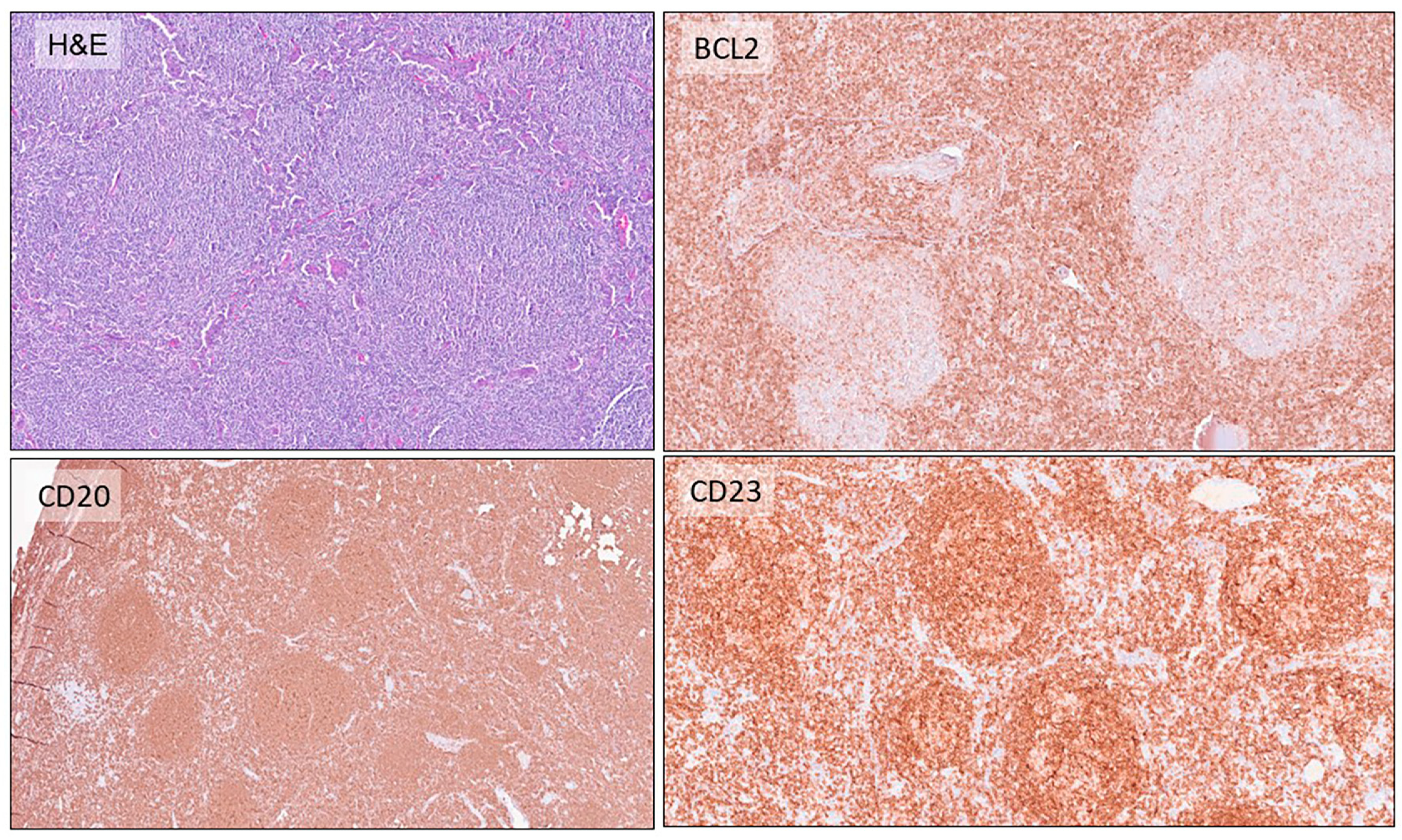
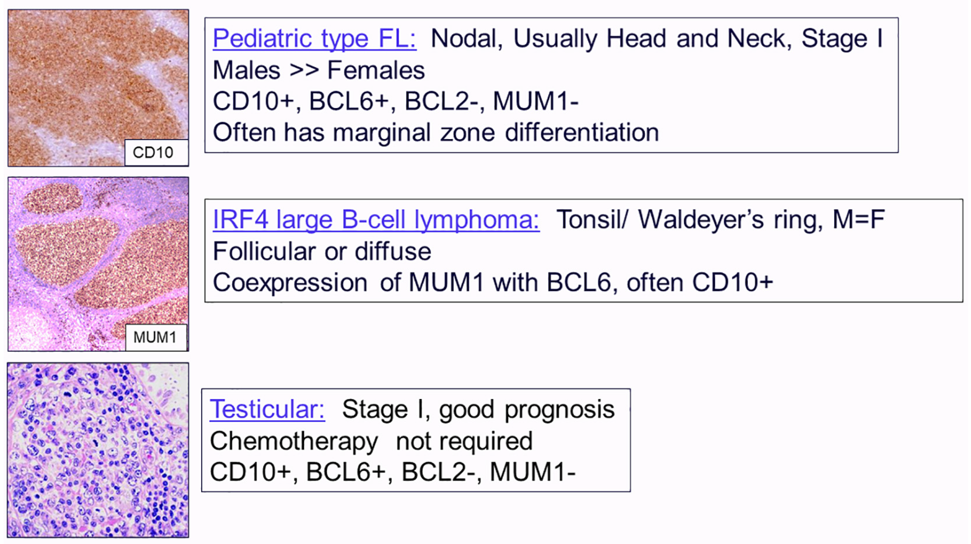
2.1. Follicular Lymphomas

Over the past few years, evolution in the spectrum of FLs has been observed. The spectrum includes both conventional FL associated with the BCL2 translocation as well as a number of alternative forms of follicle-center-derived neoplasms, which are negative for the BCL2 rearrangement. These differences are reflected in the ICC classification [4].
The ICC decided to retain the grading of FL, although the clinical significance of grading for BCL2-rearranged FL remains controversial. One of the more problematic issues is the distinction between grade 3A and grade 3B (Figure 1), since this distinction leads to different therapeutic approaches. Reliance on only cytologic features is problematic and often poorly reproducible. The ICC emphasizes that grade 3A is largely associated with the BCL2 translocation. The presence of BCL2 rearrangements and/or CD10 expression favors the diagnosis of FL 3A, whereas the expression of MUM1/IRF4 and absence of CD10 favors FL 3B [6].
Forms of histologic transformation of FL include DLBCL, high-grade B-cell lymphoma with double hit, classic Hodgkin’s lymphoma (CHL), and histiocytic sarcoma [6]. Some cases of high-grade B-cell lymphoma with double hit may show expression of TDT but are distinct from B-lymphoblastic lymphoma [7].
Among the FLs that are negative for the t(14;18), novel variants have been recognized. BCL2-rearrangement-negative, CD23-positive follicle center lymphoma was proposed as a provisional entity. It often has diffuse areas, often presents with inguinal disease, and has a high incidence of the STAT6 mutation [8]. This disease usually presents with a low stage and has a good prognosis. Figure 2 shows an example of a typical case with expression of CD23 presenting as a large-inguinal-mass lesion with stage 1 disease. The complete spectrum of t(14;18)-negative FL is clinically and genetically heterogeneous.
The ICC also recognizes characteristic variants of FL occurring mainly in young patients that include several diseases, such as pediatric-type follicular lymphoma, IRF4-rearranged large B-cell lymphoma, and testicular follicular lymphoma, all of which are recognized as separate entities in the ICC system (Figure 3) [9].
Pediatric FL is almost exclusively nodal, although some cases with similar features have been reported in the conjunctiva. Some cases show marginal zone differentiation and have been reported in the past as “pediatric marginal zone lymphoma MZL”. Genomic analysis has shown unifying features, leading to the proposal that this tumor should be designated in the future as pediatric-type FL, with and without marginal zone differentiation.
Large B-cell lymphoma with an IRF4 rearrangement often occurs in Waldeyer’s ring and occasionally in the GI tract. This disease has an excellent prognosis in comparison to other forms of aggressive B-cell lymphoma in children and young adults. For these reasons, the ICC grouped this process with follicular lymphoma. The pattern may be follicular or diffuse. Males and females are equally affected. Tumour cells co-express MUM1, BCL6, and often CD10. IRF4 breaks are generally seen but may be cryptic and difficult to detect by routine FISH studies [10,11].
Primary cutaneous follicle center lymphoma (negative for BCL2 rearrangement and negative for CD10) is classified as a specific entity. Most cases occur in the head and neck area or upper trunk. However, similar lesions have been reported in the uterine cervix and vagina, involving mucosal and cutaneous sites [12].
Table 2 summarizes some of the differences in the classification of FL between the ICC and the WHO [4,5]. In the ICC, the diseases are largely defined by clinical pathologic and genomic features, emphasizing the importance of the BCL2 rearrangement and other genomic abnormalities.

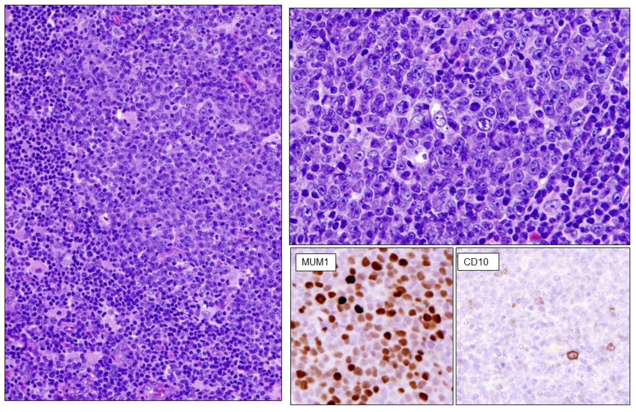
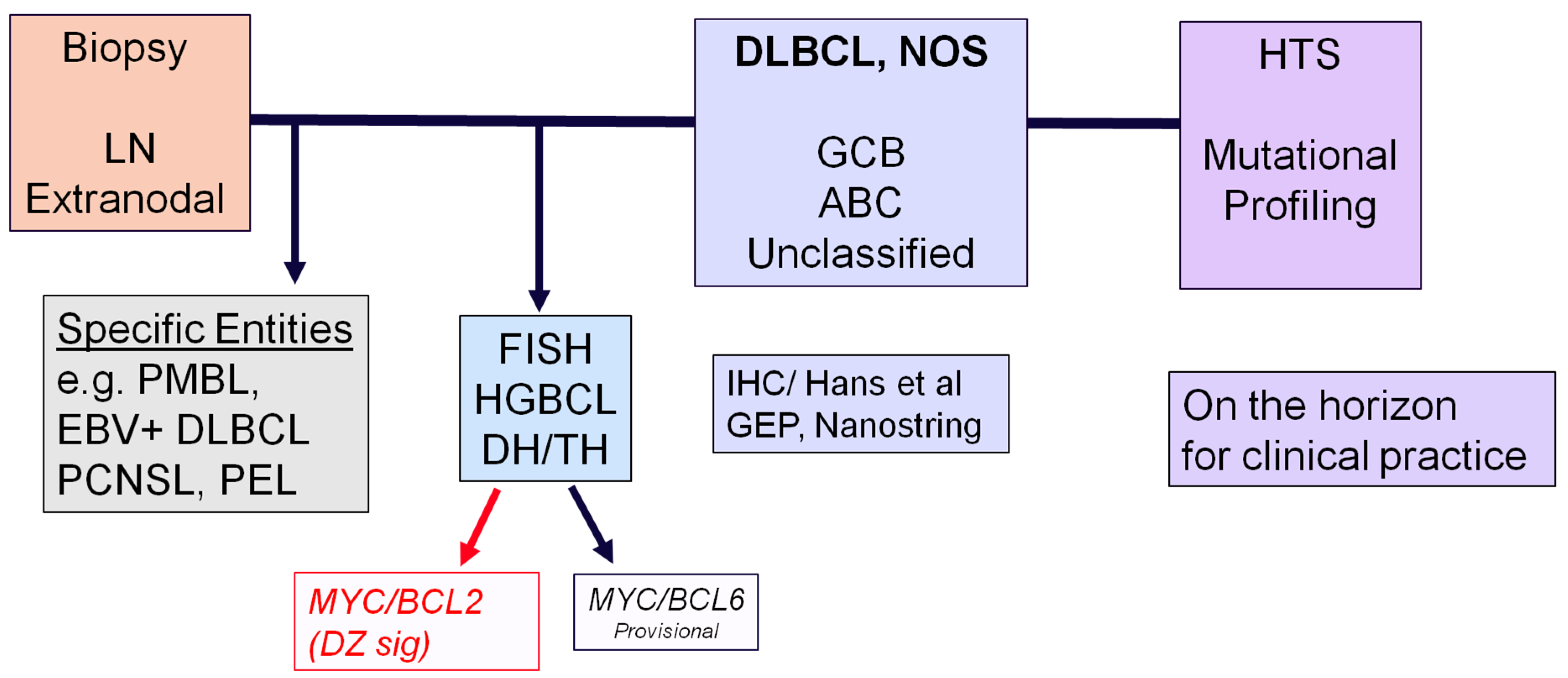
2.2. Diffuse Large B-Cell Lymphomas

The current classification of DLBCL recognizes the importance of activated B-cells (ABCs) versus germinal center B-cells (GCBs) as a basic biological distinction [13]. However, this binary classification approach fails to capture the complexity of DLBCL (Table 3). A new era is emerging with NGS and mutational profiling. This technology is on the horizon for clinical practice, although it is not currently required for the routine diagnosis of DLBCL.
Two major studies in the last few years showed that genetic heterogeneity in DLBCL can identify clinically significant genetic subgroups [14,15]. The subtype termed MCD is based on the co-occurrence of the L265P mutation in MYD88 and mutations in CD79B. This subtype is highly enriched in primary central nervous system lymphoma, testicular diffuse large B-cell lymphoma, and other aggressive extranodal DLBCLs. The subtype designated as BN2 is based on fusions involving BCL6 and mutations in NOTCH2. This subtype is heterogeneous in its gene expression profile and may represent an aggressive variant of marginal zone lymphoma. New molecular variants were uncovered, including a subset of aggressive lymphomas with a high degree of aneuploidy and a high frequency of p53 mutation and deletion [16]. The term A53 refers to these key features: p53 mutation and aneuploidy. The historical cell of origin approach (ABC or activated B-cell; GCB or germinal center B-cell) for the classification of DLBCL can be integrated in part to the new data derived from genomic profiling based on NGS studies [17]. Tumors related to activated B-cells (ABCs) are more heterogeneous, with the MCD group being a major subset. MCD tumors, as noted above, are almost always extranodal. N1, with mutations in NOTCH1, is ABC as well.
The ICC felt it was too early to implement this approach for routine clinical use. A challenge will be to make this technology accessible for daily practice in diagnosis. Notably, with current systems, many cases (35–40%) are not assigned to a specific genomic subtype.
Another issue that was discussed was whether it was time to recognize extranodal DLBCL as a separate entity (Table 4). It is recognized today that many extranodal DLBCLs of the ABC subtype share biological features.
A new category of aggressive B-cell lymphomas (WHO fifth edition) encompasses large-cell lymphomas that occur primarily in the central nervous system (PCNSL), vitreoretinal compartment (VRL), and testis, all of which are immune sanctuaries created by the structure of their unique anatomic sites [5]. These lymphomas exhibit similar morphologic, immunophenotypic, and genetic features, including concordant MYD88 and CD79B mutations. These, and other alterations, contribute to a germinal center exit phenotype and down-regulation of the immune response.
The ICC accepted DLBCL of the testis as a specific entity that is closely related to primary diffuse large B-cell lymphoma of the CNS but felt that further studies were needed to better define the interrelationships between diffuse large B-cell lymphomas in other extranodal sites.

2.3. High-Grade B-Cell Neoplasms

Another controversial issue was the classification of high-grade B-cell lymphomas with MYC and BCL2 and/or BCL6 rearrangements, which had been included in the last WHO classification. The ICC decided to retain high-grade B-cell lymphomas with MYC and BCL2 rearrangements as a specific entity (Table 5). These cases have a broad cytologic spectrum but are otherwise considered uniform. FISH break-apart probes are recommended but may miss up to 20% of cases with cryptic rearrangement. This tumor has a germinal center origin but may express TDT in some cases and has the gene expression profile of centroblasts in the so-called germinal center dark zone. The ICC decided to retain a high-grade B-cell lymphomas with MYC and BCL6 rearrangements as a provisional entity, recognizing that these cases are somewhat more heterogeneous, both in terms of cell of origin and mutational profile, while the WHO did not retain them as a category. These cases are variously incorporated as examples of DLBCL or high-grade B-cell lymphoma based on cytologic features.
Recent studies have really emphasized the importance of the dark zone signature as expanding the spectrum of double-hit lymphomas [18,19]. Twenty percent of GCB DLBCLs have either a double hit or a dark zone signature, and these cases are characterized by poor clinical outcome. This signature is shared by Burkitt’s lymphoma, but some cases with the dark zone signature are negative for the MYC rearrangement by FISH.
Another entity provisionally recognized by the ICC is large B-cell lymphoma with a 11q aberration, named as high-grade B-cell lymphoma with 11q aberration by the WHO. This is mainly a pediatric tumor that has a starry sky pattern and can look very similar to Burkitt’s lymphoma, but it usually presents with nodal disease rather than the extranodal disease more commonly seen in Burkitt lymphoma. These cases are negative for the MYC rearrangement but may have MYC protein expression, and they show gains and losses at the 11q region [20].
The ICC decided to name this entity large B-cell lymphoma with 11q rather than Burkitt-like or high grade. This was formerly Burkitt-like lymphoma with 11q aberration in the fourth edition of the WHO [3]. Further studies have shown that this tumor has generally a favorable prognosis and does not require the intensive therapy usually given for patients with Burkitt’s lymphoma. The ICC felt it was more appropriate to include it as a variant of DLBCL rather than risking more aggressive therapy for these young patients [4]. The WHO system, because this tumor looks like Burkitt’s lymphoma in many cases, retains the designation of high-grade B-cell lymphoma [5].

3. Flow Chart for the Diagnosis of Aggressive B-Cell Lymphomas

Figure 4 shows a flow chart for the diagnosis of aggressive B-cell lymphomas that provides a model for current clinical practice. The first step is a biopsy of the lymph node or extranodal lesion. The first goal is to recognize some of the specific entities, not all of which have been discussed, such as primary mediastinal large B-cell lymphoma or KSHV-/HHV8-associated lymphomas or EBV-positive large B-cell lymphomas. If the case looks like DLBCL, NOS, the next step is to carry out FISH analysis, looking for rearrangements with BCL2, BCL6, and MYC to recognize high-grade B-cell lymphomas with a double hit. Left with a case of DLBCL, NOS, gene expression profiling may be employed to recognize the ABC subtype versus GCB, or immunohistochemical approaches may be used. Mutational profiling using high-throughput sequencing is certainly on the horizon for clinical practice in the near future.

4. ICC 2022 Proposal: Hodgkin’s Lymphoma

Regarding HL, the ICC renamed nodular lymphocyte predominant HL [21] as nodular lymphocyte predominant B-cell lymphoma [4]. This entity is closely related to T-cell histiocyte-rich large B-cell lymphoma and in most instances represents a continuum. It differs in its biology and genomics features from classic HL. While some studies find suitable responses with ABVD [22], other studies have suggested a benefit for CHOP chemotherapy for patients with advanced-stage disease [23]. Notably, its treatment is also different from classic HL. Thus, to ensure appropriate therapy for this disease the change in terminology was proposed. The ICC also tried to simplify the Fan system, which has been in use for a number of years to guide clinical management and therapy [24]. The six Fan grades [24], A, B, C, D, E, and F, could be simplified as grade 1, which encompasses grades A, B, and C, and grade 2, which includes the more clinically aggressive tumors encompassing D, E, and F.
The only controversial category and only question remaining is how to deal with cases that are pattern C, which have both intrafollicular and interfollicular LP cells. The ICC decided to retain this as grade 1. Many of these patients have more localized disease and really seem to overlap Fan grades A and B. Figure 5 shows an example of nodular LP presenting in an axillary lymph node, while the bone marrow shows involvement by T-cell histiocyte-rich large B-cell lymphoma. Figure 6 shows another case of nodular lymphocyte predominant B-cell lymphoma, which was associated with transformation to DLBCL, an event seen in from 5% to 10% of cases.

5. Gray-Zone Lymphoma

Both the ICC and the WHO have a similar approach to the category of so-called gray-zone lymphoma. Recent data have emphasized differences between mediastinal and non-mediastinal cases [25,26]. The ICC adopted the same approach as the WHO, recognizing mediastinal gray-zone lymphoma as the only entity with this name. Non-mediastinal cases with overlapping features between Hodgkin’s and B-cell lymphoma have a different genomic profile. They tend to present with advanced age and are both nodal and extranodal and should be diagnosed as DLBCL in both systems.
In most cases, the B-cell program is retained with a strong expression of CD20 and other B-cell markers as well as a positivity for OCT2. Both the ICC and WHO define mediastinal gray-zone lymphoma as a distinct entity, related to primary mediastinal large B-cell lymphoma and nodular sclerosis HL. It is important to appreciate that EBV positivity in mediastinal gray-cell lymphoma is rarely seen and strongly favors a diagnosis of EBV-positive DLBCL.

6. EBV-Positive Large B-Cell Lymphoma

EBV-positive DLBCL is retained in the ICC classification (Table 3). This is mainly a disease of advanced age and is associated with immune senescence. It more often presents with extranodal disease, although nodal involvement is seen as well. The tumor has an aggressive clinical course but, interestingly, a low mutational burden. The most common alterations involve the JAK-STAT and NOTCH pathways [27]. Interestingly, some EBV-positive large B-cell lymphomas occur in young patients under the age of 45 [28]. These cases are nearly always nodal as opposed to the extranodal presentation in older patients. The immunophenotype shows evidence of a permissive immune microenvironment, and in general, these tumors have an excellent prognosis, which contrasts with EBV-positive DLBCL in the elderly. In fact, interestingly, many of the cases of EBV-positive DLBCL in young patients appear to resemble T-cell histiocyte-rich large B-cell lymphoma.

7. HHV8- and EBV-Negative Primary Effusion-Based Lymphoma

Effusion-based lymphoma was mentioned under the term HHV-8-negative primary effusion lymphoma in the differential diagnosis of primary effusion lymphoma in the WHO-HAEM fourth edition, revised, but it was not included as a distinct subtype [3]. In the upcoming WHO fifth edition classification, these lesions are recognized as a distinct subtype of DLBCL, “fluid overload-associated large B cell lymphomas”, whereas in the ICC, they are a provisional entity called “HHV8 and EBV negative primary effusion based lymphoma” [4,5]. Both classifications recognize these as lesions arising in the setting of fluid overload, often due to an underlying medical condition such as cardiac, renal, or liver failure without a preceding or concurrent tumor mass. There also appears to be a relationship with hepatitis C virus infection. Patients with this type of large-cell lymphoma are usually HIV-negative females, elderly, and with a median age of 70 [4,5,29].
These lesions are composed of cells that are usually morphologically reminiscent of the neoplastic cells in PEL. However, in contrast to PEL, the neoplastic cells are KSHV-/HHV8-negative and express B-cell antigens such as CD20, a lack expression of CD138, and occasionally contain an MYC rearrangement. In the WHO fifth edition, lesions that are EBV-positive are included in this category and account for a small proportion of the cases, whereas in the ICC, EBV positivity is an exclusion criterion [4,5]. These effusion-based lymphomas are associated with a relatively good prognosis compared with PEL. EBV positivity in effusion-based disease is associated with a more aggressive clinical course, and it would be considered within the spectrum of EBV-positive B-cell lymphomas based on other clinical and biological parameters.

8. Changes in the Classification in the T-/NK-Cell Neoplasms

Regarding T-/NK-cell neoplasms, the entities shown in bold print in Table 6 were modified or altered in the ICC classification from that published in the last edition WHO 2017. Only some entities will be discussed here. In general, there are fewer differences between the ICC and the WHO when it comes to the definition of T-cell lymphomas. Both classifications recommend that follicular helper T-cell lymphomas are a common entity and are unified under a common term. Indeed, they are the most common subtype of nodal peripheral T-cell lymphoma. The most common of these is the angioimmunoblastic type, but the ICC also recognizes the so-called follicular subtype, and then a follicular helper T-cell lymphoma is otherwise specified. These cases share a common immunophenotype expressing markers associated with Tfh cells and share a number of mutations, including TET2, DMNT3, IDH2, and RHOA [30,31,32].
Another question addressed in the ICC system was whether we can come up with a better way to subclassify peripheral T-cell lymphoma that is not otherwise specified. In a couple of publications, mainly from the ICC system [30,31], two major subtypes were recognized, one associated with Th1 cells and another with Th2 cells. Th1-positive cases express TBET, whereas Th2-positive cases express GATA3. There seem to be some clinical differences, with Th1-positive cases having a better prognosis. However, it was felt that it was premature to recommend this for routine clinical practice. This should remain a topic for further investigation.

8.1. Indolent Gastrointestinal T-/NK-Cell Neoplasms

There were some changes introduced into the classification of indolent gastrointestinal T-/NK-cell neoplasms (Table 7, Figure 7). The ICC recommends the term indolent clonal T-cell lymphoproliferative disorder of the GI tract to emphasize that demonstrating monoclonality is important to make this diagnosis in the clinical setting. Both systems recognized indolent NK-cell lymphoproliferative disorder, which is newly added to both classification systems and replaces the term NK-cell enteropathy and lymphomatoid gastropathy [33,34]. Recent mutational studies have provided evidence that this is indeed a neoplasm associated with recurrent mutations in JAK3 [35,36].

8.2. Type 2 Refractory Celiac Disease

The ICC also added type 2 refractory celiac disease to the classification and identifies it as a precursor of enteropathy-associated T-cell lymphoma (EATL). Type 2 refractory celiac disease has a poor prognosis and a high risk of progression to EATL within five years. Most of the cases have gain-of-function mutations in STAT3 and JAK 1 [37] and recurrent alterations in epigenetic regulators. These cases have an abnormal T-cell phenotype and are usually negative for both CD4 and CD8.
Regarding the EBV-negative intestinal T-cell and NK-cell lymphoproliferative disorders of the GI, it is important to remember that some cases of nasal-type EBV-positive NK-/T-cell lymphoma can present with GI tract involvement and have occult disease of the nasal cavity [38].

8.3. Anaplastic Large-Cell Lymphoma ALK-Negative

There were minor changes introduced into the classification of anaplastic large-cell lymphoma (ALCL) ALK-negative. The ICC considers cases with a DUSP-22 rearrangement as a genetic subtype of systemic ALCL ALK-negative with a good prognosis. The diagnosis is facilitated by immunohistochemistry, as these cases are positive for LEF1, while they are negative for cytotoxic markers. A minor point is that breast-implant-associated ALCL was also upgraded from a provisional to a definite entity [39,40].

9. Conclusions

This article discussed the considerable evolution in the lymphoma classification, with a major impact from clinical and genetic advances. In 2024, pathologists and hematologists are dealing with two different systems, one from the ICC 2022 and one from the WHO fifth edition. But, perhaps, these differences will be resolved, and it is hopeful that we are dealing with a consensus classification for the sixth edition of the WHO system in the future. Most of the expert hematopathologists have agreed to report their diagnoses in two or three classification systems where differences exist, reporting diagnoses in both the ICC 2022 and the proposed fifth edition of the WHO and, in some cases, also mentioning the revised fourth edition of the WHO.

Author Contributions

The manucript was written and edited by E.S.J. and A.C. It was based on lecture delivered by E.S.J. at a post-graduate course on hematopathology held in Bologna, Italy on 19–20 January 2024. All authors have read and agreed to the published version of the manuscript.

Funding

This work was supported by the intramural research program of the Center for Cancer Research, National Cancer Institute, Bethesda, MD, USA.

Conflicts of Interest

The authors declare no conflict of interest.

References

  1. Jaffe, E.S.; Harris, N.; Stein, H.; Vardiman, J.W. (Eds.) World Health Organization Classification of Tumours. In Pathology and Genetics of Tumours of Haematopoietic and Lymphoid Tissues, 3rd ed.; IARC: Lyon, France, 2001. [Google Scholar]
  2. Swerdlow, S.H.; Campo, E.; Harris, N.L.; Jaffe, E.S.; Pileri, S.A.; Stein, H.; Thiele, J.; Vardiman, J.W. (Eds.) World Health Organization Classification of Tumours of Haematopoietic and Lymphoid Tissues, 4th ed.; IARC: Lyon, France, 2008. [Google Scholar]
  3. Swerdlow, S.H.; Campo, E.; Harris, N.L.; Jaffe, E.S.; Pileri, S.A.; Stein, H.; Thiele, J.; Vardiman, J.W. (Eds.) WHO Classification of Tumours of Haematopoietic and Lymphoid Tissues; Revised 4th ed.; IARC: Lyon, France, 2017. [Google Scholar]
  4. Campo, E.; Jaffe, E.S.; Cook, J.R.; Quintanilla-Martinez, L.; Swerdlow, S.H.; Anderson, K.C.; Brousset, P.; Cerroni, L.; de Leval, L.; Dirnhofer, S.; et al. The International Consensus Classification of Mature Lymphoid Neoplasms: A Report from the Clinical Advisory Committee. Blood 2022, 140, 1229–1253. [Google Scholar] [CrossRef] [PubMed]
  5. Alaggio, R.; Amador, C.; Anagnostopoulos, I.; Attygalle, A.D.; Araujo, I.B.O.; Berti, E.; Bhagat, G.; Borges, A.M.; Boyer, D.; Calaminici, M.; et al. The 5th edition of the World Health Organization Classification of Haematolymphoid Tumours: Lymphoid Neoplasms. Leukemia 2022, 36, 1720–1748. [Google Scholar] [CrossRef] [PubMed]
  6. Jaffe, E.S.; Carbone, A. Evolution in the Definition of Follicular Lymphoma and Diffuse Large B-Cell Lymphoma: A Model for the Future of Personalized Medicine. Hemato 2022, 3, 466–474. [Google Scholar] [CrossRef]
  7. Bhavsar, S.; Liu, Y.C.; Gibson, S.E.; Moore, E.M.; Swerdlow, S.H. Mutational Landscape of TdT+ Large B-cell Lymphomas Supports Their Distinction From B-lymphoblastic Neoplasms: A Multiparameter Study of a Rare and Aggressive Entity. Am. J. Surg. Pathol. 2022, 46, 71–82. [Google Scholar] [CrossRef] [PubMed]
  8. Nann, D.; Ramis-Zaldivar, J.E.; Müller, I.; Gonzalez-Farre, B.; Schmidt, J.; Egan, C.; Salmeron-Villalobos, J.; Clot, G.; Mattern, S.; Otto, F.; et al. Follicular lymphoma t(14;18)-negative is genetically a heterogeneous disease. Blood Adv. 2020, 4, 5652–5665. [Google Scholar] [CrossRef] [PubMed]
  9. Liu, Q.; Salaverria, I.; Pittaluga, S.; Jegalian, A.G.; Xi, L.; Siebert, R.; Raffeld, M.; Hewitt, S.M.; Jaffe, E.S. Follicular lymphomas in children and young adults: A comparison of the pediatric variant with usual follicular lymphoma. Am. J. Surg. Pathol. 2013, 37, 333–343. [Google Scholar] [CrossRef] [PubMed]
  10. Ramis-Zaldivar, J.E.; Gonzalez-Farre, B.; Balague, O.; Celis, V.; Nadeu, F.; Salmeron-Villalobos, J.; Andres, M.; Martin-Guerrero, I.; Garrido-Pontnou, M.; Gaafar, A.; et al. Distinct molecular profile of IRF4-rearranged large B-cell lymphoma. Blood 2020, 135, 274–286. [Google Scholar] [CrossRef] [PubMed]
  11. Salmeron-Villalobos, J.; Egan, C.; Borgmann, V.; Müller, I.; Gonzalez-Farre, B.; Ramis-Zaldivar, J.E.; Nann, D.; Balagué, O.; Lopez-Guerra, M.; Colomer, D.; et al. PNMZL and PTFL: Morphological variants with a common molecular profile—A unifying hypothesis. Blood Adv. 2022, 6, 4661–4674. [Google Scholar] [CrossRef] [PubMed]
  12. Saksena, A.; Jain, A.; Pack, S.D.; Kim, J.; Lee, I.; Tyagi, M.; Xi, L.; Pittaluga, S.; Raffeld, M.; Jaffe, E.S. Follicle Center Lymphoma (FCL) of the Lower Female Genital Tract (LFGT): A Novel Variant of Primary Cutaneous Follicle Center Lymphoma (PCFCL). Am. J. Surg. Pathol. 2023, 47, 409–419. [Google Scholar] [CrossRef]
  13. Alizadeh, A.A.; Eisen, M.B.; Davis, R.E.; Ma, C.; Lossos, I.S.; Rosenwald, A.; Boldrick, J.C.; Sabet, H.; Tran, T.; Yu, X.; et al. Distinct types of diffuse large B-cell lymphoma identified by gene expression profiling. Nature 2000, 403, 503–511. [Google Scholar] [CrossRef]
  14. Chapuy, B.; Stewart, C.; Dunford, A.J.; Kim, J.; Kamburov, A.; Redd, R.A.; Lawrence, M.S.; Roemer, M.G.M.; Li, A.J.; Ziepert, M.; et al. Molecular subtypes of diffuse large B cell lymphoma are associated with distinct pathogenic mechanisms and outcomes. Nat. Med. 2018, 24, 679–690. [Google Scholar] [CrossRef] [PubMed]
  15. Schmitz, R.; Wright, G.W.; Huang, D.W.; Johnson, C.A.; Phelan, J.D.; Wang, J.Q.; Roulland, S.; Kasbekar, M.; Young, R.M.; Shaffer, A.L.; et al. Genetics and Pathogenesis of Diffuse Large B-Cell Lymphoma. N. Engl. J. Med. 2018, 378, 1396–1407. [Google Scholar] [CrossRef] [PubMed]
  16. Wright, G.W.; Huang, D.W.; Phelan, J.D.; Coulibaly, Z.A.; Roulland, S.; Young, R.M.; Wang, J.Q.; Schmitz, R.; Morin, R.D.; Tang, J.; et al. A Probabilistic Classification Tool for Genetic Subtypes of Diffuse Large B Cell Lymphoma with Therapeutic Implications. Cancer Cell 2020, 37, 551–568.e514. [Google Scholar] [CrossRef] [PubMed]
  17. Ennishi, D.; Hsi, E.D.; Steidl, C.; Scott, D.W. Toward a New Molecular Taxonomy of Diffuse Large B-cell Lymphoma. Cancer Discov. 2020, 10, 1267–1281. [Google Scholar] [CrossRef] [PubMed]
  18. Hilton, L.K.; Tang, J.; Ben-Neriah, S.; Alcaide, M.; Jiang, A.; Grande, B.M.; Rushton, C.K.; Boyle, M.; Meissner, B.; Scott, D.W.; et al. The double-hit signature identifies double-hit diffuse large B-cell lymphoma with genetic events cryptic to FISH. Blood 2019, 134, 1528–1532. [Google Scholar] [CrossRef] [PubMed]
  19. Alduaij, W.; Collinge, B.; Ben-Neriah, S.; Jiang, A.; Hilton, L.K.; Boyle, M.; Meissner, B.; Chong, L.; Miyata-Takata, T.; Slack, G.W.; et al. Molecular determinants of clinical outcomes in a real-world diffuse large B-cell lymphoma population. Blood 2023, 141, 2493–2507. [Google Scholar] [CrossRef] [PubMed]
  20. Horn, H.; Kalmbach, S.; Wagener, R.; Staiger, A.M.; Huttl, K.; Mottok, A.; Bens, S.; Traverse-Glehen, A.; Fontaine, J.; Siebert, R.; et al. A Diagnostic Approach to the Identification of Burkitt-like Lymphoma with 11q Aberration in Aggressive B-Cell Lymphomas. Am. J. Surg. Pathol. 2021, 45, 356–364. [Google Scholar] [CrossRef] [PubMed]
  21. Harris, N.L.; Jaffe, E.S.; Stein, H.; Banks, P.M.; Chan, J.K.; Cleary, M.L.; Delsol, G.; De Wolf-Peeters, C.; Falini, B.; Gatter, K.C.; et al. A revised European-American classification of lymphoid neoplasms: A proposal from the International Lymphoma Study Group. Blood 1994, 84, 1361–1392. [Google Scholar] [CrossRef] [PubMed]
  22. Eichenauer, D.A.; Hartmann, S. Nodular lymphocyte-predominant Hodgkin lymphoma: Current management strategies and evolving approaches to individualize treatment. Expert. Rev. Hematol. 2023, 16, 607–615. [Google Scholar] [CrossRef]
  23. Xing, K.H.; Connors, J.M.; Lai, A.; Al-Mansour, M.; Sehn, L.H.; Villa, D.; Klasa, R.; Shenkier, T.; Gascoyne, R.D.; Skinnider, B.; et al. Advanced-stage nodular lymphocyte predominant Hodgkin lymphoma compared with classical Hodgkin lymphoma: A matched pair outcome analysis. Blood 2014, 123, 3567–3573. [Google Scholar] [CrossRef]
  24. Fan, Z.; Natkunam, Y.; Bair, E.; Tibshirani, R.; Warnke, R.A. Characterization of variant patterns of nodular lymphocyte predominant hodgkin lymphoma with immunohistologic and clinical correlation. Am. J. Surg. Pathol. 2003, 27, 1346–1356. [Google Scholar] [CrossRef]
  25. Campo, E.; Jaffe, E.S. Taking gray zone lymphomas out of the shadows. Blood 2021, 137, 1703–1704. [Google Scholar] [CrossRef]
  26. Sarkozy, C.; Hung, S.S.; Chavez, E.A.; Duns, G.; Takata, K.; Chong, L.C.; Aoki, T.; Jiang, A.; Miyata-Takata, T.; Telenius, A.; et al. Mutational landscape of gray zone lymphoma. Blood 2021, 137, 1765–1776. [Google Scholar] [CrossRef]
  27. Frontzek, F.; Staiger, A.M.; Wullenkord, R.; Grau, M.; Zapukhlyak, M.; Kurz, K.S.; Horn, H.; Erdmann, T.; Fend, F.; Richter, J.; et al. Molecular profiling of EBV associated diffuse large B-cell lymphoma. Leukemia 2023, 37, 670–679. [Google Scholar] [CrossRef]
  28. Nicolae, A.; Pittaluga, S.; Abdullah, S.; Steinberg, S.M.; Pham, T.A.; Davies-Hill, T.; Xi, L.; Raffeld, M.; Jaffe, E.S. EBV-positive large B-cell lymphomas in young patients: A nodal lymphoma with evidence for a tolerogenic immune environment. Blood 2015, 126, 863–872. [Google Scholar] [CrossRef] [PubMed]
  29. Gisriel, S.D.; Yuan, J.; Braunberger, R.C.; Maracaja, D.L.V.; Chen, X.; Wu, X.; McCracken, J.; Chen, M.; Xie, Y.; Brown, L.E.; et al. Human herpesvirus 8-negative effusion-based large B-cell lymphoma: A distinct entity with unique clinicopathologic characteristics. Mod. Pathol. 2022, 35, 1411–1422. [Google Scholar] [CrossRef] [PubMed]
  30. Iqbal, J.; Wright, G.; Wang, C.; Rosenwald, A.; Gascoyne, R.D.; Weisenburger, D.D.; Greiner, T.C.; Smith, L.; Guo, S.; Wilcox, R.A.; et al. Gene expression signatures delineate biological and prognostic subgroups in peripheral T-cell lymphoma. Blood 2014, 123, 2915–2923. [Google Scholar] [CrossRef]
  31. Amador, C.; Bouska, A.; Wright, G.; Weisenburger, D.D.; Feldman, A.L.; Greiner, T.C.; Lone, W.; Heavican, T.; Smith, L.; Pileri, S.; et al. Gene Expression Signatures for the Accurate Diagnosis of Peripheral T-Cell Lymphoma Entities in the Routine Clinical Practice. J. Clin. Oncol. 2022, 40, 4261–4275. [Google Scholar] [CrossRef] [PubMed]
  32. Carbone, A.; Gloghini, A. Subclassifying peripheral T-cell lymphoma NOS. Blood 2019, 134, 2120–2121. [Google Scholar] [CrossRef]
  33. Takeuchi, K.; Yokoyama, M.; Ishizawa, S.; Terui, Y.; Nomura, K.; Marutsuka, K.; Nunomura, M.; Fukushima, N.; Yagyuu, T.; Nakamine, H.; et al. Lymphomatoid gastropathy: A distinct clinicopathologic entity of self-limited pseudomalignant NK-cell proliferation. Blood 2010, 116, 5631–5637. [Google Scholar] [CrossRef]
  34. Mansoor, A.; Pittaluga, S.; Beck, P.L.; Wilson, W.H.; Ferry, J.A.; Jaffe, E.S. NK-cell enteropathy: A benign NK-cell lymphoproliferative disease mimicking intestinal lymphoma: Clinicopathologic features and follow-up in a unique case series. Blood 2011, 117, 1447–1452. [Google Scholar] [CrossRef] [PubMed]
  35. Montes-Moreno, S.; King, R.L.; Oschlies, I.; Ponzoni, M.; Goodlad, J.R.; Dotlic, S.; Traverse-Glehen, A.; Ott, G.; Ferry, J.A.; Calaminici, M. Update on lymphoproliferative disorders of the gastrointestinal tract: Disease spectrum from indolent lymphoproliferations to aggressive lymphomas. Virchows Arch. 2020, 476, 667–681. [Google Scholar] [CrossRef] [PubMed]
  36. Yi, H.; Li, A.; Ouyang, B.; Da, Q.; Dong, L.; Liu, Y.; Xu, H.; Zhang, X.; Zhang, W.; Jin, X.; et al. Clinicopathological and molecular features of indolent natural killer-cell lymphoproliferative disorder of the gastrointestinal tract. Histopathology 2023, 82, 567–575. [Google Scholar] [CrossRef] [PubMed]
  37. Soderquist, C.R.; Lewis, S.K.; Gru, A.A.; Vlad, G.; Williams, E.S.; Hsiao, S.; Mansukhani, M.M.; Park, D.C.; Bacchi, C.E.; Alobeid, B.; et al. Immunophenotypic Spectrum and Genomic Landscape of Refractory Celiac Disease Type II. Am. J. Surg. Pathol. 2021, 45, 905–916. [Google Scholar] [CrossRef] [PubMed]
  38. Jaffe, E.S. T-cell and NK-cell neoplasms of the gastrointestinal tract—Recurrent themes, but clinical and biological distinctions exist. Haematologica 2020, 105, 1760–1762. [Google Scholar] [CrossRef] [PubMed]
  39. Laurent, C.; Nicolae, A.; Le Bras, F.; Haioun, C.; Fataccioli, V.; Amara, N.; Adélaïde, J.; Guille, A.; Schiano, J.M.; Tesson, B.; et al. Gene alterations in epigenetic modifiers and JAK-STAT signaling are frequent in breast implant-associated ALCL. Blood 2020, 135, 360–370. [Google Scholar] [CrossRef]
  40. Blombery, P.; Thompson, E.R.; Jones, K.; Arnau, G.M.; Lade, S.; Markham, J.F.; Li, J.; Deva, A.; Johnstone, R.W.; Khot, A.; et al. Whole exome sequencing reveals activating JAK1 and STAT3 mutations in breast implant-associated anaplastic large cell lymphoma anaplastic large cell lymphoma. Haematologica 2016, 101, e387–e390. [Google Scholar] [CrossRef]
Figure 1. FL 3B. Typical centrocytes are largely absent, although there is some variation in cell size. This case lacked BCL2 rearrangement, and it was positive for MUM1 and negative for CD10.
Hemato 05 00013 g001
Figure 2. The figure shows an example of BCL2-rearrangement-negative, CD23-positive follicular lymphoma variant.
Hemato 05 00013 g002
Figure 3. The figure shows variants of follicular lymphoma in young patients (usually <30 years), all of which are distinct entities in the ICC system.
Hemato 05 00013 g003
Figure 4. Flow chart for the diagnosis of aggressive B-cell lymphomas (reprinted from [6]).
Hemato 05 00013 g004
Figure 5. Upper panel: nodular lymphocyte predominant B-cell lymphoma. Lymph node involvement. Lower panel: T-cell/histiocyte-rich large B-cell lymphoma. Bone marrow involvement.
Hemato 05 00013 g005
Figure 6. Upper panel: nodular lymphocyte predominant B-cell lymphoma. Lower panel: diffuse large B-cell lymphoma.
Hemato 05 00013 g006
Figure 7. (A,B) (CD3) Indolent clonal T-cell–T-lymphoproliferative disorder (LPD). (C,D) Indolent NK-cell LPD, which is positive for CD56 (E) and CD3 (F).
Hemato 05 00013 g007
Table 1. ICC 2022: What is new in B-cell neoplasms?
Follicular Lymphomas
   -
Grading retained: Grades 1–2, 3A, 3B
   -
Recognition of novel variants (BCL2 neg, pediatric, cutaneous, testicular)
Diffuse large B-cell lymphomas
   -
“Cell of origin retained” (GCB, ABC)
   -
Genomic subtypes reserved for the future *
High-Grade B-cell neoplasms
   -
HGBCL with MYC/BCL2 R, (focal TDT expression may be seen)
   -
HGBCL with MYC/BCL6 R, considered provisional *
* Further studies required.
Table 2. Follicular lymphomas: ICC 2022/WHO fifth edition.
ICC
Follicular lymphoma (Grade 1–2; 3A, 3B)
In situ follicular neoplasia
Duodenal type FL
BCL2-R-negative, CD23-positive follicle center lymphoma (provisional)
Primary cutaneous follicle center lymphoma
Pediatric-type follicular lymphoma
Testicular follicular lymphoma
Large B-cell lymphoma with IRF4 rearrangement
WHO
Classic follicular lymphoma (no grading)
In situ follicular B-cell neoplasm
Duodenal-type follicular lymphoma
FL with unusual cytological features
FL with predominantly diffuse growth pattern (dFL)
Follicular large B-cell lymphoma
Primary cutaneous follicle center lymphoma
Pediatric-type follicular lymphoma
  •
Diseases defined by clinical, pathologic, and genomic features: BCL2 R or other alterations
  •
Classic FL is BCL2 R; other variants not clearly defined
Table 3. “Large” B-cell neoplasms in the ICC 2022.
Diffuse large B-cell lymphoma (DLBCL), not otherwise specified (NOS)
     Germinal center B-cell subtype
     Activated B-cell subtype
Large B-cell lymphoma with 11q aberration
Large B-cell lymphoma with IRF4 R
(Nodular lymphocyte predominant B-cell lymphoma)
T-cell/histiocyte-rich large B-cell lymphoma
Primary DLBCL of the central nervous system
Primary DLBCL of the testis
Primary cutaneous DLBCL, leg type
Intravascular large B-cell lymphoma
Burkitt’s lymphoma
High-grade B-cell lymphoma with MYC and BCL2 R
High-grade B-cell lymphoma with MYC and BCL6 R
High-grade B-cell lymphoma, NOS
Primary mediastinal large B-cell lymphoma
Mediastinal gray-zone lymphoma
EBV-positive mucocutaneous ulcer
EBV-positive DLBCL, NOS
DLBCL associated with chronic inflammation
     Fibrin-associated DLBCL
Lymphomatoid granulomatosis
EBV-positive polymorphic B-cell lymphoproliferative disorder, NOS
ALK-positive large B-cell lymphoma
Plasmablastic lymphoma
HHV8-associated lymphoproliferative disorder
     Multicentric Castleman’s disease
     HHV8-positive germinotropic lymphoproliferative disorder
     HHV8-positive DLBCL, NOS
     Primary effusion lymphoma
HHV-8- and EBV-negative primary effusion-based lymphoma
  • Disease with changes introduced in the ICC 2022
  • Provisional Entities
Table 4. Is it time to recognize extranodal DLBCL as a separate entity?
  • Many extranodal DLBCLs and ABCs share biological features (MCD/C5 subtypes by NGS)
  • Prototypes include primary CNS and testicular DLBCL
  • Some subtypes are also defined by their topographic site (IVLBCL)
  • Extranodal DLBCLs in other anatomic sites are more heterogeneous
    (e.g., breast, adrenal, kidney)
Proposal
  • The ICC accepts primary DLBCL of the testis as a specific entity closely related to primary DLBCL of the CNS
  • Further studies are needed to determine the interrelationships between DLBCLs in other extranodal sites
  • Unifying features: CD79B/MYD88 L265P
Table 5. ICC 2022 proposal for high-grade B-cell lymphomas.
High-Grade B-Cell Lymphomas with MYC and BCL2 Rearrangements
  –
Broad cytological spectrum but considered a single entity
  –
FISH break-apart probes recommended but may miss up to 20% cases (cryptic)
  –
Germinal center origin but may express TDT
  –
Gene expression signature of centroblasts in the GC dark zone
  –
Mutational profile similar to “aggressive” FL (BCL2, MYC, KMT2D, CREBPP, TNFRS14, EZH2)
High-grade B-cell lymphomas with MYC and BCL6 rearrangements (provisional)
  –
Heterogeneous in cell of origin and mutational profile (less FL-type, NOTCH2)
  –
Thirty percent may be “pseudo double”-hit
  –
Not retained as a category—classified as either DLBCL or HG based on cytology
Table 6. ICC classification of T-/NK-cell neoplasms: items in bold represent modified or new terms.
NodalPeripheral T-cell lymphoma, NOS
Follicular helper T-cell lymphomas
Anaplastic large-cell lymphoma, ALK-positive
Anaplastic large-cell lymphoma, ALK-negative
Primary nodal EBV-positive T-/NK-cell lymphoma
ExtranodalExtranodal NK-/T-cell lymphoma, nasal type
Enteropathy-associated T-cell lymphoma
   Type II refractory celiac disease
Monomorphic epitheliotropic intestinal T-cell lymphoma
Indolent clonal T-cell lymphoproliferative disorder of the GI tract
Indolent NK-cell lymphoproliferative disorder of the GI tract
Breast-implant-associated anaplastic large-cell lymphoma
Leukemic/SystemicT-cell prolymphocytic leukemia
T-cell large granular lymphocytic leukemia
Chronic lymphoproliferative disorder of NK-cells
Aggressive NK-cell leukemia
EBV-positive T-/NK-LPDs of childhood
Adult T-cell leukemia/lymphoma
Hepatosplenic T-cell lymphoma
CutaneousMycosis fungoides
Sézary syndrome
Primary cutaneous CD30-positive T-cell LPDs
Subcutaneous panniculitis-like T-cell lymphoma
Primary cutaneous gamma-delta T-cell lymphoma
Primary cutaneous CD8-positive aggressive epidermotropic cytotoxic T-cell lymphoma
Primary cutaneous acral CD8+ T-LPD
Primary cutaneous CD4-positive small/medium T-LPD
Hydroa vacciniforme-like LPD
Table 7. Changes in the classification of indolent gastrointestinal T-/NK-cell neoplasms.
Indolent clonal T-cell lymphoproliferative disorder of the gastrointestinal tractName changed (clonal) to emphasize importance of clonality in diagnosis.
Considered a definite entity but is heterogeneous in phenotype and genotype (CD4 or CD8) (all αβ)
May progress to more aggressive disease.
Indolent NK-cell lymphoproliferative disorder of the gastrointestinal tract
(newly added)
The term replaces both NK-cell enteropathy and lymphomatoid gastropathy.
Mutational studies provide evidence for the neoplastic origin.
Recurrent mutations in JAK3
Disclaimer/Publisher’s Note: The statements, opinions and data contained in all publications are solely those of the individual author(s) and contributor(s) and not of MDPI and/or the editor(s). MDPI and/or the editor(s) disclaim responsibility for any injury to people or property resulting from any ideas, methods, instructions or products referred to in the content.

