Abstract
Background: Teeth are the anatomical tissue with the highest resistance to the action of chemical and physical agents. This is one of the reasons that make teeth particularly useful in the identification process of skeletonized and carbonized human remains. The aim of this research is to analyze the colorimetric changes in the enamel of teeth subjected to high temperatures to develop a reproducible colorimetric cataloging method. Methods: Six groups of 21 human teeth extracted from private clinics and from a Dental School for therapeutic reasons were used and subjected to three temperature ranges in a laboratory furnace: 400 °C, 700 °C, and 1000 °C. For each temperature, two time periods of 20 min and 60 min were chosen. Each group of dental elements was analyzed using a dental spectrophotometer to extract the colorimetric data of the crown. The obtained color coordinates were subsequently converted into Red–Green–Blue (RGB) values. The two predominant colors were also selected to create average colorimetric values, which demonstrate the change in color hue according to temperature. The groups of teeth subjected to 20 min at 400 °C exhibited a dark gray coloration, while the teeth subjected to 20 min at 700 °C showed a general increase in color brightness with beige–blueish hues. Results: The teeth subjected to 20 min at 1000 °C displayed progressively lighter shades with pinkish reflections. The teeth subjected to 60 min at the same temperatures demonstrated a general increase in brightness, making differentiation more challenging, except for the group of teeth burned at 400 °C, which showed light gray–blueish tones. Conclusions: This study further supports the existing literature on the correlation between colorimetric shifts in carbonized teeth and the maximum temperature reached, providing valuable assistance to forensic pathology and the forensic dental identification of burnt human remains. Additionally, this research has led to the development of a standardized colorimetric patented scale for the observation and examination of burnt human teeth.
1. Introduction
In modern society, the role of forensic sciences in the identification of human remains is a necessary condition for social and legal reasons. The most effective methods of identification are based on the collection and comparison of primary identifiers, through the analysis of DNA, fingerprints, skeleton, and teeth, especially when unidentified human remains are severely decomposed, skeletonized, or burnt. Dental data and dentition of an individual can provide even clearer information regarding the victim’s identity, specifically in burnt human remains. Teeth are one of the most resistant tissues in the human body, less prone to modifications caused by postmortem phenomena and potential insults to which a body may be subjected, both antemortem and postmortem. Their structural uniqueness, combined with the presence of restorative materials, implants, or orthodontic appliances, makes their study crucial in the identification of victims in disasters, accidents, or crimes where bodies have been exposed to extreme conditions [1]. Fire represents one of the most destructive traumas that can affect the human body, as it damages and alters crucial evidence for the reconstruction of the victim’s identity [2,3,4,5]. There are numerous variables that influence the effects of this process and can affect the degree of body compromise. Combustion can be partial or total, complete or incomplete. From a biochemical perspective, the exposure of a body to heat, whether postmortem or antemortem, causes massive dehydration of organic components, leading to the destruction of soft tissues and the gradual exposure of the skeleton until complete combustion occurs [6]. The study of incinerated human remains poses challenges due to the destruction of most body structures except for skeletal and dental tissues and for this reason dental remains are crucial for forensic reconstructions, as the unique structure and organization within the oral cavity provide valuable information for victim identification [7]. Forensic odontologists should work at the scene to prevent loss of dental evidence and preserve fragile remains. Photographic and radiographic cataloging allows for comparison with research data correlating morphological alterations with temperature ranges. Teeth behave similarly to bone under heat, with enamel having little organic material and dentin and cementum undergoing modifications based on position and organic content [8,9]. The duration of combustion is also a crucial parameter in determining the tissue’s destruction [6]. Enamel has virtually no organic material (although it is the outermost tissue), while dentin and cementum undergo modifications based on their position and the amount of organic content present. Taking into account the histological and anatomical characteristics and adhering to the protocols for sample collection [10], forensic odontologists can analyze changes in color, alterations in dimension, and any fracture patterns that have occurred in the teeth after exposure to heat, referring to the relevant literature evidence for further analysis [11,12].
The aim of this study is to analyze the colorimetric changes in the enamel of teeth subjected to high temperatures to develop a reproducible colorimetric cataloging method as an aid in the human identification process of unidentified human remains.
2. Materials and Methods
Our study aims to analyze the color changes in human teeth subjected to charring, investigating the main color shades that dental elements assume within specific temperature ranges. The ultimate goal is to establish a color reference scale that can assist in the process of forensic dental identification of charred human remains, providing indications of the maximum thermal exposure of the remains. This study has been approved by the Bioethics Committee of the University of Turin.
We collected 63 permanent dental elements (24 anterior and 39 posterior elements) from dental clinics and the clinic of the Dental School. The selection criteria included intact teeth without dental restorations or carious lesions, extracted for periodontal, orthodontic, or impaction reasons. The collected dental elements were stored in the Personal Identification Laboratory of the Forensic Medicine department at the University of Turin (Figure 1). Three randomized groups of teeth were created and subjected to temperatures of 400 °C, 700 °C, and 1000 °C for exposure times of 20 min and 60 min (Figure 2 and Figure 3). These three temperatures were chosen based on the literature concerning charred human remains to achieve results relevant for forensic purposes.
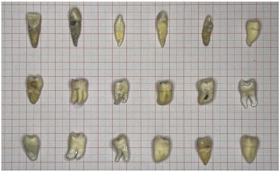
Figure 1.
Some of the selected teeth in their original state.
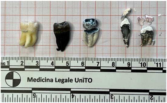
Figure 2.
Burnt teeth for 20 min. From left to right: intact tooth; tooth exposed to 400 °C; tooth exposed to 700 °C; teeth exposed to 1000 °C.
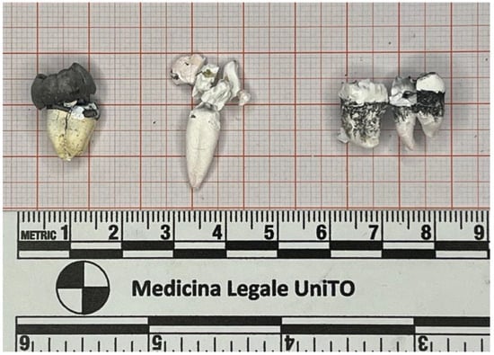
Figure 3.
Burnt teeth after 60 min. From left to right: tooth exposed to 400 °C; tooth exposed to 700 °C; teeth exposed to 1000 °C.
The teeth were subjected to heat using a laboratory dental furnace with an analog controller (model FM-74, Enrico Bruno S.r.l., Turin, Italy) with a single phase, 1.3 KW, 220 Volt, 50 HZ power supply. The maximum achievable temperature of the furnace is 1100 °C. The furnace is equipped with a control peephole and a timer. It was heated 50 °C higher than the established temperature for each group of teeth to compensate for heat loss when the furnace door was opened for inserting the dental elements. The teeth were arranged in groups inside the furnace, with spacing between each tooth, for the predetermined period of this study.
At the end of each phase, the furnace was turned off, and a gradual cooling process was allowed. On average, the cooling process involved 15 min with the furnace closed, 15 min with the furnace open, and 15 min outside the furnace.
Burnt teeth were then collected in breathable airtight plastic containers for spectrophotometric analysis at the dental prosthetics laboratory of the Dental School in Turin. To compensate for the challenging analysis of samples above 500 °C, multiple readings were taken for each individual tooth to obtain the same number of measurements. Spectrophotometric readings on the charred teeth were performed using a dental device (Vita Easy Shape V, Vita Zahnfabrik, Bad Säckingen, Germany) capable of recording the colors of natural teeth and restorations. Vita EasyShade covers the spectrum of the classic VITA A1-D4 system (colors used for direct/indirect restorations with resin or ceramic materials) and provides the coefficient values in the form of shade analysis coordinates (called Lab and LCh). These coordinates were then converted to Red–Green–Blue (RGB) coefficients to determine the colors using photo editing software through the Lab -> RGB converter on the website www.aspose.app (accessed on 4 April 2024). For each category, the colors of all the teeth used were recorded, and the average coefficients above the median value and the average coefficients below the median value were calculated to obtain two standard mean colors for the two groups of 20 min and 60 min of exposure for each temperature group (400 °C, 700 °C, and 1000 °C).
3. Results
3.1. 20 Minutes Heat Exposure
The extracted L*a*b* values obtained using Vita Easyshade V for the group of dental elements subjected to a temperature of 400 °C for a duration of 20 min were converted to RGB values and summarized in the following Table 1.

Table 1.
Values and coefficients of shade analysis coordinates (L*a*b*) converted to RGB values for the group of burnt teeth at a temperature of 400 °C for a duration of 20 min.
From these values, two average values were selected, corresponding to the predominant color shades, RGB I: 55, 55, 43 and RGB II: 91, 94, 84.
The subsequent groups of burnt samples were more challenging to analyze due to tissue fragmentation caused by the heat. By selecting only the useful coronal parts for the spectrophotometer, the issue was overcome by sampling the same elements at different points. The values obtained for the group of samples burnt at 700 °C for a duration of 20 min are reported in following Table 2.
Table 2.
Values and coefficients of shade analysis coordinates (L*a*b*) converted to RGB values for the group of burnt teeth at a temperature of 700 °C for a duration of 20 min.
From these values, two average values were selected, corresponding to the predominant color shades, RGB I: 146, 160, 159 and RGB II: 173, 191, 187.
Finally, the third group of dental elements was heated to 1000 °C for 20 min, and the obtained spectrophotometric analysis values are as follows (Table 3).
Table 3.
Values and coefficients of shade analysis coordinates (L*a*b*) converted to RGB values for the group of burnt teeth at a temperature of 1000 °C for a duration of 20 min.
From these values, two average values were selected, corresponding to the predominant color shades, RGB I; 194, 177, 170 and RGB II: 223, 218, 207.
3.2. 60 Minutes Heat Exposure
Teeth that underwent charring for 60 min experienced a colorimetric transformation that can lead to misleading results: the color shift of dental elements subjected to heat for a period longer than 30 min is difficult to analyze and reproduce, as suggested in the literature [11]. The values for the group of teeth exposed to 400 °C for 60 min are reported below (Table 4):
Table 4.
Values and coefficients of shade analysis coordinates (L*a*b*) converted to RGB values for the group of burnt teeth at a temperature of 400 °C for a duration of 60 min.
From these values, two average values were selected, corresponding to the predominant color shades, RGB I: 80, 97, 87 and RGB II: 63, 65, 59.
As regards the samples burned at 700 °C for 60 min, the analysis led to the following Lab and RGB results (Table 5):
Table 5.
Values and coefficients of shade analysis coordinates (L*a*b*) converted to RGB values for the group of burnt teeth at a temperature of 700 °C for a duration of 60 min.
From these values, the two average values were RGB I: 228, 233, 220 and RGB II: 215, 211, 197.
The last group of tooth samples was subjected to a temperature of 1000 °C for a period of 60 min. The analyzed results are the following (Table 6):
Table 6.
Values and coefficients of shade analysis coordinates (L*a*b*) converted to RGB values for the group of burnt teeth at a temperature of 1000 °C for a duration of 60 min.
From these values, the two average values were RGB I: 222, 228, 215 and RGB II: 229, 235, 220.
An additional final table with corresponding colors was created with the averages of the RGB values of the two main shades of color for each category of heat time exposure (20 and 60 min) and temperatures (400 °C and 700 °C). It can be appreciated how decisive the colors assumed by the samples are in the three charred groups for a shorter period of time (20 min), as agreed with the articles found in the literature (Table 7).
Table 7.
Final average colors derived for each category of heat time exposure (20 and 60 min) and temperatures (400 °C and 700 °C).
3.3. Burnt Teeth Colorimentric Scale (Carbodent)
The average twelve RGB values of the two main color shades for each temperature category (400 °C/752 °F; 700 °C/1292 °F; and 1000 °C/1832 °F) and time exposure (20 and 60 min), as shown in Table 1, have enabled the creation of a colorimetric scale as a reference tool for interpreting the temperature to which the teeth of a carbonized human remains were exposed.
The scale has been specifically designed for use by forensic odontologists, resembling the color scales commonly used in restorative dentistry and prosthodontics, where the color is displayed on a sample in the shape of a central incisor (Figure 4). The scale features twelve chromatic indicators divided into three temperature ranges: 400 °C/752 °F, 700 °C/1292 °F, and 1000 °C/1832 °F.
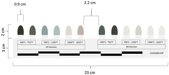
Figure 4.
Burnt teeth colorimetric scale (Carbodent) [13].
These indicators allow for visual evaluation, potentially complemented by color detection and analysis tools such as spectrophotometers and photo editing programs at the time of a dental autopsy observation. It identifies correspondences, discrepancies, and compatibility between the chromatic changes in the examined burnt teeth and those presented on the scale, thereby overcoming observer bias and enabling a more objective interpretation of the findings.
There is currently no available tool for direct use on charred human teeth that allows for colorimetric evaluation, photographic documentation, and forensic dental examinations.
4. Discussion
Scientific research on burnt dental elements has identified precise color changes in a transition that starts from the natural color of the tooth, which becomes darker at temperatures up to 400–500 °C, and then transitions to lighter shades up to 1000–1100 °C, resulting in the phenomenon of calcination, where the teeth acquire a whitish, almost pinkish color [14,15]. The literature suggests analyzing the tooth roots as they are the anatomical part that better withstands thermal insults. However, this research focused on the coronal parts of the teeth (enamel) to propose an alternative and reproducible method for interpreting the assessment of the maximum exposure temperature in the identification of burnt human remains.
The enamel tissue remained intact up to 400 °C, but at higher temperatures or longer durations, it detached from the underlying dentin, making both identification and color analysis with the available spectrophotometer difficult. For this reason, the decision was made to analyze the useful coronal tissue (dentin or enamel) in accordance with various authors in the literature who found no statistically significant differences between the color changes in dentin and enamel.
The spectrophotometric analysis [16] of the burnt samples for 20 min showed increasing color values as the temperature increased, consistent with the heat-induced modifications previously described, indicating a gradual combustion of the organic dental tissue.
The predominant average colors extrapolated from the 20 min burnt samples were dark gray and metallic for teeth subjected to 400 °C, light blue and light gray for samples subjected to 700 °C, and shades of pink and white/beige for teeth burned at 1000 °C.
Regarding the three groups of samples exposed to heat for 60 min, analytical difficulties increased due to greater tissue fragmentation, making the analysis of less intact portions more challenging. In this case, the color differences compared to the groups exposed for a shorter time flattened out, showing a rapid increase in the color value compared to the first group, making the colors assumed by the teeth charred at 700 °C and 1000 °C for 60 min extremely similar.
The elements subjected to 400 °C for 60 min, on the other hand, showed changes in color values halfway between the 400 °C group charred for 20 min and the 700 °C group charred for 20 min, assuming a coronal coloration of metallic gray with a hint of blue.
The CIELab (L*a*b*) data analyzed with Vita EasyShade V were transformed into RGB values to ensure reproducibility regardless of the system used. The colors obtained from this research allowed the identification of colorimetric indications divided into temperature intervals from 400 °C to 1000 °C, which can be useful both visually and through appropriate color detection and analysis tools (spectrophotometers and computer applications) to assist in the assessment of the time and thermal exposure of teeth in burnt human remains. However, greater reliability in interpretation is achieved when the combustion event lasts less than 30 min.
The structural, dimensional, and colorimetric changes in burnt human remains described in the literature [15,16,17,18] do not currently provide a reference associated with a reproducible color code. Therefore, the proposed new colorimetric scale for burnt human teeth (”carbodent”) will allow for visual assessment and interpretation using suitable color detection and analysis tools during the dental autopsy observation. It must be highlighted that teeth heated in different environments, such as in an oven, kiln, or open environment, as well as cooling aspect of the process, can exhibit varying types of discoloration [12]. Therefore, the proposed device is not intended to indicate a specific color after heating, but rather to facilitate the differentiation between ranges of heated teeth up to 400 °C or over 700 °C. Due to the potential variations in discoloration resulting from different heating conditions, a flexible interpretation of the data is always necessary.
The scale provides a photographic reference to identify correspondence, discrepancies, and compatibility between the chromatic modifications of the teeth of burnt human remains under examination and those present on the scale, thereby also overcoming the limitations of observer bias and a more objective interpretation of the findings.
It is recommended to use the colorimetric scale on multiple areas of the burnt teeth under examination, as is will be rare to observe uniformly colored burnt teeth. In our analysis, two average values per tooth were selected and interpreted as predominant color shades. Further studies should be conducted to explore how color patterning in burnt teeth could be affected by factors such as the position and presence of soft tissue like the cheek skin and lips, or other specific conditions or positions of the teeth during exposure to heat.
5. Conclusions
The research on burnt human teeth and colorimetric changes is of great importance in forensic odontology, pathology, and anthropology. Estimating the maximum temperature reached by burnt human remains can be one of the crucial factors in forensic dental identification. The chromatic modifications of the burnt teeth analyzed thought spectrophotometry confirms the existing literature on the correlation between colorimetric shifts in carbonized teeth and high temperatures. Specifically, it aids in the distinguishing between ranges of heated teeth up to 400 °C or over 700 °C.
The practical application of this research is the development of a standardized, patented colorimetric scale for observing and examining burnt human teeth, with a reference photographic colorimetric scale with a reproducible color code. The new scale could also overcome the observer bias and provide a more objective interpretation of findings during the dental autopsy of carbonized human remains. It is important to note that teeth heated in different environments and the cooling aspect of the process can result in various types of discoloration. Therefore, a flexible interpretation of the data is always necessary.
6. Patents
This research has resulted in a patented dental colorimetric scale “Carbodent”, Utility-Patent IT n. 202024000000781/2024.
Author Contributions
Conceptualization, E.N.; methodology, E.N. and G.D.V.; validation, E.N. and G.D.V.; resources, M.A; data curation, E.N.; writing—original draft preparation, M.A.; writing—review and editing, E.N.; supervision, E.N. and G.D.V.; All authors have read and agreed to the published version of the manuscript.
Funding
This research received no external funding.
Institutional Review Board Statement
This study was conducted in accordance with the Declaration of Helsinki and was approved by the Institutional Ethics Committee of the University of Turin, Italy, protocol n. 0340130 of the 23 June 2023.
Informed Consent Statement
Written informed consent was obtained from the patients to publish this paper, as part of the Institutional Ethics Committee of the University of Turin, Italy.
Data Availability Statement
The raw data supporting the conclusions of this article will be made available by the authors on request.
Conflicts of Interest
The authors declare no conflicts of interest.
References
- Marella, G.L.; Rossi, P. An approach to person identification by means of dental prostheses in a burnt corpse. J. Forensic Odontostomatol. 1999, 17, 16–19. [Google Scholar] [PubMed]
- Correia, P.M.M. Fire modification of bone: A review of the literature. In Forensic Taphonomy: The Postmortem Fate of Human Remains; Haglund, W.D., Sorg, M.H., Eds.; CRC Press: Boca Raton, FL, USA, 1997; pp. 275–293. [Google Scholar]
- Herrmann, N.P.; Bennett, J.L. The differentiation of traumatic and heat-related fractures in burned bone. J. Forensic Sci. 1999, 44, 461–469. [Google Scholar] [CrossRef] [PubMed]
- Reesu, G.V.; Augustine, J.; Urs, A.B. Forensic considerations when dealing with incinerated human dental remains. J. Forensic Leg. Med. 2015, 29, 13–17. [Google Scholar] [CrossRef] [PubMed]
- Wightman, J.M.; Gladish, S.L. Explosions and blast injuries. Ann. Emerg. Med. 2001, 37, 664–678. [Google Scholar] [CrossRef] [PubMed]
- Bohnert, M.; Rost, T.; Pollak, S. The degree of destruction of human bodies in relation to the duration of the fire. Forensic Sci. Int. 1998, 95, 11–21. [Google Scholar] [CrossRef] [PubMed]
- Berketa, J.W. Maximizing postmortem oral-facial data to assist identification following severe incineration. Forensic Sci. Med. Pathol. 2014, 10, 208–216. [Google Scholar] [CrossRef] [PubMed]
- Sandholzer, M.A.; Baron, K.; Heimel, P.; Metscher, B.D. Volume analysis of heat-induced cracks in human molars: A preliminary study. J. Forensic Dent. Sci. 2014, 6, 139–144. [Google Scholar] [CrossRef] [PubMed][Green Version]
- Peer, M.; Sarig, R. The effect of burning on dental tissue: A macroscopic and microscopic investigation. Forensic Sci. Int. 2024, 358, 111987. [Google Scholar] [CrossRef] [PubMed]
- Ubelaker, D.H. The forensic evaluation of burned skeletal remains: A synthesis. Forensic Sci. Int. 2009, 183, 1–5. [Google Scholar] [CrossRef] [PubMed]
- Beach, J.J. Heat-related changes in tooth color: Temperature versus duration of exposure. In The Analysis of Burned Human Remains; Schmidt, C.W., Symes, S.A., Eds.; Academic Press: Oxford, UK, 2008; pp. 137–144. [Google Scholar]
- Rahmat, R.A.; Humphries, M.A.; Austin, J.J.; Linacre, A.M.T.; Self, P. The development of a tool to predict temperature-exposure of incinerated teeth using colourimetric and hydroxyapatite crystal size data. Int. J. Legal Med. 2021, 135, 2045–2053. [Google Scholar] [CrossRef] [PubMed]
- Nuzzolese, E.; Di Vella, G.; Tattoli, L.; Aliberti, M.; Nuzzolese, D. University of Turin, Turin, Italy. Utility-Patent Italy n. 202024000000781, 22 February 2024. [Google Scholar]
- Shipman, P.; Foster, G.; Schoeninger, M. Burnt bones and teeth: An experimental study of color, morphology, crystal structure and shrinkage. J. Archaeol. Sci. 1984, 11, 307–325. [Google Scholar] [CrossRef]
- Peer, M.; Sarig, R. Color change in teeth due to burning: Spectrophotometric analysis. Forensic Sci. Int. 2023, 345, 111608. [Google Scholar] [CrossRef] [PubMed]
- Rubio, L.; Sioli, J.M.; Suarez, J.; Gaitan, M.J.; Martin-de-las-Heras, S. Spectrophotometric analysis of color changes in teeth incinerated at increasing temperatures. Forensic Sci. Int. 2015, 252, 193.e1–193.e6. [Google Scholar] [CrossRef] [PubMed]
- Deadman, W.J. The identification of human remains. Can. Med. Assoc. J. 1964, 91, 808–811. [Google Scholar] [PubMed]
- Schmidt, C.W.; Symes, S.A. (Eds.) The Analysis of Burned Human Remains; Elsevier: London, UK, 2008. [Google Scholar]


Disclaimer/Publisher’s Note: The statements, opinions and data contained in all publications are solely those of the individual author(s) and contributor(s) and not of MDPI and/or the editor(s). MDPI and/or the editor(s) disclaim responsibility for any injury to people or property resulting from any ideas, methods, instructions or products referred to in the content. |
© 2024 by the authors. Licensee MDPI, Basel, Switzerland. This article is an open access article distributed under the terms and conditions of the Creative Commons Attribution (CC BY) license (https://creativecommons.org/licenses/by/4.0/).