
New Insights into Hsp90 Structural Plasticity Revealed by cryoEM


1
Biomolecular Engineering Department, Jack Baskin School of Engineering, University of California—Santa Cruz, Santa Cruz, CA 95064, USA
2
Biomolecular Cryo-Electron Microscopy Facility, University of California—Santa Cruz, Santa Cruz, CA 95064, USA
3
Department of Chemistry and Biochemistry, University of California—Santa Cruz, Santa Cruz, CA 95064, USA
4
Sao Carlos Institute of Chemistry, University of Sao Paulo, São Carlos 13566-590, Brazil
*
Author to whom correspondence should be addressed.
†
These authors contributed equally to this work.
BioChem 2024, 4(2), 62-89; https://doi.org/10.3390/biochem4020004
Submission received: 29 November 2023
/
Revised: 29 February 2024
/
Accepted: 2 April 2024
/
Published: 4 April 2024
Abstract
:Heat Shock Protein 90 (Hsp90) acts as a crucial molecular chaperone, playing an essential role in activating numerous signaling proteins. The intricate mechanism of Hsp90 involving ATPase-coupled conformational changes and interactions with cochaperone proteins has been elucidated through biochemical and structural analyses, revealing its activation mechanism and its diverse set of “client” proteins. Despite recent advancements, certain aspects of Hsp90’s ATPase-coupled mechanism remain contentious, and the specific nature of the alterations induced by Hsp90 in client proteins remains largely undiscovered. In this review, we explore the current understanding of Hsp90’s structure and function, drawing insights from single-particle cryoEM studies. Structural studies on Hsp90 using cryoEM have provided valuable insights into the structural dynamics and interactions of this molecular chaperone. CryoEM structures have been instrumental in understanding the ATPase-coupled conformational changes that Hsp90 undergoes during its chaperone cycle. We also highlight recent progress in elucidating the structure of the ATP-bound state of the complete dimeric chaperone. Furthermore, we delve into the roles played by the multitude of cochaperones that collaborate with Hsp90, providing a glimpse into their biochemical mechanisms through the newly obtained cryoEM structures of Hsp90 cochaperone complexes.
1. Molecular Chaperones, a Brief Overview
Molecular chaperones help other proteins maintain their proper three-dimensional structure and function within the cell. These molecules are essential for maintaining protein homeostasis and are critical components of the cellular machinery that safeguards protein integrity and helps prevent the accumulation of misfolded or aggregated proteins. Their involvement in various cellular processes makes them a subject of ongoing research, particularly in the fields of cancer biology and drug development. Some of them are called “heat shock proteins”, and that name comes from the fact that their expression increases when cells are stressed, such as at elevated temperatures (heat shock). Molecular chaperones are named according to their protomer molecular weight (Hsp40—40 kDa; Hsp60—60 kDa; Hsp70—70 kDa; Hsp90—90 kDa and Hsp100—100 kDa, respectively) and are intrinsically related to several biological functions involved in macromolecule degradation, disaggregation, and refolding [1,2,3,4].
1.1. The Molecular Chaperones: Hsp40, Hsp70, Hsp100, and Hsp60
Heat Shock Protein 40 (also named J-Domain Proteins or JPDs)—Hsp40 chaperones typically work in conjunction with Hsp70 chaperones. Hsp70 and Hsp40 cooperate to assist in various cellular processes [5]. Hsp40 stimulates the ATPase activity of Hsp70, which, in turn, promotes the binding and release of client proteins. This process assists in the correct folding of newly synthesized proteins or refolding of misfolded proteins. Hsp40 and Hsp70 can guide proteins across cellular membranes, helping them reach their intended subcellular destinations. Hsp40 can also be involved in targeting misfolded or damaged proteins for degradation by the proteasome or other cellular degradation pathways [6,7].
Hsp40 chaperones contain a conserved J-domain, which is critical for their interaction with Hsp70 chaperones (Figure 1A). The J-domain facilitates the transfer of client proteins from Hsp40 to Hsp70. Additionally, Hsp40 chaperones may have other domains that enable them to interact with specific client proteins, adapt to various cellular conditions, and participate in diverse cellular processes [8].
Another heat shock protein—Hsp70, or Heat Shock Protein 70—is a highly conserved family of molecular chaperone proteins found in a wide range of organisms, from bacteria to humans [9]. Hsp70 proteins are central players in maintaining protein homeostasis within cells and are essential for various cellular functions, particularly in response to environmental stress. Hsp70 prevents protein aggregation via its recognition of and interactions with exposed hydrophobic regions on misfolded proteins, which can otherwise lead to the formation of protein aggregates or inclusion bodies. Hsp70 plays a role in the transport of proteins across cellular membranes, assisting in their translocation to specific cellular compartments (endoplasmic reticulum and mitochondria). Hsp70 can deliver misfolded or damaged proteins to proteolytic systems, such as the proteasome or lysosome, for degradation [9,10,11].
This molecular chaperone is upregulated in response to stress conditions, including temperature variation, oxidation, and other cellular stresses [11,12]. This upregulation helps protect and repair cellular proteins under adverse conditions. From a structural perspective, Hsp70 proteins contain two main domains: the N-terminal ATPase domain and the C-terminal substrate-binding domain (Figure 1B). The ATPase domain is responsible for ATP hydrolysis, which powers the chaperone’s activity. The substrate-binding domain is responsible for recognizing and interacting with client proteins. The binding and release of client proteins are regulated by the binding and hydrolysis of ATP [9,13].
The next molecular chaperone is Hsp100, also known as Clp/Hsp100 or AAA+ proteins (ATPases Associated with diverse cellular Activities), a family of molecular chaperone proteins found in various organisms, including bacteria, archaea, and eukaryotes [14,15]. These chaperones belong to the AAA+ superfamily of ATPases and play a critical role in protein quality control and cellular processes related to protein folding, disaggregation, and degradation. Hsp100 proteins are involved in handling misfolded or aggregated proteins and are particularly important during stress conditions and cellular proteostasis maintenance. Hsp100 chaperones are involved in the disaggregation of protein aggregates or inclusion bodies. They use the energy from ATP hydrolysis to mechanically unfold and disaggregate misfolded or aggregated proteins, making them amenable to refolding or degradation [16].
Similar to Hsp70 and Hsp40 chaperones, Hsp100 proteins are upregulated in response to stress conditions, such as heat shock or other environmental stresses. Their increased expression is part of the cell’s defense mechanism against protein damage during stress. Hsp100 proteins are characterized by their ATPase activity, which powers the mechanical unfolding and disaggregation of protein substrates. These proteins typically form hexameric ring structures and use the energy from ATP hydrolysis to exert mechanical force on substrates, disrupting the protein aggregates and assisting in their unfolding (Figure 1C). However, the structural information regarding Hsp100 is still not completely clear, and more studies are needed [15,16].
The next member of the chaperone family is Heat Shock Protein 60, also known as GroEL (in prokaryotes such as bacteria) and Hsp60 or Cpn60 (in eukaryotes and mitochondria), another important member of the heat shock protein family [17]. Hsp60 is primarily responsible for assisting in the proper folding of proteins within the mitochondria and chloroplasts of eukaryotic cells and the cytoplasm of prokaryotic cells, particularly bacteria [17,18].
Hsp60 forms a large, double-ring structure that functions as a molecular “cage” or chaperonin. Each ring is composed of seven Hsp60 subunits, and the two rings stack together to create a central chamber where protein folding occurs (the “cage”). Unfolded or misfolded proteins enter the central chamber and are encapsulated within the chaperonin (Figure 1D). The chamber provides a protected environment where the protein can fold correctly, shielded from aggregation or misfolding in the cellular environment. The folding cycle of Hsp60 is driven by the hydrolysis of ATP. ATP binding and hydrolysis lead to conformational changes in the chaperonin, enabling it to capture, fold, and release client proteins. Hsp60 often works in conjunction with another protein called Hsp10 (or GroES in prokaryotes). Hsp10 binds to the outer surface of the Hsp60 ring and assists in the folding process by stabilizing the encapsulated protein and facilitating its release. In prokaryotic cells, such as bacteria, GroEL and GroES play a crucial role in assisting the folding of a wide range of client proteins. These chaperonins are essential for bacterial survival, especially under stress conditions [19,20].
Hsp60 is an essential component of the cellular protein folding machinery, ensuring that newly synthesized or stress-induced denatured proteins attain their correct three-dimensional structures. A dysfunction in Hsp60 can lead to protein aggregation and various cellular disorders. Its structure and function make it a vital player in maintaining cellular proteostasis and preventing the formation of misfolded or aggregated proteins [17].
1.2. The Bright Start: Hsp90
Finally, the major focus of this review, Hsp90 (Heat Shock Protein 90 kDa), is a highly conserved molecular chaperone found in different organisms, and its members are present in cellular compartments such as the cytoplasm, mitochondria, and endoplasmic reticulum [21,22]. It is involved in cellular protein folding, stability, and quality control to maintain proteostasis.
Hsp90s exist in an equilibrium of different states, and the structure is dynamic and undergoes conformational changes in response to ATP binding and hydrolysis, alternating between open and closed states (Figure 1E) [23,24,25,26,27]. These conformational changes are essential for its chaperone functions. Hsp90 often works in concert with various cochaperones and co-factors, such as Hsp70, Hsp40, and magnesium, to assist in the folding and stabilization of client proteins [28,29]. Together, these chaperones form a complex network of protein quality control within the cell called the foldosome. The structural characteristics of Hsp90 will be fully described in the next sections [30].
In addition to molecular chaperones, cochaperones are extensively involved and play several important roles in the cellular protein folding machinery, and their presence is crucial for maintaining cellular homeostasis and preventing protein misfolding. Cochaperones often stimulate the ATPase activity of molecular chaperones. ATP hydrolysis is a key step in the chaperone cycle, and cochaperones help regulate this process. By enhancing ATPase activity, cochaperones contribute to the efficient folding of proteins and the proper functioning of the chaperone machinery.
Moreover, Minari et al. [31] delve into the crucial role of Hsp90 proteins in cellular homeostasis, their potential as therapeutic targets for diseases like cancer and malaria, and the unique characteristics exhibited by Hsp90s from different organisms, despite their high sequence identity. Their research conducts a comprehensive comparative analysis of recombinant Hsp90s from various sources using isothermal titration calorimetry, focusing on their interactions with ADP and ATP. The thermodynamic signatures of these interactions reveal distinct characteristics for each Hsp90, emphasizing specific thermodynamic properties, despite high identity. The study suggests that the design of analogs targeting the Hsp90-ADP bound state may offer a more selective approach for inhibition compared to analogs targeting the Hsp90-ATP bound state. However, the limitations of the study are acknowledged, particularly in speculating on the Hsp90 ATPase cycle under the tested conditions, due to potential conformational changes induced by ATP binding. Despite comparable affinities across all the tested Hsp90s, the thermodynamic costs associated with ADP interactions involve entropic contributions offset by substantial enthalpic contributions, indicating that ADP likely forms additional interactions with the Hsp90 nucleotide-binding site compared to ATP, although the structural analyses do not fully support this conclusion [31].
2. Hsp90 Cochaperones
While the ATP cycle model applies to all Hsp90 paralogs and isoforms, there are some particularities highlighted for each isoform. It is noteworthy that eukaryotic Hsp90 operates in a nondeterministic manner, allowing accessibility to all conformations even in the absence of nucleotides. Considering the expansion of the “cochaperome” from none in bacteria, through 12 cochaperones in yeast, to >20 identified cochaperones in humans, it reveals that cochaperones exert a growing influence on the directionality of the conformational Hsp90 cycle in eukaryotes [32].
As previously mentioned, cochaperones are proteins that work in conjunction with molecular chaperones to facilitate proper protein folding and maturation within a cell. They play a crucial role in maintaining cellular homeostasis by preventing protein misfolding and aggregation. Cochaperones, as the name suggests, are additional proteins that collaborate with molecular chaperones to enhance their activity or to provide specificity in their functions. They can modulate the chaperone’s ATPase activity, influence substrate recognition, and participate in the assembly and disassembly of chaperone–substrate complexes.
Cochaperones often have specialized roles in different cellular processes and pathways. These molecules are required in higher eukaryotes, to aid chaperones. They are responsible for client protein presentation and regulating their respective chaperone activity as well as ATPse activity [33,34]. Hsp90 cochaperones are structurally distinct, and their interaction may depend on cofactors of stoichiometry [33,34,35] as in the following examples: Hop; Ahas; p23; and SGT.
2.1. Hop
Hop (Hsp70-Hsp90 organizing protein) is a cochaperone protein that plays a crucial role in facilitating the interaction between the Hsp70 and Hsp90 chaperone systems [35]. The protein acts as a bridge and ensures the efficient handover of client proteins from one chaperone to the other. Hop is also responsible for inhibiting the ATP hydrolysis in Hsp90. This protein has been found in various organisms, and its functions may vary among species (Figure 2A). It can also interact with other cochaperones and adapt its role, depending on a specific cellular context and requirements [36,37].
The Hop sequence contains three identified TPR domains (TPR1, TPR2A, and TPR2B), and also two domains with unknown structures to date (DP1 and DP2). The TPR1 domain specifically identifies Hsp70’s heptapeptide C-terminal, while TPR2A is responsible for the recognition of Hsp90’s C-terminal pentapeptide. Both peptide sequences present an EEVD motif. The TPR–peptide complex structures were obtained using crystallography, revealing that peptides adopt an extended conformation, bridging a groove in the TPR domains. The specific peptide recognition occurs via electrostatic interactions within the EEVD motif, where the C-terminal aspartate serves as a two-carboxylate anchor. Moreover, nuclear magnetic resonance (NMR) revealed that the DP domains exhibit α-helical folds in solution (Figure 2A).
2.2. Aha-Type Cochaperone
Aha-type cochaperones are well-known to stimulate Hsp90’s ATPase activity; however, the functional regulation remains unclear [38]. Aha1 (Activator of Hsp90 ATPase 1) is important for the ATPase activity regulation of Hsp90 [39,40,41]. Aha1 is involved in enhancing the ATPase activity of Hsp90, which is essential for the chaperone’s function in assisting the folding and maturation of client proteins. A similar description can also be applied to Aha2 and Aha4, among others (Figure 2B) [38,42].
2.3. p23
The small acidic protein, p23, serves as a prominent chaperone in eukaryotes, playing a crucial role in maintaining protein homeostasis. It exhibits both physical and functional associations with various cellular systems, including those involved in ribosome biogenesis, protein transport, chromatin remodeling, and transcription activation. Despite its involvement in diverse cellular processes, p23 is most recognized as an Hsp90 cochaperone, negatively influencing the ATPase cycle and the release of client proteins within the Hsp90 chaperone machinery [43,44]. Its initial characterization linked it to progesterone receptor complexes alongside the Hsp90 chaperone system. The human p23 protein consists of an N-terminal core comprising a β-sheet-structured domain and a C-terminal acidic tail. The interaction between p23 and Hsp90 takes place primarily through the β-sheet domain of p23, which is adequate for the proper interaction. While the C-terminal tail remains poorly characterized, the β-sheet domain plays a crucial role in binding partially unfolded client proteins and engaging with Hsp90 [43].
2.4. SGTs
Small glutamine-rich tetratricopeptide repeat proteins (SGTs) are well-known to recognize and interact with Hsp70 and Hsp90 chaperones [45]. These proteins present a tetratricopeptide repeat (TPR) motif and an abundance of glutamine residues (Figure 2C). TPR motifs are structural elements that mediate protein-protein interactions and are involved in a wide range of cellular processes, including protein folding, protein transport, and signal transduction. SGT proteins are known for their association with Hsp90. By binding to Hsp90 and assisting in the recruitment of client proteins, SGT proteins help regulate the chaperone machinery’s activity [46].
3. The Importance of Hsp90
Hsp90 interacts with a diverse set of client proteins, including kinases, transcription factors, and steroid hormone receptors. The client proteins often have roles in cancer, the immune response, the virulence of parasites, and other essential cellular functions [22,24]. The chaperone function of Hsp90 is intricately linked to its capacity to bind and hydrolyze ATP. This process is tightly regulated by cochaperone proteins and post-translational modifications (PTMs), such as phosphorylation (32 different sites in the human Hsp90-α isoform), acetylation (13), SUMOylation (2), methylation (4), O-GlcNAcylation (2), ubiquitination (13), and others. These modifications significantly impact chaperone function, consequently influencing various cellular processes. The ultimate challenge lies in deciphering the comprehensive and combinatorial array of PTMs that collectively modulate the Hsp90 chaperone function—a phenomenon aptly termed the “chaperone code”.
3.1. Proteosome Relationship
Chaperones and the proteasome are interconnected in the protein quality control pathway. Chaperones recognize misfolded or damaged proteins and attempt to refold them into their native state. If the chaperones are unable to facilitate a proper folding or if the proteins are irreversibly damaged, the chaperones may target them for degradation by the proteasome [1].
The proteasome then recognizes ubiquitinated proteins, unfolds them, and translocates them into its catalytic core for degradation into small peptides. The ubiquitin moieties are recycled for further use. Chaperones can also play a role in facilitating the delivery of ubiquitinated substrates to the proteasome, acting as adaptors in the recognition process [1,4].
3.2. Foldosome
The initiation of the cycle involves the binding of a newly synthesized or misfolded protein to the Hsp70/Hsp40 complex, constituting the early complex. Subsequently, the early complex interacts with the open conformation of Hsp90, forming the intermediate complex. This interaction is facilitated by the Hop protein, which enables the transfer of client proteins between Hsp70 and Hsp90, using its middle TPR domain to deliver to the N-terminal domain and the MEEVD motif of the Hsp90 C-terminal domain. After that, the cochaperones are released, Aha1 binds Hsp90, ATP is hydrolyzed, and the folded protein is released. Hsp90 now goes to the open conformation to restart the cycle [3,47].
4. Hsp90 Structural Bases
For many years, Hsp90’s structure remained unclear due to its high flexibility and multiple conformational states (Figure 3). However, low-resolution and in-solution methods helped to evaluate Hsp90’s conformations [27,48,49]. Hsp90 typically exists as a homodimer, and each monomer consists of three major domains: The N-terminal domain (NTD) is involved in ATP binding and hydrolysis [25,27]. It contains the ATP-binding pocket and a nucleotide-binding site, which is essential for the chaperone’s function. The middle domain (MD) is crucial for client protein binding and regulation. It contains the client protein-binding site and a flexible linker region (in eukaryotes) that connects it to the N-terminal domain. The C-terminal domain (CTD) is responsible for the dimerization of the Hsp90 protomers. It is also involved in cochaperone interactions and serves as an interface for other proteins that assist in the chaperone’s activity. The exact structure of Hsp90 may vary slightly among different species, but the overall architecture and key functional domains remain conserved [50]. Hsp90 forms homodimers, with contact sites localized within the C-terminus in the open conformation of the dimer. The N-termini also come in contact with the closed conformation of the dimer [27].
All members of the Hsp90 family share a common domain structure, comprising the nucleotide-binding NTD, the MD, and the CTD. The carboxy-terminal dimerization of two Hsp90 protomers results in the formation of a V-shaped dimer characterized by substantial conformational dynamics [26]. This dynamic structure allows for transient amino-terminal dimerization, a crucial aspect of chaperone function, as highlighted by Prodromou et al. in 2000 [51]. While this overall architecture is conserved across various organisms, subtle yet functionally significant differences exist among Hsp90 paralogs and orthologs [49,50,51].
The NTD of approximately 25 kDa displays a homology among members of the ATPase/kinase GHKL (Hsp90, Gyrase, MutL, and Histidine kinase) superfamily [51,52]. A shared binding pocket for ATP and inhibitors such as geldanamycin (GA) is situated in the NTD [53]. Hsp90 becomes active only when the cellular ratio of ATP to ADP favors ATP binding. The “split ATPase” nature of GHKL ATPases necessitates conformational rearrangements in Hsp90, involving the repositioning of the NTD and MD [54]. The distinct configuration of ATP within the Hsp90 nucleotide-binding pocket offers a potential target for selective inhibition by compounds such as radicicol (RD) and GA. A charged and flexible linker plays a role in modulating interactions between domains and regulating chaperone activity. Deletion or truncation of this linker disrupts the activation of client proteins. The MD contains the binding site for both Hsp90 clients and cochaperones, whereas the CTD facilitates constitutive dimerization through its C-terminal helices. The MEEVD motif, found in cytosolic Hsp90 paralogs, facilitates binding to cochaperones containing tetratricopeptide repeat (TPR) domains [55,56,57].
After ATP binds to the NTD, the ATP lid closes over the nucleotide, marking the formation of a preliminary intermediate state. This closure initiates structural changes, resulting in two subsequent states: the amino-terminally dimerized state (closed state 1) and a reduction in the distance between the MD and NTD (closed state 2), which collectively form the ATPase-active conformation. Following ATP hydrolysis and the release of ADP and phosphate, coupled with dissociation of the N-terminal region, the Hsp90 chaperone cycle concludes. [25,27].
Hsp90’s high-resolution X-ray crystal structures have provided valuable insights into the molecular architecture of this chaperone. The first breakthroughs in this regard involved solving the crystal structures of the NTD of yeast and human Hsp90, ∼25 kDa domain, easily released from the protein through limited proteolysis, both in their apo states and in complexes with nucleotides or inhibitors [48]. The NTD structure is characterized by an α/β sandwich, featuring a pocket extending from the buried face of the anti-parallel β-sheet to the surface, forming the nucleotide-binding site. Structures with bound nucleotides confirmed Hsp90’s status as an ATP-binding protein, resolving prior controversies surrounding its ATPase activity due to its initially low basal ATPase activity [25,27,48]. The identification of key catalytic residues, coupled with mutagenesis studies, solidified the understanding of Hsp90’s ATPase activity. Crystal structures with natural products, geldanamycin, and radicicol, demonstrated the inhibition of Hsp90’s function by competitively binding to the ATP-binding pocket, hindering the essential ATPase activity of Hsp90 [48]. These structures unveiled a striking similarity in the binding modes of inhibitors and nucleotides, both adopting a kinked conformation, with conserved key interactions, including those with a tightly bound water molecule. Numerous crystal structures now exist for the NTDs of human and yeast Hsp90, revealing various inhibitor complexes.
The MD of around 40 kDa comprises three distinct regions that compress a three-layer α-β-α sandwich, a three-turn α-helix, irregular loops, and a six-turn α-helix [58,59]. The MD is responsible for client protein binding, with known interactions including PKB/Akt1, eNOS, Aha1, and Hch1 [60,61]. Cochaperone binding (e.g., by Aha1 and Hch1) to the MD increases the ATPase activity of Hsp90. The elucidation of the Hsp90 structure progressed with the resolution of the MD of yeast Hsp90 in 2003 [61]. This domain consists of two structural subdomains: an N-terminal α/β/α domain, linked to a smaller α/β/α domain through short helices.
Lastly, the CTD weighing approximately 12 kDa houses an alternative ATP-binding site, only accessible when the N-terminal primary pocket is occupied [62,63,64]. Located at the far end of the C-terminus is the recognition site for the tetratricopeptide repeat (TPR) motif, characterized by the conserved MEEVD pentapeptide. This site facilitates interactions with various cofactors, including immunophilins FKBP51 and FKBP52, Tom70, stress-induced phosphoprotein 1 (Sti1/Hop), PP5, cyclophilin-40, and others, aligning with previous biochemical findings indicating the indispensability of the CTD for Hsp90 dimerization, forming a dimer with a small mixed α/β domain [65]. The dimer interface is constructed by a pair of helices at the C-terminal end of the domain, tightly packed together to generate a four-helix bundle. In contrast to the rest of the protein, the CTD exhibits a greater divergence from the corresponding region of eukaryotic Hsp90s, featuring lower sequence similarity and two small deletions relative to the eukaryotic sequences between the secondary structural elements. Nevertheless, the overall fold of the CTDs in eukaryotic and bacterial Hsp90s is likely to be highly similar [65].
Almost two decades ago, the initial report on crystallizing full-length Hsp90 surfaced [66]. However, the attainment of well-ordered crystals with diffraction quality, capable of providing an atomic resolution structure, has proven challenging, despite ongoing efforts and notable progress. In the meantime, substantial insights have been gained through structural studies focusing on domains and extensive subconstructs. As a result, the atomic structures for nearly all the segments of the Hsp90 structure are now comprehensively understood [49]. The first full-length Hsp90 structure was published in 2006 by the Pearl group, utilizing an engineered yeast Hsp90 lacking the highly charged linker region between the N-terminal and middle domains. The engineered construct, in the presence of the non-hydrolysable ATP analog AMP–PNP and a domain of yeast p23 (sba1), formed crystals that diffracted well, trapping Hsp90 in a closed state with dimerized N-terminal domains [66]. This structure unveiled extensive interactions between domains within each monomer and between the two monomers. Notably, the N-terminal β-strands underwent domain swapping, forming interactions with the NTD of the other chain, and a conformational change in the ATP-binding lid region facilitated NTD dimerization. The structure clarified the effects of temperature-sensitive mutants affecting ATPase activity and confirmed the role of critical residues, emphasizing the significance of the catalytic arginine in the MD [66].
Additionally, the crystal structure of a truncated Hsp90 variant, referred to as MC-Hsp90, encompasses the middle segment and the CTD. The structural analysis unveils a triangular bipyramid architecture, where the hexameric assembly is built upon a dimeric unit. Solution studies using size exclusion chromatography and analytical ultracentrifugation indicate that MC-Hsp90 adopts three predominant oligomeric states: dimer, tetramer, and hexamer. Notably, the newly identified Hsp90 isoform, Hsp90N, lacking the N-terminal ATPase domain, exhibits similar oligomerization states to those observed for MC-Hsp90 construct [67].
Several complexes of cochaperone domains with their Hsp90-binding domains have provided valuable insights into how these cochaperones recognize and bind to Hsp90, as well as how they exert their effects on Hsp90’s ATPase activity. For instance, the N-terminal domain of Aha1 was crystallized in complex with the middle domain of yeast Hsp90, revealing an elongated cylindrical structure with extensive polar interactions and hydrophobic patches facilitating binding. This structure elucidated how Aha1 activates ATPase activity by inducing conformational changes in the MD [42].
However, Hsp90 is a challenging protein to study due to its dynamic nature and flexibility; therefore, researchers have been using in-solution analysis to quantify and analyze Hsp90 structural plasticity in solution [27]. The combination of several in-solution analyses allows Hsp90’s structural characterization through negative or cryo-negative staining.
Small-angle X-ray scattering (SAXS) revealed that, without nucleotides, apo-Hsp90 exhibited homogeneity in size and shape, adopting a distinctive ‘flying seagull’-shaped structure [65,68]. This unique shape was also observed in rotary-shadowing electron microscopy (EM) experiments. Despite the challenges posed by low resolution, cryoEM analysis unveiled two previously undescribed open structural states—fully open and semi-open. Two three-dimensional reconstruction algorithms were used to generate models aligned with the experimental data from both techniques, revealing a roughly 190 Å elongated ‘flying seagull’ shape for the molecule, with broken wings. However, structural disparities emerged, with the SAXS model showing smaller volumes for the CTDs and elongated NTDs compared to the cryoEM map. The SAXS model identified only the major open structure of apo-Hsp90, while cryoEM revealed a distinctive triangular domain with a flat side. Despite the inherent flexibility of the molecule causing shape variability, the SAXS model served as an illustrative representation, validating the cryoEM results. The study highlights the caution needed when using SAXS in structural studies of small molecules like Hsp90, emphasizing the complementary roles of both techniques in identifying the major and minor structural states of apo-Hsp90. This evidence shows the dynamic flexibility of the apo-Hsp90 dimer, a concept previously proposed in other studies but now clearly demonstrated for eukaryotic Hsp90 [65].
Furthermore, the authors initially proposed a reconsideration of the Hsp90 cycle, emphasizing the preponderant role of the dynamic nature of the Hsp90 dimer. In the initial step of the cycle, its intrinsic flexibility enables the apo-Hsp90 dimer to adapt its structure to client proteins, explaining its nucleotide-independent stabilization effect.
In vivo, the presentation of client proteins to Hsp90 involves a complex interplay with various cochaperones, necessitating a high degree of structural adaptability facilitated by its inherent flexibility. Upon the binding of the client protein and cochaperones, the conformational equilibrium shifts towards a semi-open state, enhancing Hsp90’s affinity for ATP. The binding of ATP stabilizes the complex, forming a mature Hsp90–ATP–cochaperone assembly, crucial for cellular protein folding. Subsequent events, such as ATP hydrolysis, the rotation of the NTD, and structural rearrangements within Hsp90, are transmitted to the bound client protein. This is followed by a relaxation of the Hsp90 dimer, leading to the release of folded client proteins and cochaperones, thereby restoring the fully open state. The inherent flexibility of the Hsp90 dimer enables it to transition smoothly into a new ATPase cycle. [69,70].
In eukaryotic organisms, including the causative protozoan for severe malaria, Plasmodium falciparum, Hsp90 plays a vital role in viability, and inhibiting Hsp90 adversely affects the growth and differentiation of these organisms [71]. This study focuses on the structural characterization of a recombinant P. falciparum Hsp90 (PfHsp90), along with its MD and NTD-MD [71].
PfHsp90 and PfHsp90NMD exhibited interactions with adenosine nucleotides through the NTD, with Mg2+ playing a crucial role in a robust binding. Solution studies revealed that PfHsp90 mostly exists as elongated and flexible dimers. On the other hand, PfHsp90MD and PfHsp90NMD behaved as globular and elongated monomers, respectively, underscoring the significance of the CTD in dimerization. Low-resolution data using SAXS unveiled PfHsp90 in an open conformation and portrayed it as a markedly elongated and flexible molecule [71].
High-resolution cryoEM has become an important method of elucidating Hsp90’s structures. Since the resolution revolution, single-particle cryoEM analysis (SPA) has become a powerful technique for visualizing the three-dimensional structures of biomolecules, but obtaining high-resolution structures of Hsp90 can be complex [49].
5. The Breakthrough in Hsp90 Complexes Analysis Using High-Resolution cryoEM
5.1. Polydispersity Analysis: TRAP1, Mitochondrion-Specific Hsp90
The observation of the formation of tetramers by the mitochondrion-specific Hsp90, TRAP1, is a departure from the typical homodimeric function of Hsp90s [72,73]. Using a combination of solution, biochemical, and cryoEM methods, the research confirms the existence of TRAP1 tetramers irrespective of nucleotide state (Figure 4A) [69]. CryoEM analysis reveals multiple tetramer conformations, including orthogonal, parallel, and anti-parallel arrangements, with the structure of one orthogonal tetrameric state resolved at 3.5 Å resolution.
In this arrangement, TRAP1 dimers adopt a symmetric closed state bound to AMP-PNP, stabilized by interactions at three separate dimer–dimer sites. The research demonstrates a connection between the formation of TRAP1 tetramers and mitochondrial oxidative phosphorylation, highlighting the presence of at least four different tetramer configurations in the closed state bound to nucleotides. The formation of the “butterfly” tetramer, identified through structural analysis, involving a TRAP1 heterodimer covalently linked to the TRAP1 client SdhB, is confirmed to occur independently of experimental artifacts. Additionally, blue native gels and analytical ultracentrifugation suggest that TRAP1 can form an open apo-state tetramer. The study proposes a mechanism for TRAP1 tetramer formation to regulate its conformational cycle without a specific cochaperone and emphasizes the significance of the TRAP1 tetramer structure in understanding its unique properties and regulation of mitochondrial metabolism. Future directions include a further structural determination of other TRAP1 tetramer states, the validation of hypothetical models, the identification of unique client protein subsets for each state, and an exploration of how each TRAP1 tetrameric state mediates client protein maturation, suggesting potential implications for other Hsp90 homologs under specific cellular conditions [73].
5.2. Hsp90: Kinase-Specific Cochaperone Complexes
Recent progress in cryoEM has provided unprecedented insights into how Hsp90 recognizes and mechanistically activates client proteins. It appears that structurally dynamic and prone-to-aggregation clients, such as glucocorticoid receptor (GR) and Cdk4, can be captured by Hsp70 or cochaperones like Cdc37. This stabilization facilitates their delivery to Hsp90. Certain cochaperones may preassemble Hsp90, ensuring its competence for binding client proteins, a role exemplified by Hop in GR activation. For inherently unfolded clients like Tau, a cochaperone loading stage may be bypassed, as their binding conformation is directly accessible to Hsp90. However, the possibility remains that an undefined cochaperone could preassemble Hsp90 for their interaction. The Hsp90 cycle ultimately reshapes the client protein toward activation, achieved either by binding a small molecule hormone (e.g., GR) or through refolding (e.g., kinases) [74].
Cdc37, a kinase-specific cochaperone, interacts with the NTD of Hsp90 through its NTD, and with the CTD of Hsp90 through its CTD. The core complex structure of the CTD of human Cdc37 and the NTD of yeast Hsp90 revealed an unusual structure for the cochaperone, highlighting hydrophobic and polar interactions. Cdc37 inhibits Hsp90’s ATPase activity through multiple mechanisms, including inserting an arginine side chain into the ATP-binding pocket and blocking ATP lid closure. It also sits between the two NTDs, preventing dimerization [75].
Additionally, the cryoEM structure at 3.9 Å resolution of the human membrane receptor guanylyl cyclase GC-C interacting with Hsp90 and its cochaperone Cdc37 was elucidated (Figure 4B). This structure sheds light on the Cdc37-mediated binding of GC-C to the Hsp90 regulatory complex, emphasizing Cdc37’s adaptability to interacting with diverse clients. As a non-kinase client of Hsp90–Cdc37, GC-C exploits regulatory mechanisms similar to active kinases for its own regulation. The findings suggest potential therapeutic avenues for conditions involving membrane receptor guanylyl cyclases, such as hypertension and inflammatory bowel disease, by targeting Hsp90. The study also highlights the crosstalk between phosphorylation and Hsp90 regulatory mechanisms in membrane guanylyl cyclase (mGC) regulation, involving factors like the phosphatase PP5. This comprehensive understanding of mGC regulation provides insights for developing targeted therapies [76].
Moreover, the collaborative role of Hsp90 and its phosphorylated Cdc37 cochaperone in the folding and activation of client kinases, focusing on CRaf kinase, was elucidated by SPA-cryoEM. While the cochaperone phosphatase PP5 is known to dephosphorylate CRaf and Cdc37 in an Hsp90-dependent manner, the findings reveal that kinases bound to Hsp90 hinder Cdc37 dephosphorylation through steric hindrance, suggesting that kinase release precedes Cdc37 dephosphorylation. The cryoEM structure of the Hsp90:Cdc37:CRaf:PP5 complex illustrates how Hsp90 both activates PP5 and serves as a scaffold for its association with CRaf, facilitating the dephosphorylation of sites near the kinase domain. This extends Hsp90’s role beyond folding and activation to include the post-translational modification of client kinases, contributing to protein homeostasis maintenance. PP5, a unique phosphatase with catalytic and regulatory domains, interacts with Hsp90 to negatively regulate various clients, and the study aimed to understand how Hsp90 activates PP5 and positions it for efficient client dephosphorylation. The complex structure reveals the separate binding of PP5 TPR and catalytic domains to Hsp90, facilitating substrate dephosphorylation by placing the phosphatase domain near its substrates. This sheds light on Hsp90’s regulatory role, suggesting that PP5 may reset kinase dephosphorylation, influencing kinase recruitment to Hsp90 by Cdc37 and enhancing the directionality of the Hsp90–kinase cycle (Figure 4C). Further research is warranted to comprehensively elucidate the mechanistic details of Cdc37pS13 dephosphorylation [77].
Furthermore, a recent structural analysis presents a 3.9 Å of the Hsp90-Cdc37-Cdk4 kinase complex, shedding light on the mechanism by which these chaperones stabilize and activate a significant portion of the human kinome. The structure reveals the surprising state of Cdk4, with fully separated lobes and an unfolded β4-β5 sheet. Cdc37 stabilizes an open kinase conformation by mimicking part of the N-lobe. Hsp90 encircles the unfolded β5 strand, forming a trapped unfolded state. Unified models propose Cdc37 as a quality control checkpoint, dissociating upon the proper folding of the N-lobe. CryoEM enables atomic modeling of human Hsp90 and Cdc37, resolving biochemical discrepancies and offering testing models for future experiments, showcasing cryoEM’s potential in exploring dynamic complexes at near-atomic resolution. [75].
Lastly, researchers presented the reconstructed cryoEM map of the full-length RAF1 in complex with Hsp90:Cdc37, providing insights into the assembly and activation of RAF1, a kinase crucial for cellular proliferation and survival through the MAPK cascade. Similarly to previous reports of other Hsp90–kinase complexes, the reconstruction reveals the unfolded N-lobe of the RAF1 kinase ensnared in the Hsp90 dimer, while Cdc37 encircles the chaperone and interacts with both the N- and C-lobes of the kinase [78]. The structure explains how Cdc37 can differentiate between RAF family members, and it shows that the folded RAF1 assembles with 14-3-3 dimers, suggesting a B-RAF-like activation process post-folding. Disrupting the Cdc37-RAF1 interaction unveils potential vulnerabilities for pharmacologically degrading RAF1 for therapeutic purposes. The study emphasizes the critical role of the Hsp90-Cdc37 system in RAF1 stability and activation, preventing degradation and maintaining RAF1 in an inactive form. The structural principles of RAF1 kinase recognition by the Hsp90-Cdc37 system are compared with those in its complex with Cdk4, revealing conserved features and highlighting the importance of the Cdc37-RAF1 interface for RAF1 stability and cellular proliferation. The Hsp90-Cdc37 system is shown to assist the folding of the B-RAF V600E oncogenic mutant, suggesting its potential role in the folding of oncogenic mutants and emphasizing its regulatory significance in controlling the dynamics of RAF1 heterodimers formed with 14-3-3, influencing cellular proliferation [78].
5.3. Hsp90:Hop and the GR-Loading Complex
Decades ago, a single-particle cryoEM reconstruction was employed to investigate the apo and nucleotide-bound forms of the three principals Hsp90s: bacterial HtpG, yeast, and human Hsp90 [48]. In this report, three distinct conformational states—apo, ATP-bound, and ADP-bound—were discovered to coexist in equilibrium, with different occupancy levels across species. Remarkably, in human Hsp90, nucleotide binding did not markedly change the conformational balance; the protein primarily retained an elongated conformation, unlike yeast Hsp90 and bacterial HtpG. Cross-linking experiments validated that all three Hsp90 variants could adopt both a closed ATP-bound state and a compact ADP-bound state, although these states were not always prevalent without cross-linking [48].
The tridimensional reconstructed map at 15-Å resolution of a human Hsp90:Hop complex was reported, with stable complexes engineered through intermolecular disulfide bridges between Hsp90 and Hop, and under mild cross-linking conditions [79]. The complex, forming a 2:2 arrangement of the Hsp90 dimer and two Hop molecules that do not interact with each other, was compared with structures resolved through X-ray crystallography, EM, and SAXS. Hop induced conformational changes in Hsp90 relative to the apo state, resulting in a small angle between the MD and CTD, and a 90° rotation of the NTD relative to the MD in the apo state. This positioning resembled the NTD:MD interface in the closed ATP-bound state. Besides not undergoing NTD dimerization, this state appeared poised for both ATP binding and ATP hydrolysis. In the Hsp90:Hop complex, exposed hydrophobic patches lined the interdimer cleft, likely constituting the binding sites for client proteins.
The primary binding site, probably housing Hsp90’s C-terminal MEEVD motif, was thought to be near the MD:CTD interface. The structure revealed Hop’s role as an ATP hydrolysis inhibitor, impeding the full MD rotation critical for NTD dimerization. Hop’s TPR1 hindered dimerization by positioning itself between Hsp90 monomers. Although the EM structure indicated a 2:2 Hsp90:Hop complex, SEC-MALS suggested a prevailing ternary (Hsp90)2:Hop complex in solution. Furthermore, a single Hsp70 can bind to either the tetrameric or trimeric Hsp90:Hop complex, indicating versatility in client interaction. These findings underscore the intricate mechanisms by which Hop modulates Hsp90 activity, providing insights into chaperone–cochaperone interactions essential for cellular homeostasis [79]. Nevertheless, the precise mechanism through which cochaperones enable client loading and influence the Hsp90 cycle remained unclear for years.
The recently obtained cryoEM structure of the GR-loading complex sheds light on the coordinated action of Hsp90 and Hsp70 during GR loading (Figure 4D). It depicts two Hsp70 proteins, one delivering GR and the other supporting Hop. Hop interacts with all the components, including GR, priming Hsp90 for ATP hydrolysis. GR assumes a partially unfolded state, recognized through a binding pocket formed by Hsp90, Hsp70, and Hop. This elucidates GR’s loading and inactivation mechanisms, complementing the previously reported GR maturation complex structure. The study outlines fundamental principles governing client recognition, inhibition, transfer, and activation within the Hsp90 and Hsp70 chaperone cycles, establishing the general principles governing client recognition, inhibition, transfer, and activation in the context of Hsp90 and Hsp70 chaperone cycles [80].
Firstly, two Hsp70 proteins bind the Hsp90 dimer, allowing a proper client protein delivery, while the other protomer scaffolds Hop. Secondly, Hop engages extensively with all subunits, including GR, surpassing the anticipated TPR–EEVD interactions. Thirdly, the interactions involving Hop–Hsp90 and Hsp70–Hsp90 determine the conformation of Hsp90, priming it for client binding and, subsequently, ATP hydrolysis and client activation. Additionally, Hsp90 utilizes one aspect of its client-binding sites to engage HopDP2, enhancing the Hsp90 luminal client-binding site and aiding in client loading from Hsp70.
The loading of GR onto Hsp90 involves the utilization of a generalized chaperone (Hsp70) and a cochaperone (Hop), making the principles gleaned from this study broadly applicable to other clients. The findings extend beyond eukaryotic systems, as Hsp70s are universally present, and HopDP2 may be substituted by the Hsp90amphi-α in bacterial and organellar compartments. The client properties dictating selectivity likely involve a nuanced balance of partial unfolding probabilities in the client, Hsp70’s ability to capture transiently exposed sites, and the likelihood of unfolding events uncovering adjacent client regions capturable by HopDP2–Hsp90. This structural understanding provides a foundation for designing experiments to predict and identify potential clients regulated by Hsp90 and Hsp70 [80].
Therefore, a prototypical client protein in this context is GR, which relies on the coordinated actions of Hsp90 and Hsp70 for its proper function. Chaperoning GR involves several steps: Hsp70-mediated inactivation, the formation of an inactive GR–Hsp90–Hsp70–Hop ‘loading’ complex, conversion to an active GR–Hsp90–p23 ‘maturation’ complex, and eventual GR release. Despite its importance, a detailed molecular understanding of this chaperone cycle has been lacking. The significance of cochaperones in Hsp90 complexes was initially observed through low-resolution cryoEM. The cryoEM structure of the human Hsp90:Hop complex, responsible for receiving client proteins from Hsp70, was elucidated. Hop stabilizes an alternative open state of Hsp90, facilitating client loading by Hsp70 and subsequent N-terminal dimerization and ATP hydrolysis. Remarkably, the Hsp90:Hop conformation remains largely unaltered upon binding a single Hsp70, unveiling distinct roles for the Hop cochaperone and providing insights into asymmetric Hsp90 regulation and client-loading mechanisms. This study exposes significant conformational changes in Hsp90 during ATP binding and hydrolysis [79,80].
5.4. Hsp90:p23 Complex and the GR Maturation Complex
Previous studies demonstrated that GR ligand binding is inhibited by Hsp70 and restored by Hsp90, facilitated by the cochaperone p23. The p23 proteins serve as small acidic molecules critical to the functional cycle of the Hsp90 molecular chaperone. Functioning as cochaperones, they temporarily inhibit Hsp90’s ATPase activity and possess intrinsic chaperone capabilities. A search within the P. falciparum genome led to the identification of two putative proteins, Pfp23A and Pfp23B, exhibiting approximately 13% identity to each other and ~20% identity to human p23 [81]. While both proteins displayed some structural similarities and dissimilarities, they exhibited distinct chemical and thermal stabilities. Notably, Pfp23A demonstrated greater stability than Pfp23B, hinting at potential divergent functions within this organism. In solution, both Pfp23 proteins behaved as elongated monomers, effectively preventing thermal-induced aggregation of model client proteins to varying degrees. Additionally, the Pfp23 proteins were observed to inhibit the ATPase activity of recombinant PfHsp90. These findings affirm the classification of the studied proteins as p23 proteins and establish their role as cochaperones of PfHsp90 [43,81].
Most recently, a cryoEM structure of the human GR maturation complex (GR–Hsp90–p23) was obtained at a resolution of 2.56 Å (Figure 4E) [82]. This structure reveals the restoration of the GR ligand-binding domain to a folded, ligand-bound conformation, concurrently threaded through the Hsp90 lumen. Additionally, p23 directly stabilizes native GR through a C-terminal helix, leading to enhanced ligand binding. The client bound to Hsp90 in a native conformation, as depicted in this structure, contrasts sharply with the unfolded kinase–Hsp90 structure. Through direct cochaperone–client interactions, Hsp90 can influence client-specific folding outcomes. Within the maturation complex, GR adeptly threads through the closed Hsp90 lumen, simultaneously adopting a native, ligand-bound conformation extensively stabilized by both Hsp90 and the p23tail helix [43]. The remarkable feature of an active, native GR within the complex stands in sharp contrast to a prior closed Hsp90–client structure that stabilized an unfolded kinase client. Although both structures demonstrate a similar passage of clients through the closed Hsp90 lumen, the differing results in client folding and function highlight Hsp90’s evolutionarily determined, client-specific conformational remodeling [82].
Contrary to prior perceptions of p23 primarily serving to stabilize closed Hsp90, the cryoEM structure unveils extensive contacts between p23 and GR through an unexplored helix in the p23 tail. This p23tail helix, crucial for enhanced GR ligand-binding activity in vitro, positions p23 not only as a cochaperone facilitating Hsp90 closure but also as a direct contributor to client maturation [83]. The preservation of the p23tail helix and the GR hydrophobic groove, along with a similar helix motif found in yeast p23, confirms the crucial nature of the p23–GR interaction. This pattern extends to other GR co-regulators, potentially employing similar helix motifs to bind GR’s hydrophobic groove and compete with p23, aiding GR release for subsequent transcription regulation. These findings emphasize the growing understanding of how Hsp90 cochaperones establish specific, direct interactions with Hsp90 clients, influencing both client recognition and function. Together with the GR-loading complex structure, this study presents the first comprehensive portrayal of the Hsp70/Hsp90 chaperone cycle for any client. These structures illustrate GR’s transformation from a partially unfolded state in the loading complex to an active, folded conformation in the maturation complex [82].
In the loading complex, Hsp70 captures GRpre-helix 1, Hop stabilizes GRhelix 1, and GRpost-helix 1 threads through the semi-closed Hsp90 lumen [9,13,82]. Subsequently, Hsp70 and Hop are released from the loading complex, driven by Hsp90 ATP binding and hydrolysis. This enables GRpre-helix 1 to slide into the Hsp90 lumen, facilitating the refolding of GRhelix 1 onto the GR core. The resulting ligand-binding-capable, native GR is stabilized by the p23tail helix. As Hsp90 transitions to a p23-stabilized closed conformation, GRpre-helix 1 is fully enclosed, potentially aiding the client in sliding and rearranging the client-binding site. The proposed sliding mechanism may present a general theme for Hsp90-dependent client remodeling, ensuring folding fidelity in multi-domain proteins and enabling the protected refolding of client domains as they exit the lumen. The precise mechanism by which eukaryotes overcome the folding challenges posed by large, multi-domain proteins remains a subject of ongoing investigation [82].
5.5. Hsp90:p23:FKBP51 Complex: An Extra Step in the Proposed Mechanism
In silico studies have been used to comprehensively profile the Hsp90 binding and allosteric interaction networks within three distinct Hsp90 maturation complexes involving cochaperones (p23 and FKBP51) and the GR client protein. The ensemble-based distance fluctuation analysis highlights the impact of p23 and the GR client on Hsp90 dynamics, illustrating that protein binding can restrict the coordinated movements of the Hsp90 dimer, thereby decelerating interdomain allosteric signals. This aligns with p23’s functional role in arresting the ATPase cycle and stabilizing the closed dimer state [84]. The structural dynamics observed in the Hsp90–FKBP51–p23 complex support a mechanism where the TPR domain helix plays a pivotal role in Hsp90 recognition specificity, enabling dynamic adjustments of the FKBP51–Hsp90 interfaces to accommodate cochaperone-specific client binding. Through a systematic mutational analysis of protein residues and mutational heat maps, essential hotspots for protein stability and binding affinity within the Hsp90 complexes are identified. Remarkably, a single W320 switch stands out as the most critical binding hotspot in the Hsp90–GR–p23 complex [85].
According to the suggested mechanistic model of Hsp90 regulation, the arrangement of the Hsp90 interaction network and the positions of the allosteric centers are fundamentally dictated by the chaperone’s dimeric architecture. Regulatory switches are activated as cochaperones and client proteins converge near crucial control points in the Hsp90 allostery [85]. These results suggest that interacting cochaperones utilize the modular arrangement and allosteric communication among the regulatory clusters within the Hsp90 chaperone to adjust the Hsp90 allostery. This allows them to regulate the ATPase cycle’s progression in a manner specific to each client. [84].
Recently, a human GR:Hsp90:FKBP52 complex cryoEM structure at 3.01 Å helped to elucidate how FKBP52 integrates into the GR chaperone cycle and directly interacts with the active client, thereby enhancing GR activity (Figure 4F) [85]. Additionally, a second human GR:Hsp90:FKBP51 complex structure at 3.23 Å unveils FKBP51 competing with FKBP52 for GR:Hsp90 binding and functioning as a potent antagonist to FKBP52. Collectively, this structure sheds light on the intricate integration of FKBP51 and FKBP52 into the GR chaperone cycle, propelling GR toward the subsequent maturation stage [85].
These structural findings support a growing concept, where Hsp90 cochaperones interact with specific Hsp90 conformations, while stabilizing particular client conformations to modulate client activity. Surprisingly, the structures also reveal a competitive allosteric mechanism where FKBP51 and FKBP52 vie with p23 to bind the GR:Hsp90 complex. They offer insights into the dynamic interactions within the GR chaperone cycle. Based on the structures of the GR:Hsp90:FKBP51 and GR:Hsp90:FKBP52 complexes, additional steps are proposed in the GR–chaperone cycle, accounting for FKBP51/52 competition with p23 and the rotated GR position. These structures elucidate the intricate interplay among Hsp90, client proteins and cochaperones, enhancing our understanding of the regulatory mechanisms governing steroid hormone receptor maturation. [85].
5.6. Hsp90:Aha1 Cochaperone Complex
To unravel the activation mechanism employed by Aha1, a comprehensive structural analysis of full-length Hsp90:Aha1 complexes in six distinct states using cryoEM was obtained (Figure 4G). These states encompass nucleotide-free semi-closed, nucleotide-bound pre-hydrolysis, and semi-hydrolyzed configurations. The structural findings unveil a complex multistep activation mechanism, wherein each of the two Aha1 domains interacts with Hsp90 in two distinct modes. This insight shows the intricate molecular steps involved in Aha1-mediated activation. Notably, these results demonstrate that Aha1 not only expedites the chemical step of ATP hydrolysis akin to a conventional enzyme but also catalyzes the rate-limiting large-scale conformational changes in Hsp90 that are indispensable for ATP hydrolysis. This unique dual role in accelerating both chemical and conformational aspects of the Hsp90 ATPase activity sets Aha1 apart in its regulatory function within the Hsp90 machinery [86].
Initially, Aha1 is brought to Hsp90 in its unbound state via interactions between Aha1NTD and Hsp90MD. Subsequently, Aha1CTD binding induces a structural transition, shifting Hsp90 from a flexible open state to a semi-closed state. The semi-closed state sets the stage for further Hsp90 conformational changes, facilitated by the undocking of Hsp90NTD in response to steric clashes with Aha1CTD. In the presence of ATP, Aha1CTD stabilizes a fully closed state, followed by Hsp90NTD dimerization. These events, orchestrated by Aha1, culminate in an asymmetric semi-hydrolyzed Hsp90 state after sequential ATP hydrolysis. Aha1’s multifaceted role is underscored by its threefold contributions: Aha1NTD aids initial recruitment, collaborates with Aha1CTD to stabilize a semi-closed Hsp90, and dictates the directionality of ATP hydrolysis. Meanwhile, Aha1CTD induces semi-closure, undocks Hsp90NTD in the apo state, and acts as an anchor point in the closed state, facilitating Aha1NTD to catalyze sequential ATP hydrolysis. Aha1’s unique feature lies in its ability to catalyze transitions across both large conformational barriers and high-energy chemical transition states. This distinctive characteristic positions Aha1 as a pivotal regulator within the Hsp90 machinery. The consequences of Aha1’s influence on the Hsp90 conformational cycle in client protein maturation are complex and context-dependent, as evidenced by conflicting reports on specific clients. The intricate interplay between Aha1, Hsp90, and client proteins highlights the need for nuanced investigations into the role of Aha1 in diverse cellular contexts [86].
5.7. Hsp90:AHR:XAP2 Unusual Cochaperone Complex
The aryl hydrocarbon receptor (AHR) serves as a ligand-dependent transcription factor, playing a crucial role in regulating various physiological and pathological processes in response to a wide array of substances such as pollutants, natural products, and metabolites. However, the limited availability of structural data has impeded our understanding of how AHR is activated by such a diverse range of compounds. Recently, a cryo-EM structure at 2.85 Å resolution of the human indirubin-bound aryl hydrocarbon receptor (AHR) complex with the chaperone Hsp90 and the cochaperone XAP2 has been elucidated, revealing a closed conformation of the Hsp90 dimer, with AHR threaded through its lumen and XAP2 providing additional support (Figure 4H) [87].
The structure offers insights into the ligand-binding specificity and promiscuity of the AHR, revealing distinct features of its ligand-binding pocket. It provides detailed structural information on the molecular events triggering AHR activation, particularly in the assembly involving AHR, Hsp90, and cochaperone XAP2. Interaction sites between AHR and its partners, notably Hsp90 and XAP2, are elucidated, addressing previous challenges in isolating AHR’s PAS-B domain. Intricate stabilizing interactions in AHR’s active form, involving the PAS-A and -B domains, as well as the C-terminal extension, are highlighted. The study suggests a stable binary complex between AHR and Hsp90, possibly involving XAP2’s scaffolding role in maintaining AHR’s structural integrity, notably its unstructured transactivation domain [87].
The cryoEM structure sheds light on Hsp90–client recognition, as AHR is threaded through the Hsp90 dimer lumen, suggesting a conserved recognition mechanism across proteins. It clarifies the overlap between AHR’s Hsp90-binding and ligand-binding sites. AHR activation involves ligand-induced conformational changes sensed by Hsp90, leading to nuclear translocation. The study elucidates AHR’s promiscuity in binding ligands, particularly planar ones, and reveals flexibility in the ligand entry site. Residues forming π-interactions with the ligand are highlighted, and the large interior cavity of the ligand-binding pocket accommodates molecules of varying sizes, explaining receptor promiscuity (Figure 4I) [88].
5.8. Hsp90:R2TP Yeast Cochaperone Complex
The R2TP complex, which consists of the Rvb1p-Rvb2p AAA-ATPases, Tah1p, and Pih1p, acts as a specialized cochaperone for Hsp90, facilitating the assembly and maturation of various multi-subunit complexes [89,90,91]. These complexes include RNA polymerase II, small nucleolar ribonucleoproteins, and those containing phosphatidylinositol-3-kinase-like kinases. The structural configuration and stoichiometry of yeast R2TP, as well as its interaction with Hsp90, have remained elusive. In 2017, researchers employed sedimentation velocity analysis and cryo-electron microscopy to elucidate the three-dimensional organization of yeast R2TP [92]. The results reveal that the 359 kDa complex is composed of a single Rvb1p/Rvb2p hetero-hexamer, wherein domains II (DIIs) form an open basket capable of accommodating a solitary copy of Tah1p-Pih1p. The binding of Tah1p-Pih1p to multiple DII domains intricately regulates the ATPase activity of Rvb1p/Rvb2p. These structural insights provide a foundation for comprehending the mechanism by which R2TP links an Hsp90 dimer to a diverse array of client proteins and complexes. The ATPase activity of the Rvb1p and Rvb2p proteins has been identified as vital for their role in maintaining yeast viability. The collaborative functioning of these two ATPase modules in stabilizing and activating the R2TP-mediated clientele of the Hsp90 chaperone system awaits further elucidation [92].
6. Drug Discovery and Mechanistic
As extensively described in this review, Hsp90 is a molecular chaperone that plays a crucial role in protein folding, stability, and function. It is involved in the maturation of a variety of client proteins, many of which are key regulators of cell growth and survival. Due to its involvement in numerous cellular processes, Hsp90 has been identified as a potential target for drug development. Inhibitors of Hsp90 have been investigated for various medical applications, particularly in the field of cancer therapy. The current improvement in Hsp90 complex structures (summarized in Table 1), especially via cryoEM at atomic resolution, has helped in the elucidation of the molecular mechanism of its function and assemblies, Figure 5. The entire structural characterization of all the intermediate steps is still unclear; however, SPA-cryoEM has shed light on most of the druggable complexes and Hsp90 conformations. Furthermore, it can be used to rationalize drug and treatment designs [22,23,24].
6.1. Cancer
Hsp90 plays a significant role in cancer by supporting the folding, stability, and function of numerous client proteins that are often associated with cancer development and progression. These client proteins, often referred to as “oncoproteins,” include key signaling molecules, transcription factors, and protein kinases. Examples of such client proteins include EGFR (epidermal growth factor receptor), HER2 (human epidermal growth factor receptor 2), BRAF (B-Raf proto-oncogene), and many others. Therefore, Hsp90 helps maintain the structural integrity of these oncoproteins, which are often essential for cancer cell growth and survival [21,27].
Hsp90’s chaperone function can also have implications for cancer therapy. In some cases, cancer cells can develop resistance to chemotherapy or targeted therapies by upregulating Hsp90 and relying on its chaperone activity to maintain the stability of oncoproteins. In addition, the Hsp90 of the tumor is more susceptible to inhibitors, and this has made Hsp90 an attractive target for cancer therapy. Various Hsp90 inhibitors have been developed and tested in preclinical and clinical trials [24,26,29]. These inhibitors disrupt Hsp90’s chaperone function, leading to the degradation of client proteins, including the oncoproteins. While Hsp90 inhibitors have shown promise in preclinical studies, their clinical success has been limited, in part due to challenges related to specificity and side effects [24].
Due to its central role in supporting cancer-related oncoproteins, Hsp90 has been explored as a therapeutic target in cancer, and understanding Hsp90’s role in cancer has significant implications for cancer biology and the development of novel cancer treatments. Comprehending the correlation between heightened expression levels of Hsp90 and oncogenesis is imperative for advancing cancer therapeutics, through the strategic manipulation of distinctions in Hsp90 mRNA and protein induction, protein activation, and the quantity of post-translational modification (PTM) sites between normal and neoplastic cells [23,93,94]. Clinical trials have investigated their efficacy in treating various types of cancers, including breast cancer, lung cancer, and melanoma.
It is worth noting that the development of Hsp90 inhibitors as drugs has faced challenges, including issues related to selectivity and potential toxicity. Researchers continue to refine and optimize these inhibitors to enhance their therapeutic efficacy while minimizing side effects. Additionally, ongoing research aims to better understand the specific client proteins and pathways affected by Hsp90 inhibitors in different disease contexts.
6.2. Neurodegenerative Diseases
Interestingly, Hsp90 is also reported as a player in Alzheimer’s disease, an area of active research, and while there is still much to learn, it appears that Hsp90 may play a protective role against Alzheimer’s disease (AD) by aiding in the clearance of misfolded proteins and promoting protein homeostasis within brain cells [95]. However, the intricate details of Hsp90’s involvement in Alzheimer’s and its potential as a therapeutic target continue to be explored by the scientific community [96,97,98]. Hsp90 inhibitors have shown promise in preclinical studies for their potential to modulate the stability and function of proteins implicated in these diseases. However, research in this area is still in the early stages, and more work is needed to fully understand the therapeutic potential [97].
6.3. Infectious Diseases
Another important Hsp90 role is related to infectious diseases [99,100,101]. In parasites, Hsp90 has several important roles, which can vary depending on the specific parasite species and its life cycle. Parasites rely on a variety of essential proteins for processes such as invasion, growth, stage conversion, host interaction, and evasion of the host’s immune system [102,103]. When parasites encounter adverse conditions, such as changes in temperature, pH, or exposure to host immune responses, Hsp90 can assist in protecting and stabilizing essential proteins to help the parasite survive and adapt.
The inhibition of Hsp90 can disrupt the life cycle of certain pathogens, making it a potential target for antiviral and antibacterial drug development. It is imperative to highlight that, while Hsp90 inhibitors show promise in preclinical studies, the development of drugs targeting Hsp90 has faced challenges in terms of specificity and potential side effects. Research in this field continues, and ongoing clinical trials will provide more insights into the safety and efficacy of Hsp90 inhibitors for various medical applications [101].
7. Conclusions and Perspectives
Hsp90 is of great importance due to its central role as a molecular chaperone in the cellular protein quality control system. Hsp90 plays a crucial role in the folding, stabilization, and activation of a diverse array of client proteins, many of which are involved in key cellular processes. The chaperone interacts with a large number of client proteins, including kinases, transcription factors, and other signaling molecules. The structure of Hsp90 provides insights into the molecular basis of these interactions and how it facilitates the folding and maturation of its clients.
During complex assemblies, Hsp90 undergoes conformational changes during its chaperone cycle, transitioning between open and closed states. Structural studies help elucidate the dynamic nature of these conformational changes, providing a detailed understanding of how Hsp90 functions in recognizing and stabilizing client proteins. Moreover, Hsp90–cochaperone complexes are undoubtedly an important target, due to the function and stability of Hsp90 in particular conformational states.
The existing structural studies on Hsp90 have provided valuable insights, but achieving higher-resolution structures would be beneficial. High-resolution structures can offer a more detailed understanding of the conformational changes that occur during the chaperone cycle and interactions with client proteins. Investigating the dynamic nature of Hsp90 is crucial. Techniques such as molecular dynamics simulations and hydrogen–deuterium exchange mass spectrometry can be employed to explore the conformational dynamics of Hsp90 under various conditions, providing a more comprehensive understanding of its flexibility and adaptation to different cellular environments.
Moreover, Hsp90 functions in concert with cochaperones, which modulate its activity. Elucidating the structures of Hsp90 in complex with different cochaperones will shed light on the molecular basis of these interactions and how they influence the chaperone cycle. This knowledge could be exploited for targeted drug development and may also include the full understanding of the structural determinants of the Hsp90–client protein complex, which is essential for unraveling its diverse roles in cellular processes. Future research should aim to identify and characterize the structural features that dictate the specificity of Hsp90 for different client proteins, providing insights into how it recognizes and stabilizes its diverse clientele.
Hsp90 is an attractive target for cancer therapy, given its role in stabilizing oncoproteins. Further research is needed to explore the structural mechanisms underlying the inhibition of Hsp90, both by endogenous regulators and synthetic inhibitors. This knowledge can guide the development of more selective and potent anti-cancer drugs.
Therefore, investigating the structural basis of Hsp90 within the cellular context is essential. Studying Hsp90 in its native environment, considering post-translational modifications, and understanding how cellular factors influence its structure and function will provide a more realistic and clinically relevant perspective. This may be possible by integrating structural information with functional studies for a comprehensive understanding of Hsp90. Correlating structural changes with specific functional outcomes, such as client protein folding or degradation, will provide a more holistic view of Hsp90’s role in cellular physiology.
In summary, Hsp90 is an attractive target for drug development, especially in the context of cancer therapy, as many cancer-related proteins depend on Hsp90 for proper folding and stability. Understanding the structure of Hsp90 provides a foundation for designing small molecules or inhibitors that can modulate its activity for therapeutic purposes.
Author Contributions
All authors contributed equally to the manuscript. Conceptualization, K.M., V.H.B.S. and J.C.B.; writing—original draft preparation, K.M., V.H.B.S. and J.C.B.; writing—review and editing, K.M., V.H.B.S. and J.C.B.; visualization, K.M., V.H.B.S. and J.C.B. All authors have read and agreed to the published version of the manuscript.
Funding
J.C.B. thanks São Paulo Research Foundation (FAPESP, 2017/26131-5) and the Conselho Nacional de Desenvolvimento Científico e Tecnológico (CNPq, 303262/2018-4 and 310927/2021-8) for financial support and research fellowship.
Data Availability Statement
No new data were created or analyzed in this review. Data sharing does not apply to this article.
Acknowledgments
The authors also acknowledge the Biomolecular cryo-Electron Microscopy Facility at the Department of Chemistry and Biochemistry of the University of California—Santa Cruz (RRID:SCR_021755) for scientific and technical assistance. Molecular graphics and analyses performed with UCSF ChimeraX, developed by the Resource for Biocomputing, Visualization, and Informatics at the University of California, San Francisco, with support from National Institutes of Health R01-GM129325 and the Office of Cyber Infrastructure and Computational Biology, National Institute of Allergy and Infectious Diseases.
Conflicts of Interest
The authors declare no conflicts of interest.
References
- Kim, Y.E.; Hipp, M.S.; Bracher, A.; Hayer-Hartl, M.; Ulrich Hartl, F. Molecular Chaperone Functions in Protein Folding and Proteostasis. Annu. Rev. Biochem. 2013, 82, 323–355. [Google Scholar] [CrossRef]
- Saibil, H. Chaperone Machines for Protein Folding, Unfolding and Disaggregation. Nat. Rev. Mol. Cell Biol. 2013, 14, 630–642. [Google Scholar] [CrossRef] [PubMed]
- Hartl, F.U.; Bracher, A.; Hayer-Hartl, M. Molecular Chaperones in Protein Folding and Proteostasis. Nature 2011, 475, 324–332. [Google Scholar] [CrossRef]
- Borges, J.C. Chaperones & Co: Roles in Protein/Nucleic Acid Homeostasis. Curr. Proteom. 2019, 16, 3–4. [Google Scholar]
- Heat Shock Protein 40—An Overview|ScienceDirect Topics. Available online: https://www.sciencedirect.com/topics/medicine-and-dentistry/heat-shock-protein-40 (accessed on 24 November 2023).
- Qiu, X.-B.; Shao, Y.-M.; Miao, S.; Wang, L. The Diversity of the DnaJ/Hsp40 Family, the Crucial Partners for Hsp70 Chaperones. Cell Mol. Life Sci. 2006, 63, 2560–2570. [Google Scholar] [CrossRef] [PubMed]
- Borges, J.C.; Seraphim, T.V.; Mokry, D.Z.; Almeida, F.C.L.; Cyr, D.M.; Ramos, C.H.I. Identification of Regions Involved in Substrate Binding and Dimer Stabilization within the Central Domains of Yeast Hsp40 Sis1. PLoS ONE 2012, 7, e50927. [Google Scholar] [CrossRef] [PubMed]
- Li, J.; Qian, X.; Sha, B. Heat Shock Protein 40: Structural Studies and Their Functional Implications. Protein Pept. Lett. 2009, 16, 606–612. [Google Scholar] [CrossRef] [PubMed]
- Da Silva, K.P.; Borges, J.C. The Molecular Chaperone Hsp70 Family Members Function by a Bidirectional Heterotrophic Allosteric Mechanism. Protein Pept. Lett. 2011, 18, 132–142. [Google Scholar] [CrossRef]
- Dores-Silva, P.R.; Beloti, L.L.; Minari, K.; Silva, S.M.O.; Barbosa, L.R.S.; Borges, J.C. Structural and Functional Studies of Hsp70-Escort Protein—Hep1—Of Leishmania Braziliensis. Int. J. Biol. Macromol. 2015, 79, 903–912. [Google Scholar] [CrossRef]
- Dores-Silva, P.R.; Minari, K.; Ramos, C.H.I.; Barbosa, L.R.S.; Borges, J.C. Structural and Stability Studies of the Human mtHsp70-Escort Protein 1: An Essential Mortalin Co-Chaperone. Int. J. Biol. Macromol. 2013, 56, 140–148. [Google Scholar] [CrossRef]
- Borges, J.C.; Ramos, C.H. Protein folding assisted by chaperones. Protein Pept. Lett. 2005, 12, 257–261. [Google Scholar] [CrossRef]
- Silva, N.S.M.; Bertolino-Reis, D.E.; Dores-Silva, P.R.; Anneta, F.B.; Seraphim, T.V.; Barbosa, L.R.S.; Borges, J.C. Structural Studies of the Hsp70/Hsp90 Organizing Protein of Plasmodium Falciparum and Its Modulation of Hsp70 and Hsp90 ATPase Activities. Biochim. Biophys. Acta (BBA) Proteins Proteom. 2020, 1868, 140282. [Google Scholar] [CrossRef]
- Heat Shock Protein 100—An Overview|ScienceDirect Topics. Available online: https://www.sciencedirect.com/topics/medicine-and-dentistry/heat-shock-protein-100 (accessed on 24 November 2023).
- Seraphim, T.V.; Houry, W.A. AAA+ Proteins. Curr. Biol. 2020, 30, R251–R257. [Google Scholar] [CrossRef] [PubMed]
- Lee, G.; Kim, R.S.; Lee, S.B.; Lee, S.; Tsai, F.T.F. Deciphering the Mechanism and Function of Hsp100 Unfoldases from Protein Structure. Biochem. Soc. Trans. 2022, 50, 1725–1736. [Google Scholar] [CrossRef] [PubMed]
- HSP60—An Overview|ScienceDirect Topics. Available online: https://www.sciencedirect.com/topics/biochemistry-genetics-and-molecular-biology/hsp60 (accessed on 24 November 2023).
- Bukau, B.; Horwich, A.L. The Hsp70 and Hsp60 Chaperone Machines. Cell 1998, 92, 351–366. [Google Scholar] [CrossRef]
- Hayer-Hartl, M.; Bracher, A.; Hartl, F.U. The GroEL-GroES Chaperonin Machine: A Nano-Cage for Protein Folding. Trends Biochem. Sci. 2016, 41, 62–76. [Google Scholar] [CrossRef]
- Chaudhuri, T.K.; Verma, V.K.; Maheshwari, A. GroEL Assisted Folding of Large Polypeptide Substrates in Escherichia coli: Present Scenario and Assignments for the Future. Prog. Biophys. Mol. Biol. 2009, 99, 42–50. [Google Scholar] [CrossRef]
- Schopf, F.H.; Biebl, M.M.; Buchner, J. The HSP90 Chaperone Machinery. Nat. Rev. Mol. Cell Biol. 2017, 18, 345–360. [Google Scholar] [CrossRef]
- Heat Shock Protein 90—An Overview|ScienceDirect Topics. Available online: https://www.sciencedirect.com/topics/medicine-and-dentistry/heat-shock-protein-90 (accessed on 24 November 2023).
- Neckers, L.; Ivy, S.P. Heat Shock Protein 90. Curr. Opin. Oncol. 2003, 15, 419–424. [Google Scholar] [CrossRef]
- Zuehlke, A.D.; Moses, M.A.; Neckers, L. Heat Shock Protein 90: Its Inhibition and Function. Philos. Trans. R. Soc. B Biol. Sci. 2017, 373, 20160527. [Google Scholar] [CrossRef]
- Biebl, M.M.; Buchner, J. Structure, Function, and Regulation of the Hsp90 Machinery. Cold Spring Harb. Perspect. Biol. 2019, 11, a034017. [Google Scholar] [CrossRef] [PubMed]
- Chiosis, G.; Digwal, C.S.; Trepel, J.B.; Neckers, L. Structural and Functional Complexity of HSP90 in Cellular Homeostasis and Disease. Nat. Rev. Mol. Cell Biol. 2023, 24, 797–815. [Google Scholar] [CrossRef] [PubMed]
- Mayer, M.P.; Le Breton, L. Hsp90: Breaking the Symmetry. Mol. Cell 2015, 58, 8–20. [Google Scholar] [CrossRef] [PubMed]
- Genest, O.; Wickner, S.; Doyle, S.M. Hsp90 and Hsp70 Chaperones: Collaborators in Protein Remodeling. J. Biol. Chem. 2019, 294, 2109–2120. [Google Scholar] [CrossRef] [PubMed]
- Hoter, A.; El-Sabban, M.E.; Naim, H.Y. The HSP90 Family: Structure, Regulation, Function, and Implications in Health and Disease. Int. J. Mol. Sci. 2018, 19, 2560. [Google Scholar] [CrossRef] [PubMed]
- Silva, K.P.; Seraphim, T.V.; Borges, J.C. Structural and Functional Studies of Leishmania Braziliensis Hsp90. Biochim. Biophys. Acta (BBA) Proteins Proteom. 2013, 1834, 351–361. [Google Scholar] [CrossRef] [PubMed]
- Minari, K.; de Azevedo, É.C.; Kiraly, V.T.R.; Batista, F.A.H.; de Moraes, F.R.; de Melo, F.A.; Nascimento, A.S.; Gava, L.M.; Ramos, C.H.I.; Borges, J.C. Thermodynamic Analysis of Interactions of the Hsp90 with Adenosine Nucleotides: A Comparative Perspective. Int. J. Biol. Macromol. 2019, 130, 125–138. [Google Scholar] [CrossRef]
- Ratzke, C.; Hellenkamp, B.; Hugel, T. Four-Colour FRET Reveals Directionality in the Hsp90 Multicomponent Machinery. Nat. Commun. 2014, 5, 4192. [Google Scholar] [CrossRef] [PubMed]
- Caplan, A.J. What Is a Co-Chaperone? Cell Stress. Chaperones 2003, 8, 105–107. [Google Scholar] [CrossRef]
- Altinok, S.; Sanchez-Hodge, R.; Stewart, M.; Smith, K.; Schisler, J.C. With or without You: Co-Chaperones Mediate Health and Disease by Modifying Chaperone Function and Protein Triage. Cells 2021, 10, 3121. [Google Scholar] [CrossRef]
- Bhattacharya, K.; Weidenauer, L.; Luengo, T.M.; Pieters, E.C.; Echeverría, P.C.; Bernasconi, L.; Wider, D.; Sadian, Y.; Koopman, M.B.; Villemin, M.; et al. The Hsp70-Hsp90 Co-Chaperone Hop/Stip1 Shifts the Proteostatic Balance from Folding towards Degradation. Nat. Commun. 2020, 11, 5975. [Google Scholar] [CrossRef] [PubMed]
- Song, Y.; Masison, D.C. Independent Regulation of Hsp70 and Hsp90 Chaperones by Hsp70/Hsp90-Organizing Protein Sti1 (Hop1). J. Biol. Chem. 2005, 280, 34178–34185. [Google Scholar] [CrossRef] [PubMed]
- Daniel, S.; Söti, C.; Csermely, P.; Bradley, G.; Blatch, G.L. Hop: An Hsp70/Hsp90 Co-Chaperone That Functions Within and Beyond Hsp70/Hsp90 Protein Folding Pathways. In Networking of Chaperones by Co-Chaperones; Springer: New York, NY, USA, 2007; pp. 26–37. ISBN 978-0-387-49308-4. [Google Scholar]
- LaPointe, P.; Mercier, R.; Wolmarans, A. Aha-Type Co-Chaperones: The Alpha or the Omega of the Hsp90 ATPase Cycle? Biol. Chem. 2020, 401, 423–434. [Google Scholar] [CrossRef] [PubMed]
- Retzlaff, M.; Hagn, F.; Mitschke, L.; Hessling, M.; Gugel, F.; Kessler, H.; Richter, K.; Buchner, J. Asymmetric Activation of the Hsp90 Dimer by Its Cochaperone Aha1. Mol. Cell 2010, 37, 344–354. [Google Scholar] [CrossRef] [PubMed]
- Meyer, P.; Prodromou, C.; Liao, C.; Hu, B.; Roe, S.M.; Vaughan, C.K.; Vlasic, I.; Panaretou, B.; Piper, P.W.; Pearl, L.H. Structural Basis for Recruitment of the ATPase Activator Aha1 to the Hsp90 Chaperone Machinery. EMBO J. 2004, 23, 1402–1410. [Google Scholar] [CrossRef] [PubMed]
- Li, J.; Richter, K.; Reinstein, J.; Buchner, J. Integration of the Accelerator Aha1 in the Hsp90 Co-Chaperone Cycle. Nat. Struct. Mol. Biol. 2013, 20, 326–331. [Google Scholar] [CrossRef] [PubMed]
- Ran, F.; Gadura, N.; Michels, C.A. Hsp90 Cochaperone Aha1 Is a Negative Regulator of the Saccharomyces MAL Activator and Acts Early in the Chaperone Activation Pathway*. J. Biol. Chem. 2010, 285, 13850–13862. [Google Scholar] [CrossRef]
- Seraphim, T.V.; Gava, L.M.; Mokry, D.Z.; Cagliari, T.C.; Barbosa, L.R.S.; Ramos, C.H.I.; Borges, J.C. The C-Terminal Region of the Human P23 Chaperone Modulates Its Structure and Function. Arch. Biochem. Biophys. 2015, 565, 57–67. [Google Scholar] [CrossRef]
- Batista, F.A.H.; Almeida, G.S.; Seraphim, T.V.; Silva, K.P.; Murta, S.M.F.; Barbosa, L.R.S.; Borges, J.C. Identification of Two P23 Co-Chaperone Isoforms in Leishmania Braziliensis Exhibiting Similar Structures and Hsp90 Interaction Properties despite Divergent Stabilities. FEBS J. 2015, 282, 388–406. [Google Scholar] [CrossRef]
- Zhang, M.; Botër, M.; Li, K.; Kadota, Y.; Panaretou, B.; Prodromou, C.; Shirasu, K.; Pearl, L.H. Structural and Functional Coupling of Hsp90- and Sgt1-Centred Multi-Protein Complexes. EMBO J. 2008, 27, 2789–2798. [Google Scholar] [CrossRef]
- Coto, A.L.S.; Seraphim, T.V.; Batista, F.A.H.; Dores-Silva, P.R.; Barranco, A.B.F.; Teixeira, F.R.; Gava, L.M.; Borges, J.C. Structural and Functional Studies of the Leishmania Braziliensis SGT Co-Chaperone Indicate That It Shares Structural Features with HIP and Can Interact with Both Hsp90 and Hsp70 with Similar Affinities. Int. J. Biol. Macromol. 2018, 118, 693–706. [Google Scholar] [CrossRef] [PubMed]
- Fatima, K.; Naqvi, F.; Younas, H. A Review: Molecular Chaperone-Mediated Folding, Unfolding and Disaggregation of Expressed Recombinant Proteins. Cell Biochem. Biophys. 2021, 79, 153–174. [Google Scholar] [CrossRef] [PubMed]
- Jackson, S.E. Hsp90: Structure and Function. Top. Curr. Chem. 2013, 328, 155–240. [Google Scholar] [CrossRef] [PubMed]
- Pearl, L.H.; Prodromou, C. Structure and Mechanism of the Hsp90 Molecular Chaperone Machinery. Annu. Rev. Biochem. 2006, 75, 271–294. [Google Scholar] [CrossRef] [PubMed]
- Chen, B.; Zhong, D.; Monteiro, A. Comparative Genomics and Evolution of the HSP90 Family of Genes across All Kingdoms of Organisms. BMC Genom. 2006, 7, 156. [Google Scholar] [CrossRef] [PubMed]
- Prodromou, C.; Panaretou, B.; Chohan, S.; Siligardi, G.; O’Brien, R.; Ladbury, J.E.; Roe, S.M.; Piper, P.W.; Pearl, L.H. The ATPase Cycle of Hsp90 Drives a Molecular ‘Clamp’ via Transient Dimerization of the N-Terminal Domains. EMBO J. 2000, 19, 4383–4392. [Google Scholar] [CrossRef] [PubMed]
- Prodromou, C.; Roe, S.M.; Piper, P.W.; Pearl, L.H. A Molecular Clamp in the Crystal Structure of the N-Terminal Domain of the Yeast Hsp90 Chaperone. Nat. Struct. Biol. 1997, 4, 477–482. [Google Scholar] [CrossRef] [PubMed]
- Panaretou, B.; Prodromou, C.; Roe, S.M.; O’Brien, R.; Ladbury, J.E.; Piper, P.W.; Pearl, L.H. ATP Binding and Hydrolysis Are Essential to the Function of the Hsp90 Molecular Chaperone In Vivo. EMBO J. 1998, 17, 4829–4836. [Google Scholar] [CrossRef] [PubMed]
- Dutta, R.; Inouye, M. GHKL, an Emergent ATPase/Kinase Superfamily. Trends Biochem. Sci. 2000, 25, 24–28. [Google Scholar] [CrossRef]
- Lee, C.-T.; Graf, C.; Mayer, F.J.; Richter, S.M.; Mayer, M.P. Dynamics of the Regulation of Hsp90 by the Co-Chaperone Sti1. EMBO J. 2012, 31, 1518–1528. [Google Scholar] [CrossRef]
- Blundell, K.L.I.M.; Pal, M.; Roe, S.M.; Pearl, L.H.; Prodromou, C. The Structure of FKBP38 in Complex with the MEEVD Tetratricopeptide Binding-Motif of Hsp90. PLoS ONE 2017, 12, e0173543. [Google Scholar] [CrossRef]
- Lott, A.; Oroz, J.; Zweckstetter, M. Molecular Basis of the Interaction of Hsp90 with Its Co-Chaperone Hop. Protein Sci. 2020, 29, 2422–2432. [Google Scholar] [CrossRef] [PubMed]
- Tsutsumi, S.; Mollapour, M.; Graf, C.; Lee, C.-T.; Scroggins, B.T.; Xu, W.; Haslerova, L.; Hessling, M.; Konstantinova, A.A.; Trepel, J.B.; et al. Hsp90 Charged-Linker Truncation Reverses the Functional Consequences of Weakened Hydrophobic Contacts in the N Domain. Nat. Struct. Mol. Biol. 2009, 16, 1141–1147. [Google Scholar] [CrossRef]
- Shiau, A.K.; Harris, S.F.; Southworth, D.R.; Agard, D.A. Structural Analysis of E. Coli Hsp90 Reveals Dramatic Nucleotide-Dependent Conformational Rearrangements. Cell 2006, 127, 329–340. [Google Scholar] [CrossRef] [PubMed]
- Huai, Q.; Wang, H.; Liu, Y.; Kim, H.-Y.; Toft, D.; Ke, H. Structures of the N-Terminal and Middle Domains of E. Coli Hsp90 and Conformation Changes upon ADP Binding. Structure 2005, 13, 579–590. [Google Scholar] [CrossRef] [PubMed]
- Meyer, P.; Prodromou, C.; Hu, B.; Vaughan, C.; Roe, S.M.; Panaretou, B.; Piper, P.W.; Pearl, L.H. Structural and Functional Analysis of the Middle Segment of Hsp90: Implications for ATP Hydrolysis and Client Protein and Cochaperone Interactions. Mol. Cell 2003, 11, 647–658. [Google Scholar] [CrossRef] [PubMed]
- Soti, C.; Vermes, A.; Haystead, T.A.J.; Csermely, P. Comparative Analysis of the ATP-Binding Sites of Hsp90 by Nucleotide Affinity Cleavage: A Distinct Nucleotide Specificity of the C-Terminal ATP-Binding Site. Eur. J. Biochem. 2003, 270, 2421–2428. [Google Scholar] [CrossRef]
- Meng, X.; Devin, J.; Sullivan, W.P.; Toft, D.; Baulieu, E.E.; Catelli, M.G. Mutational Analysis of Hsp90 Alpha Dimerization and Subcellular Localization: Dimer Disruption Does Not Impede “in Vivo’ Interaction with Estrogen Receptor. J. Cell Sci. 1996, 109 Pt 7, 1677–1687. [Google Scholar] [CrossRef] [PubMed]
- Minami, Y.; Kawasaki, H.; Suzuki, K.; Yahara, I. The Calmodulin-Binding Domain of the Mouse 90-kDa Heat Shock Protein. J. Biol. Chem. 1993, 268, 9604–9610. [Google Scholar] [CrossRef]
- Bron, P.; Giudice, E.; Rolland, J.-P.; Buey, R.M.; Barbier, P.; Díaz, J.F.; Peyrot, V.; Thomas, D.; Garnier, C. Apo-Hsp90 Coexists in Two Open Conformational States in Solution. Biol. Cell 2008, 100, 413–425. [Google Scholar] [CrossRef]
- Ali, M.M.U.; Roe, S.M.; Vaughan, C.K.; Meyer, P.; Panaretou, B.; Piper, P.W.; Prodromou, C.; Pearl, L.H. Crystal Structure of an Hsp90–Nucleotide–P23/Sba1 Closed Chaperone Complex. Nature 2006, 440, 1013–1017. [Google Scholar] [CrossRef] [PubMed]
- Lee, C.-C.; Lin, T.-W.; Ko, T.-P.; Wang, A.H.-J. The Hexameric Structures of Human Heat Shock Protein 90. PLoS ONE 2011, 6, e19961. [Google Scholar] [CrossRef] [PubMed]
- Moullintraffort, L.; Bruneaux, M.; Nazabal, A.; Allegro, D.; Giudice, E.; Zal, F.; Peyrot, V.; Barbier, P.; Thomas, D.; Garnier, C. Biochemical and Biophysical Characterization of the Mg2+-Induced 90-kDa Heat Shock Protein Oligomers. J. Biol. Chem. 2010, 285, 15100–15110. [Google Scholar] [CrossRef] [PubMed]
- Lavery, L.A.; Partridge, J.R.; Ramelot, T.A.; Elnatan, D.; Kennedy, M.A.; Agard, D.A. Structural Asymmetry in the Closed State of Mitochondrial Hsp90 (TRAP1) Supports a Two-Step ATP Hydrolysis Mechanism. Mol. Cell 2014, 53, 330–343. [Google Scholar] [CrossRef] [PubMed]
- Borges, J.C.; Seraphim, T.V.; Dores-Silva, P.R.; Barbosa, L.R.S. A Review of Multi-Domain and Flexible Molecular Chaperones Studies by Small-Angle X-Ray Scattering. Biophys. Rev. 2016, 8, 107–120. [Google Scholar] [CrossRef] [PubMed]
- Silva, N.S.M.; Torricillas, M.S.; Minari, K.; Barbosa, L.R.S.; Seraphim, T.V.; Borges, J.C. Solution Structure of Plasmodium Falciparum Hsp90 Indicates a High Flexible Dimer. Arch. Biochem. Biophys. 2020, 690, 108468. [Google Scholar] [CrossRef] [PubMed]
- Elnatan, D.; Betegon, M.; Liu, Y.; Ramelot, T.; Kennedy, M.A.; Agard, D.A. Symmetry Broken and Rebroken during the ATP Hydrolysis Cycle of the Mitochondrial Hsp90 TRAP1. eLife 2017, 6, e25235. [Google Scholar] [CrossRef]
- Liu, Y.; Sun, M.; Elnatan, D.; Larson, A.G.; Agard, D.A. Cryo-EM Analysis of Human Mitochondrial Hsp90 in Multiple Tetrameric States. bioRxiv 2020. [Google Scholar] [CrossRef]
- Prodromou, C.; Bjorklund, D.M. Advances towards Understanding the Mechanism of Action of the Hsp90 Complex. Biomolecules 2022, 12, 600. [Google Scholar] [CrossRef]
- Verba, K.A.; Wang, R.Y.-R.; Arakawa, A.; Liu, Y.; Shirouzu, M.; Yokoyama, S.; Agard, D.A. Atomic Structure of Hsp90-Cdc37-Cdk4 Reveals That Hsp90 Traps and Stabilizes an Unfolded Kinase. Science 2016, 352, 1542–1547. [Google Scholar] [CrossRef]
- Caveney, N.A.; Tsutsumi, N.; Garcia, K.C. Structural Insight into Guanylyl Cyclase Receptor Hijacking of the Kinase–Hsp90 Regulatory Mechanism. eLife 2023, 12, RP86784. [Google Scholar] [CrossRef] [PubMed]
- Jaime-Garza, M.; Nowotny, C.A.; Coutandin, D.; Wang, F.; Tabios, M.; Agard, D.A. Hsp90 Provides a Platform for Kinase Dephosphorylation by PP5. Nat. Commun. 2023, 14, 2197. [Google Scholar] [CrossRef] [PubMed]
- García-Alonso, S.; Mesa, P.; de la Puente Ovejero, L.; Aizpurua, G.; Lechuga, C.G.; Zarzuela, E.; Santiveri, C.M.; Sanclemente, M.; Muñoz, J.; Musteanu, M.; et al. Structure of the RAF1-HSP90-CDC37 Complex Reveals the Basis of RAF1 Regulation. Mol. Cell 2022, 82, 3438–3452.e8. [Google Scholar] [CrossRef] [PubMed]
- Southworth, D.R.; Agard, D.A. Client-Loading Conformation of the Hsp90 Molecular Chaperone Revealed in the Cryo-EM Structure of the Human Hsp90:Hop Complex. Mol. Cell 2011, 42, 771–781. [Google Scholar] [CrossRef]
- Wang, R.Y.-R.; Noddings, C.M.; Kirschke, E.; Myasnikov, A.G.; Johnson, J.L.; Agard, D.A. Structure of Hsp90-Hsp70-Hop-GR Reveals the Hsp90 Client-Loading Mechanism. Nature 2022, 601, 460–464. [Google Scholar] [CrossRef]
- Silva, N.S.M.; Seraphim, T.V.; Minari, K.; Barbosa, L.R.S.; Borges, J.C. Comparative Studies of the Low-Resolution Structure of Two P23 Co-Chaperones for Hsp90 Identified in Plasmodium Falciparum Genome. Int. J. Biol. Macromol. 2018, 108, 193–204. [Google Scholar] [CrossRef]
- Noddings, C.M.; Wang, R.Y.-R.; Johnson, J.L.; Agard, D.A. Structure of Hsp90-P23-GR Reveals the Hsp90 Client-Remodelling Mechanism. Nature 2022, 601, 465–469. [Google Scholar] [CrossRef]
- Biebl, M.M.; Lopez, A.; Rehn, A.; Freiburger, L.; Lawatscheck, J.; Blank, B.; Sattler, M.; Buchner, J. Structural Elements in the Flexible Tail of the Co-Chaperone P23 Coordinate Client Binding and Progression of the Hsp90 Chaperone Cycle. Nat. Commun. 2021, 12, 828. [Google Scholar] [CrossRef]
- Verkhivker, G.M. Conformational Dynamics and Mechanisms of Client Protein Integration into the Hsp90 Chaperone Controlled by Allosteric Interactions of Regulatory Switches: Perturbation-Based Network Approach for Mutational Profiling of the Hsp90 Binding and Allostery. J. Phys. Chem. B 2022, 126, 5421–5442. [Google Scholar] [CrossRef]
- Noddings, C.M.; Johnson, J.L.; Agard, D.A. Cryo-EM Reveals How Hsp90 and FKBP Immunophilins Co-Regulate the Glucocorticoid Receptor. Nat. Struct. Mol. Biol. 2023, 30, 1867–1877. [Google Scholar] [CrossRef]
- Liu, Y.; Sun, M.; Myasnikov, A.; Elnatan, D.; Delaeter, N.; Nguyenquang, M.; Agard, D. Cryo-EM Structures Reveal a Multistep Mechanism of Hsp90 Activation by Co-Chaperone Aha1. bioRxiv 2020. [Google Scholar] [CrossRef]
- Gruszczyk, J.; Grandvuillemin, L.; Lai-Kee-Him, J.; Paloni, M.; Savva, C.G.; Germain, P.; Grimaldi, M.; Boulahtouf, A.; Kwong, H.-S.; Bous, J.; et al. Cryo-EM Structure of the Agonist-Bound Hsp90-XAP2-AHR Cytosolic Complex. Nat. Commun. 2022, 13, 7010. [Google Scholar] [CrossRef] [PubMed]
- Wen, Z.; Zhang, Y.; Zhang, B.; Hang, Y.; Xu, L.; Chen, Y.; Xie, Q.; Zhao, Q.; Zhang, L.; Li, G.; et al. Cryo-EM Structure of the Cytosolic AhR Complex. Structure 2023, 31, 295–308.e4. [Google Scholar] [CrossRef] [PubMed]
- Maimoni Campanella, J.E.; Ramos Junior, S.L.; Rodrigues Kiraly, V.T.; Severo Gomes, A.A.; de Barros, A.C.; Mateos, P.A.; Freitas, F.Z.; de Mattos Fontes, M.R.; Borges, J.C.; Bertolini, M.C. Biochemical and Biophysical Characterization of the RVB-1/RVB-2 Protein Complex, the RuvBL/RVB Homologues in Neurospora Crassa. Biochimie 2021, 191, 11–26. [Google Scholar] [CrossRef] [PubMed]
- Seraphim, T.V.; Nano, N.; Cheung, Y.W.S.; Aluksanasuwan, S.; Colleti, C.; Mao, Y.-Q.; Bhandari, V.; Young, G.; Höll, L.; Phanse, S.; et al. Assembly Principles of the Human R2TP Chaperone Complex Reveal the Presence of R2T and R2P Complexes. Structure 2022, 30, 156–171.e12. [Google Scholar] [CrossRef] [PubMed]
- Seraphim, T.V.; Chakafana, G.; Shonhai, A.; Houry, W.A. Plasmodium Falciparum R2TP Complex: Driver of Parasite Hsp90 Function. Biophys. Rev. 2019, 11, 1007–1015. [Google Scholar] [CrossRef]
- Rivera-Calzada, A.; Pal, M.; Muñoz-Hernández, H.; Luque-Ortega, J.R.; Gil-Carton, D.; Degliesposti, G.; Skehel, J.M.; Prodromou, C.; Pearl, L.H.; Llorca, O. The Structure of the R2TP Complex Defines a Platform for Recruiting Diverse Client Proteins to the HSP90 Molecular Chaperone System. Structure 2017, 25, 1145–1152.e4. [Google Scholar] [CrossRef] [PubMed]
- Li, Z.-N.; Luo, Y. HSP90 Inhibitors and Cancer: Prospects for Use in Targeted Therapies (Review). Oncol. Rep. 2022, 49, 6. [Google Scholar] [CrossRef]
- Birbo, B.; Madu, E.E.; Madu, C.O.; Jain, A.; Lu, Y. Role of HSP90 in Cancer. Int. J. Mol. Sci. 2021, 22, 10317. [Google Scholar] [CrossRef]
- Tittelmeier, J.; Nachman, E.; Nussbaum-Krammer, C. Molecular Chaperones: A Double-Edged Sword in Neurodegenerative Diseases. Front. Aging Neurosci. 2020, 12, 581374. [Google Scholar] [CrossRef]
- Lackie, R.E.; Maciejewski, A.; Ostapchenko, V.G.; Marques-Lopes, J.; Choy, W.-Y.; Duennwald, M.L.; Prado, V.F.; Prado, M.A.M. The Hsp70/Hsp90 Chaperone Machinery in Neurodegenerative Diseases. Front. Neurosci. 2017, 11, 257764. [Google Scholar] [CrossRef] [PubMed]
- Luo, W.; Sun, W.; Taldone, T.; Rodina, A.; Chiosis, G. Heat Shock Protein 90 in Neurodegenerative Diseases. Mol. Neurodegener. 2010, 5, 24. [Google Scholar] [CrossRef] [PubMed]
- Gupta, A.; Bansal, A.; Hashimoto-Torii, K. HSP70 and HSP90 in Neurodegenerative Diseases. Neurosci. Lett. 2020, 716, 134678. [Google Scholar] [CrossRef] [PubMed]
- Sumi, M.P.; Ghosh, A. Hsp90 in Human Diseases: Molecular Mechanisms to Therapeutic Approaches. Cells 2022, 11, 976. [Google Scholar] [CrossRef] [PubMed]
- Lubkowska, A.; Pluta, W.; Strońska, A.; Lalko, A. Role of Heat Shock Proteins (HSP70 and HSP90) in Viral Infection. Int. J. Mol. Sci. 2021, 22, 9366. [Google Scholar] [CrossRef] [PubMed]
- Zhang, X.; Yu, W. Heat Shock Proteins and Viral Infection. Front. Immunol. 2022, 13, 947789. [Google Scholar] [CrossRef] [PubMed]
- Dutta, T.; Singh, H.; Edkins, A.L.; Blatch, G.L. Hsp90 and Associated Co-Chaperones of the Malaria Parasite. Biomolecules 2022, 12, 1018. [Google Scholar] [CrossRef]
- Shahinas, D.; Folefoc, A.; Pillai, D.R. Targeting Plasmodium Falciparum Hsp90: Towards Reversing Antimalarial Resistance. Pathogens 2013, 2, 33–54. [Google Scholar] [CrossRef]
Figure 1.
Example of heat shock proteins obtained by X-ray crystallography. (A) Bacterial Hsp40 dimer. (B) Monomeric mitochondrial Hsp70. (C) Hsp100 protomer. (D) Heptameric conformation of bacterial Hsp60. (E) Dimeric closed conformation of human Hsp90. PDB ID is represented in the figure and the molecules are colored in a rainbow from the N-(blue) to C-(red) terminal. (F) General domain organization of the Hsps (Hsp40, Hsp70, Hsp60, Hsp100, and Hsp90). For figure organization, the bar lengths remain consistent, even though the amino acid lengths vary among each molecular chaperone family.

Figure 2.
Example of cochaperone structural models obtained by X-ray crystallography or nuclear magnetic resonance (NMR). (A) Hsp70-Hsp90 organizing protein—Hop domains structure. (B) Aha-type cochaperones. (C) Small glutamine-rich tetratricopeptide repeat proteins (SGTs). (D) p23 cochaperone crystal structure. PDB ID is represented in the figure, and the molecules are colored in a rainbow from the N-(blue) to the C-(red) terminal.

Figure 3.
Hsp90’s structural plasticity was previously observed by X-ray crystallography. From top to bottom, the cartoon representations of dimeric Hsp90 in closed conformation; in ADP-bound state; open state; and wide open conformation. PDB ID is represented in the figure, and the molecules are colored in a rainbow from the N-(blue) to the C-(red) terminal.

Figure 4.
Hsp90 structural plasticity visualized using single particle cryoEM. (A) Different conformational states were observed for mitochondrial-specific TRAP1. (B) Kinase complex conformational for regulatory activity. (C) Phosphorylation regulation. (D) Molecular mechanism of loading complex (D), maturation (E), and quality control (F). (G) Other cochaperone complexes such as Aha1 and loading of AHR (H) and maturation (I). Hsp90 is highlighted in blue, and cyan is presented in the center of the complexes. PDB IDs are represented in the figure. Created with BioRender.

Figure 5.
Hsp90 molecular mechanism. Using structural biology and combining all the possible structures solved to date, it is possible to create a proposed Hsp90 molecular mechanism. Created with BioRender.
Figure 5.
Hsp90 molecular mechanism. Using structural biology and combining all the possible structures solved to date, it is possible to create a proposed Hsp90 molecular mechanism. Created with BioRender.


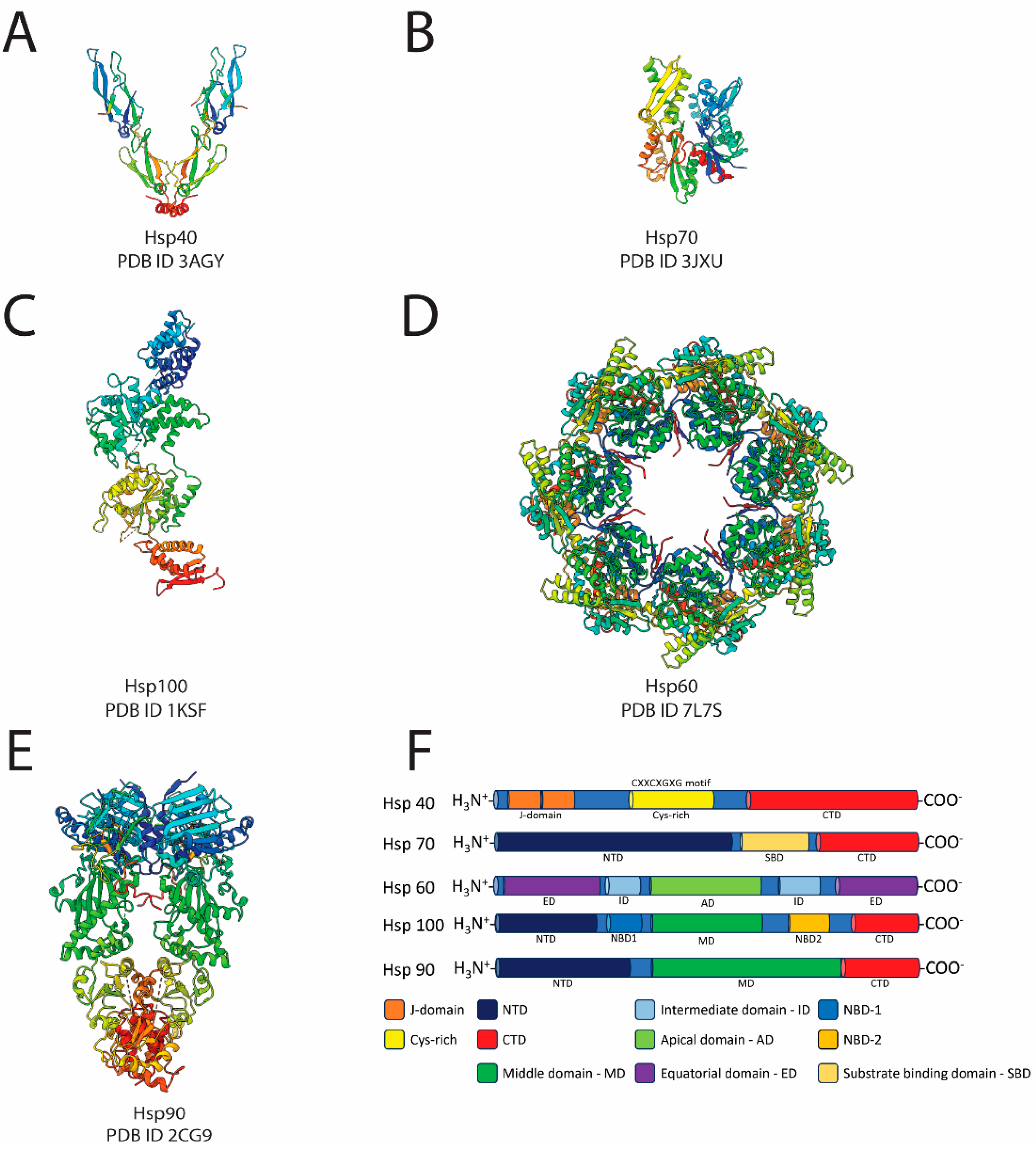
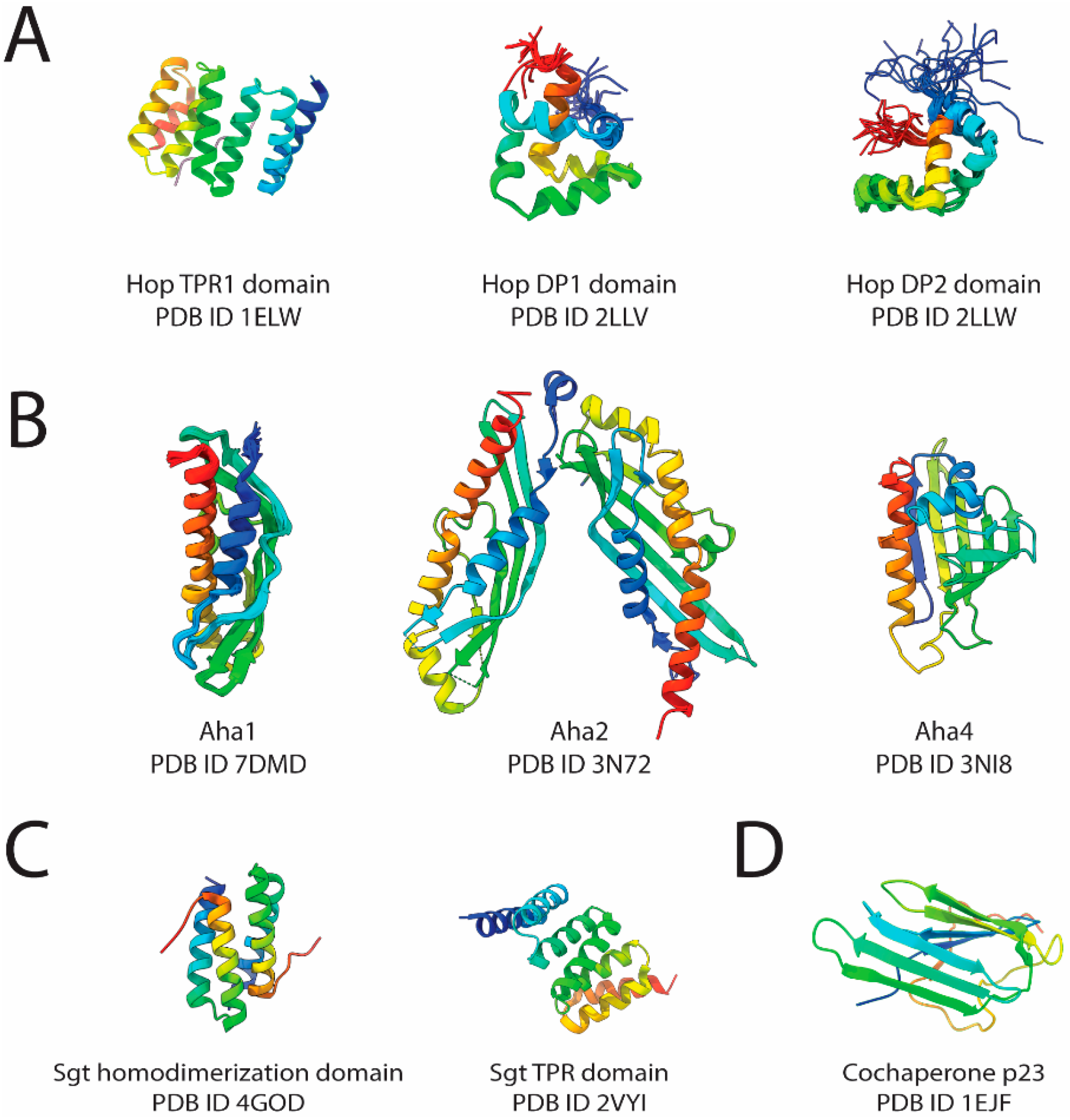
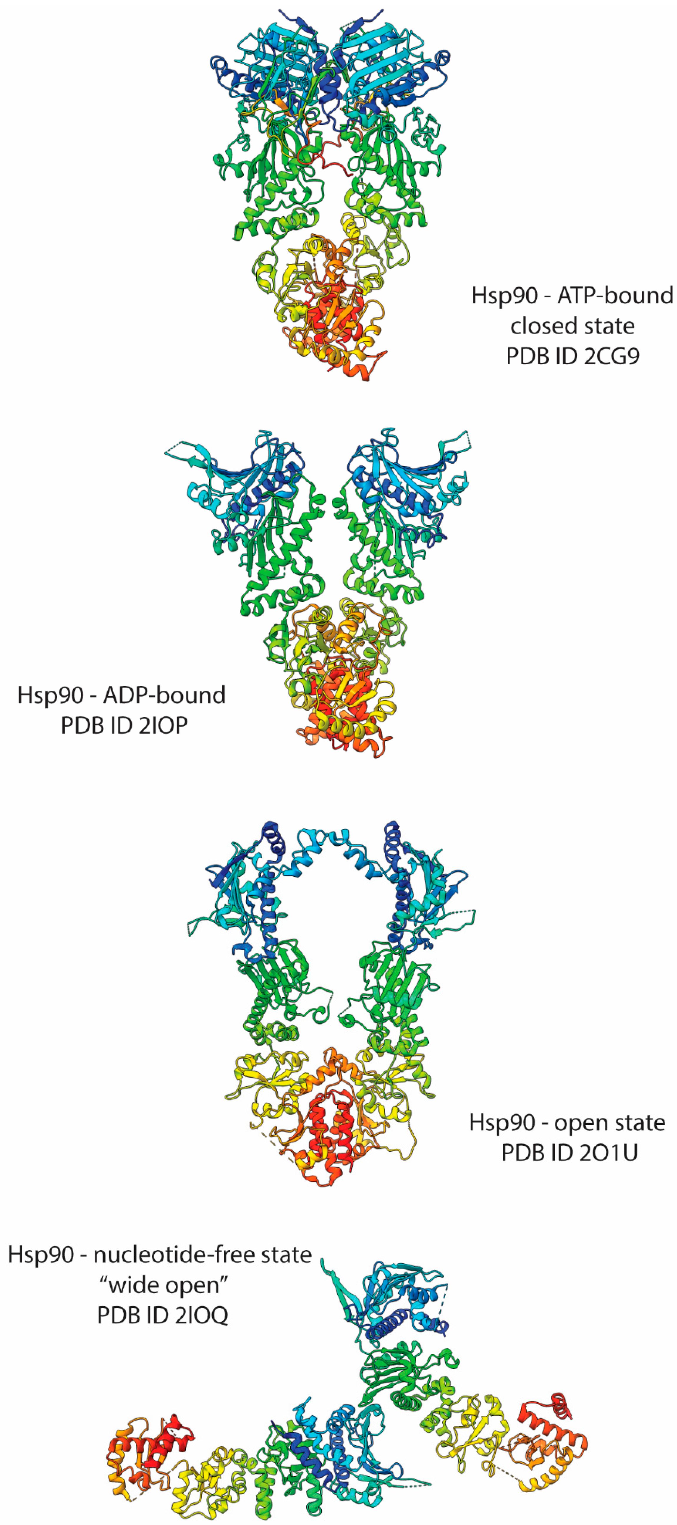
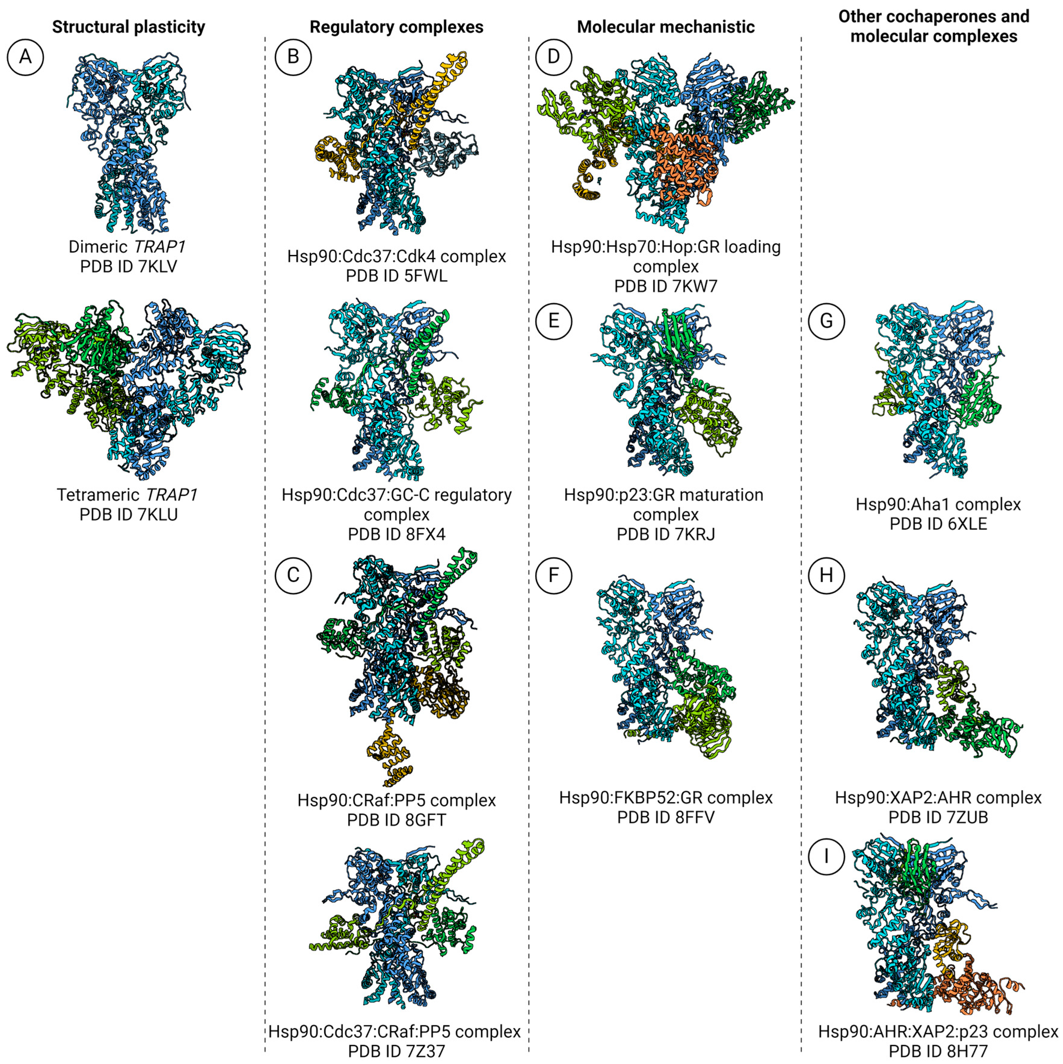
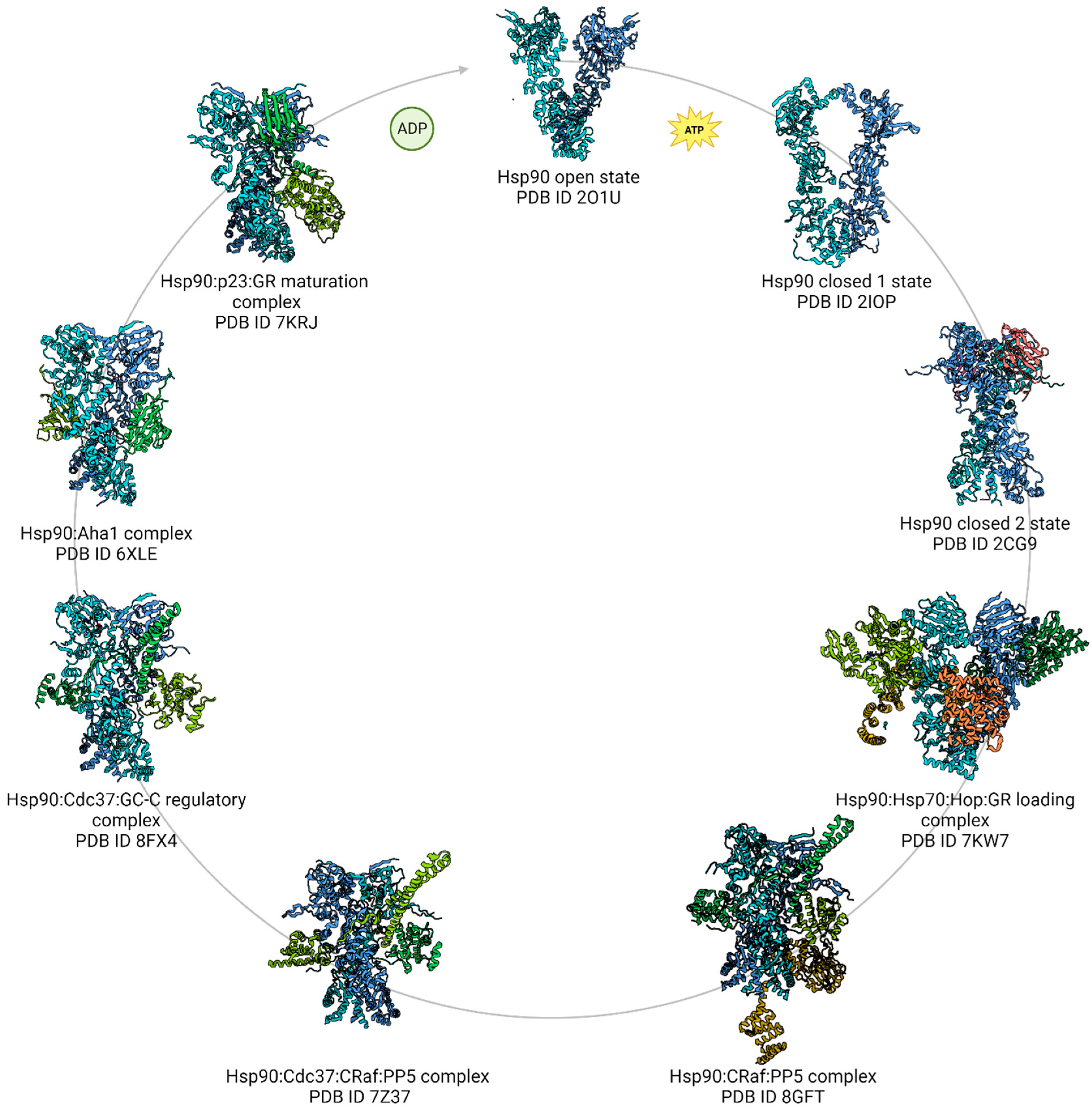
Table 1.
Molecular chaperones and Hsp90 structures summary.
| Class | Nomenclature | PDB ID | Method |
|---|---|---|---|
| Chaperones and cochaperones | Hsp40—J-Domain Proteins | 3AGY | X-ray crystallography |
| Hsp70 | 3JXU | ||
| Hsp100—Clp or AAA+ proteins | 1KSF | ||
| Hsp60—GroEL | 7L7S | ||
| Hop TPR1 domain | 1ELW | ||
| Hop DP1 domain | 2LLV | NMR | |
| Hop DP2 domain | 2LLW | ||
| Aha1 | 7DMD | ||
| Aha2 | 3N72 | X-ray crystallography | |
| Aha4 | 3NI8 | ||
| Sgt—dimerization domain | 4GOD | ||
| Sgt TPR domain | 2VYI | ||
| Cochaperone p23 | 1EJF | ||
| Hsp90 | Hsp90—ATP-bound closed state | 2CG9 | |
| Hsp90—ADP-bound | 2IOP | ||
| Hsp90—open state | 2O1U | ||
| Hsp90—nucleotide-free wide open state | 2IOQ | ||
| TRAP1 | Dimeric TRAP1 | 7KLV | SPA-cryoEM |
| Tetrameric TRAP1 | 7KLU | ||
| Hsp90 complexes | Hsp90:Cdc37:Cdk4 complex | 5FWL | |
| Hsp90:Cdc37:GC-C regulatory complex | 8FX4 | ||
| Hsp90:CRaf:PP5 complex | 8GFT | ||
| Hsp90:Cdc37:CRaf:PP5 complex | 7Z37 | ||
| Hsp90:Hsp70:Hop:GR loading complex | 7KW7 | ||
| Hsp90:p23:GR maturation complex | 7KRJ | ||
| Hsp90:FKBP52:GR complex | 8FFV | ||
| Hsp90:Aha1 complex | 6XLE | ||
| Hsp90:XAP2:AHR complex | 7ZUB | ||
| Hsp90:AHR:XAP2:p23 complex | 8H77 |
Disclaimer/Publisher’s Note: The statements, opinions and data contained in all publications are solely those of the individual author(s) and contributor(s) and not of MDPI and/or the editor(s). MDPI and/or the editor(s) disclaim responsibility for any injury to people or property resulting from any ideas, methods, instructions or products referred to in the content. |
© 2024 by the authors. Licensee MDPI, Basel, Switzerland. This article is an open access article distributed under the terms and conditions of the Creative Commons Attribution (CC BY) license (https://creativecommons.org/licenses/by/4.0/).
Share and Cite
MDPI and ACS Style
Minari, K.; Balasco Serrão, V.H.; Borges, J.C. New Insights into Hsp90 Structural Plasticity Revealed by cryoEM. BioChem 2024, 4, 62-89. https://doi.org/10.3390/biochem4020004
AMA Style
Minari K, Balasco Serrão VH, Borges JC. New Insights into Hsp90 Structural Plasticity Revealed by cryoEM. BioChem. 2024; 4(2):62-89. https://doi.org/10.3390/biochem4020004
Chicago/Turabian StyleMinari, Karine, Vitor Hugo Balasco Serrão, and Júlio César Borges. 2024. "New Insights into Hsp90 Structural Plasticity Revealed by cryoEM" BioChem 4, no. 2: 62-89. https://doi.org/10.3390/biochem4020004