Article

The Influence of Endogenous Derivatives on the Self-Assembly of Carbonized Polymer Dots

1
Department of CAS Key Laboratory of Separation Science for Analytical Chemistry, Dalian Institute of Chemical Physics, Chinese Academy of Sciences, Dalian 116023, China
2
School of Materials Science and Engineering, Guilin University of Technology, Guilin 541000, China
3
School of Biological Engineering, Dalian Polytechnic University, Dalian 116034, China
4
State Key Laboratory of Southwestern Chinese Medicine Resources, School of Pharmacy, Chengdu University of Traditional Chinese Medicine, Chengdu 611137, China
*
Authors to whom correspondence should be addressed.
These authors contributed equally to this work.
Solids 2025, 6(1), 14; https://doi.org/10.3390/solids6010014
Submission received: 17 December 2024 / Revised: 10 March 2025 / Accepted: 17 March 2025 / Published: 20 March 2025

Abstract

:
Carbonized polymer dots (CPDs) have emerged as a fascinating class of functional nanomaterials with unique physicochemical properties. However, the mechanisms governing their formation and photoluminescence remain a subject of intense debate. In this study, we conducted a systematic comparison of the structural, morphological, and optical properties of CPDs synthesized using various methods, revealing the self-assembly characteristics of low-molecular-weight CPDs with relatively complex structures. Through comprehensive structural, morphological, and optical analyses, we found that CPDs with fewer endogenous derivatives exhibited pronounced concentration-dependent self-assembly, leading to larger particle sizes and enhanced fluorescence emission at higher concentrations. In contrast, CPDs with higher proportions of endogenous derivatives showed limited self-assembly due to complex supramolecular interactions between the derivatives and polymer chains. Remarkably, the removal of endogenous derivatives using a ternary solvent extraction method significantly enhanced the self-assembly and fluorescence of the CPDs. These findings highlight the critical role of endogenous derivatives in modulating the self-assembly and photophysical properties of CPDs, paving the way for future advancements in this field.

1. Introduction

Carbonized polymer dots (CPDs) have emerged as a fascinating class of functional nanomaterials over the past two decades, captivating researchers across various fields due to their unique physicochemical properties [1,2,3,4]. However, the mechanisms governing the formation and photoluminescence of CPDs remain a subject of intense debate, leading to the development of numerous theories, including the quantum confinement effect based on graphitic core structures, the entanglement-enhanced emission effect of polymers, and the edge defect effect of graphite-polymer hybrids [5,6,7,8]. While these theories have found validation in specific experimental CPDs systems, they struggle to account for the predominance of low-molecular-weight fragments observed in the liquid chromatography-mass spectrometry (LC-MS) results of most CPDs [9,10,11]. This discrepancy has fueled speculation about alternative formation pathways for CPDs.
Recent literature has highlighted the critical role of supramolecular aggregation in achieving condensed-state luminescence, exemplified by phenomena such as aggregation-induced emission (AIE) and self-assembly-induced emission (SAIE) [12,13,14,15]. During molecular aggregation, the restriction of intramolecular motion (RIM) occurs, which suppresses non-radiative relaxation pathways for excitons through vibrational dissipation [16,17,18]. Consequently, the excited-state lifetime is prolonged, and the probability of radiative relaxation increases, resulting in enhanced fluorescence emission. Notably, RIM-induced luminescence is not limited to high-molecular-weight molecules or polymers; even small molecules can aggregate or self-assemble into nanoparticles exhibiting significant fluorescence [19,20,21]. These theories offer new insights into understanding the formation and photoluminescence mechanisms of low-molecular-weight CPDs.
In our previous work, we investigated the properties of CPDs synthesized using different methods and discovered that solvents play a crucial role in producing low-molecular-weight endogenous derivatives and suppressing polymer chain growth within the CPDs framework [22]. Specifically, CPDs obtained through airflow-assisted melt polymerization (AMP) exhibited longer polymer chains with less endogenous derivatives, and displayed significant concentration-dependent self-assembly characteristics [1,23]. In contrast, CPDs synthesized by conventional autoclave-based methods possessed similar chemical structures but had lower degrees of polymerization with significant endogenous derivatives [24,25]. However, the self-assembly property in these CPDs has not been investigated. Exploring the influence of endogenous derivatives to the polymer frameworks is crucial for elucidating the formation and photoluminescence mechanisms of those CPDs dominating by low-molecular-weight fragments [9,26,27].
In this study, we conducted a systematic comparison of the structural, morphological, and optical properties of CPDs synthesized using various methods, revealing the self-assembly characteristics of low-molecular-weight CPDs with relatively complex structures. By a systematic comparison of the structural, morphological, and optical properties, we found that the presence of endogenous derivatives significantly influences the self-assembly behavior and photoluminescence properties of CPDs. CPDs with fewer endogenous derivatives exhibit more pronounced concentration-dependent self-assembly, resulting in larger particle sizes and enhanced fluorescence emission at higher concentrations. In contrast, CPDs with a higher proportion of endogenous derivatives display limited self-assembly due to the complex supramolecular interactions between the derivatives and their polymer chains. Extraction of endogenous derivatives using a ternary solvent system can enhance the self-assembly and fluorescence emission of CPDs. These findings provide novel insights into understanding the formation and photoluminescence mechanisms of low-molecular-weight CPDs and pave the way for developing innovative functional nanomaterials with tunable self-assembly and optical properties.

2. Materials and Methods

2.1. Chemicals

m-phenylenediamine (m-PD, 99%), tricarballylic acid (TA, 99%), dimethyl sulfoxide (DMSO, 99.5%), methanol (MeOH, 99.5%), ethanol (EtOH, 99.7%), dichloromethane (99.5%), and chloroform (99.5%) were purchased from Shanghai Macklin Biochemical Co., Ltd. (Shanghai, China). Dimethyl sulfoxide-d6 (DMSO-d6, 99.8%) was purchased from Shanghai Meryer Chemical Technology Co., Ltd. (Shanghai, China). All chemicals were used without further purification.

2.2. Synthesis

2.2.1. Solvothermal Synthesis of CPDs (TM-EtOH)

To a 20 mL glass vial, 0.2 mmol TA and 0.2 mmol m-PD were added into 10 mL ethanol and sonicating for 10 min. The mixture was then transferred to a 50 mL Teflon-sealed autoclave (XD-50ML, Beijing Xingde precision Instrument Experimental Instrument Co., Ltd., Beijing, China). The autoclave was heated to 230 °C and kept for 4 h. After reaction, the autoclave was cooled down to room temperature. The product was transferred to 10 mL dialysis kit (0.5–1 kDa molecular weight cut off, Float-A-Lyzer G2, Spectrum) and dialyzed against distilled water in 2 L beaker for 48 h (changing water for every 12 h). The as-obtained product was then separated by centrifugation at 10,000 rpm (11,952× g) for 10 min. The supernatant was further purified by filtering with 0.22 μm filter membrane (Polyesthersulfone) and then collected as TM-EtOH.

2.2.2. Hydrothermal Synthesis of CPDs (TM-H2O)

TM-H2O was prepared using water as the solvent, with synthesis conditions and purification methods similar to those of TM-EtOH.

2.2.3. High Pressure Reactor Synthesis of CPDs (TM-HP)

The m-PD and TA were added to a mortar in equimolar ratios, thoroughly mixed, and homogenized before being transferred to a 50 mL Teflon-sealed autoclave (XD-50ML, Beijing Xingde precision Instrument Experimental Instrument Co., Ltd., Beijing, China). The autoclave was then heated to 230 °C and maintained at this temperature for 4 h. Following the reaction, the autoclave was cooled down to room temperature. The purification process employed was identical to that of TM-EtOH.

2.2.4. Airflow-Assisted Fusion Polymerization Synthesis of CPDs (TM)

Equimolar ratios of m-PD and TA were added to a mortar, mixed thoroughly, and ground evenly before being transferred to a quartz boat. The quartz boat was then placed in a tube furnace (SRJK-2-13, Tianjin Test Instrument Co., Ltd., Tianjin, China). After 20 min of nitrogen purging, the reaction mixture was heated to 230 °C for two hours. Subsequently, the raw product was cooled to room temperature and dispersed into DMSO with the assistance of ultrasound (using a product-to-solvent weight ratio of 1:20) followed by stirring for 10 min. Methanol was subsequently added for precipitation (MeOH:DMSO = 10:1 v/v). The precipitate was filtered and washed three times with water and Methanol consecutively. Finally, TM were obtained as a white powder immediately after drying. The yield was calculated about 75% (w/w).

2.3. Characterization

2.3.1. Ultra-High Performance Liquid Chromatography/Quadrupole Time-of-Flight Mass Spectrometer (UPLC-QTOF-MS)

Molecular characterization was performed by an ultra-performance liquid chromatography quadrupole time-of-flight mass spectrometry (UPLC-QTOF-MS) technique (Q-TOF 6540, Agilent Technologies, Santa Clara, CA, USA). The mass range for all spectra was set to m/z 0–2000. All samples were dissolved in DMSO at a concentration of 10 mg/mL.

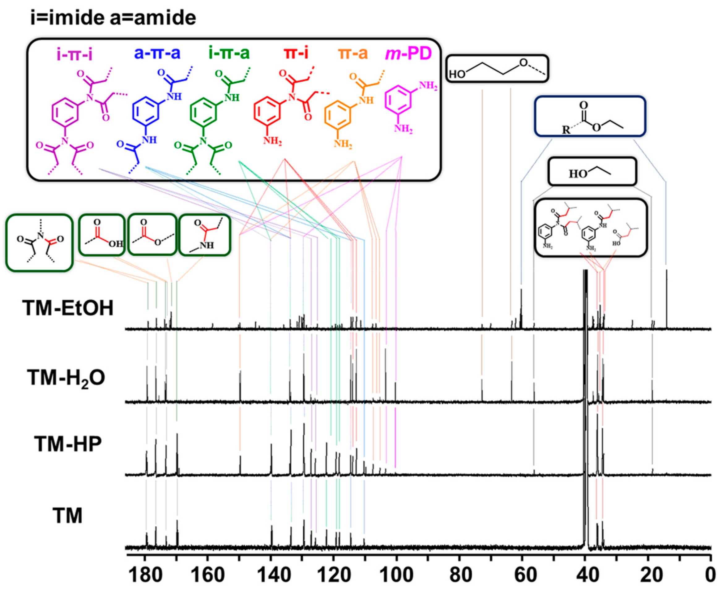
2.3.2. Nuclear Magnetic Resonance (NMR)

NMR-spectra were recorded on a Bruker AVAVCE III HD 700 MHz spectrometer (Bruker, Karlsruhe, Germany). Chemicals shifts (δ) are quoted in ppm downfield of tetramethyl silane. The residual solvent signals were used as references for 1H, 13C and HSQC NMR spectra (DMSO-d6:δH = 2.50 ppm, δC = 39.52 ppm). The samples were prepared at a concentration of 5 mg/mL for NMR experiments, excluding any additional descriptions.

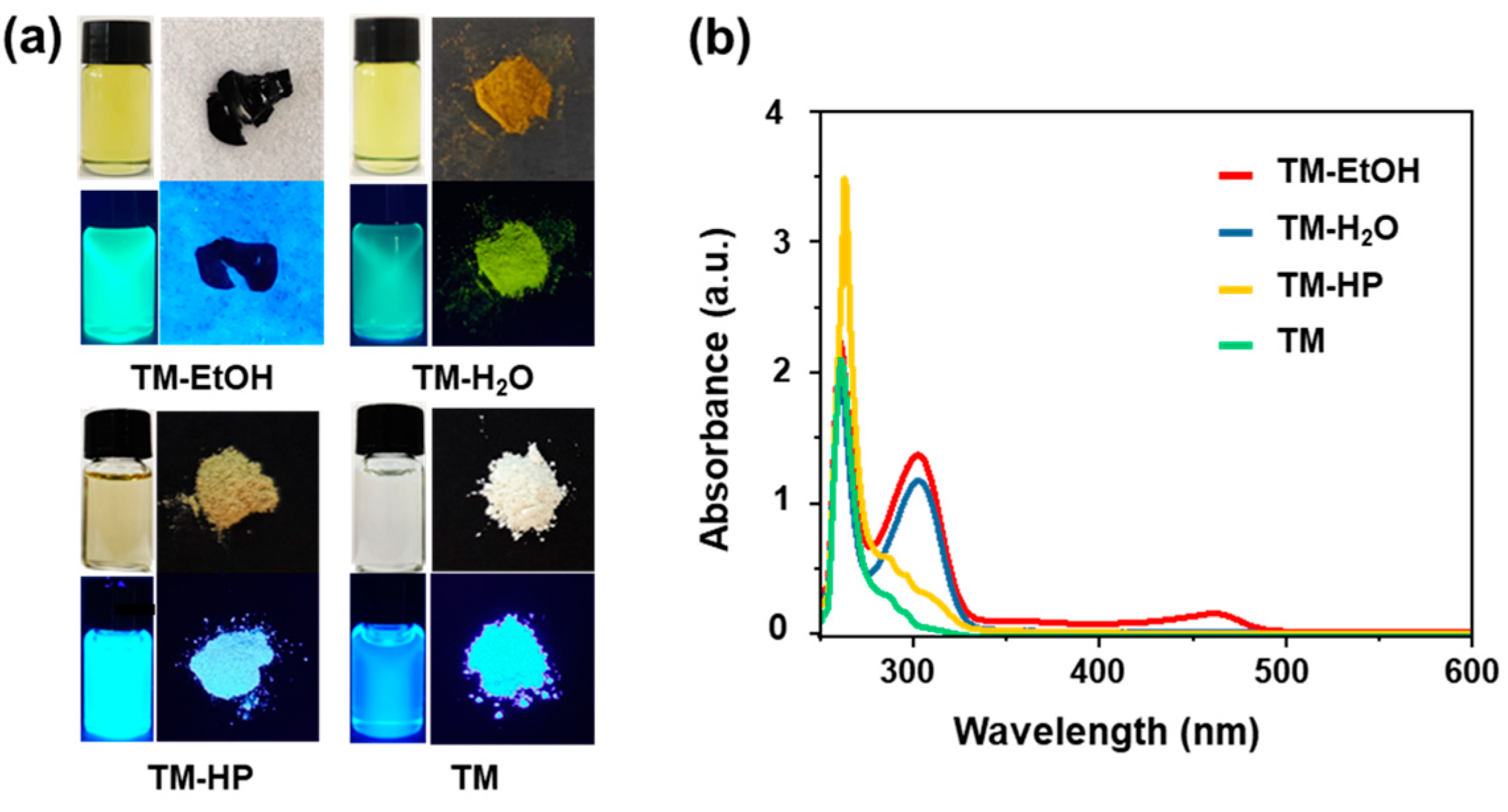
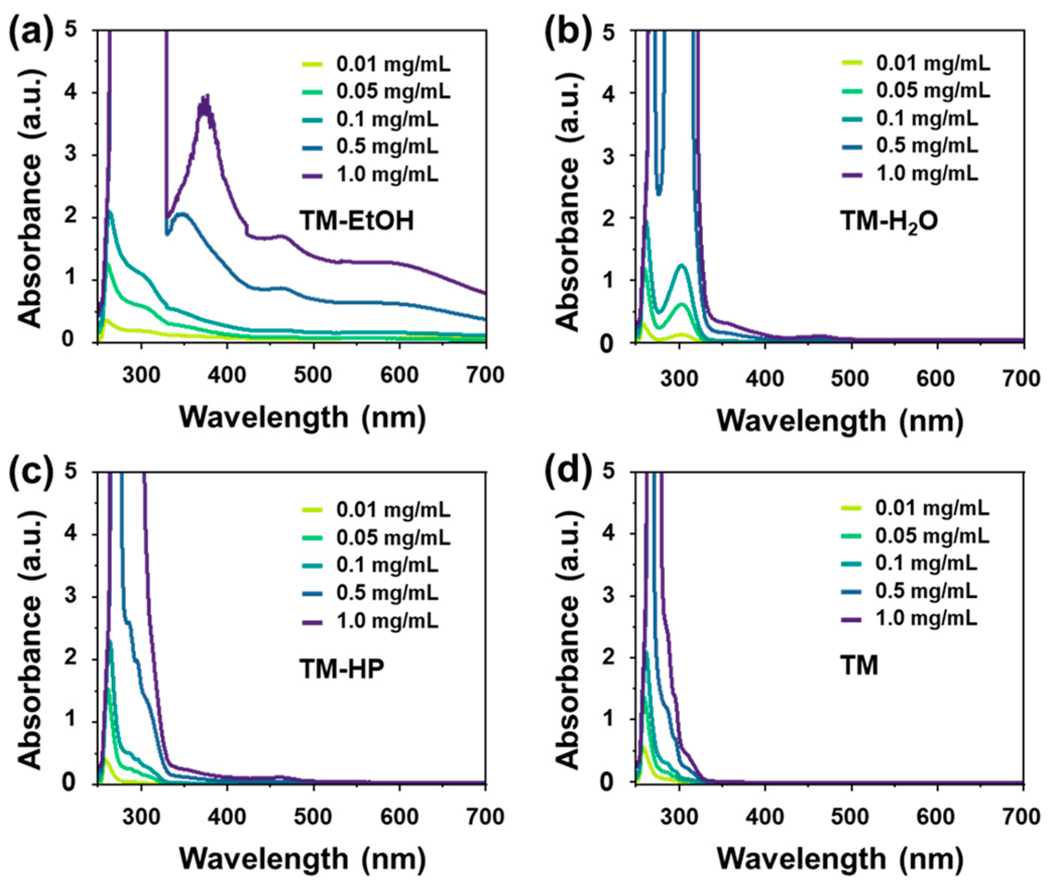
2.3.3. Optical Spectroscopy

UV-Vis spectra were recorded at room temperature on Persee TU-1901 UV-Vis spectrophotometer (Puxi General Instrument Co., Ltd., Beijing, China). Fluorescence spectra were recorded on a F-4600 (Hitachi High-Tech, Tokyo, Japan) fluorescence spectrometer. All the spectra were recorded at room temperature using 10 mm path-length cuvettes.

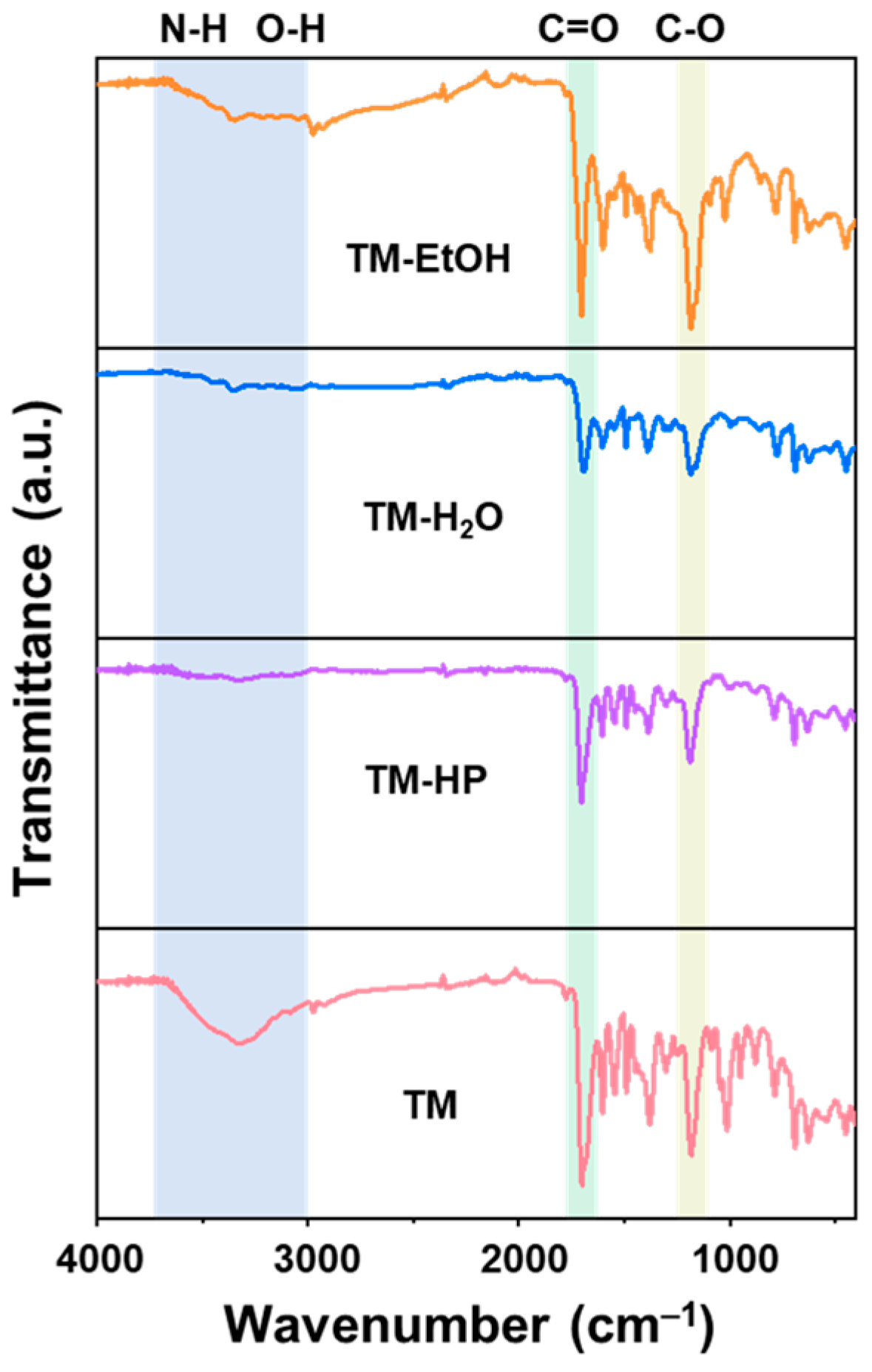
2.3.4. IR Spectroscopy

Fourier-transform infrared spectroscopy was performed by the attenuated total reflection (ATR) method on a Nicolet iS50 (Bruker, Karlsruhe, Germany) equipped with a diamond plate and ZnSe lens.

2.3.5. Transmission Electron Microscopy (TEM)

TEM samples were prepared by dispersing as-synthesized CPDs in DMSO or mixed solution, sonicating for 10 min, and then drop-casting on to carbon coated copper grids. Samples were then imaged on a FEI Tecnai G2 F20 TEM at an acceleration voltage of 200 kV (FEI, Hillsboro, OR, USA). The particles size distribution was calculated and measured by ImageJ8 software.

2.3.6. Photoluminescence Quantum Yield (QY)

The absolute fluorescence quantum yields were measured using a FLS 98 steady-state/transient fluorescence spectrometer (Edinburgh Instruments, Livingston, Scotland, UK) fluorescence system with TCSPC mode and an integrated sphere.

3. Results and Discussion

In our previous work, we compared the morphologies and chemical structures of four types of carbonized polymer dots (CPDs) [1]. All four CPDs are spherical nanoparticles with similar particle sizes and share a characteristic molecular formula of C12H12N2O4, corresponding to the repeating polymer unit R = (TA + m-PD − 2H2O), as shown in Figure S1. However, mass spectrometry (MS) analyses revealed that although these products are composed of the same polymeric units, they display differing degrees of polymerization, functionalization, and amounts of endogenous derivatives. Notably, solvothermal synthesis of CPDs (TM-EtOH) exhibited distinct features of esterification, which we previously attributed to the participation of ethanol in the CPDs formation process [22]. Additionally, the main peaks of TM-EtOH and hydrothermal synthesis of CPDs (TM-H2O) appeared at lower molecular weights compared to those of High pressure reactor synthesis of CPDs (TM-HP) and airflow-assisted fusion polymerization synthesis of CPDs (TM), suggesting that the presence of solvents, including water, can significantly promote side reactions, leading to the generation of abundant endogenous derivatives and inhibiting polymer chain propagation [28].
To elucidate the chemical structures of the four products, we performed 13C NMR analysis (Figure 1). The 13C NMR spectrum revealed that all four CPDs possess polycondensed structures based on TA and m-PD, yet they differ significantly in their substructures. Specifically, TM synthesized by airflow melt polymerization exhibits a relatively homogeneous structure, whereas the presence of solvents or condensation-generated H2O in the other three CPDs leads to the formation of impurities and byproducts, such as unreacted m-PD, TA–m-PD dimers, ethyl ester groups, and oxyethanol groups. Comparative studies of 1H NMR (Figure S2) and HSQC (Figure 2) further indicated that phenyl-derived segments are similarly present in all CPDs, albeit in varying proportions. The integrals of phenyl protons corresponding to the terminal m-PD moieties revealed that TM-HP and TM share similar main structures (structures in Figure 1), with identical ratios (1:0.4:0.4) of trimers including i-π-a (green), i-π-i (purple), and a-π-a (blue). In contrast, TM-EtOH and TM-H2O possess more dimer structures of π-i (red), π-a (orange) and monomer m-PD (carmine), which is consistent with the low degree of polymerization observed in the MS results.
The CPDs synthesized via the four different methods exhibited markedly different colors and absorption properties, suggesting that the degree of polymerization, functionalization, and presence of byproducts significantly influence the optical properties even when the CPDs share the same backbone structure. As shown in Figure 3a, TM-EtOH is a dark brown solid powder without fluorescence, whereas the other three CPDs display distinct solid-state fluorescence emissions in various colors: TM-H2O appears yellow-greenish, TM-HP is cyan, and TM is blue. The quantum yield (QY) of TM in the solid state was measured to be approximately 9.16%, which was much higher than that of TM-EtOH (0.06%), TM-H2O (5.24%), and TM-HP (0.80%) (Figure S3). When dispersed in DMSO solution, TM-EtOH, TM-H2O, and TM-HP appear light yellow under visible light and exhibit fluorescence under UV radiation: TM-EtOH and TM-H2O emit green fluorescence, while TM-HP emits cyan fluorescence. In contrast, the DMSO solution of TM is colorless under visible light but displays bright blue fluorescence under UV light. The UV-Vis absorption spectra recorded at a concentration of 0.1 mg/mL in DMSO reveal that TM and TM-HP have primary absorption bands centered at 250~260 nm, corresponding to n–π* transitions of C=O groups (Figure 3b) [29,30,31]. TM-H2O and TM-EtOH exhibit similar absorption features, with main absorption bands centered around 300 nm, possibly arising from electronic transitions of conjugated structures containing heteroatoms [32]. Notably, TM-EtOH shows a prominent absorption band at 460 nm in the visible region, indicating the possible presence of larger conjugated derivative structures within their framework.
By measuring the absorption spectra at different concentrations (Figure 4), we found that among the four CPDs, only TM showed a significant concentration-dependent increase in absorption within its intrinsic spectral range, while the other three samples developed new absorption features in the visible region. This indicates that the absorption enhancement in TM, with increasing concentration, was exclusively due to its inherent structure, but others would undergo additional photophysical courses such as reabsorption or photoinduced electron transfer, especially in TM-EtOH. At the higher concentration (0.5–1 mg/mL), the strong absorption band ranging from 320 to 700 elevated baseline of TM-EtOH indicates the evident reabsorption and scattering caused by the particle aggregations. On contract, such phenomenon was unobvious even at the concentration of 1 mg/mL, demonstrating the negligible particle aggregation. Correspondingly, all four CPDs exhibited concentration-dependent excitation and emission but in the different concentration ranges (Figure 5a–d). The dual band in the excitation and emission spectra of TM-EtOH, TM-H2O, TM-HP further implied that emissive by-product may be produced and involved in these three CPDs, with different amounts. Notably, the concentrations at which the strongest fluorescence was observed for TM-EtOH, TM-H2O, TM-HP, and TM were 1 mg/mL, 5 mg/mL, 5 mg/mL, and 37.5 mg/mL. Within the lower concentration range, the self-assembly of polymer chains induces an increase in CPDs particle size, which consequently enhances structural rigidity and suppresses nonradiative relaxation pathways. As a result, fluorescence intensity increases linearly with CPDs concentration. However, upon exceeding a critical concentration threshold, aggregation between different particles occurs, and aggregation-caused quenching (ACQ) becomes dominant, leading to a pronounced decrease in fluorescence intensity (Figure 5e–h). In our previous work, we demonstrated that the concentration-enhanced fluorescence emission of TM arises from its self-assembly properties. Therefore, we infer that these CPDs may also possess self-assembly characteristics yet being much limited.
By examining TEM images of the four CPDs at different concentrations corresponding to their peak fluorescence intensities (Figure 6), we observed that the particle sizes of all samples increased with concentration, but to varying extents. Notably, TM exhibited the most significant change, with particle sizes increasing from 3.16 nm at 1 mg/mL to 3.87 nm at 37.5 mg/mL. In contrast, the other three CPDs showed negligible size changes, increasing only from 4.01 nm (TM-HP), 3.88 nm (TM-H2O), 3.80 nm (TM-EtOH) at 1 mg/mL to 4.24 nm, 4.06 nm, 3.97 nm at 37.5 mg/mL, respectively. At a low concentration (1 mg/mL), the four types of CPDs were uniformly dispersed without visible aggregation. Increasing the concentration to 5 mg/mL resulted in self-assembly-induced particle growth, as reflected by an increased average particle size. Upon further concentration increase to 37.5 mg/mL, particle size growth became even more pronounced. Considering that all four CPDs possess identical polymer chains but differ in their substructures, it is plausible that these subtle chemical structural differences within the particles influence the self-assembly process.
Building upon our previous work, we demonstrated that introducing water or chloroform into DMSO dispersions can modulate the strength of hydrogen-bond interactions within the system, thereby guiding the self-assembly and particle aggregation of CPDs [1]. Previous experiments showed that the fluorescence of TM-EtOH, TM-H2O, TM-HP, and TM reaches its peak at concentrations of 1 mg/mL, 5 mg/mL, 5 mg/mL, and 37.5 mg/mL, respectively. To facilitate a better comparison of the differences arising from the various synthesis methods, a concentration of 5 mg/mL was chosen for further testing. Taking TM as an example, the addition of H2O promotes self-assembly, resulting in enlarged particle sizes and enhanced fluorescence with increasing water content. Conversely, when chloroform is introduced, the hydrogen-bond structures between polymer chains are disrupted, leading to particle aggregation and aggregation-induced fluorescence quenching. In the present study, we investigated the changes in the other three CPDs under identical conditions. Steady-state fluorescence spectra showed that adding chloroform to the DMSO solutions causes significant fluorescence quenching for all CPDs. In contrast, the addition of water leads to a pronounced decrease in fluorescence intensity for TM-H2O and TM-HP, whereas TM-EtOH behaves similarly to TM, exhibiting a slight increase in fluorescence followed by a decrease (Figure 7 and Figure S4).
Furthermore, the TM solution in DMSO remains transparent and colorless at 10% water content, becomes slightly turbid at 20%, and forms a distinctly milky suspension at 30%. This observation indicates that increasing water content induces solid precipitation, significantly influencing the self-assembly behavior of TM. Consequently, we selected solvent compositions containing 10% water and 10% CHCl3 for further characterization. TEM images demonstrated that in the presence of chloroform, the particles of the three CPDs exhibited morphological changes and a tendency to particle aggregation (Figure 8 and Figure S5). This finding indicates that disrupting hydrogen-bonding interactions within the system can affect the structure of framework and dispersion state of the CPDs. When water was introduced, the particle sizes of TM-H2O and TM-HP decreased significantly, from 3.92 nm and 4.08 nm, to 3.08 nm and 3.29 nm respectively, while the size of TM-EtOH increased slightly. These morphological differences are consistent with their respective trends in fluorescence changes. Enhancing hydrogen-bonding interactions in the system leads to a reduction in particle sizes for TM-H2O and TM-HP, likely due to H-bond-induced disassembly, resulting in weakened fluorescence. In contrast, the pronounced esterification in TM-EtOH reduces its sensitivity to hydrogen bonding, preventing this disassembly process and maintaining relatively stable fluorescence. Based on these findings, we explicitly distinguished between particle aggregation and polymer chain assembly. Although both phenomena inherently arise from intermolecular interactions, we define the polymer chain assembly as disordered self-assembly rather than aggregation.
Additionally, the ATR-IR spectra of the four CPDs reveal that the vibration peaks associated with C=O, C–O, O–H, and N–H groups are relatively weak in TM-H2O and TM-HP, whereas the corresponding peaks in TM and TM-EtOH are comparatively stronger (Figure 9). This observation suggests that TM-H2O and TM-HP possess stronger intrinsic hydrogen-bonding interactions within their particles, while such interactions are less pronounced in TM and TM-EtOH. Considering the presence of numerous endogenous small-molecule derivatives generated during the reaction processes of TM-H2O and TM-HP, we infer that these small molecules are incorporated into the CPDs frameworks through hydrogen bonding and other supramolecular interactions during their formation. This incorporation gives rise to a more compact framework of the CPDs, in which the motion of groups is much confined. Furthermore, although ATR-FT-IR primarily provides chemical structural information under solid-state conditions for CPDs particles, it can indirectly reflect structural evolution occurring upon solvent evaporation in solution. As solvent evaporates, the increasing polymer chain concentration promotes random self-assembly between polymer chains and aggregation between particles, which is manifested as distinct differences in their macroscopic solid-state structures.
To further validate this hypothesis, we aimed to reduce the amount of endogenous derivatives present in TM-H2O and TM-HP. Our previous studies have demonstrated that extraction using a DMSO/H2O/CHCl3 ternary solvent system can form a triphasic layer, which disrupts hydrogen bonding, hydrophilic/hydrophobic and other supramolecular interactions within the particles, thereby releasing the small molecules that are self-assembled or doped within the CPDs. In this work, we employed the same method to treat TM-H2O and TM-HP. We extracted the products separated in the aqueous phase, designated as TM-H2O-0 and TM-HP-0, and conducted 1H NMR, mass spectrometry, and ATR-IR analyses on them (Figure 10). The results of 1H NMR spectroscopy showed that the chemical shifts of proton peaks associated with the polymer chains remained almost unchanged before and after the treatment, whereas the intensities of signals related to derivatives changed significantly. This indicates that the treatment process can effectively alter the proportion of endogenous derivatives. Mass spectrometry results revealed that the ion fragment peaks representing the TA-m-PD dimer at 249.08 Da and the m-PD-TA-m-PD trimer at 339.15 Da, along with other derivative ion fragment peaks distributed between 200 ~ 400 Da, significantly decreased in intensity. In contrast, the proportion of fragment peaks corresponding to higher degrees of polymerization markedly increased. These findings demonstrate that this method effectively reduces endogenous impurities in the CPDs frameworks. Furthermore, the ATR-IR spectra of these two types of CPDs indicated that, after reducing the proportion of endogenous derivatives, the vibration peaks associated with C=O, C–O, O–H, and N–H groups in TM-H2O-0 and TM-HP-0 were significantly enhanced (Figure 11). The infrared results confirm that endogenous derivatives inhibit hydrogen bonding between CPDs polymer chains.
Based on the above experimental results, we infer that CPDs prepared by different methods possess similar oligomeric frameworks and exhibit concentration-dependent self-assembly to varying degrees [33]. The extent of self-assembly is directly related to the presence of endogenous derivatives within the particles. In the absence of endogenous derivatives (i.e., TM), the DMSO solution acting as a hydrogen-bond acceptor, can inhibit hydrogen-bonding interactions between polymer chains, allowing them to disperse uniformly in solution. During the drying process, concentration-dependent self-assembly occurs, leading to larger particle sizes and enhanced fluorescence emission at higher concentrations. In contrast, CPDs synthesized via traditional methods generate a substantial amount of endogenous derivatives during formation. These derivatives engage in significant and complex supramolecular interactions with the polymer chains as the CPDs form, preventing DMSO from fully dispersing the polymer chains in solution. As a result, these CPDs do not exhibit pronounced concentration-dependent self-assembly after drying. Instead, they show only minor changes in particle size and limited concentration-dependent fluorescence enhancement within a low concentration range.

4. Conclusions

Through systematic investigation of CPDs synthesized by different methods, we found that all CPDs possess similar oligomeric backbone structures and exhibit concentration-dependent self-assembly behavior. The presence of endogenous derivatives significantly inhibits hydrogen-bonding interactions between CPDs polymer chains, thereby affecting their dispersion in solution and self-assembly during drying. CPDs prepared via traditional methods, owing to the presence of endogenous derivatives, show only minor changes in particle size and exhibit concentration-dependent fluorescence enhancement within a low concentration range. In contrast, CPDs devoid of endogenous derivatives display pronounced concentration-dependent self-assembly, resulting in larger particle sizes and stronger fluorescence emission at higher concentrations. This study reveals the crucial role of endogenous derivatives in the self-assembly process of CPDs, providing new perspectives for understanding and regulating the properties of carbonized polymer dots.

Supplementary Materials

The following supporting information can be downloaded at: https://www.mdpi.com/article/10.3390/solids6010014/s1, Figure S1: The UPLC-Q-TOF-MS comparison of the four CPDs, Figure S2: The comparisons of the 1H NMR spectra indicate that the main structures of the four CPDs are similar. Figure S3: The fluorescence quantum yields of four CPDs solid powder samples. Figure S4: The variation in TM-EtOH, TM-H2O, TM-HP and TM emission intensity with a concentration of 5 mg/mL upon the addition of different percentages of H2O and CHCl3 in DMSO solutions. Figure S5: TEM images revealed the aggregation processes of the three CPDs.

Author Contributions

Conceptualization, Y.Q. and Y.W. (Yu Wang); methodology, W.Z., Z.L., J.C., Y.L. and L.F.; formal analysis, Y.W. (Yue Wang); investigation, Y.Q., W.Z., Z.L., J.C. and Y.W. (Yue Wang); writing—original draft preparation, Y.Q. and Y.W. (Yu Wang); writing—review and editing, M.J., Y.L., A.Q. and L.F.; supervision, A.Q. and L.F. All authors have read and agreed to the published version of the manuscript.

Funding

Supported by the Youth Innovation Promotion Association of the Chinese Academy of Sciences (No. 2020185), the AI S&T Program of Yulin Branch, Dalian National Laboratory for Clean Energy, CAS (No. DNL-YL A202203), DICP Founding (DICP I202403, DICP I202320, DICP I202331).

Data Availability Statement

The original data are available upon request from the corresponding author via email.

Conflicts of Interest

The authors declare no conflict of interest.

References

  1. Wang, Y.; Qin, Y.; Wang, F.; Zhang, H.; Huangfu, C.; Shi, Y.; Chen, X.; Wang, Z.; Tian, W.; Feng, L. The Synthesis of Functionalized Carbonized Polymer Dots via Reversible Assembly of Oligomers for Anti-Counterfeiting, Catalysis, and Gas storage. Adv. Sci. 2024, 11, 2405043. [Google Scholar]
  2. Ru, Y.; Sui, L.; Song, H.; Liu, X.; Tang, Z.; Zang, S.Q.; Yang, B.; Lu, S. Rational Design of Multicolor-Emitting Chiral Carbonized Polymer Dots for Full-Color and White Circularly Polarized Luminescence. Angew. Chem. Int. Ed. 2021, 60, 14091–14099. [Google Scholar]
  3. Shi, M.; Gao, Q.; Rao, J.; Lv, Z.; Chen, M.; Chen, G.; Bian, J.; Ren, J.; Lü, B.; Peng, F. Confinement-Modulated Clusterization-Triggered Time-Dependent Phosphorescence Color from Xylan-Carbonized Polymer Dots. J. Am. Chem. Soc. 2023, 146, 1294–1304. [Google Scholar] [PubMed]
  4. Tao, S.; Zhou, C.; Kang, C.; Zhu, S.; Feng, T.; Zhang, S.; Ding, Z.; Zheng, C.; Xia, C.; Yang, B. Confined-domain crosslink-enhanced emission effect in carbonized polymer dots. Light Sci. Appl. 2022, 11, 56. [Google Scholar]
  5. Vallan, L.; Urriolabeitia, E.P.; Ruipérez, F.; Matxain, J.M.; Canton-Vitoria, R.; Tagmatarchis, N.; Benito, A.M.; Maser, W.K. Supramolecular-Enhanced Charge Transfer within Entangled Polyamide Chains as the Origin of the Universal Blue Fluorescence of Polymer Carbon Dots. J. Am. Chem. Soc. 2018, 140, 12862–12869. [Google Scholar]
  6. Xia, C.; Zhong, J.; Han, X.; Zhu, S.; Li, Y.; Liu, H.; Yang, B. The Formation Mechanism of Carbonized Polymer Dots: Crosslinking-Induced Nucleation and Carbonization. Angew. Chem. Int. Ed. 2024, 63, e202410519. [Google Scholar]
  7. Wang, B.; Waterhouse, G.I.N.; Yang, B.; Lu, S. Advances in Shell and Core Engineering of Carbonized Polymer Dots for Enhanced Applications. Acc. Chem. Res. 2024, 57, 2928–2939. [Google Scholar]
  8. Ru, Y.; Ai, L.; Jia, T.; Liu, X.; Lu, S.; Tang, Z.; Yang, B. Recent advances in chiral carbonized polymer dots: From synthesis and properties to applications. Nano Today 2020, 34, 100953. [Google Scholar]
  9. Guo, G.; Xia, Y. General Separation of Carbon Dots by Polyamide Chromatography. Anal. Chem. 2024, 96, 5095–5105. [Google Scholar]
  10. Wang, C.; He, Y.; Huang, J.; Sui, L.; Ran, G.; Zhu, H.; Song, Q. Intramolecular hydrogen bond-tuned thermal-responsive carbon dots and their application to abnormal body temperature imaging. J. Colloid Interface Sci. 2023, 634, 221–230. [Google Scholar]
  11. Fu, R.; Song, H.; Liu, X.; Zhang, Y.; Xiao, G.; Zou, B.; Waterhouse, G.I.N.; Lu, S. Disulfide Crosslinking-Induced Aggregation: Towards Solid-State Fluorescent Carbon Dots with Vastly Different Emission Colors. Chin. J. Chem. 2023, 41, 1007–1014. [Google Scholar] [CrossRef]
  12. Xie, H.; Wang, J.; Lou, Z.; Hu, L.; Segawa, S.; Kang, X.; Wu, W.; Luo, Z.; Kwok, R.T.K.; Lam, J.W.Y.; et al. Mechanochemical Fabrication of Full-Color Luminescent Materials from Aggregation-Induced Emission Prefluorophores for Information Storage and Encryption. J. Am. Chem. Soc. 2024, 146, 18350–18359. [Google Scholar] [CrossRef] [PubMed]
  13. Kang, C.; Tao, S.; Yang, F.; Yang, B. Aggregation and luminescence in carbonized polymer dots. Aggregate 2022, 3, 269. [Google Scholar] [CrossRef]
  14. Zheng, H.; Zhang, Z.; Cai, S.; An, Z.; Huang, W. Enhancing Purely Organic Room Temperature Phosphorescence via Supramolecular Self-Assembly. Adv. Mater. 2024, 36, 2311922. [Google Scholar] [CrossRef]
  15. Yang, J.; Fang, M.; Li, Z. Organic luminescent materials: The concentration on aggregates from aggregation-induced emission. Aggregate 2020, 1, 6–18. [Google Scholar] [CrossRef]
  16. Yang, H.; Liu, Y.; Guo, Z.; Lei, B.; Zhuang, J.; Zhang, X.; Liu, Z.; Hu, C. Hydrophobic carbon dots with blue dispersed emission and red aggregation-induced emission. Nat. Commun. 2019, 10, 1789. [Google Scholar] [CrossRef]
  17. He, Z.; Shan, L.; Mei, J.; Wang, H.; Lam, J.W.Y.; Sung, H.H.Y.; Williams, I.D.; Gu, X.; Miao, Q.; Tang, B.Z. Aggregation-induced emission and aggregation-promoted photochromism of bis(diphenylmethylene)dihydroacenes. Chem. Sci. 2015, 6, 3538–3543. [Google Scholar] [CrossRef]
  18. Yang, Q.; Zhu, J.; Li, Z.; Chen, X.; Jiang, Y.; Luo, Z.; Wang, P.; Xie, H. Luminescent Liquid Crystals Based on Carbonized Polymer Dots and Their Polarized Luminescence Application. ACS Appl. Mater. Interfaces 2021, 13, 26522–26532. [Google Scholar] [CrossRef]
  19. Xu, X.; Mo, L.; Li, Y.; Pan, X.; Hu, G.; Lei, B.; Zhang, X.; Zheng, M.; Zhuang, J.; Liu, Y.; et al. Construction of Carbon Dots with Color-Tunable Aggregation-Induced Emission by Nitrogen-Induced Intramolecular Charge Transfer. Adv. Mater. 2021, 33, 2104872. [Google Scholar] [CrossRef]
  20. Wu, S.; Shi, H.; Lu, W.; Wei, S.; Shang, H.; Liu, H.; Si, M.; Le, X.; Yin, G.; Theato, P.; et al. Aggregation-Induced Emissive Carbon Dots Gels for Octopus-Inspired Shape/Color Synergistically Adjustable Actuators. Angew. Chem. Int. Ed. 2021, 60, 21890–21898. [Google Scholar] [CrossRef]
  21. Ji, C.; Zeng, F.; Xu, W.; Zhu, M.; Yu, H.; Yang, H.; Peng, Z. Hydrogen Bond-Mediated Self-Assembly of Carbon Dots Enabling Precise Tuning of Particle and Cluster Luminescence for Advanced Optoelectronic Applications. Adv. Mater. 2024, 37, 2414450. [Google Scholar]
  22. Wang, Y.; Li, Y.; Feng, L. Exploring Solvent-Related Reactions and Corresponding Band Gap Tuning Strategies for Carbon Nanodots Based on Solvothermal Synthesis. J. Phys. Chem. Lett. 2020, 11, 10439–10445. [Google Scholar] [PubMed]
  23. Wang, Y.; Qin, Y.; Tian, W.; Zhang, H.; Wang, F.; Yan, X.; Rong, S.; Huangfu, C.; Shi, Y.; Wang, Z.; et al. Dye-Incorporated Carbonized Polymer Dots with Tunable Solid-State Emission Based on Intraparticle Förster Resonance Energy Transfer. Adv. Funct. Mater. 2024, 34, 242825. [Google Scholar] [CrossRef]
  24. Xue, S.; Li, P.; Sun, L.; An, L.; Qu, D.; Wang, X.; Sun, Z. The Formation Process and Mechanism of Carbon Dots Prepared from Aromatic Compounds as Precursors: A Review. Small 2023, 19, 2206180. [Google Scholar]
  25. Liu, H.; Zhong, X.; Pan, Q.; Zhang, Y.; Deng, W.; Zou, G.; Hou, H.; Ji, X. A review of carbon dots in synthesis strategy. Coord. Chem. Rev. 2024, 498, 215468. [Google Scholar] [CrossRef]
  26. Zheng, G.; Shen, C.; Deng, Y.; Liu, K.; Zang, J.; Dong, L.; Lou, Q.; Shan, C. Rational design multi-color-emissive chemiluminescent carbon nanodots in a single solvothermal reaction. Nano Res. 2024, 17, 4651–4660. [Google Scholar]
  27. Li, P.; Xue, S.; Sun, L.; Zong, X.; An, L.; Qu, D.; Wang, X.; Sun, Z. Formation and fluorescent mechanism of red emissive carbon dots from o-phenylenediamine and catechol system. Light Sci. Appl. 2022, 11, 298. [Google Scholar] [CrossRef]
  28. Qin, Y.; Chi, J.; Zhang, W.; Pan, X.; Liu, Z.; Chen, X.; Shi, Y.; Yang, L.; Zhang, H.; Qin, A.; et al. Influence of Endogenous Derivatives on the Chemical Sensing Performance of Carbonized Polymer Dots. J. Phys. Chem. Lett. 2025, 16, 2151–2159. [Google Scholar] [CrossRef]
  29. Sun, J.; Liu, Y.; Han, Y.; Li, W.; Wang, N.; Zhang, L.; Zhang, Y.; Deng, F.; Wang, D.; Zhang, X. Enabling controllable time-dependent phosphorescence in carbonized polymer dots based on chromophore excited triplet energy level modulation by ionic bonding. Angew. Chem. Int. Ed. 2024, 64, 202415042. [Google Scholar]
  30. Xia, C.; Zhu, S.; Feng, T.; Yang, M.; Yang, B. Evolution and Synthesis of Carbon Dots: From Carbon Dots to Carbonized Polymer Dots. Adv. Sci. 2019, 6, 19013616. [Google Scholar] [CrossRef]
  31. Tao, S.; Zhu, S.; Feng, T.; Xia, C.; Song, Y.; Yang, B. The polymeric characteristics and photoluminescence mechanism in polymer carbon dots: A review. Mater. Today Chem. 2017, 6, 13–25. [Google Scholar]
  32. Li, P.; Xue, S.; Sun, L.; Ma, X.; Liu, W.; An, L.; Liu, Y.; Qu, D.; Sun, Z. Formation and Fluorescent Mechanism of Multiple Color Emissive Carbon Dots from o-Phenylenediamine. Small 2024, 20, 2310563. [Google Scholar]
  33. Ai, L.; Wang, H.; Wang, B.; Liu, S.; Song, H.; Lu, S. Concentration-Switchable Assembly of Carbon Dots for Circularly Polarized Luminescent Amplification in Chiral Logic Gates and Deep-Red Light-Emitting Diodes. Adv. Mater. 2024, 36, 210094. [Google Scholar]
Figure 1. The comparison of 13C NMR spectra of four CPDs at 5 mg/mL. The brackets in the figure denote various types of impurities or by-products that could potentially be present in the product.
Solids 06 00014 g001
Figure 2. The HSQC NMR spectra of (a) TM-EtOH, (b) TM-H2O, (c) TM-HP, and (d) TM. Different circle colors represent distinct derivatives, including end-group i-π-a, i-π-i, and a-π-a, as well as potential intermediates π-i, π-a, and monomer m-PD.
Solids 06 00014 g002
Figure 3. (a) The photographs of 5 mg/mL DMSO solutions and solid powder of four CPDs under visible light and 365 nm UV light. (b) The absorption spectra of TM-EtOH (red curve), TM-H2O (blue curve), TM-HP (yellow curve) and TM (greed curve) in DMSO solutions with a concentration of 0.1 mg/mL.
Solids 06 00014 g003
Figure 4. The absorption spectra of (a) TM-EtOH, (b) TM-H2O, (c) TM-HP and (d) TM in DMSO within the concentration range from 0.01 to 1.0 mg/mL.
Solids 06 00014 g004
Figure 5. The comparison of emission spectra of (a) TM-EtOH, (b) TM-H2O, (c) TM-HP and (d) TM within the concentration range from 0.01 to 100 mg/mL. The comparison of excitation spectra of (e) TM-EtOH, (f) TM-H2O, (g) TM-HP and (h) TM within the concentration range from 0.01 to 100 mg/mL. The illustration depicts the correlation between fluorescence intensity and the concentrations of four CPDs ranging from 0.01 to 100 mg/mL. The gradient lines transitioning from green to red in the illustration represent varying concentrations ranging from 0.01 to 100 mg/mL.
Solids 06 00014 g005
Figure 6. TEM images of (a) TM-EtOH, (b) TM-H2O, (c) TM-HP and (d) TM dispersed at different concentrations under the same magnification. (e) The diagram of TM-EtOH, TM-H2O, TM-HP and TM average particle size versus dispersing concentration. (f) Comparison of normalized fluorescence emission intensities for TM-EtOH, TM-H2O, TM-HP, and TM at various concentrations.
Solids 06 00014 g006
Figure 7. The variation in (a) TM-EtOH, (b) TM-H2O, (c) TM-HP and (d) TM emission intensity with a concentration of 5 mg/mL upon the addition of different percentages of H2O (red curve) and CHCl3 (black curve) in DMSO solutions. I0 represents the initial fluorescence emission intensity of the four CPDs, while Ii denotes the fluorescence emission intensity following the addition of varying proportions of H2O or CHCl3.
Solids 06 00014 g007
Figure 8. TEM images and the corresponding particle distribution of (a) TM-EtOH, (b) TM-H2O and (c) TM-HP in the presence of 10% H2O and 10% CHCl3 in DMSO solution. The illustration depicts the distribution of particle sizes, and the red lines indicate the average particle size and distribution characteristics of the particles, while the white circle highlights the trend of morphological changes and aggregation of CPDs particles.
Solids 06 00014 g008
Figure 9. The comparison of ATR-IR spectra of TM-EtOH (orange curve), TM-H2O (blue curve), TM-HP (purple curve) and TM (pink curve). Different background colors highlight distinct functional group contrasts.
Solids 06 00014 g009
Figure 10. (a) The comparison of 1H NMR spectra of TM-H2O-0 and TM-H2O. (b) The comparison of 1H NMR spectra of TM-HP-0 and TM-HP. (c) The comparison of UPLC-QTOF-MS spectra of TM-H2O-0 and TM-H2O. (d) The comparison of UPLC-QTOF-MS spectra of TM-HP-0 and TM-HP. TM-H2O-0 and TM-HP-0 represent the upper half of the curve in the figure, while TM-H2O and TM-HP correspond to the lower half of the curve.
Solids 06 00014 g010
Figure 11. (a) The comparison of ATR-IR spectra of TM-H2O-0 (red curve) and TM-H2O (black curve). (b) The comparison of ATR-IR spectra of TM-HP-0 (red curve) and TM-HP (black curve). Different background colors highlight distinct functional group contrasts.
Solids 06 00014 g011
Disclaimer/Publisher’s Note: The statements, opinions and data contained in all publications are solely those of the individual author(s) and contributor(s) and not of MDPI and/or the editor(s). MDPI and/or the editor(s) disclaim responsibility for any injury to people or property resulting from any ideas, methods, instructions or products referred to in the content.

