Article

Neosporosis in Naturally Infected Sheep Herds, a Prospective Cohort Study over Three Years

1
Division of Parasitology, Kimron Veterinary Institute, Beit Dagan 50200, Israel
2
Poultry Health Division, Israel Veterinary Services, Ministry of Agriculture and Rural Development, Beit Dagan 50200, Israel
*
Author to whom correspondence should be addressed.
Parasitologia 2024, 4(2), 209-221; https://doi.org/10.3390/parasitologia4020018
Submission received: 19 April 2024 / Revised: 3 June 2024 / Accepted: 5 June 2024 / Published: 12 June 2024

Abstract

:
Background: Neospora caninum is a protozoan parasite and a main cause of abortions in cattle worldwide. However, its role in abortions and decreased fertility in sheep is not completely understood, especially due to the complex, multifactorial etiology of abortions. This study aimed to perform a longitudinal field study to investigate the epidemiology of neosporosis and its effect on fertility in endemic sheep herds. Methods: Serological (IFAT) and clinical (outcome of pregnancy) data from 153 ewe-lambs was collected in four intensive management farms in Israel during three consecutive pregnancies. Results: The seroprevalence in ewe-lambs at different farms varied between 24% and 93%. The overall seroprevalence increased from 50% in ewe-lambs to 96.6% at the end of the third pregnancy. Horizontal infection was observed in all farms, with seroconversion in 59% of seronegative sheep. Abortion rates were lower (p = 0.004) in seropositive ewes in the first pregnancy and not significantly higher in seropositive sheep in consecutive pregnancies. Seropositivity or seroconversion were not associated with abortions or repeated abortions; however, many aborting ewes were removed from the flock. Conclusions: No direct short- or long-term association was found between Neopsora infection and abortions. The variations between flocks and pregnancies suggest a more complex etiology.

1. Introduction

Neospora caninum is a protozoan cyst-forming parasite affecting a wide range of animals. This parasite has a heteroxenous life cycle, with canids serving as its definitive host and other mammals as intermediate hosts [1]. Neosporosis is considered a major cause of abortion in cattle worldwide, with extensive economic impact [2,3].
In sheep, the involvement of N. caninum in abortion is not entirely clear. Neosporosis used to be considered an incidental finding in sheep [2,4]. However, it had been shown that experimental infection may lead to abortion in sheep [5] and that the gestational stage in which infection takes place has a crucial influence on the clinical outcome of pregnancy [6]. In recent years, there have been increasing reports linking Neospora seropositivity with reduced fertility, increased abortion rates, and vertical transmission in naturally infected herds [7,8,9,10,11,12,13,14]. However, these findings are inconsistent between reports, and two recent meta-analyses failed to establish a significant link between neosporosis and abortions in sheep [15,16].
In cattle, the main route of transmission of neosporosis in endemic herds is vertical transmission from infected dams to their fetuses. Due to immunomodulation in the dam during pregnancy, the dormant parasites re-emerge, leading to transient parasitemia resulting in infection of the placenta and fetus. Cases that do not result in abortion result in the delivery of infected calves, which will be persistent carriers and increase the prevalence of neosporosis in the herd, thus perpetuating the problem [17,18,19,20]. Similarly to cattle, it had been demonstrated that vertical transmission of Neospora in sheep is efficient; however, in is still unclear whether vertical or horizontal transmission is the main route of transmission in sheep [17,20,21].
Neosporosis is endemic in Israel, and it is considered a main cause of abortions in cattle [18]. A recent study from Israel reported Neospora seropositivity in 67.4% of samples from sheep submitted to diagnosis over ten years; however, seropositivity rates did not differ between aborting and non-aborting sheep [12]. On the other hand, anti-Neospora antibodies were found in 22.9% of aborted fetuses and was the most frequently diagnosed pathogen [12]. In Israel, most abortions in sheep are diagnosed when a pregnant ewe does not lamb at the end of the expected pregnancy period. In many flocks, abortions are attributed to neosporosis when several aborting ewes are seropositive. However, in several such cases, seropositivity was also prevalent in non-aborting ewes, and a more in-depth investigation is advised [22].
This study was designed as a long-term surveillance study aimed to estimate the prevalence of neosporosis in ewe-lambs and the rate of horizontal transmission during the first years of their lives and to evaluate the impact of neosporosis in infected herds on fertility, reproduction, and early culling (or removal from the flock) in three consecutive pregnancies.

2. Results

2.1. Study Population

Data collected at four sheep farms were included in this study. Between 29 and 46 ewe-lambs were sampled during the first year of the study, with a total of 153 sheep from all four farms. Due to incomplete resampling and data availability as well as the removal of some sheep from the farms, data were available for 134, 65, and 29 sheep at the end of the first, second, and third pregnancy, respectively.
All farms had intensive management. Sheep were kept indoors, with no access to pasture. Breeding management included artificial insemination with additional introduction of rams and pregnancy examinations at 40–50 days of pregnancy. Two farms were located in central Israel, one in the north and one in the south. The farms kept flocks of mixed-breed sheep, except for farm 1, which kept the Asaf breed only. The size of the flocks ranged between 220 and 1200 sheep. All sheep were routinely vaccinated against brucellosis, foot and mouth disease, peste des petits ruminants, and sheep pox, and they received a combined vaccine against clostridium perfringens type D and clostridium tetani. Three farms routinely vaccinate also against Chlamydophila and two farms against Q fever. All but one farm (farm 3) reported a history of neosporosis in the flock.
At all farms, there was possible exposure to dogs, and on one farm (farm 2) the sheep had close contact with live-in dogs. The dogs from farm 2 were also serologically tested for neosporosis. Blood was collected from 22 dogs, all of the Asian Shepherd breed, of which 6 (27.3%) tested positive, with antibody titers of 1:50 to 1:800.

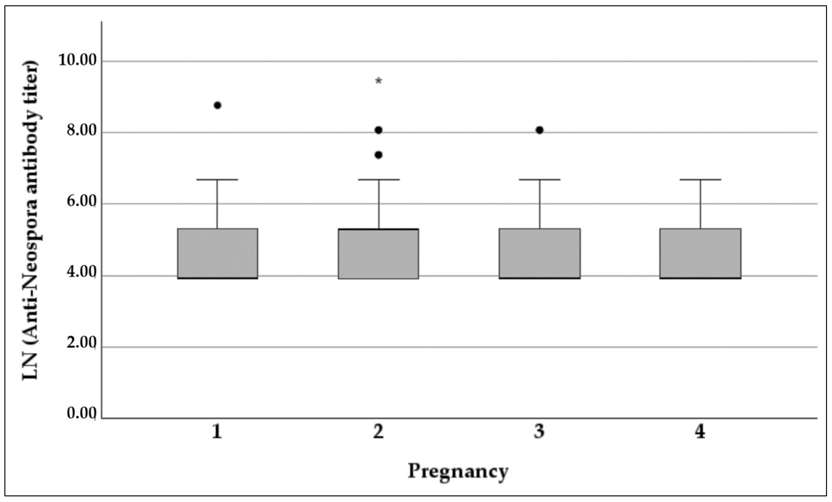
2.2. Neospora Seroprevalence

The overall seroprevalence of Neospora, using a cut-off value of 1:50, was 49.7% in ewe-lambs and gradually increased to 96.6% by the end of their third pregnancy (Spearman’s rho = 0.335, p < 0.001). When using a cut-off value of 1:200, the annual seroprevalence ranged between 23.5% and 44.8% and did not significantly correlate with age/number of pregnancy (ρ = 0.077, p = 0.061) (Table 1).
Neospora seroprevalence (1:50) in ewe-lambs ranged between 24.1% and 35.7% on three farms (farms 1, 2, and 4), while it was 93.5% on farm 3 (p < 0.001). This difference remained significant at the end of the first pregnancy, when the seroprevalence on farms 1, 2, and 4 was 50% to 69.2%, while it was 100% in farm 3 (p < 0.001). By the end of the second pregnancy the seroprevalence on farms 2 and 4 reached approximately 91%, while it remained lower (61.3%) in farm 1 (p = 0.019). Similar differences between farms were found when analyzing the seroprevalence using the higher (1:200) cut-off value (Table 1).
Antibody titers of seropositive sheep ranged between 1:50 and 1:12,800, with a median titer of 1:50 (inter-quartile range (IQR) = 150). The distribution of antibody titers of seropositive sheep did not differ between sampling dates (Kruskal–Wallis p = 0.187) (Figure 1).

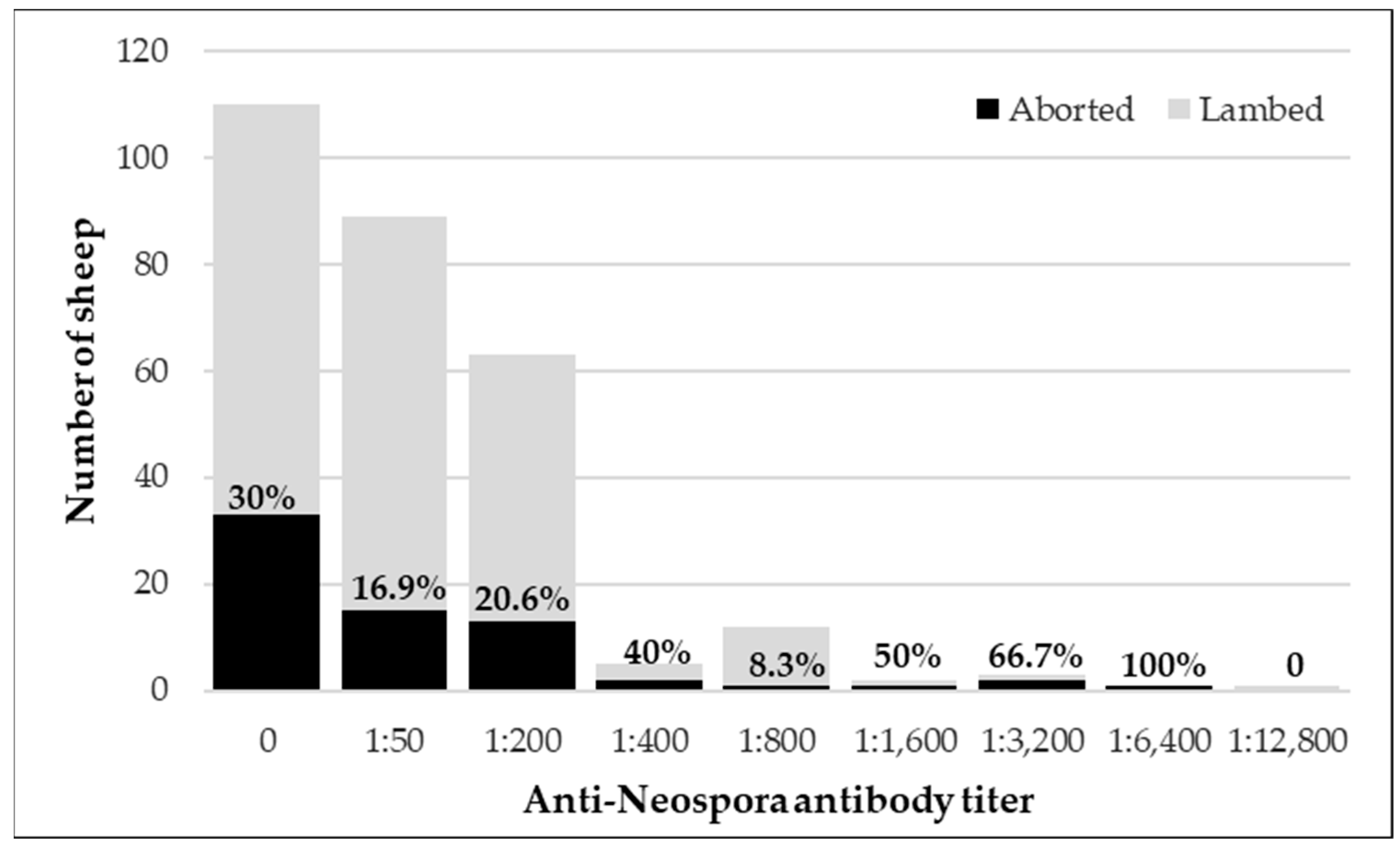
2.3. Neospora and Abortions

The outcome of pregnancy was documented for 151 sheep at the first pregnancy, 103 at the second pregnancy, and 75 at the third pregnancy. Data regarding the outcome of the pregnancy (lambing or abortion) were collected regarding all sheep that were sampled as ewe-lambs for all consecutive pregnancies, even if re-sampling was not performed. Total abortion rates were 28.5% (43 of 151 sheep), 20.4% (21 of 103 sheep), and 22.7% (17 of 75 sheep) for the first, second, and third pregnancy respectively. Abortion rates did not differ statistically between pregnancies (p = 0.308).
At the end of the first pregnancy, abortion rates were significantly higher in seronegative than seropositive sheep before the beginning of pregnancy, using a cut-off titer of 1:50 (odds ratio (OR) = 3.95% confidence interval (CI): 1.33–6.94, p = 0.004). Although abortion rates in the second and third pregnancies were higher in seropositive than in seronegative sheep, this difference was not statistically significant (Table 2). Similar trends can be observed using the cut-off titer of 1:200, with none reaching statistical significance (Table 2). There was a significant difference in total abortion rates between farms (p < 0.001), and it was higher on farm 2 than on farms 1 and 3. Abortion rates were significantly higher in seronegative than seropositive sheep on farm 4 (p = 0.04).
Abortion rates did not correlate with antibody titers (ρ = −0.69, p = 0.242). Abortion rates (regardless of the number of pregnancy) were 30% (33 of 110 sheep) in seronegative sheep, 16.9% (15 of 89 sheep) with an antibody titer of 1:50, 22.1% (15 of 68 sheep) with an antibody titer of 1:200, and 26.3% (5 of 19 sheep) with antibody titers of 1:800 or higher (p = 0.174) (Figure 2). The distribution of antibody titers did not differ between aborting and non-aborting sheep (Mann–Whitney p = 0.241). Although no statistic difference was observed, the rate of abortion in sheep with 1:400 or higher appeared to be higher.

2.4. Repeated Abortions

Of 61 sheep with data of the outcome of all three pregnancies, 41 (67.2%) had three normal lambings, 15 (24.6%) aborted once, four (6.6%) aborted twice, and one (1.6%) aborted three times. The sheep that aborted three times remained seronegative throughout all pregnancies. All four sheep that aborted twice were seropositive at 1:50 at their first sampling (one of which was over 1:200) and had an antibody titer of over 1:200 a least once during the study.

2.5. Seroconversion and Horizontal Transmission

Seroconversion of sheep from negative before pregnancy to positive at the end of pregnancy represents the rate of horizontal transmission during the course of pregnancy. During the first pregnancy, 56.3% of seronegative sheep (with a cut-off titer of 1:50) converted to positive, and a similar rate was observed during the second pregnancy (59.3%). By the end of the third pregnancy, all three available seronegative sheep had converted to positive (Table 3). However, some previously seropositive sheep tested negative at the end of pregnancy, with 5.3% to 12.7% of sheep converting from positive to negative during the course of one pregnancy (Table 3). Similar trends can be seen when using the cut-off titer of 1:200, with 17.6% to 42.1% seroconversion from negative to positive in different pregnancies, and 24.1% to 66.7% from positive to negative (Table 3).
In total, 42.66% (93 of 218 pregnancies) of serological titers remained constant during pregnancy, in 12.38% (27 of 218) of pregnancies there was a decrease in anti-Neospora antibody titer, while in 44.95% (98 of 218) of pregnancies there was an increase in antibody titer. The rate of increase in antibody titers was the same across pregnancies (p = 0.175). Abortion rates did not differ between pregnancies with a recorded increase in antibody titer (22 of 90 pregnancies, 24.4%) and pregnancies with similar or lower antibody titers at the end of pregnancy (25 of 115 pregnancies, 21.7%) (p = 0.647).

2.6. Early Selling or Culling

During the course of the study, several sheep were removed from the flocks. For some sheep, there was loss of follow-up with no documented reason. The analysis regarding removal from the flock only included sheep that the owner reported selling. No significant association was found between Neospora seropositivity and removal from the flock. However, abortion was associated with higher rates of removal immediately following the abortion (OR = 3.07, 95% CI: 1.42–6.53, p = 0.001) (Table 4).

2.7. Other Potential Causes of Abortions

A sample of 71 of the 153 ewe-lambs were also tested for toxoplasmosis to explore the chance of cross-reactivity between the two closely related parasites. Toxoplasma seropositivity was also tested by IFAT and was 46.5% (33 of 71 ewe-lambs). All antibody titers were relatively low, with a titer of 1:64 in 28 ewe-lambs (39.4%) and a titer of 1:256 in five (7%). There was no significant association between Neospora seropositivity (at 1:50) and Toxoplasma seropositivity (at 1:64, p = 0.091).
In addition, samples were sent from two of the farms in 25 cases of abortions during the first year of the study but not necessarily in the study group. These samples were tested for the presence of potential infectious causes of abortions.
Eighteen of the samples were serum samples from aborting ewes. None of these samples were seropositive for brucellosis, one was positive for Clamidophyllia, five for Coxiella, three for border disease virus, and two for simbu virus. Fifteen of the 18 samples were seropositive for Neospora, with titers ranging between 1:50 and 1:12,800, and with 44% of the positive samples having titers of 1:800 or higher. Eight of the 18 samples were seropositive for Toxoplasma, most of which had titers of 1:64 and one samples having a titer of 1:16,384. That sample also had a high antibody titer for neosporosis (1:12,800), and was seropositive for Clamidophyllia and Coxiella (Table 5).
Seven of the samples were of fetal tissues and/or placentas of aborted fetuses. None of the aborted fetuses were positive for Brucella, Mycoplasma Campylobacter, or Salmonella. One sample tested positive for Clamydophyllia, one for border disease virus, and three for simbu virus. One fetus tested seropositive for Neospora, while none tested seropositive for Toxoplasma (Table 6).

3. Discussion

This study aimed to investigate the epidemiology of neosporosis in endemic herds and its influence on reproduction. The overall prevalence in ewe-lambs prior to first insemination was 49.7% (95% CI: 41.5 57.9%). This rate is significantly higher than most surveys conducted not solely on aborting ewes [23,24,25,26,27,28], which ranged between 0.16% in Australia [25] to 32% in Spain [28], in a flock with a high occurrence of abortions. Two previous studies reported similar or higher seroprevalence, one from Brazil that reported 78% seropositivity using IFAT [29], and the other from neighboring Jordan that detected 63% seropositivity. However, a selection bias for older or sick individuals may have contributed to overestimation in the latter study [30]. The seroprevalence of neosporosis varies considerably between studies and between flocks [2]. In this study, the seroprevalence in ewe-lambs ranged between 24.1% and 93.5% in the different flocks, which may reflect differences in flock management, biosecurity, and replacement rates.
The seroprevalence in the study population increased from 49.7% in ewe-lambs to 96.6% by the end of the third pregnancy. The seroconversion at each pregnancy was around 60%, similar to the seroconversion reported over 6 months in sheep from Brazil [31]. The high incidence suggests that horizontal transmission has an important role in the epidemiology of neosporosis in sheep. In cattle, vertical transmission is the main route of infection in endemic herds, as the majority of calves born to seropositive dams are positive at birth [2,32]. In sheep, although efficient vertical transmission of up to 86% had been shown in several studies [21,28,31], it differs between studies and flocks, and it is unclear whether it is the main route of transmission in sheep.
Horizontal transmission occurs via the ingestion of sporulated oocysts secreted by canid definitive hosts [1]. On all farms included in this study, the sheep were exposed to the presence of domestic dogs, and on one farm (farm 3) the dogs resided within the sheep pen. In addition, there was possible exposure to wild canids such as jackals, foxes, and wolves. Twenty-two dogs from farm 3 were also serologically tested for neosporosis, and six (27.3%) tested seropositive. However, the presence of dogs does not necessarily increase the risk of infection. Shedding of oocysts in dogs is usually limited [2,33], and recent meta-analysis did not detect a significant link between the exposure to canines and Neospora infection in sheep [16]. The source of horizontal transmission is more likely contaminated feed or water [33].
The association between neosporosis and abortions in sheep is also not fully elucidated. Whereas it had been demonstrated that Neospora infection may lead to abortion when sheep are infected during pregnancy, the timing of infection influenced the clinical manifestation, and abortions were observed when sheep were infected during the first or second trimester of pregnancy [6]. Moreover, when sheep were inoculated with live tachyzoites prior to pregnancy, the exposure did not lead to abortions and provided a degree of protection against subsequent infection during pregnancy [5]. Indeed, unlike cattle, where carriage of Neospora may lead to abortions, and repeated abortions in subsequent pregnancies [18], the influence of persistent carriage on abortion in sheep is less clear. Although several studies in sheep linked Neospora seropositivity with higher abortion rates [21,28] and parasite DNA was found in aborted fetuses [2,12,15] other studies, including meta-analyses, failed to demonstrate such a link [12,15,16,25,31]. A recent report from Israel described the epidemiological investigation of abortion waves in two flocks and highlighted the challenge to determine neosporosis as the cause of abortions based on serology [22]. The results of the current study also did not suggest a direct link between Neospora serological status and abortion rates. Abortion rates were even significantly higher in seronegative ewes in their first pregnancy, and although abortion rates were higher in seropositive ewes during their second and third pregnancies, this difference was not statistically significant, probably due to the smaller sample size. Moreover, no evidence of repeated abortions in seropositive ewes was found. Therefore, unlike cattle, it is possible that carriage in sheep is more likely to have a protective effect than a pathological effect.
As in this study, fluctuations in anti-Neospora antibody titers have been observed in cattle, in horses, and in sheep [31,34,35,36]. Here, antibody titers of infected ewes differed between sampling dates in both directions (both increases and decreases in titers have been noted). Moreover, some seropositive ewes tested negative at some point. Such a change may reflect true clearance from parasites in cases of primoinfection, but not likely in cases of chronic infection. In these cases, antibody titers probably drop below the cut-off value for seropositivity, but the animal may still harbor parasites within tissue cysts. These variations in antibody titers present a challenge in the diagnosis of carrier animals, especially as a part of control programs that aim to remove positive animals from the flock. This also highlights the significance of the timing when performing serological screening for positivity [34,35].
In cattle, anti-Neospora antibody titers correlate with the chance of abortion. The higher the antibody titer during pregnancy, the higher the chance that the pregnancy will result in abortion [18,37]. It had been speculated that the pathogenesis of abortions in carriers is a result of immunosuppression during pregnancy, which leads to re-emergence of parasites from the tissue cysts into the bloodstream, placenta, and fetus [34]. Previous studies in sheep also suggested a link between antibody titers and the risk of abortion. Anti-Neospora antibody titers in aborting ewes tend to be higher than those of apparently healthy ewes [12], and higher antibody titers were observed in aborting ewes that in non-aborting ewes in the same flock during an investigation of an abortion storm in the flock [22]. However, in the current study, the distribution of antibody titers did not differ between pregnancies, and there was no association between antibody titer and the risk of abortion. In addition, using a higher serological cut-off value for positivity did not change the results and did not improve the chance of detecting an association with abortions.
The method used for the diagnosis of neosporosis in this study was IFAT. Serological methods are most suited to detecting Neospora infection or exposure, since parasitemia is short and transient, and long-term carriage of parasites is within tissue cysts [1,2]. The most commonly used serological tests are IFAT and enzyme-linked immunosorbent assay (ELISA). The results of both tests may vary but may be interpreted together, as they do not lead to significant data heterogeneity [16]. Both assays have been used in our laboratory. We have recently made an in-house evaluation of the performance of the commercial ELISA kit (IDVet, Grables, France) in comparison to the IFAT assay using a local antigen [38] and found that the ELISA assay is in moderate agreement (Cohen’s kappa = 0.591) with the IFAT results for titers of 1:800 or higher but in fair agreement (Cohen’s kappa = 0.395) for lower titers. Since the cut-off titer for seropositivity in sheep is debatable, we have chosen to use the more sensitive method and analyze the results using different cut-off titers in order to possibly identify the clinically relevant cut-off titer. We presented the results using two potential cut-off titers. The seroprevalence was higher when a low cut-off was used, but the results were similar concerning the impact of seropositivity in abortions. The efficacy of the IFAT methods was also evaluated by performing a parallel diagnosis of toxoplasmosis. In this work, most antibody titers against toxoplasmosis were low, and no association was detected between seropositivity to toxoplasmosis and neosporosis, implying that cross-reactivity between the tests is unlikely, as previously reported [15]. Although Neospora and Toxoplasma parasites are closely related, a study in dogs that were naturally or experimentally infected with N. caninum did not detect cross-reactivity to T. gondii by IFAT when using 1:50 dilutions as a cut-off [39]. In addition, Godim et al. (2017) [40] suggested that cross-reactivity is probably limited to apical antigens, as the apex of these parasites is highly conserved between various Apicomplexan parasites, including T. gondii. Thus, a complete peripheral fluorescence of the parasite viewed in IFAT is considered a positive and specific response [41].
The main limitation of this study was lack of follow-up. Out of 153 ewe-lambs sampled at the beginning of the study, only 29 remained available until the end of their third pregnancy. None of the farms implemented a control program against neosporosis, and the removal of sheep was according to the owners’ discretion, as this study was only observational, with no interference with farm management. Although the removal of sheep from the flock was not associated with Neospora seropositivity, it was significantly associated with abortion. The access removal of aborting ewes may have influenced the estimation of abortion rates in the second and third pregnancies and repeated abortions in the study population. Further studies in naturally infected animals in endemic areas should be performed during consecutive pregnancies, with a focus on this point.
Another challenge that we faced during this study was the lack of collaboration of the farmers to send samples in cases of abortions in the flock during the study period to exclude other aborting agents. During the study, only 24 samples of either serum from the dam or fetal tissues were sent. Various pathogens were identified in some of these cases from three of the farms, including Chlamydophila, Coxiella, and Toxoplasma. However, no significant abortion storm or outbreak was observed on any of the farms during the study period. As previously published, the link of neosporosis as the causative agent of abortion in complex and comprehensive epidemiological investigation is warranted, including paired samples from aborting and non-aborting ewes, for reliable interpretation of the results [12,22].
The evidence linking neosporosis to abortions in sheep, as clearly demonstrated in experimental infections on one hand [6] and the ambiguous results from different studies investigating infections in the field on the other, may suggest a more complex epidemiology of neosporosis. Since abortions are a multifactorial problem, it is sometimes difficult to diagnose the exact cause. When neosporosis is endemic in a flock, positive serologic results in an aborting ewe do not necessarily imply this is the cause of abortion. As observed in this study, the impact of neosporosis in sheep significantly differs between different farms. Thus, the combination of management factors and several pathogens that may be simultaneously circulating in a flock may influence the dynamics between the parasites and the immune system of infected individuals and influence the chance of Neospora-related abortion. This hypothesis may explain the differences in prevalence and relation to abortions between flocks and between studies.

4. Materials and Methods

4.1. Study Design

The study was conducted during 2018–2021 on four intensive-management sheep farms. The farms were selected based on their willingness to participate in this study and based on the quality of their record keeping (to allow for follow-up of lambing data). At each farm, between 30 and 50 ewe-lambs aged 6 to 9 months were included in the study. On one farm, where dogs co-resided with the sheep, blood was collected from the dogs and tested for serologic exposure to neosporosis.
Initially, blood samples were collected from ewe-lambs prior to first insemination and serologically tested for neosporosis. Additional blood samples were collected from the same group at the end of their first, second, and third pregnancies (if possible). Each pregnancy was confirmed by ultrasonic examination at day 50 by the attending veterinarian of each farm. Data of the outcome of each pregnancy (lambing or abortion) and of removal of animals from the herd were collected periodically from the farm owners.
Horizontal transmission of neosporosis was estimated as the incidence of seroconversion from negative to positive during the study period. The effect of neosporosis on abortions was evaluated by the association between Neospora seropositivity at the beginning of each pregnancy and the outcome of the same pregnancy.
During the first sampling, almost half of the samples (71 of 153) were also evaluated for exposure to toxoplasmosis in order to evaluate the risk of cross-reactivity between these closely related parasites. In addition, the participating farms were encouraged to report and sample cases of abortions on the farms. These samples were screened for the presence of various abortive pathogens to evaluate the role of other pathogens circulating on the farms as possible causes of abortions.
Sample collection was performed under the farm owners’ consent and with the ethical approval of the KVI experimental animal use committee number 2018-9.

4.2. Sample Collection and Serological Testing

During each sampling, blood was collected from the jugular vein of each sheep and the cephalic vein of each dog in sterile serum collection tubes using vacutainer tubes and needles. Serum was separated following centrifugation at 1500 rpm for 10 min.
Serological testing for Neospora exposure was performed on all samples using an indirect fluorescence antibody test (IFAT), as previously described [38]. Sera were tested starting at 1:50 dilution with bovine serum albumin (BSA) as a cut-off value for screening [42] and at 1:2 serial dilutions from 1:200 up to a final dilution of 1:12,800.
Serological testing for Toxoplasma was performed on 71 (randomly selected) samples of ewe-lambs using IFAT, as previously described [43]. Screening for other pathogens was performed by the bacteriology and virology departments of the Kimron Veterinary Institute as a routine diagnosis in case of ovine abortions. Screening for specific pathogens was performed on serum samples from aborting ewes using serological methods, and on fetal tissues by isolation of specific bacterial species and by serology (when fluids were available) or polymerase chain reaction (PCR) for specific parasitic or viral pathogens (https://www.gov.il/BlobFolder/reports/doch-shnati-vet-2020/he/vet_doch-shnati-vet-2020.pdf, last accessed on 2 May 2024).

4.3. Statistical Analysis

Neospora seropositivity was analyzed as a dichotomous parameter using two cut-off values for seropositivity, 1:50 and 1:200. The correlation between Neospora seropositivity and the number of pregnancy was evaluated using Spearman’s rho. The association between Neospora seropositivity and abortion (or other categorial parameters such as the farm, the number of pregnancy, and removal from the herd) was analyzed using Chi-square or Fisher’s exact test, as appropriate, and odds ratios were calculated. The distribution of antibody titers between aborting and non-aborting ewes was compared using the Mann–Whitney U-test, while the distribution of antibody titers in different pregnancies was compared using the Kruskal–Wallis test. Statistical significance was set at p < 0.05. The analysis was performed using SPSS 25.0® (IBM Corp, Armonk, NY, USA) and Win Pepi 11.43® (Abramson, J.H., WINPEPI updated: computer programs for epidemiologists, and their teaching potential. Epidemiologic Perspectives & Innovations, 2011) statistical software.

5. Conclusions

The results of this study revealed high seroprevalence of neosporosis in Israeli sheep, with significant differences between flocks. Horizontal transmission seems to be a major route of infection in sheep. No direct short- or long-term association was found between Neopsora infection or antibody titer and abortions. The variations between flocks and pregnancies suggest a more complex etiology of neosporosis.

Author Contributions

Conceptualization, M.L.M.; formal analysis, M.P.M. and S.T.-L.; investigation, O.A., D.Y.L. and E.B.; resources, M.L.M.; writing—original draft preparation, S.T.-L.; writing—review and editing, M.L.M. and S.T.-L.; visualization, S.T.-L.; supervision, M.L.M.; funding acquisition, M.L.M. All authors have read and agreed to the published version of the manuscript.

Funding

This research was funded by The Dairy Board, grant number: 851-352-22.

Institutional Review Board Statement

The animal study protocol was approved by the Internal Research Committee of the Kimron Veterinary Institute (number 9-2018).

Informed Consent Statement

Not applicable.

Data Availability Statement

All relevant data was included in the manuscript.

Acknowledgments

The authors wish to thank Shmuel Zamir for his clinical assistance on the farms; Igor Savitsky and Binyamin Leibovitz for their assistance in the laboratory; and the breeders and farm owners for their willingness to participate in this study.

Conflicts of Interest

The authors declare no conflicts of interest.

References

  1. Dubey, J.; Hemphill, A.; Calero-Bernal, R.; Schares, G. Neosporosis in Animals; CRC Press: Boca Raton, FL, USA, 2017. [Google Scholar]
  2. Dubey, J.P.; Schares, G. Neosporosis in animals—The last five years. Vet. Parasitol. 2011, 180, 90–108. [Google Scholar] [CrossRef]
  3. Reichel, M.P.; Ayanegui-Alcérreca, M.A.; Gondim, L.F.; Ellis, J.T. What is the global economic impact of Neospora caninum in cattle–the billion dollar question. Int. J. parasitol. 2013, 43, 133–142. [Google Scholar] [CrossRef] [PubMed]
  4. Buxton, D. Protozoan infections (Toxoplasma gondii, Neospora caninum and Sarcocystis spp.) in sheep and goats: Recent advances. Vet. Res. 1998, 29, 289–310. [Google Scholar] [PubMed]
  5. Buxton, D.; Wright, S.; Maley, S.; Rae, A.; Lunden, A.; Innes, E. Immunity to experimental neosporosis in pregnant sheep. Parasite Immunol. 2001, 23, 85–91. [Google Scholar] [CrossRef]
  6. Arranz-Solis, D.; Benavides, J.; Regidor-Cerrillo, J.; Fuertes, M.; Ferre, I.; Ferreras Mdel, C.; Collantes-Fernandez, E.; Hemphill, A.; Perez, V.; Ortega-Mora, L.M. Influence of the gestational stage on the clinical course, lesional development and parasite distribution in experimental ovine neosporosis. Vet. Res. 2015, 46, 19. [Google Scholar] [CrossRef]
  7. Buxton, D.; Henderson, D. Infectious abortion in sheep. Practice 1999, 21, 360–368. [Google Scholar] [CrossRef]
  8. Carson, A. Abortion in sheep: An update. Vet. Rec. 2018, 183, 528–529. [Google Scholar] [CrossRef] [PubMed]
  9. Dubey, J.P.; Lindsay, D.S. Neospora caninum induced abortion in sheep. J. Vet. Diagn. Investig. 1990, 2, 230–233. [Google Scholar] [CrossRef] [PubMed]
  10. Hässig, M.; Sager, H.; Reitt, K.; Ziegler, D.; Strabel, D.; Gottstein, B. Neospora caninum in sheep: A herd case report. Vet. Parasitol. 2003, 117, 213–220. [Google Scholar] [CrossRef] [PubMed]
  11. Szeredi, L.; Janosi, S.; Tenk, M.; Tekes, L.; Bozso, M.; Deim, Z.; Molnar, T. Epidemiological and pathological study on the causes of abortion in sheep and goats in Hungary (1998–2005). Acta Vet. Hung. 2006, 54, 503–515. [Google Scholar] [CrossRef]
  12. Tirosh-Levy, S.; Savitsky, I.; Blinder, E.; Mazuz, M.L. The involvement of protozoan parasites in sheep abortions—A ten-year review of diagnostic results. Vet. Parasitol. 2022, 303, 109664. [Google Scholar] [CrossRef] [PubMed]
  13. Gonzalez-Warleta, M.; Castro-Hermida, J.A.; Calvo, C.; Perez, V.; Gutierrez-Exposito, D.; Regidor-Cerrillo, J.; Ortega-Mora, L.M.; Mezo, M. Endogenous transplacental transmission of Neospora caninum during successive pregnancies across three generations of naturally infected sheep. Vet. Res. 2018, 49, 106. [Google Scholar] [CrossRef] [PubMed]
  14. West, D.M. Ovine abortion in New Zealand. N. Z. Vet. J. 2002, 50, 93–95. [Google Scholar] [CrossRef]
  15. Nayeri, T.; Sarvi, S.; Moosazadeh, M.; Daryani, A. The Global Prevalence of Neospora caninum Infection in Sheep and Goats That Had an Abortion and Aborted Fetuses: A Systematic Review and Meta-Analysis. Front. Vet. Sci. 2022, 9, 870904. [Google Scholar] [CrossRef] [PubMed]
  16. Romanelli, P.R.; Caldart, E.T.; Martins, F.D.C.; Martins, C.M.; de Matos, A.M.R.N.; Pinto-Ferreira, F.; Mareze, M.; Mitsuka-Breganó, R.; Freire, R.L.; Navarro, I.T. Seroprevalence and associated risk factors of ovine neosporosis worldwide: A systematic review and meta-analysis. Semin. Ciências Agrárias 2021, 42, 2111–2126. [Google Scholar] [CrossRef]
  17. Goodswen, S.J.; Kennedy, P.J.; Ellis, J.T. A review of the infection, genetics, and evolution of Neospora caninum: From the past to the present. Infect. Gen. Evol. 2013, 13, 133–150. [Google Scholar] [CrossRef] [PubMed]
  18. Mazuz, M.L.; Fish, L.; Reznikov, D.; Wolkomirsky, R.; Leibovitz, B.; Savitzky, I.; Golenser, J.; Shkap, V. Neosporosis in naturally infected pregnant dairy cattle. Vet. Parasitol. 2014, 205, 85–91. [Google Scholar] [CrossRef] [PubMed]
  19. Mazuz, L.; Fish, L.; Molad, T.; Savitsky, I.; Wolkomirsky, R.; Leibovitz, B.; Shkap, V. Neospora caninum as causative-pathogen of abortion in cattle. Isr. J. Vet. Med. 2011, 66, 14–18. [Google Scholar]
  20. Regidor-Cerrillo, J.; Arranz-Solís, D.; Benavides, J.; Gómez-Bautista, M.; Castro-Hermida, J.A.; Mezo, M.; Pérez, V.; Ortega-Mora, L.M.; González-Warleta, M. Neospora caninum infection during early pregnancy in cattle: How the isolate influences infection dynamics, clinical outcome and peripheral and local immune responses. Vet. Res. 2014, 45, 10. [Google Scholar] [CrossRef] [PubMed]
  21. González-Warleta, M.; Castro-Hermida, J.A.; Regidor-Cerrillo, J.; Benavides, J.; Álvarez-García, G.; Fuertes, M.; Ortega-Mora, L.M.; Mezo, M. Neospora caninum infection as a cause of reproductive failure in a sheep flock. Vet Res 2014, 45, 88. [Google Scholar] [CrossRef]
  22. Tirosh-Levy, S.; Wolkomirski, R.; Savitsky, I.; Kenigswald, G.; Fridgut, O.; Bardenstein, S.; Blum, S.; Mazuz, M.L. Neospora-related abortions in sheep in Israel—A serological diagnostic challenge in an endemic area. Vet. Parasitol. Reg. Stud. Rep. 2022, 36, 100809. [Google Scholar] [CrossRef] [PubMed]
  23. Alcala-Gomez, J.; Medina-Esparza, L.; Vitela-Mendoza, I.; Cruz-Vazquez, C.; Quezada-Tristan, T.; Gomez-Leyva, J.F. Prevalence and risk factors of Neospora caninum and Toxoplasma gondii infection in breeding ewes from central western Mexico. Trop. Anim. Health Prod. 2022, 54, 225. [Google Scholar] [CrossRef] [PubMed]
  24. Bartova, E.; Sedlak, K.; Literak, I. Toxoplasma gondii and Neospora caninum antibodies in sheep in the Czech Republic. Vet. Parasitol. 2009, 161, 131–132. [Google Scholar] [CrossRef]
  25. Clune, T.; Lockwood, A.; Hancock, S.; Bruce, M.; Thompson, A.N.; Beetson, S.; Campbell, A.J.; Glanville, E.; Brookes, D.; Trengove, C. Neospora caninum is not an important contributor to poor reproductive performance of primiparous ewes from southern Australia: Evidence from a cross-sectional study. Parasitol. Res. 2021, 120, 3875–3882. [Google Scholar] [CrossRef] [PubMed]
  26. Gazzonis, A.L.; Garcia, G.A.; Zanzani, S.A.; Mora, L.M.O.; Invernizzi, A.; Manfredi, M.T. Neospora caninum infection in sheep and goats from north-eastern Italy and associated risk factors. Small Rumin. Res. 2016, 140, 7–12. [Google Scholar] [CrossRef]
  27. Nasir, A.; Ashraf, M.; Khan, M.S.; Javeed, A.; Yaqub, T.; Avais, M.; Reichel, M.P. Prevalence of Neospora caninum antibodies in sheep and goats in Pakistan. J. Parasitol. 2012, 98, 213–215. [Google Scholar] [CrossRef]
  28. Sanchez-Sanchez, R.; Vazquez-Calvo, A.; Fernandez-Escobar, M.; Regidor-Cerrillo, J.; Benavides, J.; Gutierrez, J.; Gutierrez-Exposito, D.; Crespo-Ramos, F.J.; Ortega-Mora, L.M.; Alvarez-Garcia, G. Dynamics of Neospora caninum-Associated Abortions in a Dairy Sheep Flock and Results of a Test-and-Cull Control Programme. Pathogens 2021, 10, 1518. [Google Scholar] [CrossRef] [PubMed]
  29. Rossi, G.F.; Cabral, D.D.; Ribeiro, D.P.; Pajuaba, A.C.; Correa, R.R.; Moreira, R.Q.; Mineo, T.W.; Mineo, J.R.; Silva, D.A. Evaluation of Toxoplasma gondii and Neospora caninum infections in sheep from Uberlandia, Minas Gerais State, Brazil, by different serological methods. Vet. Parasitol. 2011, 175, 252–259. [Google Scholar] [CrossRef] [PubMed]
  30. Abo-Shehada, M.N.; Abu-Halaweh, M.M. Flock-level seroprevalence of, and risk factors for, Neospora caninum among sheep and goats in northern Jordan. Prev. Vet. Med. 2010, 93, 25–32. [Google Scholar] [CrossRef]
  31. Filho, P.; Oliveira, J.M.B.; Andrade, M.R.; Silva, J.G.; Kim, P.C.P.; Almeida, J.C.; Porto, W.J.N.; Mota, R.A. Incidence and vertical transmission rate of Neospora caninum in sheep. Comp. Immunol. Microbiol. Infect. Dis. 2017, 52, 19–22. [Google Scholar] [CrossRef]
  32. Schares, G.; Peters, M.; Wurm, R.; Barwald, A.; Conraths, F.J. The efficiency of vertical transmission of Neospora caninum in dairy cattle analysed by serological techniques. Vet. Parasitol. 1998, 80, 87–98. [Google Scholar] [CrossRef] [PubMed]
  33. Dubey, J.P.; Schares, G.; Ortega-Mora, L.M. Epidemiology and control of neosporosis and Neospora caninum. Clin. Microbiol. Rev. 2007, 20, 323–367. [Google Scholar] [CrossRef] [PubMed]
  34. Nogareda, C.; Lopez-Gatius, F.; Santolaria, P.; Garcia-Ispierto, I.; Bech-Sabat, G.; Pabon, M.; Mezo, M.; Gonzalez-Warleta, M.; Castro-Hermida, J.A.; Yaniz, J.; et al. Dynamics of anti-Neospora caninum antibodies during gestation in chronically infected dairy cows. Vet. Parasitol. 2007, 148, 193–199. [Google Scholar] [CrossRef]
  35. Takashima, Y.; Takasu, M.; Yanagimoto, I.; Hattori, N.; Batanova, T.; Nishikawa, Y.; Kitoh, K. Prevalence and dynamics of antibodies against NcSAG1 and NcGRA7 antigens of Neospora caninum in cattle during the gestation period. J. Vet. Med. Sci. 2013, 75, 1413–1418. [Google Scholar] [CrossRef] [PubMed]
  36. Leszkowicz Mazuz, M.; Mimoun, L.; Schvartz, G.; Tirosh-Levy, S.; Savitzki, I.; Edery, N.; Blum, S.E.; Baneth, G.; Pusterla, N.; Steinman, A. Detection of Neospora caninum Infection in Aborted Equine Fetuses in Israel. Pathogens 2020, 9, 962. [Google Scholar] [CrossRef] [PubMed]
  37. Mazuz, M.L.; Leibovitz, B.; Savitsky, I.; Blinder, E.; Yasur-Landau, D.; Lavon, Y.; Sharir, B.; Tirosh-Levy, S. The Effect of Vaccination with Neospora caninum Live-Frozen Tachyzoites on Abortion Rates of Naturally Infected Pregnant Cows. Vaccines 2021, 9, 401. [Google Scholar] [CrossRef] [PubMed]
  38. Shkap, V.; Reske, A.; Pipano, E.; Fish, L.; Baszler, T. Immunological relationship between Neospora caninum and Besnoitia besnoiti. Vet. Parasitol. 2002, 106, 35–43. [Google Scholar] [CrossRef] [PubMed]
  39. Dubey, J.P.; Hattel, A.L.; Lindsay, D.S.; Topper, M.J. Neonatal Neospora caninum infection in dogs: Isolation of the causative agent and experimental transmission. J. Am. Vet. Med. Assoc. 1988, 193, 1259–1263. [Google Scholar] [PubMed]
  40. Gondim, L.F.; Mineo, J.R.; Schares, G. Importance of serological cross-reactivity among Toxoplasma gondii, Hammondia spp., Neospora spp., Sarcocystis spp. and Besnoitia besnoiti. Parasitology 2017, 144, 851–868. [Google Scholar] [PubMed]
  41. Paré, J.; Hietala, S.K.; Thurmond, M.C. Interpretation of an indirect fluorescent antibody test for diagnosis of Neospora sp. infection in cattle. J. Vet. Diag. Investig. 1995, 7, 273–275. [Google Scholar] [CrossRef]
  42. Figliuolo, L.P.; Kasai, N.; Ragozo, A.M.; de Paula, V.S.; Dias, R.A.; Souza, S.L.; Gennari, S.M. Prevalence of anti-Toxoplasma gondii and anti-Neospora caninum antibodies in ovine from Sao Paulo State, Brazil. Vet. Parasitol. 2004, 123, 161–166. [Google Scholar] [CrossRef] [PubMed]
  43. Margalit Levi, M.; Bueller-Rosenzweig, A.; Horowitz, I.; Bouznach, A.; Edery, N.; Savitsky, I.; Fleiderovitz, L.; Baneth, G.; Mazuz, M. Clinical toxoplasmosis in two meerkats (Suricata suricatta) in Israel. Isr. J. Vet. Med. 2017, 72, 49–54. [Google Scholar]
Figure 1. Boxplot of the distribution of logarithmic antibody titers of seropositive sheep before their first pregnancy (0) and at the end of their first three pregnancies (1–3). The bold lines represent the median titer, the box represents the IQR, the whiskers mark the range, and the asterisks mark outliers.
Parasitologia 04 00018 g001
Figure 2. Abortion rates of sheep (in all pregnancies) according to their anti-Neospora antibody titer prior to pregnancy. Columns represent the number of sheep. Aborting sheep appear in black. The percentage of aborting sheep in each category appears above each column.
Parasitologia 04 00018 g002
Table 1. The number (N) of sheep sampled at each of four farms, and Neospora caninum (Neo) seropositivity among them, starting at the age of 6–9 months, prior to first insemination (0) and at the end of each of three pregnancies (1–3). Serological exposure was determined by IFAT, with cut-off titers of 1:50 and 1:200. The difference in seroprevalence between farms at each cut-off titer was evaluated using Chi-square or Fisher’s exact tests, and the statistical significance is presented (p).
Pregnancy0 1 2 3
NNeo 1:50 (%)Neo 1:200 (%)NNeo 1:50 (%)Neo 1:200 (%)NNeo 1:50 (%)Neo 1:200 (%)NNeo 1:50 (%)Neo 1:200 (%)
Farm 13611 (30.6%)3 (8.3%)3417 (50%)7 (20.6%)3119 (61.3%)12 (38.7%)0nana
Farm 2297 (24.1%)5 (17.2%)2717 (63%)7 (25.9%)1110 (90.9%)3 (27.3%)109 (90%)5 (50%)
Farm 34643 (93.5%)23 (50%)3434 (100%)29 (85.3%)0nana0nana
Farm 44215 (35.7%)5 (11.9%)3927 (69.2%)7 (17.9%)2321 (91.3%)1 (4.3%)1919 (100%)8 (42.1%)
p <0.001<0.001 <0.001<0.001 0.0190.01 0.3450.714
Total15376 (49.7%)36 (23.5%)13495 (70.9%)50 (37.3%)6550 (76.9%)16 (24.6%)2928 (96.6%)13 (44.8%)
na—not available.
Table 2. Abortion rates (ARs) of sheep during their first three pregnancies, according to their serological status against Neospora caninum, using cut-off titers of 1:50 or 1:200. The number of sheep in each group and titer is specified (N). The difference between abortion rates of seropositive and seronegative sheep was evaluated using Chi-squared or Fisher’s exact test, and the statistical significance is presented (p).
PregnancyNeospora StatusN (1:50)Abortions
N (%)
pN (1:200)Abortions
N (%)
p
1Negative7730 (39%) 11635 (30.2%)
Positive 74 (49%)13 (17.6%)0.00435 (23.2%)8 (22.9%)0.401
2Negative222 (9.1%) 539 (17%)
Positive72 (76.6%)17 (23.6%)0.22441 (43.6%)10 (24.4%)0.375
3Negative111 (9.1%) 304 (13.3%)
Positive30 (73.2%)5 (16.7%)111 (26.8%)2 (18.2%)0.651
TotalNegative11033 (30%) 19948 (24.1%)
Positive176 (61.5%)35 (19.9%)0.05187 (30.4%)20 (23%)0.836
Table 3. Serological testing of neosporosis in sheep before their first pregnancy and at the end of their first three pregnancies. The rates of consistent results and of seroconversion are presented for each pregnancy, with their statistical significance (p) calculated for consistency between test results before and at the end of each pregnancy.
Pregnancy After 1st p After 1st p
1:50NegativePositive 1:200NegativePositive
Before 1stNegative31 (43.7%)40 (56.3%) Negative77 (73.3%)28 (26.7%)
Postitive8 (12.7%)55 (87.3%)<0.001Postitive7 (24.1%)22 (75.9%)<0.001
Pregnancy After 2nd p After 2nd p
1:50NegativePositive 1:200NegativePositive
Before 2ndNegative11 (40.7%)16 (59.3%) Negative42 (82.4%)9 (17.6%)
Postitive3 (8.6%)32 (91.4%)0.005Postitive4 (36.4%)7 (63.6%)0.004
Pregnancy After 3rd p After 3rd p
1:50NegativePositive 1:200NegativePositive
Before 3rdNegative03 (100%) Negative11 (57.9%)8 (42.1%)
Postitive1 (5.3%)18 (94.7%)1Postitive2 (66.7%)1 (33.3%)1
Table 4. The rates of early culling or selling (OUT) of sheep regarding their Neospora caninum serological status with cut-off antibody titers of 1:50 and 1:200, and recent history of abortion. The statistical significance (p) of Chi-square or Fisher’s exact test is presented.
StatusN OUT (%)p
Negative 1:5010812 (11.1%)
Positive 1:50172 (61.4%)34 (19.8%)0.057
Negative 1:20020029 (14.5%)
Positive 1:20080 (28.6%)17 (21.3%)0.168
Lambed21121 (10%)
Aborted71 (25.2%)18 (25.4%)0.001
Table 5. Serological diagnosis of potential infectious causes of abortions in aborting ewes from the farms included in this study during the first year of the study.
SampleFarmNeosporaToxoplasmaBrucellaChlamydophilaCoxiellaBorderSimbu
11800000001
2112,80016,38401100
313200000000
41506400000
518006400000
61200000000
71506400000
83500001nana
93320064000nana
10300001nana
11300001nana
123500000nana
133320064001nana
143800000000
1538006400010
1632006400011
1730000100
18350000010
na—not available.
Table 6. Serological and bacteriological diagnosis of potential infectious causes of abortions in aborted fetuses and fetal tissues from the farms included in this study during the first year of the study.
SampleFarmNeosporaToxoplasmaBrucellaChlamydophilaMycoplasmaCampylobacterSalmonellaBorderSimbu
13000000001
23000000011
33000000011
46000000000
56000000000
66000000000
76100100000
Disclaimer/Publisher’s Note: The statements, opinions and data contained in all publications are solely those of the individual author(s) and contributor(s) and not of MDPI and/or the editor(s). MDPI and/or the editor(s) disclaim responsibility for any injury to people or property resulting from any ideas, methods, instructions or products referred to in the content.

