Communication

Cell-Type-Specific Effect of Innate Immune Signaling on Stress Granules

1
Department of Microbiology and Immunology, The University of Texas Medical Branch, Galveston, TX 77555, USA
2
Department of Biochemistry and Molecular Biology, The University of Texas Medical Branch, Galveston, TX 77555, USA
*
Author to whom correspondence should be addressed.
These authors contributed equally to this work.
Stresses 2024, 4(3), 411-420; https://doi.org/10.3390/stresses4030027
Submission received: 10 May 2024 / Revised: 21 June 2024 / Accepted: 3 July 2024 / Published: 5 July 2024
(This article belongs to the Collection Feature Papers in Human and Animal Stresses)

Abstract

:
Stress granules (SGs) are cytoplasmic membraneless compartments that can form in stressed cells. There is an intricate relationship between SGs and innate immune signaling pathways. A previous study reported that the innate immune signaling mediated by Toll-like receptors (TLRs) can inhibit SGs induced by endoplasmic reticulum stress (ER stress) in bone-marrow-derived macrophages (BMDMs) and the chemotherapy drug oxaliplatin in B16 melanoma cells. We wanted to test if this observation can be generalized to other cell types. First, we recapitulated the results from the previous study showing TLR signaling-mediated inhibition of SGs in BMDMs induced by ER stress. However, SGs formed in response to ER stress were either not inhibited or only very weakly inhibited by TLR4 stimulation in human lung cancer-derived A549 cells, murine immortalized mouse lung fibroblasts (iMLFs) and primary murine mouse lung fibroblasts. This correlated with a weak induction of IKK complex kinase activity by TLR4 stimulation in these cells. SGs formed by sodium arsenite treatment also remained unaffected by TLR4 signaling. Our results indicate that the innate immune signaling-mediated inhibition of SGs is cell-type-dependent, thus opening a new avenue for mechanistic studies of the crosstalk between innate immune and stress signaling pathways.

1. Introduction

Stress granules (SGs) can be formed in response to some physicochemical stressors [1,2]. SGs were originally discovered in plants as electron-dense structures formed in response to heat stress [3]. They were also observed in mammalian cells in response to heat shock as well as sodium arsenite treatment [4]. Research on SGs accelerated after the discovery of TIA1 and TIAR as critical components [2]. This was followed by a study showing that SGs are distinct from other cytoplasmic membraneless compartments called P-bodies [5]. Several studies also showed that inhibition of translation initiation is associated with SG formation [5,6,7]. Since then, a multitude of studies have revealed the importance of SGs in maintaining cellular and organismal homeostasis [1,8,9,10]. SGs can promote resistance to chemotherapy [11,12]. Mutations in SG components are associated with several neurological diseases [13,14,15]. SGs can inhibit several regulated cell death pathways [16,17,18]. SGs also modulate host response to viral infection in a complex and incompletely understood manner [19,20,21]. Although these studies have highlighted some of the ways in which SGs affect biological processes, many important knowledge gaps remain to be filled.
Microbial infections induce the innate immune response in parallel with activation of cellular stress signaling, suggesting that crosstalk between these pathways must be important for mounting an optimal host response [17,22]. Understanding the molecular mechanism and consequences of this crosstalk has biomedical importance. A previous study reported that innate immune signaling through Toll-like receptors (TLRs) can inhibit SG formation induced by endoplasmic reticulum stress (ER stress) in bone-marrow-derived macrophages (BMDMs) [23]. This inhibition was dependent on the kinase activity of the Inhibitor of kappa b kinase complex (IKK complex).
Since SGs promote resistance to cancer therapy, their inhibition by TLR signaling could be a means to increase treatment efficacy by combinatorial treatment strategies involving simultaneous TLR stimulation. Therefore, in this study we endeavored to check whether SG inhibition by TLR signaling occurred in A549 human lung cancer derived cell line. Our results suggest that this is not a general phenomenon that happens in every cell type. We found that there is a stressor- and cell-type-dependent effect of TLR signaling on SG biology.

2. Results

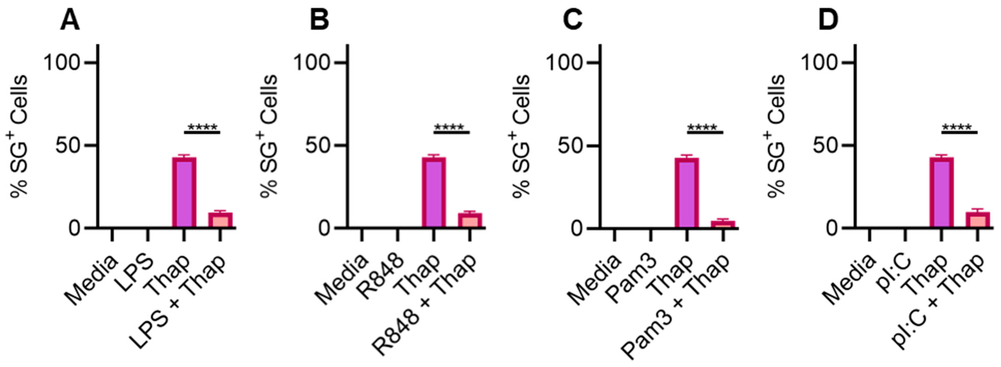
2.1. TLR Signaling Inhibits SGs in Bone-Marrow-Derived Macrophages

We started by repeating experiments from the published study that showed TLR-mediated stress granule inhibition in BMDMs to establish our experimental conditions [23]. We fully recapitulated the results of the study (Figure 1). We used 2 µg/mL thapsigargin treatment for 90 min to induce ER stress and subsequent SG formation. We used G3BP1 and DDX3X as SG markers. We chose G3BP1 as a marker because it has been used extensively to visualize SGs and its demonstrated specificity [24]. We decided to use DDX3X as an additional marker because it is one of the essential proteins for SG assembly [7,17]. Use of these two markers allowed us to specifically label SGs in the cytoplasm. We found that TLR4 stimulation by its ligand lipopolysaccharide (LPS; 100 ng/mL) for 6 h inhibited ER stress-induced SGs (Figure 1A). Similarly, stimulation of TLR7 by R848 (Figure 1B), stimulation of TLR2 by Pam3CSK4 (Figure 1C) and stimulation of TLR3 by polyinosinic–polycytidylic acid (poly(I:C)) (Figure 1D) also inhibited SGs induced by ER stress in BMDMs. Since TLR3 only uses TRIF as an adaptor, this result suggests that TRIF-mediated signaling is sufficient for SG inhibition. Similarly, TLR2 and TLR7 only use MYD88 as an adaptor, and our results suggest that MYD88-mediated signaling is sufficient for SG inhibition. Finally, TLR2 localizes to the plasma membrane, while TLR3 and TLR7 are present in the endosome. Since all three of them can inhibit SGs, our results suggest that sub-cellular localization of a TLR does not affect its ability to inhibit SG formation. In conclusion, we recapitulated the results from the previous study showing that MYD88- and TRIF-mediated signaling are redundant for SG inhibition induced by ER stress in BMDMs.

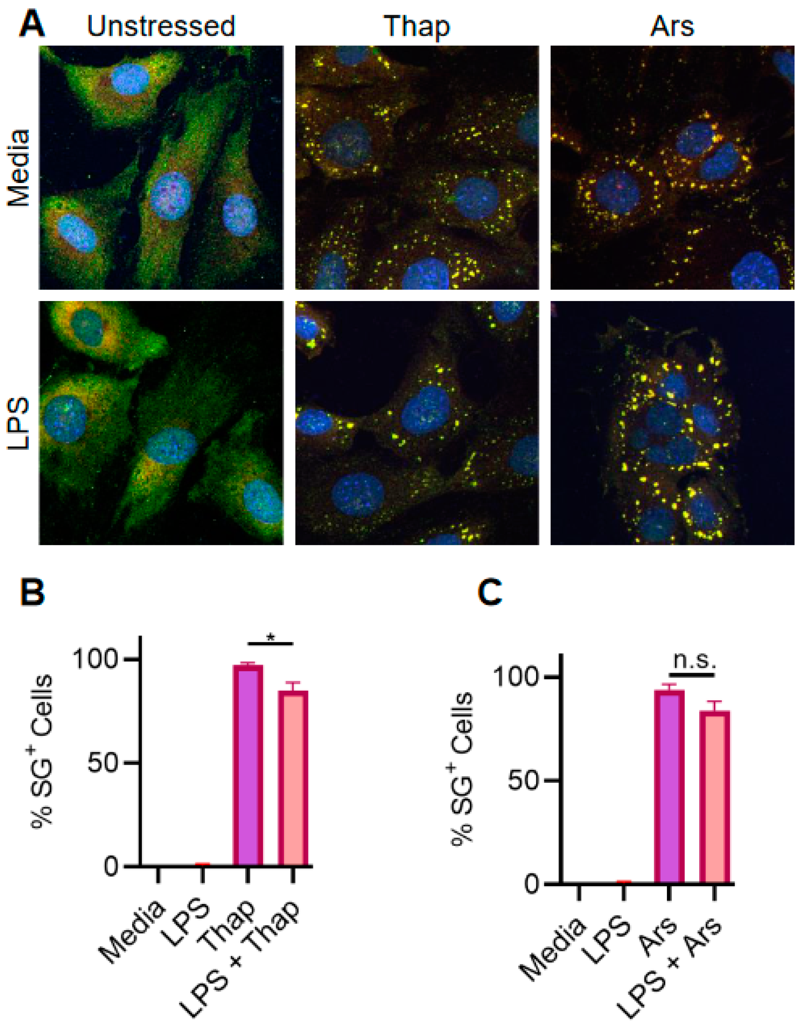
2.2. TLR4 Signaling Has a Very Weak Effect on ER Stress-Induced SGs in A549 Lung Cancer Cells

To test the possibility of SG inhibition in A549 lung cancer cells, we used 1 µg/mL thapsigargin treatment for 1 h. We used a lower a dose and duration of stress because we found that these conditions were sufficient to induce SGs robustly and reproducibly in A549 cells. We were surprised to see only a minor effect of 1 µg/mL LPS treatment for 6 h on SGs (Figure 2A,B), even though the concentration of LPS was 10 times higher than that which we had used for BMDMs. LPS treatment on its own did not induce SGs, similar to what has been observed previously (Figure 2A,B) [17,23]. We also used 500 µM sodium arsenite treatment for 1 h and tested the effect of LPS (Figure 2A–C). There was, again, only a minor effect of LPS on sodium arsenite-mediated SG assembly. In fact, LPS stimulation seemed to promote SGs in A549 cells. To show that it is possible to inhibit assembly in A549 cells, we used cycloheximide treatment to stabilize the polysomes that is known to interfere with SG assembly (Figure 2D) [25]. We used puromycin as a control for translation inhibition that does not involve stabilization of polysomes. As expected cycloheximide inhibited SG assembly, while puromycin did not. Taken together, our results demonstrate a minor effect of TLR signaling on SG assembly in A549 cells.

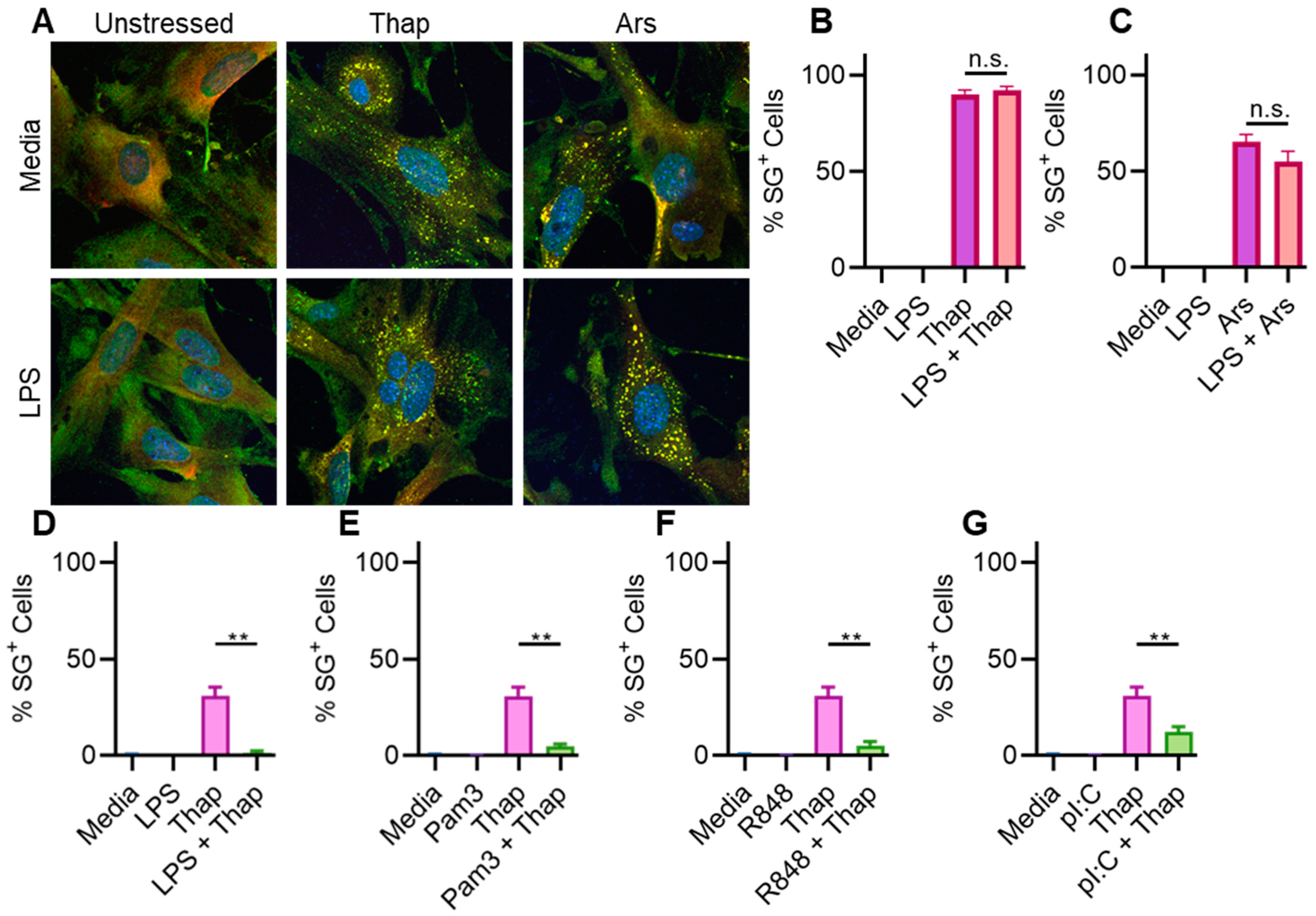
2.3. TLR4 Signaling Has a Very Weak Effect on ER Stress-Induced SGs in Immortalized Mouse Lung Fibroblasts

Since A549 cells are of human origin while BMDMs are from mice, observed differences in the effect of TLR signaling on SGs could be due to the species difference. To test this possibility, we performed an experiment using immortalized mouse lung fibroblasts (iMLFs). We used 1 µg/mL LPS stimulation for 6 h prior to 1 µg/mL thapsigargin treatment for 1 h to induce ER stress and SGs. Similar to A549 cells, LPS stimulation only had a very weak effect on stress granules (Figure 3A,B). LPS stimulation did not significantly affect SGs induced by the 500 mM sodium arsenite treatment (Figure 3A–C). Our results suggest that the inability of TLR stimulation to inhibit SGs induced by ER stress was not due to species-specific differences between mice and humans.

2.4. TLR4 Signaling Does Not Significantly Affect ER Stress-Induced SGs in Immortalized Mouse Lung Fibroblasts

Since the A549 cell line is a lung cancer cell line, it is likely to express oncogenes. Oncogenes have been reported to promote SG assembly [12]. Our immortalization protocol involved the expression of viral oncogenes that were adapted from the BMDM immortalization protocol [26]. Therefore, it is possible that the inability of TLR signaling to inhibit SGs could be due to the expression of oncogenes. To test this possibility, we repeated the experiment performed with iMLFs using primary mouse lung fibroblasts (MLFs). Our data show that LPS stimulation did not significantly affect SGs induced by either ER stress or sodium arsenite treatment (Figure 4A–C). Additionally, TLR signaling was able to inhibit SG assembly in immortalized BMDMs (iBMDMs) that expressed the oncogenes (Figure 4D–G). These results indicate a cell-intrinsic effect that is independent of oncogenic expression program.

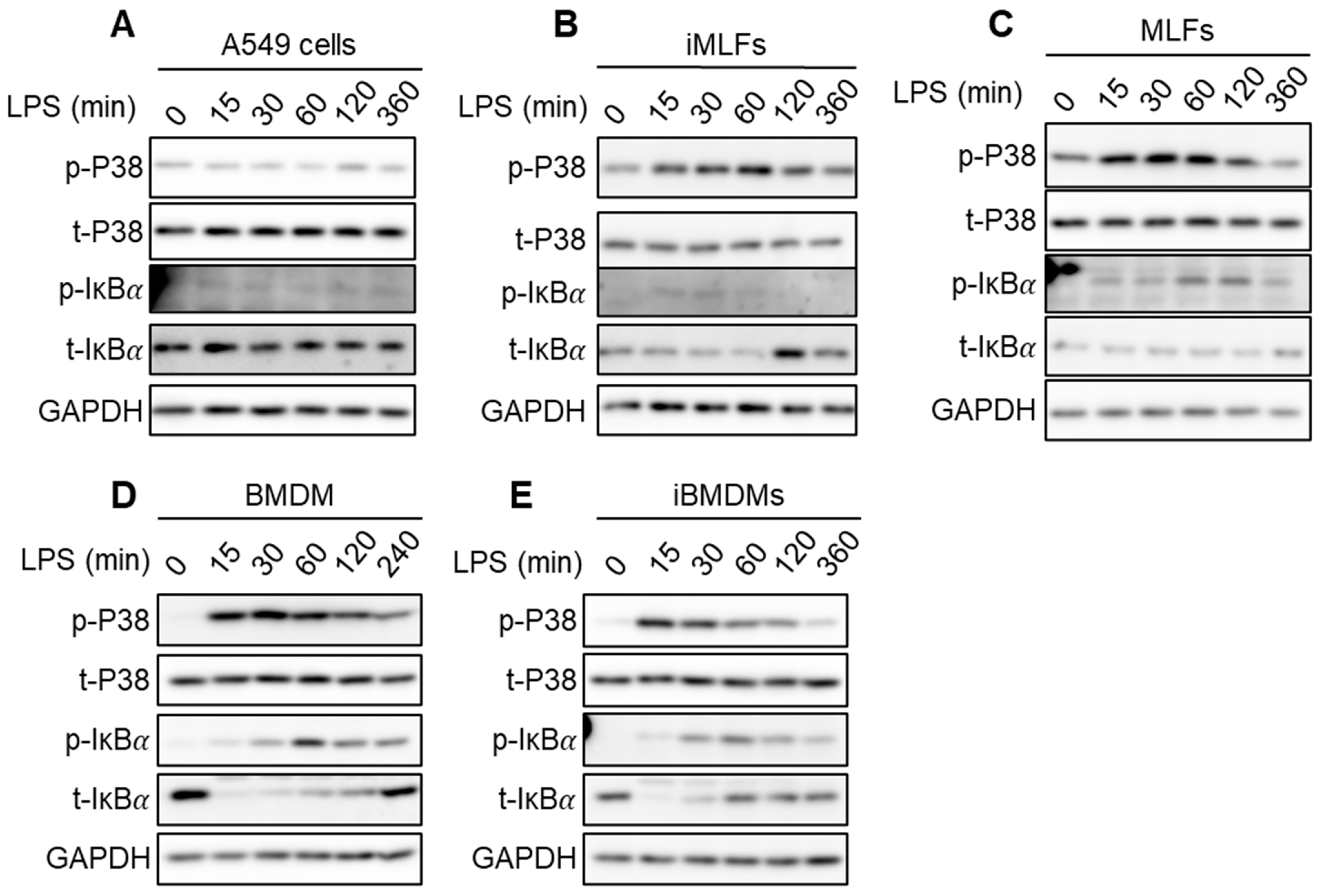
2.5. Strength of IKK Complex Kinase Activity Induction Is Weak in Cells Where It Fails to Inhibit SGs

TLR signaling inhibits SGs through the kinase activity of the IKK complex. Therefore, we tested whether LPS stimulation leads to a differential induction of IKK kinase activity. We used phosphorylation of a well-established IKK complex substrate, Inhibitor of Kappa B alpha (Iκbα), as a marker for its kinase activity (Figure 5A–E) [27,28,29]. Our data show no accumulation of phosphorylated Iκbα in A549 cells and only a small increase in iMLFs and MLFs, suggesting a weak induction of IKK complex kinase activity in these cells (Figure 5A–C). In contrast, there was a noticeably greater increase in the amount of phosphorylated Iκbα in BMDMs and iBMDMs, indicating a strong IKK kinase activity induction (Figure 5D,E). Since phosphorylated Iκbα is targeted for proteasomal degradation, the amount of total Iκbα alpha decreases upon IKK complex activation. Therefore, a decrease in the amount of total Iκbα is complementary evidence for activation of the IKK complex. We observed only minor changes in the abundance of total Iκbα in A549, iMLF and MLF cells, while there was a drastic decrease in BMDMs and iBMDMs at early timepoints and recovery at later timepoints. Our results indicate a correlation between the strength of IKK kinase activity induction and the ability of TLR signaling to inhibit SGs.

2.6. Discussion and Conclusions

In this study, we uncovered a cell-type-specific effect of TLR signaling on SGs depending on the strength of IKK kinase activity induction. Our data show that TLR signaling can inhibit ER stress-induced SGs in BMDMs and iBMDMs but not in A549, iMLF and MLF cells. There was a strong induction of IKK complex kinase activity in BMDMs and iBMDMs but only a minor induction in A549, iMLF and MLF cells. Although the exact reason for this is unclear, it is possible that there is a stronger basal IKK complex kinase activity in A549, iMLF and MLF cells which makes them less responsive to external stimulation. Another reason could be the differences in the expression of TLRs and downstream signaling components. Single-cell transcriptomic studies have revealed large differences in expression, which makes this explanation more likely. However, there is another observation that does not have an easy explanation. We found that the effect of TLR signaling on sodium arsenite-induced SGs is minor. Both ER stress- and sodium arsenite-triggered SG assembly require eIF2α phosphorylation [18]. However, our data show that there is a differential effect of TLR signaling on SGs induced by the two stressors. In fact, in a previous study we observed that eIF2α was phosphorylated in the presence of ER stress, but there was no SG assembly if TLR signaling was active. This means that there is another hitherto undiscovered step in SG assembly. Discovery of this step will not only be an important milestone in cell biology research, but it could also help develop SG modulation strategies with strong biomedical implications.
P-bodies are another type of membraneless cytoplasmic compartment that can form in cells [11,21]. Both SGs and P-bodies contain translation initiation factors, RNA-binding proteins and 40S ribosomal proteins. An important difference between the two is that P-bodies also contain RNAse enzymes. Based on this, it has been hypothesized that P-bodies can be a site for RNA degradation in stressed cells [30]. Directional transport of RNA from SGs to P-bodies has been observed as well. The effect of TLR signaling on P-bodies remains unknown and is an exciting area for research in the future.
In summary, our observations indicate that insights gained from one cell type or stressor may not be applicable in the context of another cell type or stressor. This calls for a more comprehensive analysis of SGs in different cell types and stressors. It also cautions researchers against over-generalizing concepts in SG biology. Finally, all cells tested by us and others are capable of forming SGs. However, the dose and duration of stress required can vary between cell types. The reason for this heterogeneity remains unknown. Future research should be able to shed light on the molecular mechanism driving cell-type-specific differences in stress responses. These differences could be exploited for devising therapeutic interventions that target SG biology.

3. Materials and Methods

3.1. Mice

BL6/J WT mice were acquired from the Jackson Laboratories. The mice were bred at the University of Texas Medical Branch at Galveston. Animal studies were conducted in accordance with protocols approved by the UTMB Institutional Animal Care and Use Committee (IACUC).

3.2. Cell Culture and Stimulations

MLFs, iMLFs and iBMDMs were cultured in DMEM medium supplemented with 10% FBS (S1620; Biowest USA, Bradenton, FL, USA) and 1% penicillin and streptomycin (Sigma-Aldrich, St. Louis, MO, USA). BMDMs were immortalized using the protocol described in [26]. The same protocol was followed for the immortalization of primary MLFs, with one difference. Primary MLFs were transduced only once with the J2 retrovirus after 3 passages. Primary BMDMs were grown for 6 days in BMDM medium. The BMDM medium was prepared by supplementing DMEM (D5671; Sigma-Aldrich, St. Louis, MO, USA) with 10% FBS, 30% L929-conditioned medium (the base medium was IMDM and 1% penicillin and streptomycin (Sigma-Aldrich, St. Louis, MO, USA). BMDMs were seeded in 12-well plates at a density of 1 × 10⁶ cells per well and allowed to attach overnight before stimulation for Western blot analysis. To induce SG formation, BMDMs were washed with PBS and incubated with DMEM supplemented with 10% FBS for 30 min, followed by the treatments indicated in the figure legends. Pam3CSK4 (tlrl-pms; InvivoGen, San Diego, CA, USA) at a concentration of 1 μg/mL was used to stimulate TLR2. Low-molecular-weight poly(I:C) (tlrl-picw-250; InvivoGen, San Diego, CA, USA) at a concentration of 50 μg/mL was used to stimulate TLR3. Ultrapure LPS (Salmonella minnesota; tlrl-smlps; InvivoGen, San Diego, CA, USA) at a concentration of 100 ng/mL was used to stimulate TLR4 in BMDMs. The LPS concentration for stimulating other cell types was 1 µg/mL. R848 (resiquimod) (tlrl-r848-5; InvivoGen, San Diego, CA, USA) at a concentration of 1 μg/mL was used to stimulate TLR7/8. A concentration of 2 µg/mL Thapsigargin (Cayman Chemicals Inc., Ann Arbor, MI, USA) was used to stimulate BMDMs, while a concentration of 1 µg/mL was used for the other cell types. The sodium arsenite concentration used was 500 µM.

3.3. Western Blot Analysis

Western blot analysis was performed as described in [23]. The primary Abs used were anti–phospho-P38 (1:1000; catalog no. 9211; Cell Signaling Technology, Danvers, MA, USA), anti-P38 (1:1000; catalog no. 9212; Cell Signaling Technology, Danvers, MA, USA), anti–phospho-IκBα (1:1000; catalog no. 2859; Cell Signaling Technology, Danvers, MA, USA), anti-IκBα (1:1000; catalog no. 9242; Cell Signaling Technology, Danvers, MA, USA) and anti-GAPDH (1:10,000, catalog no. 60004-1-Ig; Proteintech, Rosemont, IL, USA). The secondary antibodies used were Peroxidase-conjugated AffiniPure Goat Anti-Rabbit IgG, F(ab’)2 Fragment Specific (1:1000, catalog no. 111-035-047; Jackson Immunoresearch, West Grove, PA, USA) and Peroxidase-conjugated AffiniPure Rabbit Anti-Mouse IgG, F(ab’)2 (1:10,000, catalog no. 315-035-047; Jackson Immunoresearch, West Grove, PA, USA).

3.4. Confocal Microscopy Imaging and Analysis

Following stimulation, the cells were fixed in 4% paraformaldehyde (sc-281692, Santa Cruz Biotechnology, Inc., Dallas, TX, USA) at room temperature for 15 min and washed with PBS. Blocking was performed in 2% BSA in PBS (Sigma-Aldrich, St. Louis, MO, USA). BMDMs were stained with the following Abs overnight at 4 °C: anti-G3BP1 (1:1000; 66486-1-Ig; Proteintech, Rosemont, IL, USA) and anti-DDX3X (1:1000; A300-474A; Bethyl Laboratories, Montgomery, AL, USA). BMDMs were incubated with the following secondary Abs: Alexa Fluor 488–conjugated anti-mouse IgG (1:1000; A32766; Life Technologies, Carlsbad, CA, USA), Alexa Fluor 555–conjugated anti-rabbit IgG (1:1000; A32794, Life Technologies) and DAPI (5 μg/mL, Cayman Chemicals Inc.). Confocal images were acquired using either a Nikon AXR (Nikon Instruments Inc., Melville, NY, USA) or an ImageXpress® Micro Confocal (Molecular Devices, San Jose, CA, USA) confocal microscope. Images were analyzed using ImageJ Fiji v1.5 [31]. AI-assisted nuclei counting was performed using the Stardist plugin v0.85.

3.5. Statistical Analysis

The statistical significance of the data was determined by the two-tailed Mann–Whitney test with an alpha level of 0.05. Sample means are reported, and error bars represent SEMs. GraphPad Prism v8 software was used for statistical analysis and generating plots.

Author Contributions

P.S. designed the experiments. P.P.L., A. and P.S. performed the experiments. X.X. and P.S. supervised the study. P.S. wrote the first draft of the manuscript. All authors have read and agreed to the published version of the manuscript.

Funding

This work was funded by UTMB startup package to P.S. (Grant numbers P86059 and P86276).

Data Availability Statement

All data included in the manuscript will be made available upon reasonable request by the corresponding author.

Acknowledgments

We thank all members of the Department of Microbiology and Immunology at the University of Texas Medical Branch for their advice and encouragement.

Conflicts of Interest

The authors declare no conflict of interest.

References

  1. Protter, D.S.W.; Parker, R. Principles and Properties of Stress Granules. Trends Cell Biol. 2016, 26, 668–679. [Google Scholar] [CrossRef] [PubMed]
  2. Kedersha, N.L.; Gupta, M.; Li, W.; Miller, I.; Anderson, P. RNA-Binding Proteins Tia-1 and Tiar Link the Phosphorylation of Eif-2α to the Assembly of Mammalian Stress Granules. J. Cell Biol. 1999, 147, 1431–1442. [Google Scholar] [CrossRef] [PubMed]
  3. Nover, L.; Scharf, K.D.; Neumann, D. Formation of Cytoplasmic Heat Shock Granules in Tomato Cell Cultures and Leaves. Mol. Cell. Biol. 1983, 3, 1648–1655. [Google Scholar]
  4. Collier, N.C.; Schlesinger, M.J. The Dynamic State of Heat Shock Proteins in Chicken Embryo Fibroblasts. J. Cell Biol. 1986, 103, 1495–1507. [Google Scholar] [CrossRef] [PubMed]
  5. Hoyle, N.P.; Castelli, L.M.; Campbell, S.G.; Holmes, L.E.A.; Ashe, M.P. Stress-Dependent Relocalization of Translationally Primed mRNPs to Cytoplasmic Granules That Are Kinetically and Spatially Distinct from P-Bodies. J. Cell Biol. 2007, 179, 65–74. [Google Scholar] [CrossRef] [PubMed]
  6. Kedersha, N.; Cho, M.R.; Li, W.; Yacono, P.W.; Chen, S.; Gilks, N.; Golan, D.E.; Anderson, P. Dynamic Shuttling of Tia-1 Accompanies the Recruitment of mRNA to Mammalian Stress Granules. J. Cell Biol. 2000, 151, 1257–1268. [Google Scholar] [CrossRef] [PubMed]
  7. Hilliker, A.; Gao, Z.; Jankowsky, E.; Parker, R. The DEAD-Box Protein Ded1 Modulates Translation by the Formation and Resolution of an eIF4F-mRNA Complex. Mol. Cell 2011, 43, 962–972. [Google Scholar] [CrossRef] [PubMed]
  8. Kedersha, N.; Ivanov, P.; Anderson, P. Stress Granules and Cell Signaling: More than Just a Passing Phase? Trends Biochem. Sci. 2013, 38, 494–506. [Google Scholar] [CrossRef] [PubMed]
  9. Kedersha, N.; Anderson, P. Chapter 4 Regulation of Translation by Stress Granules and Processing Bodies. In Translational Control in Health and Disease; Hershey, J.W.B., Ed.; Academic Press: Cambridge, MA, USA, 2009; Volume 90, pp. 155–185. [Google Scholar]
  10. Buchan, J.R.; Parker, R. Eukaryotic Stress Granules: The Ins and Outs of Translation. Mol. Cell 2009, 36, 932–941. [Google Scholar] [CrossRef] [PubMed]
  11. Anderson, P.; Kedersha, N.; Ivanov, P. Stress Granules, P-Bodies and Cancer. Biochim. Biophys. Acta (BBA)—Gene Regul. Mech. 2015, 1849, 861–870. [Google Scholar] [CrossRef] [PubMed]
  12. Grabocka, E.; Bar-Sagi, D. Mutant KRAS Enhances Tumor Cell Fitness by Upregulating Stress Granules. Cell 2016, 167, 1803–1813.e12. [Google Scholar] [CrossRef] [PubMed]
  13. Ash, P.E.A.; Vanderweyde, T.E.; Youmans, K.L.; Apicco, D.J.; Wolozin, B. Pathological Stress Granules in Alzheimer’s Disease. Brain Res. 2014, 1584, 52–58. [Google Scholar] [CrossRef] [PubMed]
  14. Li, Y.R.; King, O.D.; Shorter, J.; Gitler, A.D. Stress Granules as Crucibles of ALS Pathogenesis. J. Cell Biol. 2013, 201, 361–372. [Google Scholar] [CrossRef] [PubMed]
  15. Dewey, C.M.; Cenik, B.; Sephton, C.F.; Johnson, B.A.; Herz, J.; Yu, G. TDP-43 Aggregation in Neurodegeneration: Are Stress Granules the Key? Brain Res. 2012, 1462, 16–25. [Google Scholar] [CrossRef] [PubMed]
  16. Arimoto, K.; Fukuda, H.; Imajoh-Ohmi, S.; Saito, H.; Takekawa, M. Formation of Stress Granules Inhibits Apoptosis by Suppressing Stress-Responsive MAPK Pathways. Nat. Cell Biol. 2008, 10, 1324–1332. [Google Scholar] [CrossRef] [PubMed]
  17. Samir, P.; Kesavardhana, S.; Patmore, D.M.; Gingras, S.; Malireddi, R.S.; Karki, R.; Guy, C.S.; Briard, B.; Place, D.E.; Bhattacharya, A. DDX3X Acts as a Live-or-Die Checkpoint in Stressed Cells by Regulating NLRP3 Inflammasome. Nature 2019, 573, 590–594. [Google Scholar] [CrossRef] [PubMed]
  18. Lamichhane, P.P.; Samir, P. Cellular Stress: Modulator of Regulated Cell Death. Biology 2023, 12, 1172. [Google Scholar] [CrossRef] [PubMed]
  19. White, J.P.; Lloyd, R.E. Regulation of Stress Granules in Virus Systems. Trends Microbiol. 2012, 20, 175–183. [Google Scholar] [CrossRef] [PubMed]
  20. Eiermann, N.; Haneke, K.; Sun, Z.; Stoecklin, G.; Ruggieri, A. Dance with the Devil: Stress Granules and Signaling in Antiviral Responses. Viruses 2020, 12, 984. [Google Scholar] [CrossRef] [PubMed]
  21. Reineke, L.C.; Lloyd, R.E. Diversion of Stress Granules and P-Bodies during Viral Infection. Virology 2013, 436, 255–267. [Google Scholar] [CrossRef] [PubMed]
  22. Kesavardhana, S.; Samir, P.; Zheng, M.; Malireddi, R.S.; Karki, R.; Sharma, B.R.; Place, D.E.; Briard, B.; Vogel, P.; Kanneganti, T.-D. DDX3X Coordinates Host Defense against Influenza Virus by Activating the NLRP3 Inflammasome and Type I Interferon Response. J. Biol. Chem. 2021, 296, 100579. [Google Scholar] [CrossRef] [PubMed]
  23. Samir, P.; Place, D.E.; Malireddi, R.K.S.; Kanneganti, T.-D. TLR and IKK Complex–Mediated Innate Immune Signaling Inhibits Stress Granule Assembly. J. Immunol. 2021, 207, 115–124. [Google Scholar] [CrossRef] [PubMed]
  24. Kedersha, N.; Stoecklin, G.; Ayodele, M.; Yacono, P.; Lykke-Andersen, J.; Fritzler, M.J.; Scheuner, D.; Kaufman, R.J.; Golan, D.E.; Anderson, P. Stress Granules and Processing Bodies Are Dynamically Linked Sites of mRNP Remodeling. J. Cell Biol. 2005, 169, 871–884. [Google Scholar] [CrossRef] [PubMed]
  25. Blasi, E.; Mathieson, B.J.; Varesio, L.; Cleveland, J.L.; Borchert, P.A.; Rapp, U.R. Selective Immortalization of Murine Macrophages from Fresh Bone Marrow by a Raf/Myc Recombinant Murine Retrovirus. Nature 1985, 318, 667–670. [Google Scholar] [CrossRef]
  26. Israël, A. The IKK Complex, a Central Regulator of NF-κB Activation. Cold Spring Harb. Perspect. Biol. 2010, 2, a000158. [Google Scholar] [CrossRef] [PubMed]
  27. Akira, S.; Takeda, K. Toll-like Receptor Signalling. Nat. Rev. Immunol. 2004, 4, 499–511. [Google Scholar] [CrossRef] [PubMed]
  28. Fitzgerald, K.A.; Kagan, J.C. Toll-like Receptors and the Control of Immunity. Cell 2020, 180, 1044–1066. [Google Scholar] [CrossRef] [PubMed]
  29. Corbet, G.A.; Parker, R. RNP Granule Formation: Lessons from P-Bodies and Stress Granules. Cold Spring Harb. Symp. Quant. Biol. 2019, 84, 203–215. [Google Scholar] [CrossRef] [PubMed]
  30. Schindelin, J.; Arganda-Carreras, I.; Frise, E.; Kaynig, V.; Longair, M.; Pietzsch, T.; Preibisch, S.; Rueden, C.; Saalfeld, S.; Schmid, B.; et al. Fiji: An Open-Source Platform for Biological-Image Analysis. Nat. Methods 2012, 9, 676–682. [Google Scholar] [CrossRef] [PubMed]
  31. Schmidt, U.; Weigert, M.; Broaddus, C.; Myers, G. Cell Detection with Star-Convex Polygons. In Medical Image Computing and Computer Assisted Intervention—MICCAI 2018; Frangi, A.F., Schnabel, J.A., Davatzikos, C., Alberola-López, C., Fichtinger, G., Eds.; Springer International Publishing: Cham, Switzerland, 2018; pp. 265–273. [Google Scholar]
Figure 1. TLR signaling inhibits ER stress-induced SGs in BMDMs. (A) Effect of TLR4 stimulation using 100 ng/mL LPS for 6 h on ER stress-induced SGs. (B) Effect of TLR7 stimulation using 1 µg/mL R848 for 6 h on ER stress-induced SGs. (C) Effect of TLR2 stimulation using 1 µg/mL Pam3CSK4 for 6 h on ER stress-induced SGs. (D) Effect of TLR3 stimulation using 50 µg/mL poly(I:C) for 6 h on ER stress-induced SGs. Abbreviations: pI:C = poly(I:C), 2 µg/mL Thap = thapsigargin for 90 min, Pam3 = Pam3CSK4. DAPI was used to mark nuclei. **** denotes p < 0.0001 using the two-tailed Mann–Whitney test.
Stresses 04 00027 g001
Figure 2. TLR signaling only weakly inhibits ER stress-induced SGs in A549 cells. (A) Effect of TLR4 stimulation using 1 µg/mL LPS for 6 h on ER stress- and sodium arsenite-induced SGs. (B) Quantification of SG assembly induced by ER stress. (C) Quantification of SG assembly induced by sodium arsenite. Abbreviations: Thap = 1 µg/mL thapsigargin for 60 min, Ars = 500 µM sodium arsenite for 60 min. Blue: DAPI, green: G3BP1, red: DDX3X. ** denotes p < 0.01 using the two-tailed Mann–Whitney test. (D) Effect of translation inhibition on arsenite-induced SG assembly in A549 cells. Cycloheximide was used to stabilize polysomes and inhibit SG assembly. Puromycin did not stabilize polysomes, resulting in no SG inhibition. Blue: DAPI, green: G3BP1.
Stresses 04 00027 g002
Figure 3. TLR signaling only weakly inhibits ER stress-induced SGs in iMLFs. (A) Effect of TLR4 stimulation using 1 µg/mL LPS for 6 h on ER stress- and sodium arsenite-induced SGs. (B) Quantification of SG assembly induced by ER stress. (C) Quantification of SG assembly induced by sodium arsenite. Abbreviations: Thap = 1 µg/mL thapsigargin for 60 min, Ars = 500 µM sodium arsenite for 60 min. Blue: DAPI, green: G3BP1, red: DDX3X. * denotes p < 0.05 using the two-tailed Mann–Whitney test.
Stresses 04 00027 g003
Figure 4. TLR signaling fails to inhibit SGs in primary MLFs. (A) Effect of TLR4 stimulation using 1 µg/mL LPS for 6 h on ER stress- and sodium arsenite-induced SGs. (B) Quantification of SG assembly induced by ER stress. (C) Quantification of SG assembly induced by sodium arsenite. Quantification of SG assembly induced by 2 µg/mL thapsigargin (Thap) treatment for 90 min in immortalized BMDMs stimulated with (D) LPS, (E) Pam3CSK4 (Pam3), (F) R848 and (G) poly(I:C) (pI:C) for 6 h. Abbreviations: Thap = 1 µg/mL thapsigargin for 60 min, Ars = 500 µM sodium arsenite for 60 min. Blue: DAPI, green: G3BP1, red: DDX3X. The two-tailed Mann–Whitney test was used to calculate statistical significance. ** denotes p-value < 0.01, n.s. denotes non-significant p-value (>0.05).
Stresses 04 00027 g004
Figure 5. Effect of TLR4 activation on IKK kinase activity in A549 cells, MLFs, iMLFs and BMDMs. (A) Quantification of phosphorylated P38 (p-P38), total P38 (t-P38), phosphorylated IκBα (p-IκBα) and total IκBα (t-IκBα) in A549 cells following stimulation with 1 µg/mL LPS for indicated durations. GAPDH was used as loading control. (B) Quantification of phosphorylated P38 (p-P38), total P38 (t-P38), phosphorylated IκBα (p-IκBα) and total IκBα (t-IκBα) in iMLFs following stimulation with 1 µg/mL LPS for indicated durations. GAPDH was used as loading control. (C) Quantification of phosphorylated P38 (p-P38), total P38 (t-P38), phosphorylated IκBα (p-IκBα) and total IκBα (t-IκBα) in MLFs following stimulation with 1 µg/mL LPS for indicated durations. GAPDH was used as loading control. (D) Quantification of phosphorylated P38 (p-P38), total P38 (t-P38), phosphorylated IκBα (p-IκBα) and total IκBα (t-IκBα) in BMDM cells following stimulation with 100 ng/mL LPS for indicated durations. GAPDH was used as loading control. (E) Quantification of phosphorylated P38 (p-P38), total P38 (t-P38), phosphorylated IκBα (p-IκBα) and total IκBα (t-IκBα) in immortalized BMDM cells following stimulation with 100 ng/mL LPS for indicated durations. GAPDH was used as loading control. Abbreviations: iMLF = immortalized mouse lung fibroblast, MLF = primary mouse lung fibroblast, BMDM = bone-marrow-derived macrophage, iBMDM = immortalized bone-marrow-derived macrophage.
Stresses 04 00027 g005
Disclaimer/Publisher’s Note: The statements, opinions and data contained in all publications are solely those of the individual author(s) and contributor(s) and not of MDPI and/or the editor(s). MDPI and/or the editor(s) disclaim responsibility for any injury to people or property resulting from any ideas, methods, instructions or products referred to in the content.

