Review

The Relationships Among Perineural Invasion, Tumor–Nerve Interaction and Immunosuppression in Cancer

by
Jozsef Dudas
*,
Rudolf Glueckert
,
Maria do Carmo Greier
and
Benedikt Gabriel Hofauer
Department of Otorhinolaryngology and Head and Neck Surgery, Medical University Innsbruck, 6020 Innsbruck, Austria
*
Author to whom correspondence should be addressed.
Submission received: 26 March 2025 / Revised: 11 May 2025 / Accepted: 20 May 2025 / Published: 23 May 2025

Simple Summary

One of the most important events in epithelial tumors is the change in phenotype of the tumor cells from epithelial to mesenchymal, which is accompanied by the release of junctions with neighboring cells and the start of migration into the local tissue environment, but also further afield. Such invasive tumor cells reach lymph or blood vessels, but several tumors, such as cervical, pancreatic, colorectal, and head and neck cancers, also invade nerves. In addition, an intensive, mutually beneficial “co-existence” develops between tumor cells and nerves. This cooperation between nerve components and tumor cells has a suppressive effect on the immune system and helps tumor cells evade anti-tumor immune attacks.

Abstract

Tumor cells and the tumor microenvironment (TME) produce factors, including neurotrophins, that induce axonogenesis and neurogenesis, and increase local nerve density. Proliferative growing cancer cell clusters and disseminated invasive tumor cells undergoing partial epithelial-to-mesenchymal transition (pEMT) can invade peripheral nerves. In the early stages of tumor–nerve interactions, Schwann cells (SCs) dedifferentiate, become activated and migrate to cancer cell nests; later, they induce pEMT in tumor cells and activate tumor cell migration along nerves. The SC–tumor–nerve interaction attracts myeloid-derived suppressor cells (MDSCs) and inflammatory monocytes, and the latter differentiate into macrophages. SCs and MDSCs are responsible for the activation of transforming growth factor-beta (TGF-beta) signaling. Intra-tumoral innervation is followed by perineural invasion (PNI), which has an unfavorable prognosis. What are the interventional options against PNI: local reduction in tumor nerves or inhibition of TGF-beta-related events, inhibition of downstream signaling of TGF-beta or immune activation, or intervention against immunosuppression? This systematic review is based on the Prisma 2009 search method and provides an overview of tumor–nerve interaction.

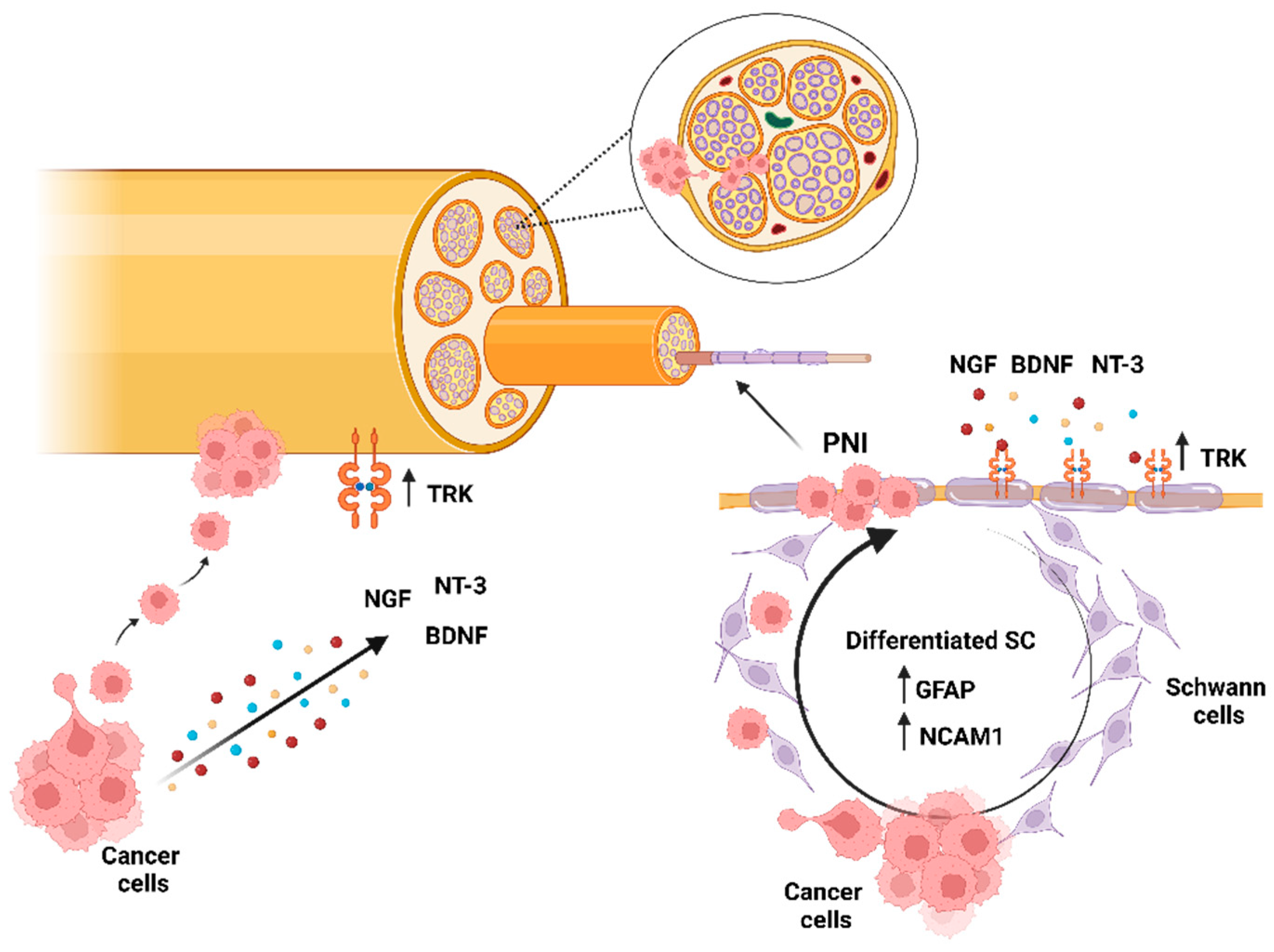
1. Introduction (Perineural Invasion, the Original Clinical Context)

Perineural invasion (PNI) is a form of cancer metastasis by which tumor cells travel along nerve tracts [1]. PNI occurs in several cancers with significant high frequency, but not in all cancers. The occurrence of PNI in uterine cervical cancer [2] is between 7 and 41.7%. PNI in colorectal cancer has been found in 67.3% of cases [3]; in pancreatic cancer, it has been found in 70-100% [4].
PNI is associated with poor prognosis in many malignancies [4]; in the case of pancreatic cancer [4], prostate cancer [5,6,7], colon cancer [8], head and neck cancer [9] and cervical cancer [10], intensive research is available on PNI.
In head and neck squamous cell carcinoma (HNSCC), PNI is associated with decreased survival (23% with PNI vs. 49% without PNI), increased local recurrence (23% with PNI vs. 9% without PNI) and increased disease-specific mortality (54% with PNI vs. 25% without PNI) [11,12].
PNI occurs when a tumor invades around, along and throughout different layers of nerves [12]. In HNSCC, PNI can be asymptomatic but microscopically detected, and in this case, it represents a possible criterion for intensification of (adjuvant) therapy [13], which might have a negative impact on the patient’s further quality of life. In advanced-stage disease, PNI can be associated with significant morbidity, as in HNSCC, the cranial nerves become more grossly involved in the disease [12].
PNI is evaluated by hematoxylin–eosin (HE) staining, but other, mainly immunohistochemical methods have been developed. The use of S-100 immunostaining is widespread [14]; an alternative is the use of PGP9.5 [15]. According to Fukuda et al. (2022) [14], S-100 immunohistochemistry detected PNI in 67.3% of the examined tissue samples of 279 colorectal cancer patients, whereas HE staining detected PNI in 18.5% of the cases [14]. The independently found negative prognostic value was associated with many S-100 positive cells [14]. Their paper also showed the negative relationship between positive S-100 IHC in PNI and stromal lymphocytic reaction. This indicates the immunosuppressive nature of PNI [14]. This observation has also been published by other research groups [3,16]. As seen here, PNI as an “invasive” disease is associated with a negative prognosis and is a clinically relevant factor. Literature sources mention that denervation reduces cancer risk [16] and tumor–nerve interaction provides significant support for tumor growth [17]. With improved immunohistochemical detection of PNI [15,18,19], clinical diagnostics of the presence of Schwann cells and factors they may elute becomes more relevant. Moreover, intensive research revealed that PNI is not “simply tumor cells attacking nerve fibers” but a complex tumor–nerve interaction [20,21]. The first steps might involve tumor cell dissemination, but, based on newer sources, it is more likely that the first players are the Schwann cells (SCs) that migrate toward the cancer cell nests. The nerve path metastasis [1] seems to be more efficient for tumor spread than other pathways (Figure 1). Possible interactions of this complex model will be discussed in the following sections.

2. Materials and Methods

The authors conducted searches using Endnote X9 by PubMed (NLM) and Web of Science SCI (Clarivate) search engines. The first keyword search was performed with “cancer, perineural invasion, immunosuppression” and resulted in 32 papers. Only 4 papers were kept, as 28 papers were found to be outside the scope of our review. The second search with the keywords “myeloid-derived suppressor cells, neural progenitor cells, neurotrophin, cancer” found two papers: one was a duplicate of the previous search and the other was outside the scope of our review. The third search contained “TGF, perineural invasion (PNI), cancer” and resulted in 16 references, 3 of which were kept. The fourth search used the keywords “Schwann cells, perineural invasion and cancer”, found 58 papers, of which 18 were kept. The fifth search used the keywords “cancer, peripherial nerve damage, perineurium, endoneurium, macrophages” and found 3 papers, none of which were kept. There was no publication date limit given to the searches; both original papers and review articles were considered, but original works were preferred. Older publications and overlapping earlier results from the same research group were removed. Several resources were found by following citations in the considered papers. The content of the review article dynamically changed according to the found literature resources. The search strategy and found resources are summarized in Figure 2. All found references and search details and all results are detailed in Supplementary Table S1.

3. Nerves and Cancer

3.1. The Cancer Gets Nervous

The tumor microenvironment (TME) has been studied for decades, and in addition to cancer cells, stromal fibroblasts—especially carcinoma-associated fibroblasts (CAFs) [22]—and extracellular matrix (ECM) remodeling—especially fibrotic changes [22], vessels, and immune components—have been described. In the last 25 years, the peripheral nerve has also received attention as a component of the TME [6,7,10,17,23].
Tumor–nerve interaction is not independent from other components of the TME. Co-culture models and three-dimensional outgrowth assays have provided insight into tumor–nerve interactions [9,10,12,13,20,24,25,26,27,28], yielding important results but also contradictions.
Two main modes of tumor–nerve interaction were described and reviewed by Gregory et al. in 2020 [29]. The first is the interaction of neuronal components with tumor cells leading to tumor innervation and the second is perineural invasion (PNI) [17,29]. Tumor innervation was discussed, such as axonogenesis and neurogenesis, by Ayala in a recent review in 2023 [17], and these processes occur earlier than PNI, which is a subsequent event.

3.2. From the Tumor Side

The original hypothesis was that cancer invades nerves via the path of least resistance in the perineural space. This hypothesis is contradicted by the fact that the nerve sheath is composed of multiple layers of collagen made by fibroblasts of the perineurium and basement membrane produced by Schwann cells for a microenvironment of neural tissue (Figure 3). This biochemical and mechanical barrier presents big obstacles for any invasion [13]. A more recent theory introduces reciprocal signaling between tumor cells and nerves. Nerve tissue releases trophic factors that stimulate cancer cells, and cancer cells release neurotrophic factors. Ein et al. investigated this relationship in HNSCC [13]. In their original head and neck article [12], they mention that HNSCCs secrete nerve growth factor (NGF), brain-derived neurotrophic factor (BDNF) and neurotrophin-3 (NT-3), which induce neurite outgrowth [12]. The importance of the BDNF and its receptor tropomyosin receptor kinase (Trk) B (TrkB) pathway has been previously investigated by our research group in HNSCC [30,31]. In accordance with Ein et al. [12], we found that this pathway was associated with aggressive behavior, poor prognosis and resistance to therapy. Moreover, the BDNF/TrkB signaling pathway has also been described as a possible epithelial-to-mesenchymal transition (EMT)-inducing pathway [31]. Interestingly, neurotrophin expression is not restricted to tumor cells, but carcinoma-associated fibroblasts (CAFs) may be significantly involved in neuro-attraction, as they release, e.g., BDNF in HNSCC [30,32]. The cells, which might respond to the tumor-produced neurotrophin, are probably Schwann cells (SCs), which express high levels of neurotrophin receptors, Trks: TrkA, TrkB and TrkC. These receptors respond to NGF, BDNF and NT-3, respectively [12] (Figure 3). The NT-3 activation of TrkC in Schwann cells induces their migration [33].
Several authors highlight that Schwann cells (SCs), which are the key regulators of peripheral nerve repair, promote axonal regrowth and are attracted first to tumor cells [34]. In addition to their role in nerve repair, they also affect non-neural wound healing. Tumor-produced factors are required for the SCs to gain their repair-like feature, which is similar to their function in damaged nerves [34]. This nerve injury response happens without damage of the nerve by tumor cells at this stage. Dedifferentiated SCs re-express proteins such as GFAP and NCAM1 during nerve repair [12] and secrete a pro-regenerative extracellular matrix [35]. According to Deborde et al. 2016 [21], NCAM1 in SCs coordinates the intercalation of SCs and cancer cell clusters, and directs cancer cell migration toward nerves (Figure 3). NCAM1-deficient mice showed decreased neural invasion and less paralysis [21]. Similarly, Ein et al. in 2019 [13] found that before cancer cells invade nerves, SCs dedifferentiate and intercalate between them. The cancer cell dissemination is promoted later by the attracted SCs [36], similar to axonal regrowth where migrating SCs direct axons [37]. In addition, Silva et al. in 2019 [38] and other authors [20,21] mention a direct contact between SCs and tumor cells.
An important step in tumor cell invasion is the cadherin switch [39], which implies downregulation of E-cadherin and induction of N-cadherin. N-cadherin is involved in heterotypic contacts, which is also required for PNI [40]. These changes in the cancer cells are comparable with EMT, which is achieved by TGF-beta [41], but SC-derived CCL2 also contributes to tumor cell EMT [10,20]. Moreover, SC-produced CCL2 recruits CCR2-expressing inflammatory monocytes to the sites of PNI, where they differentiate into macrophages and further potentiate nerve invasion [20]. Depending on the tumor type and experimental system, the tumor cell–SC interaction might induce EMT in tumor cells but also the opposite process, the mesenchymal-to-epithelial transition (MET) [42]. These data contradict each other in the literature, which indicates that both processes might be present depending on what we observe, if we work with an in vitro experimental model or use inhibitor treatments [12,13]. The MET findings of Fujii-Nishimura et al. are based on immunohistochemical investigation of the tissue samples of 168 PDAC patients, and the MET process is confined to tumor cells colonizing the pancreatic nerves, which are late steps of PNI [42]. The initial movement of the tumor cells toward nerves requires EMT [10,41], but the final nerve-colonizing tumor cells return to MET [42]. The metastatic cancer cells have elevated EMT activity, and are even more likely to invade neural structures. Metastatic cancer cells develop enzymatic functions invadopodia and podosomes, the latter of which includes expression of actin and required structural proteins [43]. Neuronal growth cones form protrusions that share the molecular, structural and functional characteristics of invadosomes [43]. Based on related research, we see that in addition to invadosomes, the activation of Wnt signaling [44] and the guidance by semaphorin 3c axon guidance molecules are shared between neuronal growth and expansion and metastatic tumor cells with sustained EMT activity [45].
In relation to tumor cells’ migration to nerves, the colorectal cancer microenvironment represents a special situation with nonmyelinated intrinsic enteric nervous system (ENS) [46]. Primary tumor epithelial cells (TECs) from human colon adenocarcinomas and cell lines were co-cultured with primary cultures of ENS and cultures of human ENS plexus explants. TECs adhered directly to enteric neurons. Blocking N-cadherin and L1CAM decreased TEC migration along ENS structures, which indicates similar adhesion mechanisms, as discussed above [40,46,47].
Interestingly, the findings of the perineural invasion of tumors provide valuable information for the spread of central nervous system invasion, which is defined as distant progression. A recent 2024 paper by Bruschi et al. [48] is a good example of the approach taken in this research field. The topic is diffuse midline glioma (DMG), which is a pediatric tumor with 2-year survival after diagnosis. The authors used patient-derived glioma stem cells (GSCs) to create patient-specific 3D avatars to model invasion and elucidate the cellular supporting mechanisms. They described two modes of migration, mesenchymal and ameboid-like, and associated the ameboid-like modality with GSCs derived from the most invasive tumors. They characterized the invasive ameboid-like tumors as oligodendrocyte progenitor-like, with highly contractile cytoskeleton and reduced adhesion ability driven by overexpression of bone morphogenetic pathway 7 (BMP7). These migration mechanisms, such as the ameboid migration of the PNI and the BMP pathways, are also described [44,48] in relation to invasion of metastatic tumor cells; we feel that integrative knowledge of metastasis invasion research of solid tumors such as HNSCC, PDAC and melanoma, and the research of glioma stem cells might generate hypotheses for each other and lead to possibilities for developing therapeutic inhibitors and modulators.

3.3. From the Perspective of Innervation

Demir et al. in 2014 [25] and other papers by Ayala and co-workers [6,7,17,23] consider that nerves may reach cancer cells even in the tumor stage, when most cancer cells are still not invasive, and the nerves contribute to the spread of cancer. Tumor cell migration toward peripheral neurons occurs afterwards. Both [25] and [12] highlight that inhibition of the TrkA or TrkB signal pathways, i.e., neurotrophin signaling [49], decreases the SC migration toward cancer nests. In a paper by Ein et al. [12], SCs are presented in two forms. If they were BDNF-treated, they did not express GFAP, and were in a so-called myelinating state. When they were treated with ANA-12, a new TrkB inhibitor, they became motile and dedifferentiated [13]. These data contradict previous reports suggesting that inhibition of TrkB signaling decreases SC migration, and here, ANA-12-treated SCs became motile. Dynamic differentiation changes in SCs in reaction to the TME and tumor-cells-derived factors seem to be key prerequisites for a PNI. The authors of these in vitro studies mention as a limitation of their work that they did not include neurons in their studies, which would increase their complexity [12,13]. Another limitation includes differences in commercial cell lines in the affinity to SCs or to nerves. Differences in the neurotrophic response of relevant models, including SC sources (human or rat-derived primary cells from spinal nerves such as primary cells [25] or rat SC lines (RSC96) [12]) may influence such in vitro models. The understanding of the behavior of SCs interacting with tumor cells might be partly based on in vitro experimentation, but for the investigation of innervation, tissue-based 3D culture systems should be favored. In this respect, we will discuss other models using dorsal root ganglion and other neuronal cultures, which might better represent the involved complexity.
A recent review article by Ayala (2023) [17] highlights bilateral interactions in the early embryonic development of nerves and epithelial tissues that show similarities to epithelial cancer and nerve interplay. Ayala explains axonogenesis as the requirement for an increased epithelial volume [17]. The increased axon density and neurite counts in cancer are also discussed in a recent paper published by Khorani et al. [9]. Axonogenesis and nerve-sprouting are mediated by neurotrophic factors. In addition to the above-mentioned NGF, BDNF and NT-3, neurturin [50] and semaphorins play important roles [6,23] here. Cancer cells also release axon guidance molecules such as Ephrin B1, which promotes axonogenesis, neurogenesis and neuronal reprogramming [9].
In a 2023 review article, Ayala explains the changes in the number of neurons by migration of “neoneurons” and by activation of neuronal stem cells from ganglia around the primary organs [17]. Moreover, Mezey et al. in 2000 discussed “how blood turns into brain” by differentiation of bone marrow stromal cells into cells having neuronal markers [28]. Mechanisms of neurogenesis or activation of the neural stem cell pool is beyond the scope of this article, and the reader is recommended to refer to the 2023 review article of Ayala [17]. Progenitor cell maintenance and neurogenesis in peripheral ganglia are regulated by Notch signaling [9], which has also been found in a special SC-high HNSCC gene expression profile [9]. Clinical observations indicated that in patients with spinal cord injury, the pancreatic cancer development risk was reduced by 65% [51]. In a paper by Ayala and co-workers [7] published in 2001, the authors experimentally investigated the interaction between human prostate cancer cells and mouse dorsal root ganglia (DRG). The prostate cancer cells were tracked, and co-cultured with DRG in Matrigel matrix. DRG neurites projected to cancer cell colonies. Later cancer colonies and neurites formed a network [7].
Novel findings evidenced functional connections between nerves and tumor cells. This was highlighted by higher electrical activity in malignant tumors compared to benign and normal tissues. Furthermore, tumors implanted in transgenic animals lacking nociceptor neurons had reduced electrical activity. Not only was the neuropeptide Substance P (SP) present in the tumor space, but tumor cells expressed the SP receptor NK1R. This SP-NK1R interaction promoted tumor cell proliferation and migration [52]. In HNSCC, a similar finding is related to human papillomavirus (HPV)-positive oropharyngeal squamous cell carcinoma (OSCC). The overall prognosis for HPV-positive HNSCC is better than that for HPV-negative HNSCC. The reasons for this are complex, but one possible explanation is that HPV-positive OSCCs have fewer SCs but also reduced expression of synaptic markers and electrical activity within the tumor, and consequently a lower frequency of PNI [53]. Since nerves and electrical activity support tumor growth, less nerve involvement in HPV-positive HNSCC also contributes to a better prognosis.
At the same time, nerves have advantages in interaction with tumor cells due to better survival [17,24,54].
The tumor-induced changes in nerves are highlighted by matrix remodeling, which allows PNI. Matrix remodeling in nerves occurs by cancer cell-derived signals, which trigger the expression of metalloproteinases (MMPs), including MMP2, MMP9, and MMP12 in SCs, promoting SCs to dissolve matrix [10]. Na’ara et al. [47] described similar findings in pancreatic ductal adenocarcinoma (PDAC). PDAC cancer cells overexpressed the L1 cell adhesion molecule (L1CAM), which was also overexpressed in adjacent Schwann cells (SCs) and in invaded nerves. L1CAM secreted from SCs was a strong chemoattractant to cancer cells. Moreover, L1CAM also upregulated expression of metalloproteinase-2 (MMP-2) and MMP-9 by PDAC cells [47].

3.4. From the Perspective of the Tumor Microenvironment

The tumor–nerve interaction is fully integrated in the tumor microenvironment, where several immunosuppressive components such as carcinoma-associated fibroblasts (CAFs), macrophages and myeloid-derived suppressor cells (MDSCs) are present.
CAFs have similar functions to the above-described neural plexus. Xue et al. found in 2023 [55] that SCs affect both tumor cells and CAFs in pancreatic ductal adenocarcinoma. SCs drive CAFs into the inflammatory subtype. A recently frequently discussed interesting point is the interaction of physiological stress, activation of beta-adrenergic pathways and development of immunosuppressive TME.
Within the TME, tumor and stromal cells harbor beta-adrenergic receptors whose activation via the sympathetic nervous system often results in metastasis of solid epithelial tumors and dissemination of hematopoietic malignancies. The activating catecholamines are derived from local sympathetic nerve fibers (norepinephrine) and from the circulating blood (epinephrine). Tumor-associated macrophages are important targets of this beta-adrenergic regulation [56]. Several papers discuss stress conditions, where epinephrine for example facilitates the switch to M2 macrophages. These data confirm the effects of physiological stress on the immune system, and mechanistically underline the immune suppressive role of stress [57]. This point is extended by a paper about propranolol, a nonselective beta blocker [58], which could have been used for the treatment of metastatic angiosarcoma. The effects included a reduction in MDSCs, an increase in T cell infiltration and a reduction in tumor angiogenesis. Moreover, programmed cell death 1 ligand 1 (PD-L1) was upregulated on tumor-associated macrophages. A further study using a sleep-deprived tumor-bearing mouse model evidenced that in non-small cell lung cancer (NSCLC), sleep deprivation leads to upregulation of beta2-adrenergic receptor (ADRB2), and facilitates pro-tumoral M2 macrophage polarization [59].
In addition to the activating catecholamines, SC-derived factors are active in immunosuppressive TME. SCs upregulated factors such as IL-1Ra, TNF-α, CCL3 (MIP-1α), CCL4 (MIP-1β), CXCL2 (MIP-2), CXCL12 (SDF-1) and CXCL13 (BCA-1) when treated with tumor cell lines in vitro [60]. These factors are involved in the chemoattraction of MDSCs that enhance the immunosuppressive and tolerogenic potential of the tumor immune environment. In addition, SCs are potent modulators of immune cell activity. Melanoma tumor cells treated with SCs, but not normal cells, significantly enhance the ability of MDSCs to suppress T cell proliferation in vitro, which is a potentiation of an existing suppressive function. Increased expression of myelin-associated glycoprotein (MAG) in SCs is responsible for this phenomenon [60]. Cervantes-Villagrana et al. in 2020 extended this repertoire of factors with IL-1beta, CCL2 and PGE2, and showed that tumor-associated nerve fibers attract immunosuppressive cells, in addition to the above-mentioned MDSCs, T-regulatory (Tregs) cells and M2 macrophages [27]. This mechanism acts in addition to the tumor cell-activated PD-L1/PD1 and CD80/CTLA-4 immune checkpoints [27,36]. Mu, Wang and Zoller in 2019 [36] showed that although pancreatic cancer is rich in immune cells, these cells are only immunosuppressive to nearby tumor cells. Moreover, these cells hamper effector immune cells entering the tumor stroma. TGF-beta was mentioned above as being intensively involved in the PNI process. TGF-beta signaling is known to suppress the function of adaptive and innate immune cells [61], and neural involvement has been found to enhance tumor aggressiveness by upregulating TGF-β signaling and PD-L1 expression in OSCC [9]. Denervation of OSCC inhibited tumor growth, which was accompanied by reduced TGF-beta signaling, enhanced CD8+ T cell activity and improved efficacy toward anti-PD-1 immunotherapy [16]. SC-rich HNSCC contained high levels of TGF-β signaling [9].

3.5. Possible Treatments

Tumor cells and nerves form a mutually beneficial symbiosis with survival advantages for both components [17], but this complementary relationship is unfortunately associated with poor patient survival [9]. The question is whether this problem can be solved. Some approaches have been reported. One such approach is related to a natural product, Honokiol (HNK), and it was used for experiments in pancreatic cancer [4]. Honokiol is derived from Magnolia tree species. Here, the previously mentioned cancer cell–dorsal root ganglion co-culture model was used, extended with a subcutaneous tumor model and sciatic nerve invasion model, and established in transgenic engineered mice harboring spontaneous pancreatic cancer. HNK had negative effects on pancreatic cancer cells by inhibiting their migration and invasion. In fact, the main effect was mediated by the inhibition of SMAD2/3 phosphorylation, which interfered with the TGF-beta-1 signaling. The multiple effects caused by inhibition of the TGF-beta-1 pathway also comprise the inhibition of PNI and the inhibition of EMT in pancreatic cancer cells [4]. These results indicate that one possible way to combat PNI is to interfere with TGF-beta-1 activation as it is one of the main drivers. Another PNI driver is the CCL2-CCR2 signaling of SCs, tumor cells and inflammatory monocytes (which differentiate into macrophages at the PNI sites). Blocking of CCL2-CCR2 signaling or cathepsin B produced by macrophages may significantly impair PNI [20].
A further approach is the local inhibition of SC activity following cutaneous sensory nerve transection, e.g., in melanoma orthotopic models. Such experimental treatments significantly decrease the rate of tumor growth [34]. The previously mentioned propranolol, which is a nonselective beta blocker [58], is also instrumental against immunosuppression, for reducing tumor angiogenesis and for increasing T cell infiltration.

4. Discussion

In this review, new findings and recently discussed mechanisms of PNI are discussed. Recent works confirm that SCs migrate to tumor cells first [9,12,13,20,26], and axons project in the direction of tumor cells [7,17,24]. However, in PNI, tumor cell migration to nerves and metastatic dissemination of tumor cells are important [1]. In the first interaction between SCs and tumor cells/TME, the importance of neurotrophins and their receptors is highlighted. In addition, inhibition of the TrkA or TrkB signaling pathways experimentally reduced SC migration toward cancer cell nests [12,25,49]. The neurotrophin/neurotrophin receptor interaction between tumor cells and SCs describes a probable biomechanistic background, but these events have already been completed at the time of diagnosis. The clinical efficacy of blocking neurotrophin signaling may not prevent the initial steps of axonogenesis and neurogenesis, but interferes with the mutual survival interaction between nerve fibers and tumor cells. TrkB inhibitors are used for the treatment of NTRK fusion-related tumors [56], which are not the subject of this review. In contrast, NTRK-inhibitors for clinical implications for PNI-based tumor spread are not yet available. Nevertheless, experimental approaches consider this interference to be triggered by neurotrophin-supported survival [26,39], and the NTRK fusion tumor therapy provides clinically relevant inhibitors. These approaches might enable options for PNI suppression. Although blocking the neurotrophin-based maintenance of the tumor–nerve-suppressed immune microenvironment is certainly a central topic, the system might switch easily to other pathways. In relation to this, Notch signaling gained importance [9], proven both experimentally and with molecular analysis of patient-derived tissues. The invasive tumor cells adhere to nerve fiber structures by heterotypic contacts [40] via N-cadherin, NCAM1 and L1CAM. Experimental inhibition of N-cadherin and L1CAM decreased the migration of epithelial tumor cells along nerve fibers [40,46,47].
The tumor–nerve interaction allows a sustained TGF-beta level in the TME, which maintains the invasive phenotype of tumor cells and enables immunosuppression, which supports tumor cells’ survival and growth. Experimental data indicate that inhibition of TGF-beta-activated SMAD signaling results in effective PNI suppression [4]. Interestingly, denervation, i.e., reducing the nerves in a tumor, was accompanied by reduced TGF-beta signaling, which resulted in lower immunosuppression, enhanced CD8+ T cell activity and improved outcome of immunotherapy [16]. In animal models, locally limited denervation as the abrogation of the tumor–nerve interaction qualifies as an alternative cancer treatment strategy [62], which might also have some supportive effects for immunotherapy.

5. Conclusions

Tumor–nerve interactions are initiated by differentiation changes in SCs, and axonal projection to cancer cell nests, followed by PNI. PNI maintains sustained TGF-beta levels and tumor–nerve interaction recruits immunosuppressive cells that secure tumor survival and growth. In future, a locally limited denervation [17] and inhibition of TGF-beta pathways combined with immunotherapy [63] will contribute to an effective therapeutic approach for cancer types where PNI is significantly involved.

Supplementary Materials

The following supporting information can be downloaded at https://www.mdpi.com/article/10.3390/onco5020025/s1: Table S1: Details of found literature resources.

Author Contributions

Conceptualization, J.D., R.G. and B.G.H.; methodology, M.d.C.G. and J.D.; resources, J.D. and R.G.; writing—original draft preparation, J.D. and R.G.; writing—review and editing, B.G.H. and R.G.; visualization, M.d.C.G.; supervision, B.G.H.; project administration, J.D.; funding acquisition, J.D. All authors have read and agreed to the published version of the manuscript.

Funding

This research received no external funding.

Conflicts of Interest

The authors declare no conflicts of interest.

References

  1. Binmadi, N.O.; Basile, J.R. Perineural invasion in oral squamous cell carcinoma: A discussion of significance and review of the literature. Oral Oncol. 2011, 47, 1005–1010. [Google Scholar] [CrossRef] [PubMed]
  2. Horn, L.C.; Meinel, A.; Fischer, U.; Bilek, K.; Hentschel, B. Perineural invasion in carcinoma of the cervix uteri—Prognostic impact. J. Cancer Res. Clin. Oncol. 2010, 136, 1557–1562. [Google Scholar] [CrossRef] [PubMed]
  3. Litchman, G.H.; Fitzgerald, A.L.; Kurley, S.J.; Cook, R.W.; Rigel, D.S. Impact of a prognostic 40-gene expression profiling test on clinical management decisions for high-risk cutaneous squamous cell carcinoma. Curr. Med. Res. Opin. 2020, 36, 1295–1300. [Google Scholar] [CrossRef]
  4. Qin, T.; Li, J.; Xiao, Y.; Wang, X.; Gong, M.; Wang, Q.; Zhu, Z.; Zhang, S.; Zhang, W.; Cao, F.; et al. Honokiol Suppresses Perineural Invasion of Pancreatic Cancer by Inhibiting SMAD2/3 Signaling. Front. Oncol. 2021, 11, 728583. [Google Scholar] [CrossRef] [PubMed]
  5. Zhao, J.; Chen, J.; Zhang, M.; Tang, X.; Sun, G.; Zhu, S.; Liu, J.; Zhang, H.; Zhang, X.; Yin, X.; et al. The clinical significance of perineural invasion in patients with de novo metastatic prostate cancer. Andrology 2019, 7, 184–192. [Google Scholar] [CrossRef]
  6. Ayala, G.E.; Dai, H.; Powell, M.; Li, R.; Ding, Y.; Wheeler, T.M.; Shine, D.; Kadmon, D.; Thompson, T.; Miles, B.J.; et al. Cancer-related axonogenesis and neurogenesis in prostate cancer. Clin. Cancer Res. 2008, 14, 7593–7603. [Google Scholar] [CrossRef]
  7. Ayala, G.E.; Wheeler, T.M.; Shine, H.D.; Schmelz, M.; Frolov, A.; Chakraborty, S.; Rowley, D. In vitro dorsal root ganglia and human prostate cell line interaction: Redefining perineural invasion in prostate cancer. Prostate 2001, 49, 213–223. [Google Scholar] [CrossRef]
  8. Huang, Y.; He, L.; Dong, D.; Yang, C.; Liang, C.; Chen, X.; Ma, Z.; Huang, X.; Yao, S.; Liang, C.; et al. Individualized prediction of perineural invasion in colorectal cancer: Development and validation of a radiomics prediction model. Chin. J. Cancer Res. 2018, 30, 40–50. [Google Scholar] [CrossRef]
  9. Khorani, K.; Burkart, S.; Weusthof, C.; Han, R.; Liang, S.; Stogbauer, F.; Hess, J. Context-Dependent Regulation of Peripheral Nerve Abundance by the PI3K Pathway in the Tumor Microenvironment of Head and Neck Squamous Cell Carcinoma. Cells 2024, 13, 1033. [Google Scholar] [CrossRef]
  10. Huang, T.; Fan, Q.; Wang, Y.; Cui, Y.; Wang, Z.; Yang, L.; Sun, X.; Wang, Y. Schwann Cell-Derived CCL2 Promotes the Perineural Invasion of Cervical Cancer. Front. Oncol. 2020, 10, 19. [Google Scholar] [CrossRef]
  11. Fagan, J.J.; Collins, B.; Barnes, L.; D’Amico, F.; Myers, E.N.; Johnson, J.T. Perineural invasion in squamous cell carcinoma of the head and neck. Arch. Otolaryngol. Head Neck Surg. 1998, 124, 637–640. [Google Scholar] [CrossRef]
  12. Ein, L.; Bracho, O.; Mei, C.; Patel, J.; Boyle, T.; Monje, P.; Fernandez-Valle, C.; Bas, E.; Thomas, G.; Weed, D.; et al. Inhibition of tropomyosine receptor kinase B on the migration of human Schwann cell and dispersion of oral tongue squamous cell carcinoma in vitro. Head Neck 2019, 41, 4069–4075. [Google Scholar] [CrossRef] [PubMed]
  13. Ein, L.; Mei, C.; Bracho, O.; Bas, E.; Monje, P.; Weed, D.; Sargi, Z.; Thomas, G.; Dinh, C. Modulation of BDNF-TRKB Interactions on Schwann Cell-induced Oral Squamous Cell Carcinoma Dispersion In Vitro. Anticancer Res. 2019, 39, 5933–5942. [Google Scholar] [CrossRef] [PubMed]
  14. Fukuda, Y.; Tanaka, Y.; Eto, K.; Ukai, N.; Sonobe, S.; Takahashi, H.; Ikegami, M.; Shimoda, M. S100-stained perineural invasion is associated with worse prognosis in stage I/II colorectal cancer: Its possible association with immunosuppression in the tumor. Pathol. Int. 2022, 72, 117–127. [Google Scholar] [CrossRef]
  15. Sigorski, D.; Gulczynski, J.; Sejda, A.; Rogowski, W.; Izycka-Swieszewska, E. Investigation of Neural Microenvironment in Prostate Cancer in Context of Neural Density, Perineural Invasion, and Neuroendocrine Profile of Tumors. Front. Oncol. 2021, 11, 710899. [Google Scholar] [CrossRef]
  16. Tao, Z.Y.; Wang, L.; Zhu, W.Y.; Zhang, G.; Su, Y.X. Lingual Denervation Improves the Efficacy of Anti-PD-1 Immunotherapy in Oral Squamous Cell Carcinomas by Downregulating TGFbeta Signaling. Cancer Res. Commun. 2024, 4, 418–430. [Google Scholar] [CrossRef]
  17. Ayala, G. Neuroepithelial Interactions in Cancer. Annu. Rev. Pathol. 2023, 18, 493–514. [Google Scholar] [CrossRef]
  18. Dewanto, A.; Dudas, J.; Glueckert, R.; Mechsner, S.; Schrott-Fischer, A.; Wildt, L.; Seeber, B. Localization of TrkB and p75 receptors in peritoneal and deep infiltrating endometriosis: An immunohistochemical study. Reprod. Biol. Endocrinol. 2016, 14, 43. [Google Scholar] [CrossRef] [PubMed]
  19. Li, J.; Ma, Q.; Liu, H.; Guo, K.; Li, F.; Li, W.; Han, L.; Wang, F.; Wu, E. Relationship between neural alteration and perineural invasion in pancreatic cancer patients with hyperglycemia. PLoS ONE 2011, 6, e17385. [Google Scholar] [CrossRef]
  20. Bakst, R.L.; Xiong, H.; Chen, C.H.; Deborde, S.; Lyubchik, A.; Zhou, Y.; He, S.; McNamara, W.; Lee, S.Y.; Olson, O.C.; et al. Inflammatory Monocytes Promote Perineural Invasion via CCL2-Mediated Recruitment and Cathepsin B Expression. Cancer Res. 2017, 77, 6400–6414. [Google Scholar] [CrossRef]
  21. Deborde, S.; Omelchenko, T.; Lyubchik, A.; Zhou, Y.; He, S.; McNamara, W.F.; Chernichenko, N.; Lee, S.Y.; Barajas, F.; Chen, C.H.; et al. Schwann cells induce cancer cell dispersion and invasion. J. Clin. Investig. 2016, 126, 1538–1554. [Google Scholar] [CrossRef] [PubMed]
  22. Federspiel, J.; Steinbichler, T.B.; Vorbach, S.M.; Eling, M.T.; Borena, W.; Seifarth, C.; Hofauer, B.G.; Dudas, J. Patient-Derived Cancer-Associated Fibroblasts Support the Colonization of Tumor Cells in Head and Neck Squamous Cell Carcinoma. Biomedicines 2025, 13, 358. [Google Scholar] [CrossRef]
  23. Ding, Y.; He, D.; Florentin, D.; Frolov, A.; Hilsenbeck, S.; Ittmann, M.; Kadmon, D.; Miles, B.; Rowley, D.; Ayala, G. Semaphorin 4F as a critical regulator of neuroepithelial interactions and a biomarker of aggressive prostate cancer. Clin. Cancer Res. 2013, 19, 6101–6111. [Google Scholar] [CrossRef] [PubMed]
  24. Bapat, A.A.; Munoz, R.M.; Von Hoff, D.D.; Han, H. Blocking Nerve Growth Factor Signaling Reduces the Neural Invasion Potential of Pancreatic Cancer Cells. PLoS ONE 2016, 11, e0165586. [Google Scholar] [CrossRef]
  25. Demir, I.E.; Boldis, A.; Pfitzinger, P.L.; Teller, S.; Brunner, E.; Klose, N.; Kehl, T.; Maak, M.; Lesina, M.; Laschinger, M.; et al. Investigation of Schwann cells at neoplastic cell sites before the onset of cancer invasion. J. Natl. Cancer Inst. 2014, 106, dju184. [Google Scholar] [CrossRef]
  26. Allen, J.K.; Armaiz-Pena, G.N.; Nagaraja, A.S.; Sadaoui, N.C.; Ortiz, T.; Dood, R.; Ozcan, M.; Herder, D.M.; Haemmerle, M.; Gharpure, K.M.; et al. Sustained Adrenergic Signaling Promotes Intratumoral Innervation through BDNF Induction. Cancer Res. 2018, 78, 3233–3242. [Google Scholar] [CrossRef]
  27. Cervantes-Villagrana, R.D.; Albores-Garcia, D.; Cervantes-Villagrana, A.R.; Garcia-Acevez, S.J. Tumor-induced neurogenesis and immune evasion as targets of innovative anti-cancer therapies. Signal Transduct. Target. Ther. 2020, 5, 99. [Google Scholar] [CrossRef] [PubMed]
  28. Mezey, E.; Chandross, K.J.; Harta, G.; Maki, R.A.; McKercher, S.R. Turning blood into brain: Cells bearing neuronal antigens generated in vivo from bone marrow. Science 2000, 290, 1779–1782. [Google Scholar] [CrossRef]
  29. Gregory, E.; Dugan, R.; David, G.; Song, Y.H. The biology and engineered modeling strategies of cancer-nerve crosstalk. Biochim. Biophys. Acta Rev. Cancer 2020, 1874, 188406. [Google Scholar] [CrossRef]
  30. Dudas, J.; Riml, A.; Tuertscher, R.; Pritz, C.; Steinbichler, T.B.; Schartinger, V.H.; Sprung, S.; Glueckert, R.; Schrott-Fischer, A.; Johnson Chacko, L.; et al. Brain-Derived Neurotrophin and TrkB in Head and Neck Squamous Cell Carcinoma. Int. J. Mol. Sci. 2019, 20, 272. [Google Scholar] [CrossRef]
  31. Dudas, J.; Bitsche, M.; Schartinger, V.; Falkeis, C.; Sprinzl, G.M.; Riechelmann, H. Fibroblasts produce brain-derived neurotrophic factor and induce mesenchymal transition of oral tumor cells. Oral Oncol. 2011, 47, 98–103. [Google Scholar] [CrossRef] [PubMed]
  32. Jiffar, T.; Yilmaz, T.; Lee, J.; Miller, Y.; Feng, L.; El-Naggar, A.; Kupferman, M.E. Brain derived neutrophic factor (BDNF) coordinates lympho-vascular metastasis through a fibroblast-governed paracrine axis in the tumor microenvironment. Cancer Cell Microenviron. 2017, 4, e1566. [Google Scholar] [CrossRef] [PubMed]
  33. Yamauchi, J.; Chan, J.R.; Shooter, E.M. Neurotrophin 3 activation of TrkC induces Schwann cell migration through the c-Jun N-terminal kinase pathway. Proc. Natl. Acad. Sci. USA 2003, 100, 14421–14426. [Google Scholar] [CrossRef] [PubMed]
  34. Shurin, G.V.; Kruglov, O.; Ding, F.; Lin, Y.; Hao, X.; Keskinov, A.A.; You, Z.; Lokshin, A.E.; LaFramboise, W.A.; Falo, L.D., Jr.; et al. Melanoma-Induced Reprogramming of Schwann Cell Signaling Aids Tumor Growth. Cancer Res. 2019, 79, 2736–2747. [Google Scholar] [CrossRef]
  35. Pascual, G.; Dominguez, D.; Elosua-Bayes, M.; Beckedorff, F.; Laudanna, C.; Bigas, C.; Douillet, D.; Greco, C.; Symeonidi, A.; Hernandez, I.; et al. Dietary palmitic acid promotes a prometastatic memory via Schwann cells. Nature 2021, 599, 485–490. [Google Scholar] [CrossRef]
  36. Mu, W.; Wang, Z.; Zoller, M. Ping-Pong-Tumor and Host in Pancreatic Cancer Progression. Front. Oncol. 2019, 9, 1359. [Google Scholar] [CrossRef]
  37. Min, Q.; Parkinson, D.B.; Dun, X.P. Migrating Schwann cells direct axon regeneration within the peripheral nerve bridge. Glia 2021, 69, 235–254. [Google Scholar] [CrossRef]
  38. Silva, V.M.; Gomes, J.A.; Tenorio, L.P.G.; de Omena Neta, G.C.; da Costa Paixao, K.; Duarte, A.K.F.; da Silva, G.C.B.; Ferreira, R.J.S.; Koike, B.D.V.; de Sales Marques, C.; et al. Schwann cell reprogramming and lung cancer progression: A meta-analysis of transcriptome data. Oncotarget 2019, 10, 7288–7307. [Google Scholar] [CrossRef]
  39. Ingruber, J.; Dudas, J.; Savic, D.; Schweigl, G.; Steinbichler, T.B.; Greier, M.D.C.; Santer, M.; Carollo, S.; Trajanoski, Z.; Riechelmann, H. EMT-related transcription factors and protein stabilization mechanisms involvement in cadherin switch of head and neck squamous cell carcinoma. Exp. Cell Res. 2022, 414, 113084. [Google Scholar] [CrossRef]
  40. De Wever, O.; Mareel, M. Role of tissue stroma in cancer cell invasion. J. Pathol. 2003, 200, 429–447. [Google Scholar] [CrossRef]
  41. Roger, E.; Martel, S.; Bertrand-Chapel, A.; Depollier, A.; Chuvin, N.; Pommier, R.M.; Yacoub, K.; Caligaris, C.; Cardot-Ruffino, V.; Chauvet, V.; et al. Schwann cells support oncogenic potential of pancreatic cancer cells through TGFbeta signaling. Cell Death Dis. 2019, 10, 886. [Google Scholar] [CrossRef]
  42. Fujii-Nishimura, Y.; Yamazaki, K.; Masugi, Y.; Douguchi, J.; Kurebayashi, Y.; Kubota, N.; Ojima, H.; Kitago, M.; Shinoda, M.; Hashiguchi, A.; et al. Mesenchymal-epithelial transition of pancreatic cancer cells at perineural invasion sites is induced by Schwann cells. Pathol. Int. 2018, 68, 214–223. [Google Scholar] [CrossRef]
  43. Santiago-Medina, M.; Gregus, K.A.; Nichol, R.H.; O’Toole, S.M.; Gomez, T.M. Regulation of ECM degradation and axon guidance by growth cone invadosomes. Development 2015, 142, 486–496. [Google Scholar] [CrossRef]
  44. Sinnberg, T.; Levesque, M.P.; Krochmann, J.; Cheng, P.F.; Ikenberg, K.; Meraz-Torres, F.; Niessner, H.; Garbe, C.; Busch, C. Wnt-signaling enhances neural crest migration of melanoma cells and induces an invasive phenotype. Mol. Cancer 2018, 17, 59. [Google Scholar] [CrossRef]
  45. Xu, X.; Zhao, Z.; Guo, S.; Li, J.; Liu, S.; You, Y.; Ni, B.; Wang, H.; Bie, P. Increased semaphorin 3c expression promotes tumor growth and metastasis in pancreatic ductal adenocarcinoma by activating the ERK1/2 signaling pathway. Cancer Lett. 2017, 397, 12–22. [Google Scholar] [CrossRef] [PubMed]
  46. Duchalais, E.; Guilluy, C.; Nedellec, S.; Touvron, M.; Bessard, A.; Touchefeu, Y.; Bossard, C.; Boudin, H.; Louarn, G.; Neunlist, M.; et al. Colorectal Cancer Cells Adhere to and Migrate Along the Neurons of the Enteric Nervous System. Cell Mol. Gastroenterol. Hepatol. 2018, 5, 31–49. [Google Scholar] [CrossRef] [PubMed]
  47. Na’ara, S.; Amit, M.; Gil, Z. L1CAM induces perineural invasion of pancreas cancer cells by upregulation of metalloproteinase expression. Oncogene 2019, 38, 596–608. [Google Scholar] [CrossRef]
  48. Bruschi, M.; Midjek, L.; Ajlil, Y.; Vairy, S.; Lancien, M.; Ghermaoui, S.; Kergrohen, T.; Verreault, M.; Idbaih, A.; de Biagi, C.A.O.J.; et al. Diffuse midline glioma invasion and metastasis rely on cell-autonomous signaling. Neuro Oncol. 2024, 26, 553–568. [Google Scholar] [CrossRef] [PubMed]
  49. Misztal, C.I.; Green, C.; Mei, C.; Bhatia, R.; Velez Torres, J.M.; Kamrava, B.; Moon, S.; Nicolli, E.; Weed, D.; Sargi, Z.; et al. Molecular and Cellular Mechanisms of Perineural Invasion in Oral Squamous Cell Carcinoma: Potential Targets for Therapeutic Intervention. Cancers 2021, 13, 6011. [Google Scholar] [CrossRef]
  50. Wang, K.; Demir, I.E.; D’Haese, J.G.; Tieftrunk, E.; Kujundzic, K.; Schorn, S.; Xing, B.; Kehl, T.; Friess, H.; Ceyhan, G.O. The neurotrophic factor neurturin contributes toward an aggressive cancer cell phenotype, neuropathic pain and neuronal plasticity in pancreatic cancer. Carcinogenesis 2014, 35, 103–113. [Google Scholar] [CrossRef]
  51. Barbonetti, A.; D’Andrea, S.; Martorella, A.; Felzani, G.; Francavilla, S.; Francavilla, F. Risk of prostate cancer in men with spinal cord injury: A systematic review and meta-analysis. Asian J. Androl. 2018, 20, 555–560. [Google Scholar] [CrossRef] [PubMed]
  52. Restaino, A.C.; Walz, A.; Vermeer, S.J.; Barr, J.; Kovacs, A.; Fettig, R.R.; Vermeer, D.W.; Reavis, H.; Williamson, C.S.; Lucido, C.T.; et al. Functional neuronal circuits promote disease progression in cancer. Sci. Adv. 2023, 9, eade4443. [Google Scholar] [CrossRef] [PubMed]
  53. Weusthof, C.; Burkart, S.; Semmelmayer, K.; Stogbauer, F.; Feng, B.; Khorani, K.; Bode, S.; Plinkert, P.; Plath, K.; Hess, J. Establishment of a Machine Learning Model for the Risk Assessment of Perineural Invasion in Head and Neck Squamous Cell Carcinoma. Int. J. Mol. Sci. 2023, 24, 8938. [Google Scholar] [CrossRef] [PubMed]
  54. Jurcak, N.; Zheng, L. Signaling in the microenvironment of pancreatic cancer: Transmitting along the nerve. Pharmacol. Ther. 2019, 200, 126–134. [Google Scholar] [CrossRef]
  55. Xue, M.; Zhu, Y.; Jiang, Y.; Han, L.; Shi, M.; Su, R.; Wang, L.; Xiong, C.; Wang, C.; Wang, T.; et al. Schwann cells regulate tumor cells and cancer-associated fibroblasts in the pancreatic ductal adenocarcinoma microenvironment. Nat. Commun. 2023, 14, 4600. [Google Scholar] [CrossRef]
  56. Cole, S.W.; Sood, A.K. Molecular pathways: Beta-adrenergic signaling in cancer. Clin. Cancer Res. 2012, 18, 1201–1206. [Google Scholar] [CrossRef]
  57. Qin, J.F.; Jin, F.J.; Li, N.; Guan, H.T.; Lan, L.; Ni, H.; Wang, Y. Adrenergic receptor beta2 activation by stress promotes breast cancer progression through macrophages M2 polarization in tumor microenvironment. BMB Rep. 2015, 48, 295–300. [Google Scholar] [CrossRef]
  58. Fjaestad, K.Y.; Romer, A.M.A.; Goitea, V.; Johansen, A.Z.; Thorseth, M.L.; Carretta, M.; Engelholm, L.H.; Grontved, L.; Junker, N.; Madsen, D.H. Blockade of beta-adrenergic receptors reduces cancer growth and enhances the response to anti-CTLA4 therapy by modulating the tumor microenvironment. Oncogene 2022, 41, 1364–1375. [Google Scholar] [CrossRef]
  59. Yin, S.; Wang, J.; Jia, Y.; Wang, X.; Zhao, Y.; Liu, T.; Lv, W.; Duan, Y.; Zhao, S.; Wang, S.; et al. Sleep deprivation-induced sympathetic activation promotes pro-tumoral macrophage phenotype via the ADRB2/KLF4 pathway to facilitate NSCLC metastasis. iScience 2025, 28, 112321. [Google Scholar] [CrossRef]
  60. Martyn, G.V.; Shurin, G.V.; Keskinov, A.A.; Bunimovich, Y.L.; Shurin, M.R. Schwann cells shape the neuro-immune environs and control cancer progression. Cancer Immunol. Immunother. 2019, 68, 1819–1829. [Google Scholar] [CrossRef]
  61. Tauriello, D.V.F.; Sancho, E.; Batlle, E. Overcoming TGFbeta-mediated immune evasion in cancer. Nat. Rev. Cancer 2022, 22, 25–44. [Google Scholar] [CrossRef] [PubMed]
  62. Mitsou, J.D.; Tseveleki, V.; Dimitrakopoulos, F.I.; Konstantinidis, K.; Kalofonos, H. Radical Tumor Denervation Activates Potent Local and Global Cancer Treatment. Cancers 2023, 15, 3758. [Google Scholar] [CrossRef] [PubMed]
  63. Naing, A.; McKean, M.; Tolcher, A.; Victor, A.; Hu, P.; Gao, W.; Nogueira Filho, M.A.F.; Kitzing, T.; Gleicher, S.; Holland, D.; et al. TIGIT inhibitor M6223 as monotherapy or in combination with bintrafusp alfa in patients with advanced solid tumors: A first-in-human, phase 1, dose-escalation trial. J. Immunother. Cancer 2025, 13, e010584. [Google Scholar] [CrossRef] [PubMed]
Figure 1. In the early stages of tumor–nerve interaction, Schwann cells (SCs) dedifferentiate and migrate to cancer cell nests. SC-produced factors such as TGF-beta initiate partial epithelial-to-mesenchymal transition (pEMT) in tumor cells, which results in migrating invasive tumor cells that move to the nerves. Not only SCs but also nerves project to cancer cell nests by axonogenesis and neurogenesis. The altered tumor–SC–nerve microenvironment attracts immunosuppressive cells as myeloid-derived suppressor cells (MDSCs), which contribute to immunosuppressive conditions. Tumor cells and nerves survive under protection from attacks of the immune system. Blocking of TGF-beta-signaling and local denervation seem to be promising approaches in targeting tumor–nerve survival. Created in BioRender. Dudas, J. et al. (2025) https://BioRender.com/2dhjbbo. URL acessed on 21 May 2025.
Onco 05 00025 g001
Figure 2. Prisma search strategy.
Onco 05 00025 g002aOnco 05 00025 g002b
Figure 3. The nerve sheath is composed of multiple layers of collagen and a basement membrane produced by Schwann cells. Invasion of tumor cells to the nerves requires active bidirectional signaling between innervating nerve fibers and tumor cells. Nerves release trophic factors that stimulate cancer cells, and cancer cell nests release neurotrophic factors. Three major neurotrophins are NGF, BDNF and NT-3. SCs respond to neurotrophic factors as they express neurotrophin receptors (TRK) and they migrate to the cancer cell nests. The axonogenesis is also induced by the neurotrophins. A more precise navigation of the axons to the cancer cells is achieved by axon guidance molecules such as Ephrin B1, which promotes axonogenesis and neurogenesis. SCs are also able to attach to tumor cells, for which the cadherin switch in tumor cells from E-cadherin to N-cadherin is required. In SCs, an increase in GFAP and NCAM1 was detected. Created in BioRender. Dudas, J. et al. (2025) https://BioRender.com/gpkcl0u. acessed on 21 May 2025.
Onco 05 00025 g003
Disclaimer/Publisher’s Note: The statements, opinions and data contained in all publications are solely those of the individual author(s) and contributor(s) and not of MDPI and/or the editor(s). MDPI and/or the editor(s) disclaim responsibility for any injury to people or property resulting from any ideas, methods, instructions or products referred to in the content.

