Article

Comparative Analysis of Ligninolytic Potential among Pleurotus ostreatus and Fusarium sp. with a Special Focus on Versatile Peroxidase

by
Manisha Parmar
1,
Sayeed A. H. Patel
2,
Urmila Gupta Phutela
3 and
Manish Dhawan
1,4,*
1
Department of Microbiology, College of Basic Science and Humanity, Punjab Agricultural University, Ludhiana 141004, Punjab, India
2
Department of Vegetable Science, College of Horticulture and Forestry, Punjab Agricultural University, Ludhiana 141004, Punjab, India
3
Department of Renewable Energy Engineering, College of Agricultural Engineering & Technology, Punjab Agricultural University, Ludhiana 141004, Punjab, India
4
Trafford College, Altrincham, Manchester WA14 5PQ, UK
*
Author to whom correspondence should be addressed.
Appl. Microbiol. 2024, 4(3), 1348-1361; https://doi.org/10.3390/applmicrobiol4030093
Submission received: 13 June 2024 / Revised: 10 September 2024 / Accepted: 11 September 2024 / Published: 18 September 2024

Abstract

:

Highlights

  • The lignin in paddy and wheat straws is highly resistant to degradation, and burning the same is a global warming issue.
  • Dye degrading ability and expression of three vp genes were studied in Pleurotus ostreatus and Fusarium sp. cultures.
  • High enzymatic activity and the vp2 gene were observed in the Fusarium sp. culture, which makes it an alternate fungal species for lignin degradation.

Abstract

Lignocellulosic biomass is contemplated to be an inexpensive and copious feedstock that can be used for numerous industrial applications. However, lignin forms the lignin sheath and provides a physical barrier to enzymatic hydrolysis. In addition, lignin physically blocks cellulase, preventing it from being combined with the substrate in a process known as non-productive binding. Therefore, the depletion of lignin is a crucial method for obtaining fermentable sugars from the lignocellulosic biomass. Different white-rot fungi secrete different sets of lignin-mineralizing enzymes and each fungus secretes one or more of the three enzymes essential for lignin degradation. Among efficient redox enzymes, versatile peroxidase is extensively studied for its ability to degrade aromatics without the need for a mediator or polyvalent catalytic site. However, the presence of versatile peroxidase in F. spp. has not been studied. This study was planned with the objective of screening and comparing the production of versatile peroxidase enzymes from F. spp. and a standard culture of Pleurotus ostreatus MTCC-142. These fungal strains were first screened on solid media containing tannic acid, malachite green, or bromocresol green. The potency index for the tannic acid, malachite green, and bromocresol green on the 16th day of incubation was reported to be 1.28, 1.07, 1.09, and 1.10, respectively. Versatile peroxidase production patterns were investigated under solid state fermentation conditions for a period of 25 days at different temperatures ranging from 10 to 35 °C. The highest versatile peroxidase activity (592 UL−1) in F. sp. was observed at 30 °C after the 7th day of incubation. The molecular confirmation showed the presence of the vp gene in F. sp. along with Pleurotus ostreatus MTCC-142. The results determined that F. sp. possesses a versatile peroxidase enzyme and is able to degrade lignin efficiently, and thus it could be utilized as an alternative to other ligninolytic enzyme-producing fungi.

1. Introduction

Lignocellulosic biomass, reaped from the non-edible parts of food crops, is the most copious bio-renewable biomass on Earth [1]. Sugarcane bagasse, rice husk, paddy straw, wheat straw, and corn stover have acquired considerable attention as potential feedstocks with numerous industrial applications [2]. Paddy straw is the most preferred lignocellulosic biomass among all the agricultural residues, attributable to its surplus availability, its minimal utilization, and the pollution caused by the inefficient burning of straw, which has encouraged research for its valorization [3]. Around 700 million tons of lignocarbohydrates are generated through various agricultural practices and agro-based industries, with 122.6 million tons of residue coming from paddy straw alone [4]. In India, rice is a major crop, yielding approximately 130.84 million metric tons of grain from 2022 to 2023. The natural decomposition of rice straw is exceptionally slow, prompting farmers to often burn the straw in the fields to prepare the land for the next crop. This practice leads to smog, pollution, health hazards, and the loss of soil fertility and nutrients [5].
Cellulose, hemicellulose, and lignin are major constituents of lignocellulosic biomass, with trace amounts of other components like acetyl groups, minerals, and phenolic substituents [6]. Cellulose microfibrils are a major structural attribute of plant cell walls and the prime contributors to their strength. Hemicellulose functions as a link between cellulose fibers and lignin, thus strengthening the whole network [7].
In addition to cellulose and hemicellulose, lignin is an important constituent of lignocellulosic biomass. This lignin–carbohydrate matrix gives rise to a highly resistant and recalcitrant structure, which is a major obstruction to utilizing lignocellulosic biomass for commercial purposes [8]. Additionally, paddy straw is encrusted within a silicified cuticular layer, thus creating a composite structure impeding hydrolysis by the enzymes [9]. Therefore, pretreatment is a requisite step to ameliorate the feedstock structure, unveil the cellulose portion [10] from lignocellulosic biomass, and make it accessible to microbial or enzymatic attack. These methods are mainly classified as biological and non-biological methods.
Non-biological methods include physical, chemical, and physico-chemical methods [11]. Most of these pretreatment processes require specific instruments and demand high energy for operation. Biological pretreatment, however, is conducted under mild conditions with low chemical inputs and energy requirements [12]. It is carried out through the actions of microorganisms or their enzymes [13] on the lignocellulose biomass, wherein extracellularly secreted ligninolytic enzymes depolymerize lignin. Owing to their sturdy enzymatic competency, fungi provide a potential biotechnological application. Microfungi are the primary degraders of lignocelluloses under aerobic conditions and play a significant part in the biogeochemical cycling of organic carbon [14]. White-rot fungi, among wood-rot fungi, are regarded as being most potent in the pretreatment of various kinds of biomasses [15].
The efficient degradation of lignocellulosic waste in nature, particularly during the wood decay process, has sparked interest among researchers [16]. Various fungi and bacteria with the ability to produce ligninolytic enzymes, which play a key role in breaking down lignocellulosic waste, have been identified. The degradation of cellulose and hemicellulose is a complex process involving multiple enzymatic pathways, with some of these components existing in the form of insoluble crystalline fibers [17].
The degradation of crystalline cellulose is an enzymatic process involving β–glucosidases, cellobiohydrolases, and β1-4-glucanases. It has been reported that various aerobic and anaerobic microorganisms can degrade cellulose and hemicellulose, resulting in the production of carbon dioxide and glucose. However, the degradation of hemicelluloses requires a different enzyme system, including acetyl xylan esterases, α-glucuronidases, β-D-xylosidases, and endo-1,4-β-xylanases [18]. Lignin-degrading enzymes have been classified into two major groups: lignin-degrading auxiliary enzymes and lignin-modifying enzymes. The lignin-degrading auxiliary enzymes cannot degrade lignin on their own and require additional enzyme involvement for complete degradation. This group includes cellobiose dehydrogenase, aryl alcohol oxidase, glyoxal oxidase, glucose oxidase, and pyranose 2-oxidase. Lignin-modifying enzymes, also known as ligninolytic enzymes, produced by various microorganisms, are grouped as laccase and heme-containing peroxidase, such as lignin peroxidase (LiP), manganese peroxidase (MnP), versatile peroxidase (VP), and feruloyl esterase [19]. These enzymes have gained significant attention as biological agents for the degradation of lignocellulosic waste compounds and other organic pollutants. It has been reported that ligninolytic enzymes are effective in the treatment of industrial waste and other xenobiotic compounds through biodegradation and the decolorization process.
Several fungal species, including Termetes versicolor, Pleurotus ostreatus, and Phanerochaete chrysosporium, have been reported for their degradation of lignocellulosic waste. However, information regarding ligninolytic enzyme properties, their mechanism of action, and their purification process is incomplete, which limits their commercial application. Therefore, more extensive research is needed to understand their production and broad range industrial application [17]. This unique ability makes fungi the subject of extensive research for their dye decolorization potential.
Fusarium spp. are economically important species, although they are barely studied for lignin degradation. They are a superficial intercellular wood-colonizing fungus and a cosmopolitan species, having both pathogenic and non-pathogenic strains [20]. This genus preferentially breaks down cellulose, hemicellulose, and lignin in wood with the aid of different enzymes, i.e., cellulases, endoglucanases, laccases, and xylanases [21], but is also capable of mineralizing lignin [22], degrading humic substances, and subsequent increases in metal solubilization and the bioaccessibility of plants [23].
F. spp. are reported to produce laccase and heme peroxidases (MnPs and LiPs); however, to date, VPs were not reportedly produced. VPs, which belong to the class II peroxidase family, exhibit a unique combination of characteristics derived from LiPs and MnPs [24]. VPs show oxidation capabilities for both phenolic and non-phenolic dimers, as well as aromatic alcohols. The functional promiscuity of VPs can be attributed to the presence of three distinct active sites for substrate oxidation: a site for the oxidation of Mn2+ to Mn3+, which acts as a diffusible mediator, a low-redox potential heme-dependent binding pocket, and a high-redox potential surface-reactive tryptophan radical, connected to the heme through a long-range electron-transfer pathway. To date, only a VP from Pleurotus eryngii (VPL) has been fully characterized biochemically and structurally [25].
Consequently, F. spp. can be considered an alternative to white and brown rot fungi for lignin degradation, owing to the high growth rate and flexibility of the fungi. F. spp. are known to secrete much higher amounts of proteins, thereby significantly increasing the productivity of the biosynthetic approach and evidencing how it is capable of both saccharification and fermentation of sugars for the production of bioethanol [26].
Hence, the objective of the present study was to detect the capability of F. sp. to degrade cellulosic waste materials, viz., paddy straw and decolorized organic dyes. The present study also focuses on the ability of F. sp. to synthesize the VP enzyme, along with the molecular confirmation of a VP-linked gene in it.

2. Method and Materials

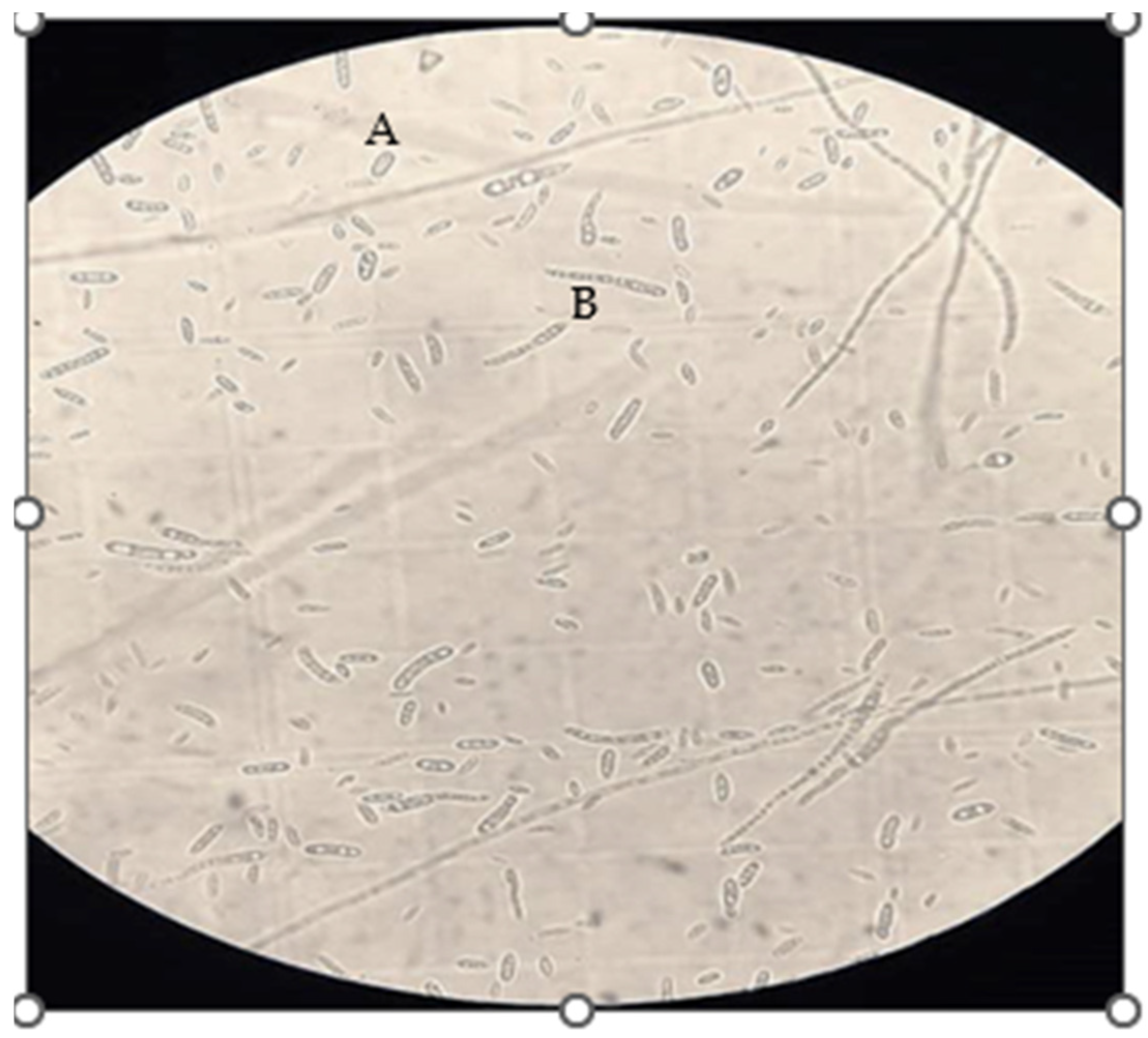
2.1. Fungal Isolation

The arable soil from the agricultural fields of Punjab Agricultural University was used to isolate the Fusarium sp. The fungus was isolated using the serial dilution method, wherein 0.1 mL of sample was poured on a potato dextrose agar medium. The plate was incubated at 25 ± 2 °C for 72 h. Mycelial tips of the fungal isolate grown on the medium were transferred to new PDA plates. The colony characteristics of the fungal isolate were observed after the culture was grown on a PDA for 5 days. The fungal isolate was further purified using the hyphal tip or single-spore method [27] and was identified on the basis of the presence of macro-conidia, micro-conidia, and chlamydospores, as described by Nelson et al. [28] and Leslie and Summerell [29]. The isolate was stored for subsequent screening on PDA plates at 4 °C.
The standard culture of Pleurotus ostreatus MTCC 142 was obtained from the Institute of Microbial Technology in Chandigarh, India.

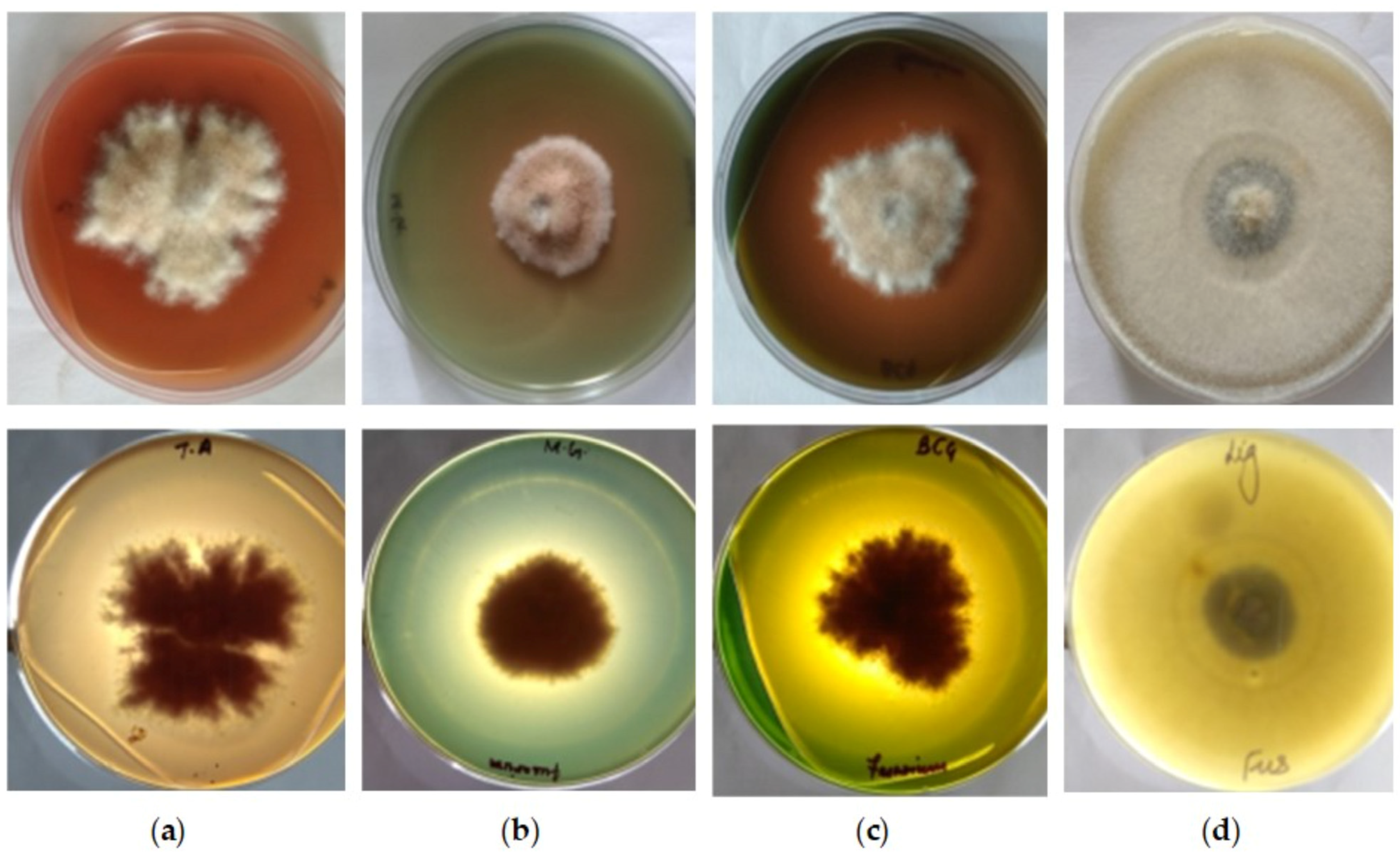
2.2. Qualitative Screening

To study the ability of any pathogen to colonize and to degrade organic dye resembling a plant lignin structure, a plate assay technique was performed for qualitative screening. Thus, a glucose yeast extract agar (GYEA) procured from HiMedia Laboratories in Punjab, India, was prepared by dissolving 1% yeast extract, 2% glucose, and 2% agar in distilled water, and the volume was 1000 mL. The pH was maintained at 7 and sterilized by autoclaving for 20 min at 121 °C at a pressure of 15 psi. Organic dyes (malachite green 0.05%, bromocresol green 0.1%, and tannic acid 0.2%) were used for the initial screening of the isolate. The diameter of the clear zone and fungal growth were measured quantitatively, and the potency index was calculated with the following formula:
P o t e n c y   i n d e x = a r e a   o f   c l e a r a n c e   z o n e   c m 2 a r e a   o f   c o l o n y   c m 2
The ability of the selected fungal cultures to degrade lignin was carried out by inoculating each one on a glucose yeast extract medium (containing 0.5% Glucose) supplemented with lignin (0.5%) as a carbon and energy source. The lignin powder used to determine the Ligninolytic Index (LI%) was purchased from Sigma Aldrich Chemical Private Limited in Punjab, India. The plates were incubated at 25 ± 2 °C for 16 days and were compared to the control with glucose. The ability of Pleurotus ostreatus MTCC 142 and Fusarium sp. to degrade lignin was estimated using the LI.
L I % = C o l o n y   d i a m e t e r   L i g n i n   m e d i a × 100 C o l o n y   d i a m e t e r   c o n t r o l

2.3. Quantitative Estimation of Enzyme Activity

Different flasks containing 100 mL of a Yeast glucose extract broth (YGEB) supplemented with 5% paddy straw were inoculated with 4–5 bits of 8 mm diameter agar plugs consisting of Pleurotus ostreatus MTCC 142 and F. sp., separately. The flasks were incubated at 28 ± 2 °C for 25 days. The supernatant was filtered with Whatman No. 1 filter paper and centrifuged for 20 min at 10,000 rpm (4 °C). All the experiments were performed in triplicate.

2.4. Analytical Procedure

The reaction mixture for determining versatile peroxidase activity contains 0.2 mL of MnSO4 (final concentration of 10 mM as the substrate and 2 mL of sodium tartarate buffer (pH 5.0)), and 0.2 mL of the enzyme filtrate. The reaction was initiated by adding H2O2 (0.2 mL), and the change in the absorbance was recorded every 10 s up to 180 s at 238 nm against a blank without H2O2 [30].
The enzyme activity was calculated according to the following formula:
Z = ( Δ A × V × 1000 ) ( ε × d × Δ t )
where
  • Z = Enzyme activity (UL−1); ∆A: is change in OD per unit of time;
  • V = assay volume in mL;
  • ∆t = change in time (minutes); ε = Extinction Coefficient (M−1cm−1); and
  • d = path length (cm).

2.5. DNA Isolation and Molecular Identification

The fungal mycelium from a seven- to ten-day-old culture, grown in a potato dextrose broth was filtered. Then, using a sterile mortar and pestle, the mycelium was ground in liquid nitrogen [28]. The genomic DNA extraction was performed using the cetyl trimethyl ammonium bromide (CTAB) method [31], purified through chloroform, and precipitated with chilled isopropanol [32]. The working concentration of fungal DNA was maintained at 50 ng/µL for effective molecular confirmation of the VP gene.
Molecular confirmation of ligninolytic peroxidase genes was performed with standard reference to P. ostreatus MTCC 142 [33]. Three VP genes were used for this study. Amplification reactions were made in a volume of 20 µL, which included fungal template DNA (50 ng/µL), primers (0.5 mM), dNTPs (0.25 mM), MgCl2 (1.5 mM), a 1.0 × buffer (20 mM Tris-HCl, pH 8.4, 50 mM KCl), and 0.45 U of Taq DNA polymerase (Invitrogen, Carlsbad, CA, USA). PCR amplifications were carried out in a 9700 thermocycler (Perkin Elmer, Boston, MA, USA) with an initial denaturation for 4 min at 94 °C, followed by 35 cycles of 45 s at 94 °C, 45 s at 57 °C, and 1 min at 72 °C, and a final step of 10 min at 72 °C. PCR products were separated by 2.0% agarose gel electrophoresis using ethidium bromide (50 ng/µL) as a staining agent and visualized under the UV-light spectrum with the AlphaImager 2000 image analysis system (GelDoc2000, BioRad, Hercules, CA, USA). The primers used to identify vp genes are given in Table 1.

3. Results and Discussion

3.1. Qualitative Estimation of Enzyme Production

The colony formed with white aerial mycelia, the microconidia produced on the microconidiophores had an elliptical shape and no septate. The macroconidia were abundantly formed, these were straight to slightly curved in shape with three septa (Figure 1).
The initial screening was generally performed to determine the potential of any fungal culture to produce ligninolytic enzymes and decolorize the organic dyes. A quantitative method in terms of potency index was developed to estimate the production of ligninolytic enzymes. P. ostreatus MTCC 142 and the isolated F. sp. were inoculated on screening media, and the plates were incubated for a period of 16 days. Plates were observed for the development of colored zones, and areas of the colony and halo were measured in terms of square centimeters. A positive response was observed with the appearance of a halo region around the fungal colony, suggesting the ability of ligninolytic enzymes to colorize the dyes present (Figure 2).
A similar study was conducted to investigate the relationship between the enzyme activity of different saprotrophic fungi, and their capacity to decolorize two chemically distinct synthetic dyes, Orange G and RBBR. The results demonstrated a positive correlation between the production of ligninolytic enzymes and synthetic dye decolorization and they concluded that the ligninolytic enzymes cooperated in the decolorization process [35].
In a study conducted in the Sundarban Mangrove ecosystem in West Bengal, India, natural bacterial isolates were examined for their ability to produce ligno-cellulolytic enzymes. All isolates were qualitatively screened based on clear zone formations on selective media supplemented with specific indicators, indicating their ligninolytic, cellulolytic, and pectinolytic activities. One of the isolates, GD1, exhibited laccase activity on the ABTS plate and was able to decolorize congo red, malachite green, and methylene blue, respectively, indicating its laccase property. The degradation of the dyes was confirmed through a FTIR analysis, which revealed the breakdown products based on their functional groups. Additionally, a UV–spectral shift analysis supported the decolorization process, confirming the effects of the potential enzymes produced by the isolated bacterial strains [36].
In a research study, the white-rot fungi Pleurotus ostreatus, P. sapidus, and P. florida were examined for their ability to produce ligninolytic enzymes and degrade dyes. They were tested with azo textile disperse dyes coralene golden yellow, coralene navy blue, and coralene dark red in a liquid medium, at concentrations of 50, 100, and 200 (mg/mL). Additionally, this study involved a HPTLC analysis of the azo dyes and their breakdown products. All three Pleurotus species effectively removed color from all three dyes, and the HPTLC analysis showed the breakdown of the dyes into intermediate products. This study documented various factors, such as dye concentration, pH, and protein and sugar content, that influenced the ability of the fungi to degrade the dyes. It was concluded that the dye degradation was due to microbial action and was not affected by the changes in pH [37].
Table 2, Table 3 and Table 4 and Figure 3a–c show the comparative potency index of a standard culture of P. ostreatus MTCC 142 and F. sp. for tannic acid, malachite green, and bromocresol green. The plates were incubated for 16 days at 30 ± 2 °C. The intensity of the halo around the colony was observed to be in correlation with the ligninolytic enzyme activity. Table 2 represents the results of tannic acid oxidation by selected fungal cultures. The appearance of a yellow- to light-brown-colored region around the colony indicates a positive reaction. F. sp. showed 46% colonization of the plate on the 16th day of incubation, compared to P. ostreatus MTCC 142, which showed 66% colonization after the 16th day of incubation. Meanwhile, F. sp. resulted in 56% and 58% oxidation of tannic acid on the 14th and 16th days of incubation, while P. ostreatus resulted in 62% and 72% oxidation on the 14th and 16th days of incubation, respectively. The results were in agreement with the findings of Sharma et al. [38].
Table 3 shows the potency index for malachite green oxidation. The F. sp. colonized 72% of the plate area on the 16th day, while P. ostreatus MTCC 142 showed 65% colonization after the 16th day of incubation. Meanwhile, the F. sp. resulted in 68% and 79% oxidation of malachite green, while P. ostreatus MTCC 142 resulted in 74% and 77% oxidation on the 14th and 16th days of incubation, respectively. In a similar study, the decolorization potential of the F. spp. HUIB02 strain in minimal medium supplemented with 0.01% malachite green was investigated. It was reported that the F. spp. HUIB02 showed 75% dye decolorization after 15 days of incubation [39].
Abedin [40] reported that F. solani (isolated from the dye-containing effluents) was able to decolorize a relatively high concentration of malachite green (2.5 mg/L) with significant decolorization (96%) after two days of incubation in the nutrient-growing medium. In a study carried out by Eichlerová et al. [41], the ability of Pleurotus calyptratus to decolorize several synthetic dyes of different chemical groups was studied, and it was reported that P. calyptratus decolorized only 5% of the dye after 14 days of cultivation, as white-rot fungi have a low triphenylmethane dye decolorization ability.
Neelamegam et al. [42] studied the effect of the initial concentrations of dyes (such as malachite green, congo red, etc.) on their decolorization pattern using Pleurotus ostreatus MTCC 142. For malachite green, it was observed that the maximum percentage of decolorization was 96% after 10 days of incubation, and also that dye concentration had a positive effect on decolorization, which increased with the increase in concentration.
In a similar study, two synthetic dyes were selected to evaluate the dye decolorization potential of spent mushroom compost (SMC) from Pleurotus compared to commercial laccase. Among the SMCs of Pleurotus spp., P. eryngii SMC showed the highest decolorization activity. The SMC decolorized the diazo-like dye congo red without a mediator. After 3 h, P. eryngii SMC and commercial laccase exhibited similar decolorization abilities, with approximately 93.04% of RBBR decolorized [43].
The positive results for bromocresol green oxidation were demonstrated by the appearance of the yellow-colored halo around the fungal colony. Table 4 and Figure 3c show the potency index for Pleurotus ostreatus MTCC 142 and Fusarium sp. The F. sp. colonized 54% of plate area on the 14th and 15th day of incubation and oxidized bromocresol green from the 7th day onwards, with 60% oxidation on the 16th day. In contrast, the P. ostreatus MTCC 142 showed 66% colonization of the plate after the 16th day of incubation, which resulted in 74% oxidation on the 16th day of incubation. Plate assays revealed the production of ligninolytic enzymes by the F. sp. and therefore, a quantitative estimation of the versatile peroxidase was carried out.
In a similar study, the activity of lignin peroxidase, manganese activity, and laccase activity were determined under in vitro conditions using acid black, congo red, methyl orange, methyl red, and phenol red. Test samples for the enzyme assay were taken from a liquid culture containing 0.02% dye inoculated with selected Pleurotus species. An enzymatic analysis was carried out at 5-day intervals. The four most promising species of Pleurotus (P. flabellatus, P. ostreatus, P. sajorcaju, and P. citrinopileatus) were selected, and dye decolorization was carried out for the five azo dyes (acid black, congo red, methyl orange, methyl red, and phenol red) along with observation of the activities of the ligninolytic (LiP, MnP, and laccase) enzymes. The highest activity of LiP was observed in P. citrinopileatus in a liquid media supplemented with acid black, followed by P. flabellatus on congo red, phenol red, and methyl orange, while the lowest activity was recorded on methyl red by P. citrinopileatus. P. citrinopileatus exhibited the maximum amount of MnP activity in a liquid media containing methyl orange and acid black, followed by P. flabellatus on phenol red and congo red; whereas the minimum amount of MnP activity was recorded on congo red by P. flabellatus. In the methyl orange supplemented liquid media, P. ostreatus showed maximum laccase activity, followed by P. citrinopileatus on acid black, phenol red, and methyl red, with the minimum laccase activity in the congo red-containing media. Among the five dyes, methyl orange decolorization by P. citrinopileatus was faster and earlier, reaching 96.98% on the 20th day of incubation, while the remaining four dyes took 25 days for decolorization. A total of 93.33% of phenol red was decolorized by P. citrinopileatus on the 25th day of incubation [44].
In a similar study, three soil fungal isolates, A. niger, F. sp., and T. lignorum, were chosen to study the biodegradation of the azo dye congo red, as well as the triphenylmethane dyes, crystal violet and methylene blue. The biodegradation study was conducted using three methods: agar overlay and liquid media under stationary and shaking conditions at 25 °C over a period of 10 days. The findings revealed that A. niger achieved the highest decolorization of congo red (74.07%), followed by crystal violet (33.82%) and methylene blue (22.44%), under liquid medium stationary conditions. Meanwhile, T. lignorum achieved the maximum decolorization of crystal violet (92.7%), methylene blue (48.3%), and congo red (35.25%) under the same conditions, marking the first use of T. lignorum as a dye biodegrader or decolorizer. Additionally, F. spp. exhibited superior performance under shaking conditions compared to the stationary and overlay methods [45].
The decrease in color of the dyes can be related to the growth of the P. ostreatus MTCC 142 and Fusarium sp. The discoloration is an indication that Fusarium sp. has the ability to degrade the dyes.
Besides observing the potency index, the present study also observed the ligninolytic index of the F. sp. and P. ostreatus MTCC 142 (Figure 4). However, P. ostreatus MTCC 142 showed the highest ligninolytic index of 97% on the 16th day of incubation, which was comparable to F. sp. which showed the maximum ligninolytic index of 89% on the 16th day of incubation. The ligninolytic index showed that both the cultures were able to produce ligninolytic enzymes and utilize lignin as carbon source.
Similarly, a study was conducted to investigate the impact of lignocellulose and lignin on the growth of Lentinula edodes mushroom mycelium. The cultivation of the mushroom was carried out in a liquid medium for 28 days and on a solid medium for 16 days at a temperature of +22 °C. The liquid nutrient medium was prepared by diluting the malt extract broth twice. The solid nutrient medium was prepared using the same diluted liquid medium with the addition of 20 g/L of agar. This study revealed that the cultivation of L. edodes mycelium in a liquid nutrient medium with the addition of 0.25–0.5% kraft lignin led to an approximately two-fold increase in mycelium biomass yield compared to the reference conditions without the addition of lignin [46]. Lignin-derived phenols and polymeric lignin can promote the growth of fungi. The addition of glucose and lignin to the nutrient growth medium promotes fungal growth, and glucose is necessary for the manifestation of lignin growth-stimulating activity.
In a study, it was shown that lignin can be efficiently degraded by Lentinula edodes. However, the process of lignin degradation and utilization by L. edodes has not been thoroughly discussed. The basic liquid medium was a modified potato dextrose broth (PDB) medium containing 2% (w/v) hemicellulose. Lignin was added to the medium at the beginning of cultivation, providing the final concentrations of 0.05%, 0.10%, and 0.20% (w/v). This study revealed that a concentration of 0.10% lignin was most effective in accelerating mycelia growth, resulting in the highest biomass production [47].

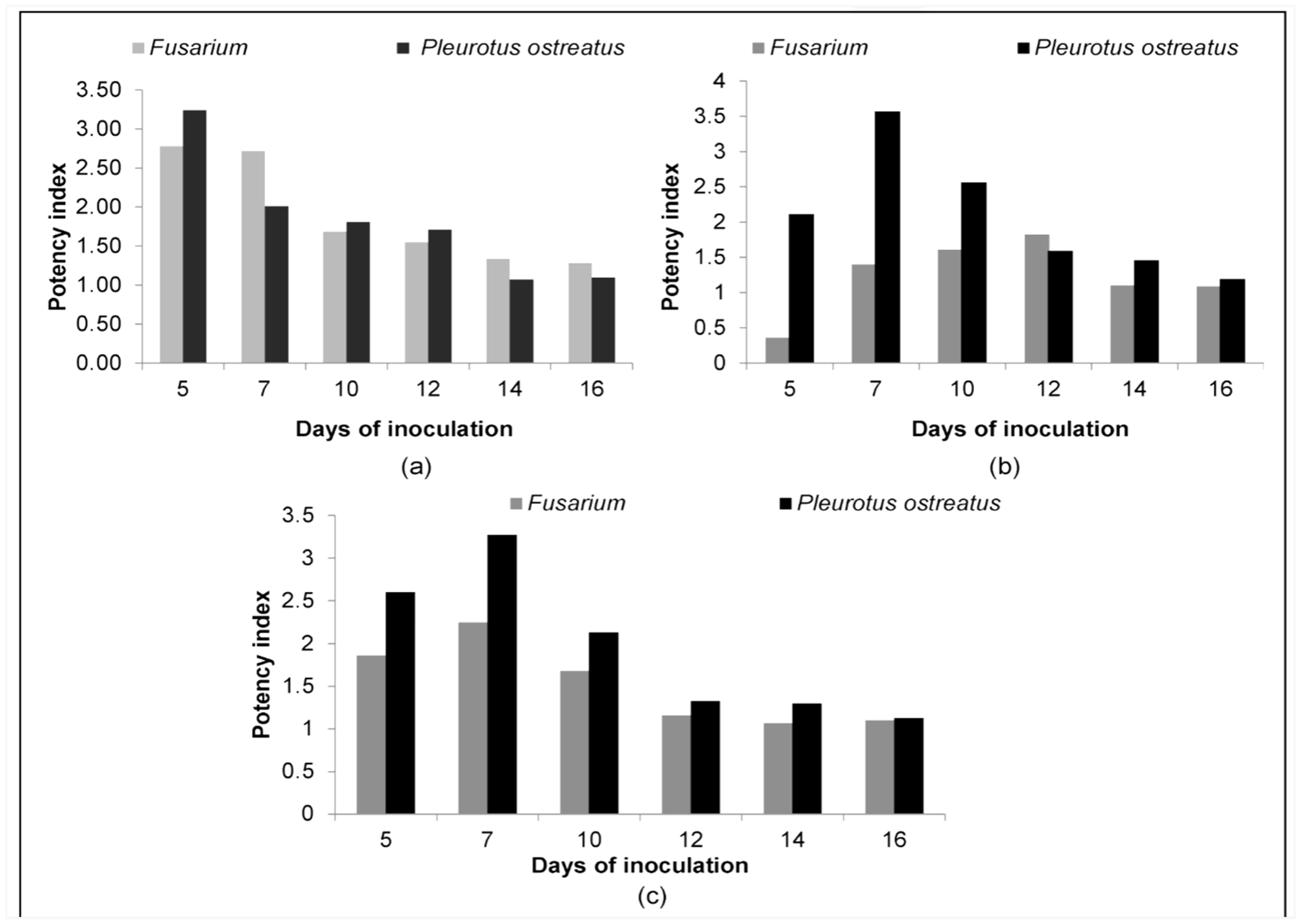
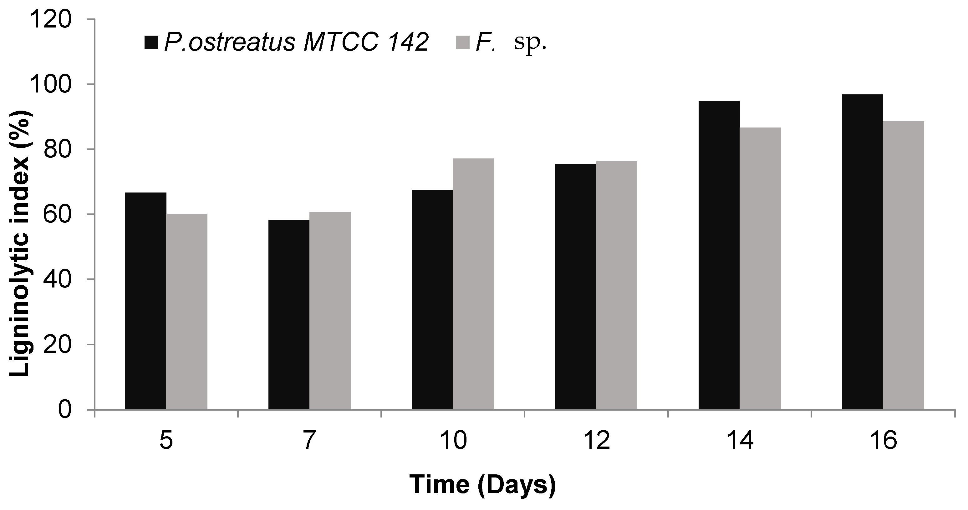
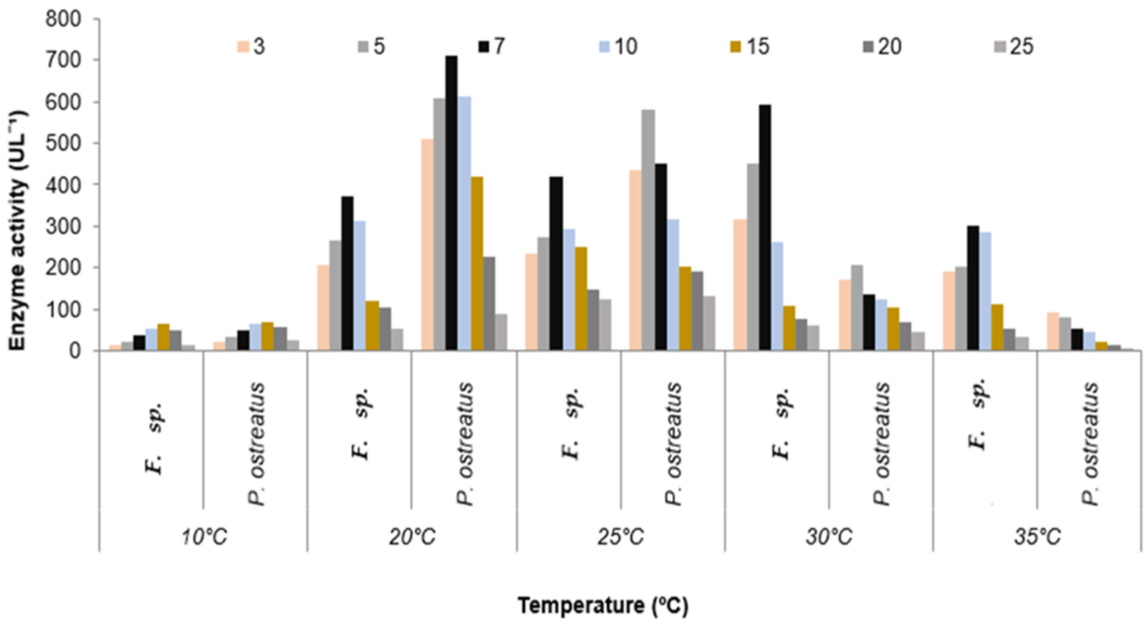
3.2. Quantitative Estimation of Enzyme Production

The versatile peroxidase (VP) activity of F. sp. and P. ostreatus MTCC 142 was recorded and compared over different temperature regimes i.e., 10–35 °C (Figure 5). The effect of the temperature on VP production has not been analyzed systematically, but it is considered that the optimal temperature range for growth is not certainly the optimum range for the expression of enzymes. In the F. sp., the VP activity increased with the increasing temperature until 25–30 °C, while the production at 10 °C was observed to be the lowest. The highest VP activity of 592 UL−1 was recorded at 30 °C on the 7th day after inoculation (DAI). In contrast, in P. ostreatus MTCC 142, the production of versatile peroxidase was favored by low temperatures (Figure 5).
Figure 5 represents the effect of temperature on the VP activity of F. sp. and P. ostreatus MTCC 142 using paddy straw as growth substrate. In the F. sp., the highest VP activity of 592 UL−1 was observed at 30 °C, followed by 419 UL−1 at 25 °C on the 7th DAI. It was noticed that temperature had a remarkable effect on VP activity as little activity was observed at the temperatures below 20 °C and above 35 °C; evidently owed to the fact that increasing temperature inhibits the growth of fungus and consequently, results in decreased enzyme activities. The optimal temperature range for VP activity was found to be 25–35 °C.
The optimum temperature range for the VP activity of P. ostreatus MTCC 142 was observed to be 20–25 °C; however, VP obtained its maximum activity of 711 UL−1 at 20 °C on the 7th DAI, followed by 582 UL−1 25 °C on the 5th DAI and a slight decrease in enzyme activity was noticed with further incubation. The lowest VP activity of 70 UL−1 was observed at 10 °C. The results were similar to those obtained by Eibes et al. [48] while evaluating the temperature effect on the production recombinant VP of P. eryngii in Aspergillus hosts. The experiment was performed at five temperatures; 16 °C, 19 °C, 25 °C, 28 °C, and 31 °C and it was observed that the peak activity of VP (466 UL−1) was obtained at 19 °C, while the lowest activity of 24 UL−1 was obtained at 31 °C. The VP activity reduced slowly as the temperature was increased to 25 °C, while a sharp decline in the activity was noticed by decreasing temperature from 19 °C to 16 °C.

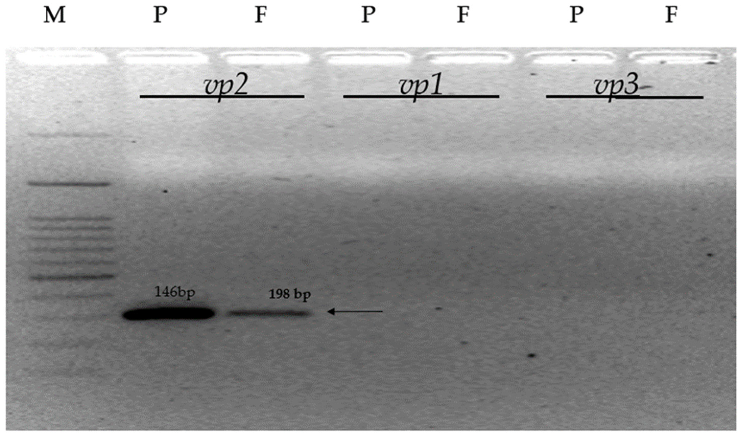
3.3. Confirmation of VP Gene Specific Sequences

The molecular confirmation of the ligninolytic peroxidases genes viz. the versatile peroxidase (VP) gene in F. sp. and P. ostreatus MTCC 142 is shown in Figure 6. SSR primers were used to detect the presence of VP specific genes. P. ostreatus is considered a model organism for lignin degradation, whose genome consists of nine genes of ligninolytic peroxidases, which encode six manganese peroxidase and three versatile peroxidase isoenzymes. Based on the published report [48], the expected size of the PCR product was about 198 bp, 146 bp, and 195 bp for vp1, vp2, and vp3 genes, respectively. Out of the three vp genes, the vp2 gene was present in both the P. ostreatus MTCC-142 and F. sp., when grown on paddy straw, which acted as the substrate, while the vp1 and vp3 gene were not present. However, the presence of one gene out of three could be due to genomic development or the evolution of non-pathogenic F. sp. in this specific region. The preliminary data showed the presence of a PCR product corresponding to the vp2 gene, which requires further confirmation. The observation requires amplicon sequencing to determine the similarities between this and the P. ostreaus gene.

4. Conclusions

In recent times, interest in exploring ligninolytic enzymes to discover more efficient systems for various biotechnological and industrial applications has risen. Earlier studies demonstrated the presence of the ligninolytic enzymes laccase and manganese peroxidase in F. spp., but none have reported the presence and production of versatile peroxidase (VP) from the genus Fusarium. The utilization of paddy straw as a substrate for VP production by F. sp. and its molecular confirmation were the basis of this study. The ability of the F. spp. strain to remove and decolorize synthetic dyes such as malachite green, bromocresol green, and tannic acid was studied, and it was found that F. sp. was able to decolorize and degrade the dyes. Quantitative estimation revealed that the highest VP activity of 592 UL−1 was recorded at 30 °C on the 7th day of incubation, and the optimal temperature range for VP activity was 25–35 °C. To the best of our knowledge, this was the first report using molecular confirmation to identify the presence of VP gene-specific sequences in F. sp. It is concluded from the findings of this study that the strategy to produce VP from F. sp. was successful, and hence, this study paves the way for the use of such a strain in various biotechnological applications.

Author Contributions

Conceptualization, M.P. and U.G.P.; methodology, M.P., S.A.H.P. and U.G.P.; software, M.D., M.P. and S.A.H.P.; validation, U.G.P. and M.P.; formal analysis, M.D., S.A.H.P. and U.G.P.; resources, U.G.P.; original draft preparation, M.P.; review and editing, U.G.P., M.D. and S.A.H.P.; supervision, U.G.P.; project administration, U.G.P.; funding acquisition, M.D. All authors have read and agreed to the published version of the manuscript.

Funding

This research received no external funding.

Institutional Review Board Statement

Not Applicable.

Informed Consent Statement

Not Applicable.

Data Availability Statement

Data are available upon reasonable request.

Conflicts of Interest

The authors declare no conflicts of interest.

References

  1. Marriott, P.E.; Gómez, L.D.; McQueen-Mason, S.J. Unlocking the Potential of Lignocellulosic Biomass through Plant Science. New Phytol. 2015, 209, 1366–1381. [Google Scholar] [CrossRef] [PubMed]
  2. Ahorsu, R.; Medina, F.; Constantí, M. Significance and Challenges of Biomass as a Suitable Feedstock for Bioenergy and Biochemical Production: A Review. Energies 2018, 11, 3366. [Google Scholar] [CrossRef]
  3. Satlewal, A.; Agrawal, R.; Bhagia, S.; Das, P.; Ragauskas, A.J. Rice Straw as a Feedstock for Biofuels: Availability, Recalcitrance, and Chemical Properties. Biofuels Bioprod. Biorefining 2017, 12, 83–107. [Google Scholar] [CrossRef]
  4. Tu, W.-C.; Hallett, J.P. Recent Advances in the Pretreatment of Lignocellulosic Biomass. Curr. Opin. Green Sustain. Chem. 2019, 20, 11–17. [Google Scholar] [CrossRef]
  5. Pandey, P. RRI’s Commitment to Care and Vulnerability of Agrarian Systems: The Problem of Rice Straw Burning in India. Sci. Technol. 2020, 25, 240–255. [Google Scholar] [CrossRef]
  6. Anwar, Z.; Gulfraz, M.; Irshad, M. Agro-Industrial Lignocellulosic Biomass a Key to Unlock the Future Bio-Energy: A Brief Review. J. Radiat. Res. Appl. Sci. 2014, 7, 163–173. [Google Scholar] [CrossRef]
  7. Kaur, K.; Phutela, U.G. Enhancement of Paddy Straw Digestibility and Biogas Production by Sodium Hydroxide-Microwave Pretreatment. Renew. Energy 2016, 92, 178–184. [Google Scholar] [CrossRef]
  8. Giummarella, N.; Pu, Y.; Ragauskas, A.J.; Lawoko, M. A Critical Review on the Analysis of Lignin Carbohydrate Bonds. Green Chem. 2019, 21, 1573–1595. [Google Scholar] [CrossRef]
  9. Ayeni, A.O.; Adeeyo, O.A.; Oresegun, O.M.; Oladimeji, T.E. Compositional Analysis of Lignocellulosic Materials: Evaluation of an Economically Viable Method Suitable for Woody and Non-Woody Biomass. Am. J. Eng. Res. 2015, 4, 14–19. [Google Scholar]
  10. Alvira, P.; Tomás-Pejó, E.; Ballesteros, M.; Negro, M.J. Pretreatment Technologies for an Efficient Bioethanol Production Process Based on Enzymatic Hydrolysis: A Review. Bioresour. Technol. 2010, 101, 4851–4861. [Google Scholar] [CrossRef]
  11. Mosier, N.; Wyman, C.; Dale, B.; Elander, R.; Lee, Y.Y.; Holtzapple, M.; Ladisch, M. Features of Promising Technologies for Pretreatment of Lignocellulosic Biomass. Bioresour. Technol. 2005, 96, 673–686. [Google Scholar] [CrossRef] [PubMed]
  12. Sindhu, R.; Binod, P.; Pandey, A. Biological Pretreatment of Lignocellulosic Biomass—An Overview. Bioresour. Technol. 2016, 199, 76–82. [Google Scholar] [CrossRef] [PubMed]
  13. Saritha, M.; Arora, A. Lata Biological Pretreatment of Lignocellulosic Substrates for Enhanced Delignification and Enzymatic Digestibility. Indian J. Microbiol. 2011, 52, 122–130. [Google Scholar] [CrossRef] [PubMed]
  14. Sahrawat, R.; Garg, A.P. Rice Straw Degradation with Mixed Cultures of Microfungi and Their Enzymes. Int. J. Environ. Clim. Chang. 2023, 13, 2097–2104. [Google Scholar] [CrossRef]
  15. Shi, J.; Chinn, M.; Sharmashivappa, R. Microbial Pretreatment of Cotton Stalks by Solid State Cultivation of Phanerochaete Chrysosporium. Bioresour. Technol. 2008, 99, 6556–6564. [Google Scholar] [CrossRef]
  16. Janusz, G.; Pawlik, A.; Sulej, J.; Świderska-Burek, U.; Jarosz-Wilkołazka, A.; Paszczyński, A. Lignin Degradation: Microorganisms, Enzymes Involved, Genomes Analysis and Evolution. FEMS Microbiol. Rev. 2017, 41, 941–962. [Google Scholar] [CrossRef]
  17. Sani, R.K.; Krishnaraj, R.N. Extremophilic Enzymatic Processing of Lignocellulosic Feedstocks to Bioenergy; Springer International Publishing: Cham, Switzerland, 2017. [Google Scholar]
  18. Kumar, A.; Chandra, R. Ligninolytic Enzymes and Its Mechanisms for Degradation of Lignocellulosic Waste in Environment. Heliyon 2020, 6, e03170. [Google Scholar] [CrossRef]
  19. Shahid, A.; Singh, J.; Bisht, S.; Teotia, P.; Kumar, V. Biodegradation of Textile Dyes by Fungi Isolated from North Indian Field Soil. EnvironmentAsia 2013, 6, 51–57. [Google Scholar]
  20. Ruiz-Dueñas, F.J.; Camarero, S.; Pérez-Boada, M.; Martínez, M.J.; Martínez, A.T. A New Versatile Peroxidase from Pleurotus. Biochem. Soc. Trans. 2001, 29, 116. [Google Scholar] [CrossRef]
  21. Pessôa, M.G.; Paulino, B.N.; Mano, M.C.R.; Neri-Numa, I.A.; Molina, G.; Pastore, G.M. Fusarium Species—A Promising Tool Box for Industrial Biotechnology. Appl. Microbiol. Biotechnol. 2017, 101, 3493–3511. [Google Scholar] [CrossRef]
  22. Gordon, T.R.; Martyn, R.D. The Evolutionary Biology of Fusarium oxysporum. Annu. Rev. Phytopathol. 1997, 35, 111–128. [Google Scholar] [CrossRef] [PubMed]
  23. Escobosa, A.R.C.; Figueroa, J.A.L.; Corona, J.F.G.; Wrobel, K.; Wrobel, K. Effect of Fusarium Oxysporum f. Sp. Lycopersici on the Degradation of Humic Acid Associated with Cu, Pb, and Ni: An in Vitro Study. Anal. Bioanal. Chem. 2009, 394, 2267–2276. [Google Scholar] [CrossRef] [PubMed]
  24. Sellami, K.; Couvert, A.; Nasrallah, N.; Maachi, R.; Abouseoud, M.; Amrane, A. Peroxidase Enzymes as Green Catalysts for Bioremediation and Biotechnological Applications: A Review. Sci. Total Environ. 2022, 806, 150500. [Google Scholar] [CrossRef]
  25. Barber-Zucker, S.; Mindel, V.; Garcia-Ruiz, E.; Weinstein, J.J.; Alcalde, M.; Fleishman, S.J. Stable and Functionally Diverse Versatile Peroxidases Designed Directly from Sequences. J. Am. Chem. Soc. 2022, 144, 3564–3571. [Google Scholar] [CrossRef] [PubMed]
  26. Gupta, K.; Chundawat, T.S. Zinc Oxide Nanoparticles Synthesized Using Fusarium Oxysporum to Enhance Bioethanol Production from Rice-Straw. Biomass Bioenergy 2020, 143, 105840. [Google Scholar] [CrossRef]
  27. Alsohaili, S.A.; Bani-Hasan, B.M. Morphological and Molecular Identification of Fungi Isolated from Different Environmental Sources in the Northern Eastern Desert of Jordan. Jordan J. Biol. Sci. 2018, 3, 329–337. [Google Scholar]
  28. Choi, Y.W.; Hyde, K.D.; Ho, W.H. Single Spore Isolation of Fungi. Fungal Divers. 1999, 3, 29–38. [Google Scholar]
  29. Nelson, P.E.; Toussoun, T.A.; Marasas, W.F.O. Fusarium Species. In An Illustrated Manual for Identification; Pennsylvania State University Press: University Park, PA, USA, 1983. [Google Scholar]
  30. Knop, D.; Levinson, D.; Makovitzki, A.; Agami, A.; Lerer, E.; Mimran, A.; Yarden, O.; Hadar, Y. Limits of Versatility of Versatile Peroxidase. Appl. Environ. Microbiol. 2016, 82, 4070–4080. [Google Scholar] [CrossRef]
  31. Leslie, J.F.; Summerell, B.A. Fusarium Laboratory Workshops—A Recent History. Mycotoxin Res. 2006, 22, 73–74. [Google Scholar] [CrossRef]
  32. Fernández-Fueyo, E.; Castanera, R.; Ruiz-Dueñas, F.J.; López-Lucendo, M.F.; Ramírez, L.; Pisabarro, A.G.; Martínez, A.T. Ligninolytic Peroxidase Gene Expression by Pleurotus Ostreatus: Differential Regulation in Lignocellulose Medium and Effect of Temperature and pH. Fungal Genet. Biol. 2014, 72, 150–161. [Google Scholar] [CrossRef]
  33. Wu, Z.H.; Wang, T.H.; Huang, W.; Qu, Y.B. A Simplified Method for Chromosome DNA Preparation from Filamentous Fungi. Mycosystema 2001, 20, 575. [Google Scholar]
  34. Doyle, J.; Doyle, J. A Rapid DNA Isolation Procedure for Small Quantities of Fresh Leaf Tissue. Phytochem. Bull. 1987, 19, 11–15. [Google Scholar]
  35. Eichlerová, I.; Baldrian, P. Ligninolytic Enzyme Production and Decolorization Capacity of Synthetic Dyes by Saprotrophic White Rot, Brown Rot, and Litter Decomposing Basidiomycetes. J. Fungi 2020, 6, 301. [Google Scholar] [CrossRef] [PubMed]
  36. Das, S.; Sinha, N.; Sen, M.; Ghosh, D. Isolation and Screening of Dye Degrading Lignocellulolytic Bacteria from Sundarban Mangrove Ecosystem, West Bengal, India. J. Pure Appl. Microbiol. 2023, 17, 609–626. [Google Scholar] [CrossRef]
  37. Kunjadia, P.D.; Sanghvi, G.V.; Kunjadia, A.P.; Mukhopadhyay, P.N.; Dave, G.S. Role of Ligninolytic Enzymes of White Rot Fungi (Pleurotus spp.) Grown with Azo Dyes. SpringerPlus 2016, 5, 1487. [Google Scholar] [CrossRef]
  38. Ashktorab, H.; Cohen, R.J. Facile Isolation of Genomic DNA from Filamentous Fungi. PubMed 1992, 13, 198–200. [Google Scholar]
  39. Huy, N.D.; Ha, D.T.T.; Khoo, K.S.; Lan, P.T.N.; Quang, H.T.; Loc, N.H.; Park, S.-M.; Veeramuthu, A.; Show, P.L. Synthetic Dyes Removal by Fusarium Oxysporum HUIB02 and Stimulation Effect on Laccase Accumulation. Environ. Technol. Innov. 2020, 19, 101027. [Google Scholar] [CrossRef]
  40. Goud, B.S.; Cha, H.L.; Koyyada, G.; Kim, J.H. Augmented Biodegradation of Textile Azo Dye Effluents by Plant Endophytes: A Sustainable, Eco-Friendly Alternative. Curr. Microbiol. 2020, 77, 3240–3255. [Google Scholar] [CrossRef]
  41. Eichlerová, I.; Homolka, L.; Nerud, F. Ability of Industrial Dyes Decolorization and Ligninolytic Enzymes Production by Different Pleurotus Species with Special Attention on Pleurotus Calyptratus, Strain CCBAS 461. Process Biochem. 2006, 41, 941–946. [Google Scholar] [CrossRef]
  42. Neelamegam, R.; Baskaran, V.; Dhanasekar, R.; Viruthagiri, T. Decolourization of Synthetic Dyes Using Rice Straw Attached Pleurotus Ostreatus. Indian J. Chem. Technol. 2004, 11, 622–625. [Google Scholar]
  43. Lim, S.-H.; Lee, Y.-H.; Kang, H.-W. Efficient Recovery of Lignocellulolytic Enzymes of Spent Mushroom Compost from Oyster Mushrooms, Pleurotus spp., and Potential Use in Dye Decolorization. Mycobiology 2013, 41, 214–220. [Google Scholar] [CrossRef] [PubMed]
  44. Singh, M. Bioremediation of Synthetic Dyes Pollution by White Rot Fungi (Pleurotus spp.); Reference No. (F. No. 41-1126/2012 (SR)); UGC: New Delhi, India, 2012. [Google Scholar]
  45. Singh, L. Biodegradation of Synthetic Dyes: A Mycoremediation Approach for Degradation/Decolourization of Textile Dyes and Effluents. J. Appl. Biotechnol. Bioeng. 2017, 3, 430–435. [Google Scholar] [CrossRef]
  46. Matjuškova, N.; Okmane, L.; Zaļā, D.; Rozenfelde, L.; Puķe, M.; Krūma, I.; Vederņikovs, N.; Rapoport, A. Effect of Lignin-Containing Media on Growth of Medicinal Mushroom Lentinula Edodes. Proc. Latvian Acad. Sci. Sect. B 2017, 71, 38–42. [Google Scholar] [CrossRef]
  47. Wu, F.; Wang, H.; Chen, Q.; Pang, X.; Jing, H.; Yin, L.; Zhang, X. Lignin Promotes Mycelial Growth and Accumulation of Polyphenols and Ergosterol in Lentinula Edodes. J. Fungi 2023, 9, 237. [Google Scholar] [CrossRef] [PubMed]
  48. Eibes, G.M.; Lú-Chau, T.A.; Ruiz-Dueñas, F.J.; Feijoo, G.; Martínez, M.J.; Martínez, A.T.; Lema, J.M. Effect of Culture Temperature on the Heterologous Expression of Pleurotus Eryngii Versatile Peroxidase in Aspergillus Hosts. Bioprocess Biosyst. Eng. 2008, 32, 129–134. [Google Scholar] [CrossRef] [PubMed]
Figure 1. Morphological characteristics of F. sp. (A) Oval to kidney-shaped 162 microconidia; (B) sickle-shaped, thin-walled macroconidia.
Applmicrobiol 04 00093 g001
Figure 2. In vitro growth of Fusarium sp. on GYEA medium supplemented with (a) tannic acid (top and bottom view); (b) malachite green (top and bottom view); (c) bromocresol green (top and bottom view); and (d) lignin (top and bottom view).
Applmicrobiol 04 00093 g002
Figure 3. Potency index of Fusarium sp. and P. ostreatus MTCC 142 on GYEA medium containing tannic acid (a), malachite green (b), and bromocresol green (c).
Applmicrobiol 04 00093 g003
Figure 4. The % Ligninolytic index of F. sp. and P. ostreatus MTCC 142.
Applmicrobiol 04 00093 g004
Figure 5. Variation in VP activity in F. sp. and P. ostreatus (MTCC—142) at different temperatures. The figure shows the production of versatile peroxidase after 3, 5, 7, 10, 15, 20, and 25 days after inoculation.
Applmicrobiol 04 00093 g005
Figure 6. Molecular confirmation of versatile peroxidase gene in P. ostreatus MTCC-142 and F. sp. (M = 50 bp ladder, P = P. ostreatus MTCC-142, and F = F. sp.; vp 1, vp 2, vp3 = peroxidase genes encoding the versatile peroxidase). The arrow represents the band indicating the presence of the vp2 gene.
Applmicrobiol 04 00093 g006
Table 1. Primers used to detect vp genes (based on P. ostreatus sequences sourced from Fernández-Fueyo et al. [34]).
Gene CodeForward (5′-3′)Reverse (5′-3′)Amplicon Size (bp)
vp1CTCCTGACAACAAGGGAGAAGTCCCAATCAGTTTGCTCTTGTCCTGG198
vp2TATCGCTCGTCACAACATCAGTCTGGGACAAGACCATCAGGTGGA146
vp3TGGATTCTCTCCCACCAAAGGGGCAGTTGGAAACACCTAA195
Table 2. Potency index of cultures for tannic acid oxidation.
DaysPotency Index
Pleurotus ostreatus MTCC 142Fusarium sp.
A1A2IndexA1A2Index
50.640.203.200.200.072.78
72.271.132.016.162.272.71
1014.528.041.8118.1010.751.68
1220.4311.951.7124.6315.901.55
1431.17 (62%) *29.22 (58%)1.0728.27 (56%)21.24 (42%)1.33
1636.32 (72%)33.18 (66%)1.0929.22 (58%)22.90 (46%)1.28
A1: Area of clearance zone, A2: Area of colony. * The percent indicates % colonization of plate.
Table 3. Potency index of cultures for malachite green oxidation.
DaysPotency Index
Pleurotus ostreatus MTCC 142Fusarium sp.
A1A2IndexA1A2Index
50.680.322.110.070.200.36
76.651.863.575.313.801.40
1016.196.332.5617.3510.751.61
1234.3221.571.5926.4214.521.82
1437.39 (74%)25.61 (51)1.4634.21 (68%)31.17 (62%)1.10
1638.48 (77%)32.47 (65)1.1939.59 (79%)36.32 (72%)1.09
Table 4. Potency index of cultures for bromocresol green oxidation.
DaysPotency Index
Pleurotus ostreatus MTCC 142Fusarium sp.
A1A2IndexA1A2Index
56.612.542.60 1.770.951.86
711.343.463.277.073.142.25
1018.868.872.139.625.731.68
1220.5115.411.3325.5222.061.16
1429.22 (58%)22.40 (45)1.3029.22 (58%)27.34 (54%)1.07
1637.39 (74%)33.18 (66)1.1330.19 (60%)27.34 (54%)1.10
Disclaimer/Publisher’s Note: The statements, opinions and data contained in all publications are solely those of the individual author(s) and contributor(s) and not of MDPI and/or the editor(s). MDPI and/or the editor(s) disclaim responsibility for any injury to people or property resulting from any ideas, methods, instructions or products referred to in the content.

