Review

Recent Progress in Terrestrial Biota-Derived Anti-Biofilm Agents for Medical Applications

by
Todorka G. Vladkova
1,*,
Younes Smani
2,3,
Boris L. Martinov
4 and
Dilyana N. Gospodinova
5
1
Department of Polymer Engineering and Laboratory for Advanced Materials Research, University of Chemical Technology and Metallurgy, 8 “Kl. Ochridski” Blvd., 1756 Sofia, Bulgaria
2
Andalusian Center of Developmental Biology, CSIC, Junta de Andalusia, University of Pablo de Olavide, 41013 Seville, Spain
3
Department of Molecular Biology and Biochemical Engineering, Andalusian Center of Developmental Biology, CSIC, Junta de Andalusia, University of Pablo de Olavide, 41013 Seville, Spain
4
Department of Biotechnology, University of Chemical Technology and Metallurgy, 8 “Kl. Ochridski” Blvd., 1756 Sofia, Bulgaria
5
Faculty of Electrical Engineering, Technical University of Sofia, 8 “Kl. Ochridski” Blvd., 1756 Sofia, Bulgaria
*
Author to whom correspondence should be addressed.
Appl. Microbiol. 2024, 4(3), 1362-1383; https://doi.org/10.3390/applmicrobiol4030094
Submission received: 5 August 2024 / Revised: 19 August 2024 / Accepted: 23 August 2024 / Published: 18 September 2024

Abstract

:
The terrestrial biota is a rich source of biologically active substances whose anti-biofilm potential is not studied enough. The aim of this review is to outline a variety of terrestrial sources of antimicrobial agents with the ability to inhibit different stages of biofilm development, expecting to give some ideas for their utilization in improved anti-biofilm treatments. It provides an update for the last 5 years on anti-biofilm plant products and derivatives, essential oils, antimicrobial peptides, biosurfactants, etc., that are promising candidates for providing novel alternative approaches to combating multidrug-resistant biofilm-associated infections. Based on the reduction in bacterial adhesion to material and cell surfaces, the anti-adhesion strategy appears interesting for the prevention of bacterial attachment in combating a broad range of mono- and multispecies bacterial biofilms. So far, few studies have been carried out in this direction. Anti-biofilm coatings made by or containing biologically active products from terrestrial biota have scarcely been studied although they are of significant interest for a reduction in infections associated with medical devices. Combination therapy with commercial antibiotics and natural products is accepted now as a promising base for future advances in anti-biofilm treatment. In vivo testing and clinical trials are necessary for clinical application.

1. Introduction

Biofilms are communities of microorganisms attached to biotic or abiotic surfaces embedded in a self-elaborated extracellular matrix (ECM). Non-desirable biofilm formation as a reservoir of microbial contamination is a hazard to human health, and a persistent problem for the healthcare, marine, and water-based industrial productions. The need to control biofilm formation promotes international interest in the development of mitigating strategies that include anti-biofilm agents and the redesign of material surfaces.
Providing an environment for the most stable microbial life, medical biofilms cause extensive antibiotic resistance, morbidity, and mortality, and substantial economic loss. Biofilms could be the leading cause for the failure of antibiotic treatments and play an important role in infections associated with both medical devices and tissues, many of them chronic [1].
The use of orthopedic implants, cardiovascular and ureteral tract devices, orthodontia prosthetics, and other devices is sometimes associated with infections due to the detachment of microorganisms from the biofilm on the medical device. The very common urinary tract infections are an example. Microorganisms may colonize the outside surface of the catheter by direct inoculation during catheterization or by migration through the surrounding mucous sheath. The microbial biofilm on the inner catheter surface in patients with long-term catheterization hampers antibiotic treatment and causes chronic infection [2]. Biofilm formation on long-term medical implants, such as pacemakers and heart valves, prosthetic joints, and breast implants, leads to postoperative complications. Infections could cause inflammation and tissue destruction around the implant that sometimes are life-threatening. The use of large doses of antibiotics that may cause toxicity in the host, and/or removal of the infected device, are the only possible options, which may have a lethal impact for some patients [3]. Furthermore, infections associated with medical devices due to microbial attachment and biofilm formation on their surface have a high economic cost. For example, biofilm infections associated with prosthetic devices affect more than 4.1 million patients per year, with a total cost of the treated complications of around 7 billion euros in Europe [4,5].
Microorganisms adhering to biotic surfaces form biofilms in different tissues of the host, like epidermal cells and teeth, mucus on mucosal membranes, and chronic wounds. Multispecies biofilms could be formed in host intestines. Oral multispecies biofilm formation is very common. Gastrointestinal infections could also be caused by multispecies biofilm formation. Biofilm-associated persistent inflammations and chronic infections hide an increased risk of cancer development, like gallbladder cancer [6].
Numerous reports highlight alarming antimicrobial resistance rates [6,7]. The role of biofilms in antimicrobial resistance (AMR) is complex and significantly increasing resistance. The cells in a biofilm have intrinsic characteristics different from those of planktonic cells. Bacteria living in a biofilm can cause an increase of about 10- to 1000-fold in antibiotic resistance as compared to similar bacteria living in a planktonic state [8].
Biofilms act as physical barriers against drugs and host immune responses. They reduce the possibility of eradicating infections and cause relapses after the traditional appropriate treatment. The eradication of biofilms is challenging and more complicated in the predominant multispecies microbial biofilms. Many researchers are working to address biofilm-related infections. Some anti-biofilm strategies were developed and identified as promising. However, after about one hundred years since the first evidence of the mode of development and decades of intensive investigations, there are no therapeutic solutions to eradicate bacterial biofilms and their biomedical-related issues [9]. The failure of conventional antibiotic therapies and multidrug treatments indicates that biofilm control needs a significant upgrade [10].
Utilization of natural biologically active substances having relatively low toxicity, high efficiency, and little or no capacity to induce antimicrobial resistance rose as a new trend in combating biofilms during recent decades. Our former review [11] presented a five-year update, up to 2022, of marine organism-derived anti-biofilm agents. The terrestrial biota is also rich in biologically active substances whose anti-biofilm potential has not been studied enough up to now. The focus of this review is on medical bacterial biofilms and anti-biofilm approaches based on the utilization of natural products from the terrestrial biota. Its aim is to outline the variety of terrestrial sources and new antibacterial agents with the ability to inhibit different stages of biofilm formation, expecting to give birth to some ideas for their utilization in improved anti-biofilm treatments. This review is an update of the previously published review, from 2019 until now, on anti-biofilm plant products and derivatives, essential oils, antimicrobial peptides, biosurfactants, etc., and their anti-biofilm ability. A brief presentation is also included here of the anti-biofilm strategies based on the current knowledge about the mode of development and composition of the bacterial biofilm.

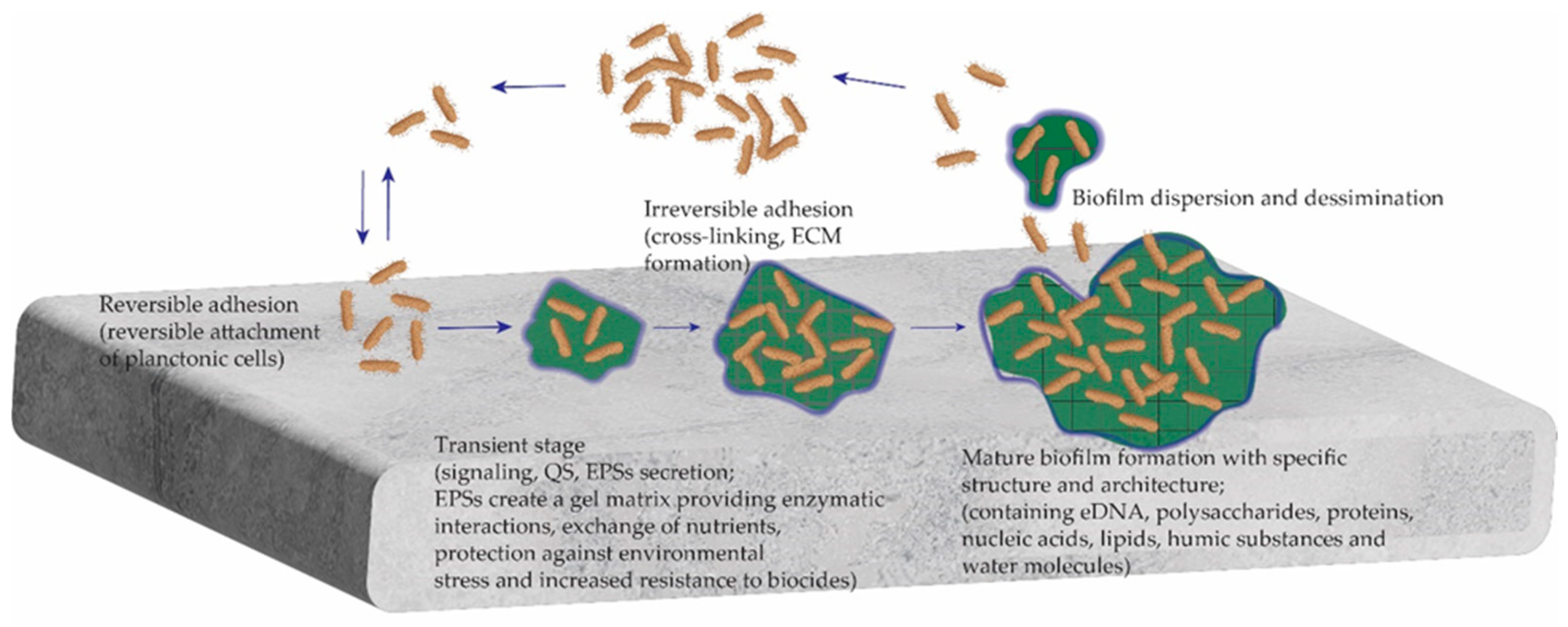
2. Composition and Mode of a Bacterial Biofilm Development

A bacterial biofilm is a complex, surface-attached community of microorganisms engaged in extracellular polymeric substances (EPSs) that create a gel matrix providing enzymatic interactions, exchange of nutrients, protection against environmental stress, and increased resistance to biocides. The EPSs produced by microorganisms are a mixture of biopolymers consisting of polysaccharides, proteins, nucleic acids, lipids, and humic substances. EPSs make up the intercellular space of microbial aggregates and form the structure and architecture of the biofilm extracellular matrix (ECM). Key functions of the EPSs comprise the mediation of the initial attachment of microbial cells to different substrates and protection against environmental stress and dehydration. In a biofilm state, the microbes are more resistant to antibiotic and multidrug treatments [12,13,14].
A generalized concept of microbial biofilm formation includes several stages (Figure 1). Initially, planktonic cells reversibly attach to a surface (reversible adhesion) and remain in this transient state until signaling by an environmental cue. This is a cell-to-cell communication mechanism, a phenomenon known as quorum sensing (QS). It plays an important role in biofilm development and balances the environment when the bacterial density becomes high. Once microorganisms begin to secrete EPSs, the biofilm develops into an irreversible process due to a cross-linking. In the mature biofilm, the cells are already engaged in an ECM composed mainly of proteins, exopolysaccharides, and extracellular DNA (eDNA).
The development of anti-biofilm strategies should take into account all possible interactions used by the microbes to adhere and develop a biofilm, including the competitive relationships in multispecies biofilms. Any event included in the biofilm formation could be a target of its control, but combating the biofilm seems to be easiest at the initial stage of its development (reversible attachment, reversible adhesion), and this could be an interesting opportunity to limit microbial growth prior to human infection [15,16,17,18,19,20]. As the bacterial biofilm is perceived as a dynamic multistage process in which bacterial attachment on solid surfaces is the prerequisite for biofilm formation, the interference with the attachment is the most promising environmentally benign option to be inhibited [15].

3. Principal Anti-Biofilm Approaches

The principal anti-biofilm approaches can be divided into three main groups (Figure 2):
(i)
Biocidal, including killing of pathogenic, biofilm-forming microbes and development of both contact killing or biocide-releasing surfaces, in the case of infections associated with medical devices;
(ii)
Non-biocidal approaches, including mechanical removal of the biofilm, if it is possible; minimizing the reversible microbial cell attachment and development of anti-adhesive material surfaces in the case of infections associated with medical devices; QS disruption; inhibition of the cross-linking of the EPSs; ECM disruption; and activation of biofilm dispersal;
(iii)
Combination treatments, including treatment by combinations of different anti-biofilm agents; combinations of antibiotics and terrestrial biota-derived anti-biofilm agents; utilization of multi-functional anti-biofilm agents; and development of anti-adhesive material surfaces that deliberate antimicrobial agents [16,17,18].
The focus of this review is on bacterial biofilms and anti-biofilm approaches (biocidal, non-biocidal, and combinatorial) based on the use of different natural products: plant-derived products, essential oils, natural quorum sensing inhibitors, enzymes and other biologically active ECM-degrading substances, anti-biofilm peptides, biosurfactants, etc., called by some authors “biological approaches”, unlike the chemical and physical ones [20].

3.1. Biocidal Approaches

The biocidal approaches are based on the use of antimicrobial agents that kill bacterial cells (biocides). They include development of contact killing or antimicrobial agent (biocide)-deliberating biomaterial surfaces of medical devices. Contact killing surfaces are usually created by grafting of an antimicrobial agent or deposition of a coating containing such an agent; the surfaces of both types kill microbial cells in contact with them. Antimicrobial agent (biocide)-deliberating biomaterial surfaces are usually created by deposition of gel or another type of coating that deliberates biocide and kills microbial cells in contact with them. Anti-biofilm agents that kill pathogenic microbial cells are enzymes, antimicrobial peptides inspired by natural chemical compounds, and other natural biocides [21].
Examples of enzymes that kill microbial cells are peptidoglycan-degrading enzymes and phage-derived polysaccharide depolymerase [22]; combined DNase and proteinase, which are effective against multispecies oral biofilms [23]; proteinase K and lysozyme, with proven anti-biofilm activity; and others. Some researchers believe that the complete elimination of heterogeneous biofilms needs amalgamation of hydrolytic enzymes that can degrade proteins, polysaccharides, eDNA, and QS molecules [24]. Several features of the enzymes limit their practical application as anti-biofilm agents in the creation of anti-biofilm biomaterial surfaces: (i) Carrying out the function while keeping the own natural activity. Any enzyme should be structurally stable and have some structural mobility in aqueous solutions. Simultaneously, the enzyme should be released in water media, which is difficult to provide when it is included in an anti-biofouling coating. (ii) The enzyme activity increases, whereas its stability decreases, with a temperature increase. Finding an optimal balance between enzyme activity and stability is a challenge in enzyme-based anti-biofouling coatings [18].
Immobilization of antimicrobial peptides or other biologically active substances that kill microbial cells, and deposition of antimicrobial coatings and deliberating anti-microbial agents, are largely studied approaches for the creation of contact killing or biocide-releasing surfaces in the case of biofilms associated with medical devices [13].

3.2. Non-Biocidal Approaches

The non-toxic control of biofouling is based on approaches that exclude the killing of microbial cells. The non-biocidal approaches include mechanical removal of biofilm, if possible, and affecting the biofilm development stages, i.e., minimization of cell–surface adhesion; hampering and disruption of cell-to-cell signaling, i.e., quorum sensing; inhibition of the cross-linking of EPSs; disruption of the formed ECM; and biofilm dispersion. All these are studied to combat medical biofilms and infections associated with medical biofilms, and have lately used a variety of terrestrial biota-derived biologically active substances [25].

3.2.1. Mechanical Removal of Biofilms

Mechanical removal of dental and some other biofilms has achieved limited success. Irrigation with water jets and debridement followed by aggressive antimicrobial therapy have been widely carried out for oral, wound, and prosthetic joint biofilm-related infections, although it was found that biofilms spread across the surface and cause a persistence of bacteria on surfaces after these treatments [26]

3.2.2. Minimization of Initial Adhesion

Bacterial attachment on solid surfaces is a prerequisite for biofilm formation, and minimization of the initial cell adhesion is accepted as the most promising environmentally benign option to antifouling [15]. Minimization of the initial adhesion could be achieved by creation of a strong hydrophilic (with a water contact angle (WCA) approaching zero) or a strong/super hydrophobic surface (with surface tension, γ, approaching zero) [16,18,27,28]. The presence of a surfactant/biosurfactant on such a surface contributes to additional minimization of the reversible microbial cell’s attachment [29,30]. ECM-degrading enzymes, i.e., D Nase I, dispersin B, and lysostaphin, also inhibit microbial adhesion to surfaces [1]. Attempts were made to coat biomaterials or to immobilize biologically active substances aimed at decreasing the adhesion of pathogenic microorganisms to the surface of indwelling medical devices. Some examples are the anti-adhesive coatings based on cyanobacterium Cyanothece spp. derivatives, which are active against Proteus mirabilis, E. coli, and Candida albicans biofilms in urinary tract infections [31].
Generally, terrestrial biota-derived anti-biofilm agents are not sufficiently studied as anti-adhesive coatings, or a component of composite coatings, although phyto-compounds and plant extracts are known to play a significant role in bacterial adhesion. For example, the crude extract of Adiantum philippense, which shows a promising role in decreasing the content of biofilm exopolysaccharides, restrains biofilm formation at the initial stage by targeting adhesive proteins [32]. The inhibition of bacterial adhesion and anti-biofilm effects of biosurfactants (BSs) extracted from Bacillus cereus and Serratia nematodiphila were found against Staphylococcus aureus and Pseudomonas aeruginosa. The combination of BSs from B. cereus and S. nematodiphila demonstrates synergistic effects, e.g., a decrease of about 60% in the adhesion of S. aureus and P. aeruginosa, and a decrease of about 80% in the biofilm mass, which indicate an increased inhibitory potential against pathogenic microbial attachment and biofilm eradication [33].

3.2.3. Quorum Sensing Inhibition

Pathogenic microbial cells forming biofilms on indwelling medical devices (orthopedic implants, urinary catheters and stents, cardiovascular devices, etc.), human skin, oral cavities, and other surfaces communicate through quorum sensing (QS) signals that regulate both microbial biofilm development and pathogenesis. Numerous natural bioactive agents that are able to hamper cell-to-cell communication have been identified so far, including enzymes degrading autoinducer compounds, blocking autoinducer production, or blocking their receptors through addition of some “inhibitor” compounds mimicking them [34]. Interference with QS was already identified as a novel potential therapeutic approach to the treatment of bacterial infections. The effect of QS molecules, like Farnesol (a natural 15-carbon organic compound which is an acyclic sesquiterpene alcohol), was demonstrated on mixed biofilms of C. albicans and S. aureus [35]. The first compound identified as a broad-spectrum QS inhibitor is a sulfur-rich molecule from garlic Ajoene. It decreases the expression of small regulatory RNAs in both Gram-negative (P. aeruginosa) and Gram-positive (S. aureus) bacteria [36]. Metalloproteinase AHL-lactonase from cell-free lysate of endophytic Enterobacter species blocks cell-to-cell communication in pre-formed biofilms and regulates virulence factor production, and thus significantly inhibits biofilm formation by Aeromonas hydrophila [37].
Diversity of bacteria, bacterial enzymes, and secondary metabolites known to interfere with the QS mechanisms of pathogenic bacteria are studied as anti-biofilm and anti-QS drugs against pathogenic bacteria [38]. Natural products, based on quorum quenching (QQ) enzymes, i.e., AiiA, AHL lactose, AHL acylase, and others, could block cell-to-cell communication and regulate virulence factor production. They disrupt QS systems in two main ways: (i) inhibiting and degrading signal molecules, and (ii) mimicking the signal molecules to inhibit their binding to corresponding receptors. QS inhibitors are discussed by some researchers as the best anti-biofilm alternative to antibiotics [39]. Lactobacillus crustorum ZHG 2-1 is reported as a QQ bacterium that functions as a P. aeruginosa anti-biofilm agent [40].
Essential oils (EOs) and other plant-based natural products are found to demonstrate anti-QS activities against pathogenic bacteria and are accepted as promising anti-biofilm agents [36,37]. A number of BSs of microbial origin were found to destroy cell-to-cell communication [41]. Two main types are rhamnolipieds, produced predominantly by the Gram-negative bacteria P. aeruginosa, and sophorolipieds, produced mainly by Candida bombicola species [42]. In 2023, Adnan et al. [43] demonstrated that a BS derived from probiotic strain Lactobacillus acidophilus inhibits QS-regulated virulence against three Gram-negative bacteria and exhibits broad-spectrum anti-biofilm activity.

3.2.4. Inhibition of the Cross-Linking of Exopolymeric Substances, Degradation of the Extracellular Matrix (ECM), Biofilm Dispersal

A complex mixture of biopolymers secreted by the pathogenic bacteria, EPSs mediate the initial attachment of cells to different substrates and form the extracellular matrix (ECM) after cross-linking [12,13,14,15,44]. Many anti-biofilm approaches are focused on the inhibition of EPS cross-linking, degradation of the ECM, and finally biofilm dispersal. All these can be achieved via the use of natural products, such as antioxidants and enzymes. Supposing that the cross-linking of bacterial EPSs is carried out by an oxidative mechanism (as was demonstrated earlier for some macro-biofoulers), the effect of several antioxidants was tested as a component of low-adhesion siloxane coatings. It was found that some antioxidants significantly inhibit, whereas others stimulate, the biofilm formation on the coated surface. The effect depends on the chemical nature of the antioxidant, the type of microfouler, and the conditions under which the mono-spaces or multispecies biofilm develop [29,30,45]. The complexity and heterogeneity of the microbial EPSs and the ECM composition require hydrolases and leases in order to achieve efficient enzymatic degradation [18].
Biofilm dispersal is often assisted by surfactants or/and other substances, like enzymes, that are able to break already established cross-links [16]. Enzymes, such as beta-N-acetylglucosaminidase and dispersin B, secreted by the Gram-negative periodontal pathogen Actinobacillus actinomycetemcomitans, disintegrate mature biofilms of S. epidermidis [24]. The design of enzymatic biofilm dispersal using nanotechnology demonstrates improved anti-biofilm activity and achieves good results [46,47].

3.3. Combination Approaches

Combination approaches include simultaneous application of antibiotics and anti-biofilm agents, the combination of several anti-biofilm agents, or minimizing the cell–surface adhesion and application of antibiotics and/or anti-biofilm agents.
Biofilm formation is a dynamic and complex process starting with the initial reversible attachment of microbial cells, and this stage is accepted as the easiest for the inhibition of the biofilm development. However, the large diversity of both microbial species and adhesive substances secreted by them, and the variety of material surfaces on which a biofilm develops, make the complete elimination of the initial (reversible) adhesion difficult. In addition, the same microorganisms secrete different EPSs against different surfaces [16]. For example, EPSs produced by E. coli demonstrate a predominant amount of chemically bound free EPSs under different growth conditions, with over 500 proteins identified in the free EPSs [48]. Combating an already developed biofilm is much more complicated and requires a complex strategy. Creation of surfaces that reduce the adhesion of microbes and provide biocidal activity or combinatory effects emerges as a major global strategy in combating bio-contamination. The function of natural anti-biofilm agents appears to be more effective in combination approaches as compared with their single use in terms of antibacterial activity, decreased toxicity, etc. [10].

3.3.1. Antibiotic Treatment Simultaneous with Natural Medicine

Antibiotic treatment simultaneous with natural medicine seems to be a promising therapy for bacterial biofilm infection. For example, combined with levofloxacin, a plant-derived anti-neuropeptidesodium, houttuyfonate better inhibits P. aeruginosa biofilm formation [49]. A pre-treatment by naringin (a flavanone glycoside, extracted from citruses) followed by ciprofloxacin and tetracycline antibiotic-treatment is more effective against P. aeruginosa biofilms than individual treatment with the same antibiotics. The effect of naringin is due to the depletion of biofilm EPSs and facilitation of the diffusion of antibiotics, thereby reducing pellicle formation and decreasing the flagella movement of bacteria on medical device surface [50]. Nisin, an FDA-approved AMP, produced by certain Gram-positive bacteria (Lactococcus and Streptococcus spp.), and found in plants and in dairy environments, acts as anti-biofilm agent synergistically with conventional antibiotics against methicillin-resistant S. aureus, S. pneumoniae, Enterococci, and Clostridium difficile [51]. AMPs produced by insects can act synergistically with classical antibiotics for a double-pronged attack on infections [52]. Although combinations of plant-based products and antibiotics have been studied and discussed widely, they are currently poorly explored and for a reduced number of bacterial species only [9].

3.3.2. Natural Products with Multiple Anti-Biofilm Activities

Some natural products demonstrate multiple anti-biofilm activities. Significant anti-QS and biofilm dispersal activities were found in honey against multi-species bacterial biofilms [53]. Disruption of established bacterial and fungal biofilms by a nutraceutical enzyme and botanical extract blend (NEBB) was demonstrated in vitro for five microbial strains, C. albicans, S. aureus, S. simulants (coagulase-negative, penicillin-resistant), Borrelia burgdorferi, and P. aeruginosa, causing chronic human illnesses. The mass of post-treated biofilm (evaluated by crystal violet staining) and the metabolic activity (measured by MTT assay) were significantly influenced by NEBB treatment [54].

3.3.3. Nanoparticles Containing Natural Products

Unlike antibiotics, which are used to treat infected patients, nanomaterials provide an interesting opportunity to limit microbial growth prior to human infection. This led to the development of engineered antimicrobial materials, targeting different applications with combinations of natural products and biogenic synthetized metal, metal oxide, or composite nanoparticles as the active antimicrobial agents [55,56].
The pandemic coronavirus disease 2019 (COVID-19) motivated a survey focused on nanotechnology-based strategies to develop functional antimicrobial and antiviral surface coatings that include combinations of biologically active compounds [57]. Use of biologically active polymers, such as chitosan in combination with silver, zinc oxide, and others, attracts attention to combat persistent, antimicrobial-resistant bacterial biofilms [58].

3.4. Some Considerations for the Design of Anti-Biofilm Surfaces

Based on a large number of literature sources and the authors’ own experience, some conclusions about the design of anti-biofilm surfaces were made. Since biofilm formation on a medical device is a complex phenomenon, it requires a complex strategy. The most important prerequisite for a material surface to be clean from biofilm is to be low-adhesion, i.e., strongly hydrophilic, water-like or strongly/super hydrophobic, flat and slippery, or with special micro/nanotopography, that is, a protein-rejective surface [16,27].
When microbial cells contact any surface, they start to secret EPSs containing adhesive proteins, which support their attachment to the surface. If one is able to stop the protein and EPS adsorption, the biofilm development can be stopped. Unfortunately, the proteins have a versatile nature and, in front of a complementary surface, they attach to a different mechanism. This makes it almost impossible to completely stop the protein adsorption. Therefore, a sharp reduction in biofilm, but not a complete lack, is observed on such surfaces [27,28,59]. In addition to low adhesion, it will be beneficial for the anti-biofilm surface to contain a relevant surfactant and/or inhibitor of both QS and EPS cross-linking [30], as well as agents breaking already established cross-links and biofilm dispersal, to avoid the insufficient microbial settlement of the low-adhesion surfaces [59].

4. Anti-Biofilm Agents from the Terrestrial Biota

Anti-biofilm agents from the terrestrial biota are a subject of numerous reviews, original papers, and books aiming to highlight new prospects for the development of efficient alternatives to the currently used but insufficiently effective antibiotic and multidrug treatments. The potential anti-biofilm agents from the terrestrial biota include plants and herbal extracts, essential oils, biosurfactants (BSs), and antimicrobial peptides (AMPs), which target different stages of biofilm formation on different mechanisms [1]. With or without considerable structural alterations, they could be applied to the treatment of biofilm-associated infections, as well as to the development of anti-biofilm surfaces. The design of effective anti-biofilm molecules requires their minimum inhibitory concentrations to eradicate biofilm-associated infections without causing toxic effects. The knowledge accumulated in the last decades demonstrates that many natural antibacterial products are more efficient for inhibition of biofilm development and have less side effects than their chemically synthesized counterparts. Natural anti-biofilm compounds can attack one or more stages of biofilm formation in combination, or not, with antibiotics [60].
Identified natural products with anti-biofilm activity include elligic acid glycosides, hama melitannin, carolacton, skyllamycins, promysalin, phenazines, bromoageliferin, flustramine C, meridianin D, and brominated furanones. Medicinal chemistry programs facilitate structure confirmation, identification of critical structural motifs, better understanding of mechanistic pathways, and the development of more potent, more accessible, or more pharmacologically favorable derivatives of anti-biofilm natural products [61]. Natural polymers in different forms are reported to help antibiotics transport to the targeted site, mainly for periodontal biofilm-forming pathogens [62]. the anti-biofilm potential of Actinobacterial compounds from different terrestrial or aquatic microbial species against various pathogenic bacteria is supposed to be due to the cell–surface and cell–cell interaction [63]. Xu et al. [64] recently reported targeting only biofilm-forming pathogenic microorganisms without influencing the normal microflora. Chi et al. [65] summarized the importance, functions, and anti-biofilm effects of natural products from traditional medicine, targeting multi-species oral biofilms. The anti-biofilm activity of 11 natural compounds and their derivatives (phenyl propenes and phenolic aldehydes, eugenol, ferulic acid, sinapic acid, salicyl aldehyde, vanillin, cinnamoyl acid, and aldehydes) was tested against 32 clinical isolates of S. aureus. Using both qualitative and quantitative assays, followed by qPCR analysis to examine the differences in the expression levels of biofilm-forming genes (ica-A, fnb-B, clf-A, and cna) in treated and untreated clinical isolates, it was found that the tested natural compounds and their analogues exhibit significant antimicrobial and anti-biofilm activity against S. aureus [66].
Lately, the potential value of new neutral deep eutectic solvents (NADESs) as anti-biofilm agents was demonstrated. NADESs are considered “natural” because their constituent components are primary metabolite groups (which are naturally used by the plants for their self-survival), such as sugars, organic acids and bases, and amino acids. A potential use of NADESs as anti-biofilm agents is proposed due to their ability to solubilize and stabilize biological macromolecules [67]. The potential capacity of a huge number of natural and synthetic compounds as inhibitors of bacterial biofilm formation is widely presented. This includes the natural chemical compounds resisting bacterial biofilm growth by different mechanisms; the stage of biofilm formation at which the chemical compound is introduced into the biofilm system; polymer matrix formation inhibition; suppression of the cell adhesion and cell attachment to itself or to an external surface; and interruption of the extracellular polymeric matrix generation and decrease in virulence factor production [68].

4.1. Plant Products and Derivative Compounds

Long-known medicinal plants rich in bioactive compounds with diverse structures and functional groups are presented now as demonstrating anti-biofilm properties. Plant-based products are usually crude or fraction extracts, derived anti-biofilm compounds, and secondary metabolites [69,70].
A crude extract of Adiantum philippense L. was found to decrease the content of biofilm exopolysaccharides. It restrains biofilm at the initial stages by targeting adhesion proteins, deforming the pre-formed biofilms and obstructing EPS production [32]. The anti-biofilm activity of extracts from the Moringa oleifera plant and Citrus sinensis fruit (known to treat multiple human infections) was evaluated against pathogenic P. aeruginosa and S. aureus biofilms. The experimental results show that the peel extract of C. sinensis and the flesh extract of M. oleifera efficiently inhibit the formation of such biofilms by sub-inhibitory concentrations. The comparative study reveals a better anti-biofilm activity of the M. oleifera extract than that of C. sinensis against mixed cultures [71].
Several classes of natural compounds with anti-biofilm properties are known: phenolics, essential oils, terpenoids, lectins, alkaloids, polypeptides, and polyacetylenes [72]. Malaysian plant species are known to control biofilm infection by QS pathway inhibition, disruption of the ECM, alteration of cell permeability, and reduction in the hydrophobicity of the cell surface. The phytocompounds having a curlicide and pilicide nature can be exploited in therapeutic strategies against Enterobacterales biofilm development [73]. Guzzo et al. [74] presented the anti-biofilm activity and molecular mechanisms of action of plant-derived natural products against P. aeruginosa and S. aureus, together with an update of anti-biofilm properties of terpenes, flavonoids, alkaloids, and phenolic compounds. A survey of natural and synthetic plant compounds as anti-biofilm agents against seropato-type E. coli (STEC) biofilm was directed to a development of antibacterial drugs against STEC-induced infections [75]. In search of alternatives to the conventional antibiotics and safe therapies, natural plant-derived phytomedicines are discussed now as promising candidates to combat microbial biofilms. The efficacy, characteristics, and corresponding mechanisms of action of phytochemicals, biosurfactants, antimicrobial peptides, and their sources were included [76].
Plant secondary metabolites are also studied as anti-biofilm agents. Secondary metabolites from seeds of Annona senegalensis were tested for their antimicrobial, anti-biofilm, and anti-QS activities using three test microorganisms (S. aureus, E. coli, and Chromobacterium violaceum). Column chromatography of the seed extract affords N-cerotoyltryptamine, asimicin and ent-19-carbomethoxykauran-17-oic acid. The testing of their antimicrobial, anti-biofilm, and anti-QS activities demonstrates that these compounds inhibit biofilm formation of the test microorganisms to different extents in a dose-dependent manner [77]. For the first time, the activity of 20 secondary metabolites produced by pathogenic fungi of forest plants (belonging to different classes of natural compounds) was evaluated against clinical isolates of antibiotic-resistant Gram-negative and Gram-positive bacteria. It appears that epiepoformin, sphaeropsidone, and sphaeropsidin A show antimicrobial activity against all test strains. Furthermore, sphaeropsidin A decreases biofilm formation of methicillin-resistant S. aureus and P. aeruginosa at sub-inhibitory concentrations (as quantified by crystal violet staining). Mixtures of sphaeropsidin A and epiepoformin show a synergistic effect and a reduction in cytotoxicity against human immortalized keratinocytes [78]. Small carbohydrate derivatives are discussed as potential anti-biofilm agents that hold promise in addressing the problem of biofilm-related infections. Sugar scaffolds and potent synthetic carbohydrate-based molecules are accepted as valuable entities for the development of anti-biofilm agents because of their structural diversity and specificity [79].
The biological activity of honey has been known for a long time. Recently, the activity of honey samples from two Pakistan bees (Apis dorsata and Apis cerana) was studied in the context of an anti-QS and dispersal agent against multispecies bacterial biofilms. Multi-species biofilms were grown in batch cultures of P. aeruginosa, E. coli, S. aureus, M. morganii, and K. pneumonia by a mixture of equal volumes. Significant anti-biofilm, anti-QS, and biofilm dispersal potential was found for the two honey samples, by crystal violet staining and the culture supernatant method, respectively [53].
Based on published studies, it can be concluded that plant-based products and isolated compounds that detach or dismantle biofilms on the World Health Organization’s list of priority human pathogens (ESKAPE pathogens: E. faecium, S. aureus, K. pneumoniae, Acinetobacter baumannii, P. aeruginosa, and Enterobacter spp.) could be promising potential therapeutics for biofilm-associated infections. Flavonoids and phenolic compounds seem to be the most effective anti-biofilm agents [9].

Essential Oils

Essential oils (EOs) are volatile mixtures of compounds found in medicinal herbs. All parts of the medicinal herbs, like roots, stems, leaves, buds, flowers, seeds, and fruits, are used to extract essential oils by hydro or steam distillation and other technologies. For centuries, EOs have been used in medicine and lately they have been studied as anti-biofilm agents because they have fewer side effects, broad-spectrum biological activity, and a diverse range of pharmacological characteristics [80]. Aromatic plants and derivatives prepared from them are known for their antiseptic, bactericidal, fungicidal, anti-inflammatory, antioxidant, antiviral, and smooth muscle relaxant properties. EO components easily penetrate biological membranes and exert therapeutic effects due to their lipophilicity and small molecular size. Currently research is underway on the use of EOs for the treatment of inflammatory diseases, viral infections, and cancer prevention [81,82]. Of interest is also the scarcely studied anti-biofilm potential of EOs and their compounds. Purkait et al. [83] evaluated the anti-biofilm efficacy of EO components β-caryophyllene, cinnamaldehyde, and eugenol alone and in combination against Listeria monocytogenes and Salmonella typhimurium biofilm formation. The results demonstrate that a cinnamaldehyde/eugenol combination has potential for the design of a novel, more efficient natural anti-biofilm blend at sufficiently low concentrations. Lobos et al. [84] studied anti-biofilm and antimicrobial properties of Laurelia sempervirens (Chilean laurel) EO on human pathogenic strains C. albicans and S. aureus. Their results indicate the ability for the domestic use of Chilean laurel EO as an antimicrobial agent and provide knowledge about the anti-biofilm properties of L. sempervirens. EOs obtained from clove, oregano, thymus, cinnamon bark, rosemary, eucalyptus, and lavender have been shown to present significant inhibitory effects on bacteria, fungi, and viruses. Enhanced efficacy was exhibited by the combinations EO–EO or EO–antibiotic [85].

4.2. Antimicrobial Peptides

Natural antimicrobial peptides (AMPs) are extracted from different kinds of live organisms, including vertebrates, invertebrates, plants, and bacteria. AMPs can also be produced by chemical synthesis. Compared to conventional antibiotics, both natural and synthetic AMPs play a broad range of antimicrobial roles without inducing development of antibiotic resistance. Only a few AMPs can affect biofilms, and some of them show anti-biofilm activity below the MIC, such as the human cathelicidin peptide LL-37. The latter presents very weak anti-planktonic cell activity, while its anti-biofilm activity is much higher [86].
The anti-biofilm actions of AMPs include (i) membrane-associated activity through pore formation and/or membrane disruption; (ii) penetration into the cytoplasm of bacteria and suppression of cell wall, enzyme, or protein synthesis; (iii) degradation or destabilization of the extracellular matrix; and (iv) prevention of cell attachment and promotion of existing cell dispersion in the early stages of biofilm development [1,86,87].
Terrestrial plants and animals have protective factors to defend themselves against constant attacks by a wide range of pathogens. Their innate immunity is provided from different AMPs that are categorized in families according to the sequence similarity, the number and order of amino acid residues, and the tertiary structure of the mature peptide. AMPs are used by the terrestrial biota for fine-tuning their responses to biotic and abiotic factors. Along with activity against planktonic bacteria, many AMPs show anti-biofilm activity. AMPs are accepted now as new potential antibiotics that are less vulnerable to microbial resistance and that could be used individually or in combinations with conventional antibiotics or other antimicrobial agents. AMPs are already being discussed as anti-biofilm agents for human therapy and prophylaxis because they have a wide range of inhibitory effects against both Gram-positive and Gram-negative bacteria, fungi, parasites, and viruses [88]. Efforts have been made to design peptide mimetics as potential therapeutics targeting oral bacteria and oral biofilms, i.e., a novel specifically targeted multi-domain AMP composed of a species-targeting peptide linked to a broad-spectrum antimicrobial killing peptide domain [89]. Numerous AMPs with anti-biofilm properties have been represented lately that are potentially good candidates for the development of new anti-biofilm drugs because they can act at different stages of biofilm formation, including initial bacterial adhesion, QS factor regulation, and pre-formed biofilm disruption. It is thought that the QS inhibiting potential of natural AMPs could be utilized in an alternative antibiotic-free approach to overcome biofilm-associated infections [90].
Bacteriocins are ribosomally synthesized peptides that are produced by lactic acid bacteria. Some bacteriocins produced by almost all groups of bacteria, such as the colicins and microcins, present antibacterial activity against S. aureus, P. fluorescens, P. aeruginosa, E. coli, Salmonella typhi, Listeria monocytogenes, and others. Colicins and colicin-like bacteriocins are highly effective at killing target strains growing in a biofilm state. Microcins were used to fight a P. aeruginosa biofilm, and the killing activity of microcins against planktonic and mature biofilm cells was proven. The use of bacteriocins is expected to become a new strategy for biofilm treatment. However, they could interact with eukaryotic host cells, inducing some degree of host DNA damaging, and this effect might limit their application in anti-biofilm therapy [91,92].
Different combination strategies based on AMPs were evaluated, such as AMPs combined with nanoparticles that could penetrate the barrier of biofilms, and low doses of AMPs could be used to overcome their disadvantages and potential toxicities [93]. Some AMPs combined with traditional antibiotics show a synergistic effect via the promotion of improved antibiotic uptake [94].
Bose et al. [95] developed machine learning models to identify the distinguishing characteristics of known anti-biofilm peptides, and to mine peptide databases from diverse habitats to classify new peptides with potential anti-biofilm activities. Thus, a new in silico approach was demonstrated for predicting anti-biofilm efficacy, and for identifying promising new candidates for biofilm eradication.
Until the year 2020, more than 3000 AMPs had been discovered, but the FDA [96] has approved only 7 of them. Terrestrial sources of AMPs are different plants, worms, amphibians, and others.

4.2.1. Plant Antimicrobial Peptides

Several plant AMPs are reported as being studied for anti-biofilm activity. The effect of plant-derived neuropeptide sodium houttuyfonate (SH) was investigated against the dispersion of P. aeruginosa biofilm by an in vitro model. The results show that it can penetrate into and repress the biofilm life cycle [49]. The relationship between the charge of corn-derived AMPs and their activity in media with elevated salt concentrations was investigated using plant defensins. A recombinant defensin ZmD32 derived from Zea mays, (with a predicted charge of +10.1 at pH 7, the highest of any defensins) demonstrates that it is active against C. albicans and both Gram-negative and Gram-positive bacterial biofilms in the presence of salt, whereas the anti-biofilm activity of many other defensins is lost under these conditions [97].
Peptides are common components in the seeds of Capsicum baccatum (red pepper). Gomes Von Borowski et al. [98] reported an anti-biofilm strategy based on the development of capsicumicine, a natural peptide that strongly controls biofilm formation by the most prevalent pathogen in device-related infections, S. epidermidis. Capsicumicine prevents Staphylococcal biofilm in vitro and in vivo via a new matrix anti-assembly mechanism of action. Since capsicumicine is not cytotoxic, it is a promising candidate for complementary treatment of infectious diseases.

4.2.2. Skin Antimicrobial Peptides

Amphibian skin is a source of many AMPs effective against various biofilm-developing microorganisms. Yuan et al. isolated an AMP, Japonicin-2LF, from skin secretions of the Fujian large-headed frog (Limnonectes fujianensis), which inhibits multidrug-resistant biofilms by membrane permeabilization [99].
The frog-skin-isolated AMP, esculentin 1a, and its D-amino acid-containing diastereomer, inhibit P. aeruginosa biofilm formation by membrane perturbing activity and show potential activity against chronic lung Pseudomonas infections of cystic fibrosis patients [100]. In addition, it was reported that the human skin AMP β-defensin 2 inhibits biofilm production of P. aeruginosa without compromising the metabolic activity [101]. The inhibitory effect of the human β-defensin 2 is due to structural changes in the biofilm, alteration in the outer membrane protein profile, and interference with the transfer of biofilm precursors into the extracellular space. The anti-biofilm peptide human cathelicidin LL-37 affects the bacterial cell signaling system and inhibits P. aeruginosa biofilm formation at 0.5 µg/mL by downregulating genes of the QS system [90]. The anti-biofilm potential of AMPs from other terrestrial sources has also been investigated.

4.2.3. Honey Bee and Insect Antimicrobial Peptides

Khozani et al. [102] performed a kinetic study of simultaneous biofilm degradation and eradication of multidrug-resistant P. aeruginosa isolates by melittin (26-amino-acid, α-helical peptide from the honeybee Apis mellifera venom). The experimental results show that the melittin degrades about 90–95% of P. aeruginosa biofilm biomass at 50 µg concentrations in 24 h. This gives a reason for its further investigation as a novel drug in an animal model of biofilm-associated burn infection.
AMPs present in insect venom prevent the spread of infections that may be a source of pathogens [103]. The insect AMP cecropin A is reported as destroying a planktonic and sessile biofilm forming uropathogenic E. coli cells, either alone or in combination with the antibiotic nalidixic acid, synergistically influencing the infection without cytotoxicity [104]. Sahoo et al. [52] discussed insect-derived AMPs as a novel therapeutic option focused on anti-biofilm-based strategies. Existing antibiotics cannot eradicate most biofilms, especially of ESKAPE pathogens (E. faecium, S. aureus, K. pneumoniae, Acinetobacter baumannii, P. aeruginosa, and Enterobacter spp.). Generally, AMPs, produced by most insects, have broad-spectrum activity and a potential to bypass the resistance mechanisms of classical antibiotics. Such AMPs may well act synergistically with classical antibiotics for a double-pronged attack on infections.

4.2.4. Others

Although many AMPs share common structural characteristics, for example, having an overall size between 10 and 100 amino acids, a net positive charge, a γ-core motif, or a high content of cysteines, they greatly differ in coding sequence because of multiple parallel evolutions in the face of pathogens. This is especially the case of annelids (ringed worms). Bruno et al. [105] surveyed the primary structures and functions of AMPs encountered to date in annelids and nematodes, and presented a selection of AMPs that are promising sources of antibiotics.
Surface modification of plasma pre-treated polydimethylsiloxane (PDMS) was performed by cataractous eye protein isolate, aimed at decreasing the PDMS hydrophobicity. Antimicrobial activity was not tested and the measurements of the dielectric properties of the modified samples indicate that they behave as capacitors at a lower frequency range, while demonstrating resistive characteristics at a higher frequency. They provide preliminary ideas for developing flexible devices for potential applications in diverse areas such as thermo-elective wireless switching devices and energy storage materials [106].
The AMPs studied during the last 5 years that can affect biofilms are summarized in Table 1.

4.3. Biosurfactants

Biosurfactants (BSs) are amphiphilic compounds synthesized by plants and microorganisms, and are capable of lowering the surface tension of liquids. Their hydrophilic moieties are made of acids, cationic peptides, anions, or sugar (monosaccharide, disaccharide, or polysaccharide). The hydrophobic moieties are made of hydrocarbon or fatty acid chains. Classification of the BSs is based on their charge (non-ionic, cationic, and anionic) and molecular structure. Plants and microorganisms generate numerous simple glycolipids, comprising hydrophobic groups linked directly to complex carbohydrate moieties via C-, N-, O-, and S-glycosidic bonds. Owing to the amphiphilic nature of the natural glycolipids, they remain an integral part of the membrane in cell walls and other functional parts. The BSs sophorolipids and rhamnolipids are widely studied because of their high biological activity, inherent biodegradability, low toxicity, and sustainable synthesis. In addition, sophorolipids and rhamnolipids are active under extreme temperatures, pH, and salinity [108]. Recently, BSs have been discussed as promising new-generation biocompatible anti-adhesive and antimicrobial agents that could be used for coating implantable medical devices to improve their anti-biofilm properties. BSs hinder biofilm formation by altering the cell attachment ability through alteration of the hydrophilic/hydrophobic balance on the cell surface, cell membrane disruption, and inhibition of the electron transport chain [1].

Biosurfactants from Microbes

Due to their great metabolic versatility, bacteria are the most traditional and well-known microbial BS producers, with Bacillus and Pseudomonas spp. being their typical representatives. BSs obtained from microbes are generally anionic or neutral, while a few are cationic. Bacterial surfactants show interesting properties for a range of applications, including in the oil industry, food, agriculture, pharmaceuticals, cosmetics, bioremediation, and, more recently, nanotechnology and as anti-biofilm agents [109]. Different BS classes of microbial origin exhibit anti-biofilm activity by destroying the QS. Rhamnolipid BSs are produced by several types of microbes such as bacteria, fungi, and yeast. Rhamnolipids are predominantly produced from the Gram-negative bacteria P. aeruginosa, whereas sophorolipids are produced mainly by C. bombicola species [42]. BSs are discussed as appropriate coating agents for indwelling medical devices, such as bone implants and urinary catheters, to inhibit pathogenic organism biofilm formation without using synthetic drugs. Rhamnolipids and sophorolipids are reported to have the potential to inhibit the development of Gram-negative and Gram-positive microbial biofilms [110]. Abdullahi et al. [111] found that the rhamnolipids produced from P. aeruginosa MN1 have higher anti-adhesive and anti-biofilm activity than those of the trade product surfactin.
A variety of BSs have been derived from Lactobacilli. Their potential as an anti-adhesive agent on the surface of various biomedical devices is the focus of numerous studies. The production, purification, and properties of key components of the anionic, glycolipoprotein-type, cell-associated BS from L. acidophilus have been reported. It was found that this BS inhibits biofilm formation of P. vulgaris and S. aureus on polydimethylsiloxane (PDMS) implants [112]. Liposomes loaded with BS derived from Lactobacillus demonstrate greater ability to inhibit methicillin-resistant S. aureus biofilm formation as compared to the free BS [113]. Yan et al. [107] evaluated the effect of BSs from L. plantarum and Pediococcus acidilactici on QS signaling molecules and expressions of biofilm-linked genes in S. aureus. They found that the studied BSs reduce the growth in S. aureus biofilm by regulating the expression of the biofilm-related genes dltB, icaA, cidA, etc. Recently, it was demonstrated that the BS derived from the probiotic strain L. acidophilus exhibits broad-spectrum anti-biofilm activity and inhibits the QS-regulated virulence. The testing against three Gram-negative bacteria clearly demonstrates that the BS derived from L. acidophilus significantly inhibits virulence factors of Gram-negative pathogenic bacteria, thus providing an effective method to inhibit the formation of biofilms by Gram-negative bacteria [43]. Anti-biofilm activity was shown for an exopolysaccharide BS (a hetero-polysaccharide produced by Pandoraea pnomenusa MS5, with two functional carbonyl and hydroxyl groups) against Burkholderia cepacia [114].
Lipopeptide BSs are also investigated as anti-biofilm agents. An anionic lipopeptide BS from A. junii was identified that self-aggregates to form β-sheet rich vesicles. Its antimicrobial, anti-biofilm, and antiproliferative activities were studied. It was found that, simultaneous with high anti-biofilm activity, this BS has high thermostability and low toxicity [115]. In vitro and ex vivo anti-biofilm activity testing of lipopeptide BS produced by the entomo-pathogenic Beauveria bassiana strain demonstrates good activity against the dermatophyte Microsporum canis biofilm, which is otherwise very difficult to eradicate. The results show that it this due to disruption of the cell membrane integrity and interference with the cell membrane permeability [116]. Janek et al. [117] demonstrated the in vitro efficacy of the cyclic lipopeptide BS surfactin-C15 from B. bassiana, and its complexes with divalent counter ions, to inhibit C. albicans biofilm and hyphal formation. This BS controls the expression of hyphal specific genes, acts mainly by decreasing cell surface hydrophobicity, and is effective against C. albicans biofilm-related infections. Surfactin C15 is promising for recalcitrant treatment of dermatophytosis because, in addition, it is cheap as it is produced from corn steep liquor. Achita et al. [118] characterized and evaluated the antibacterial and anti-biofilm potential of BS produced by the endophyte Burkholderia spp. WYAT7 (from Artemisia Nilagirica Pamp) and reported that it has significant activity against S. aureus biofilm formation. The anti-biofilm potential of a BS extracted from the A. indicus M6 strain, which contains broad-spectrum glycolipoproteins rich in Leu-His-Trp amino acids, was found to have the capacity to remove 82.5% of a biofilm at a concentration of 500 µg/mL [119,120].
The inhibitory effects of lipopeptides and glycolipids (lipopeptide AC7BS, rhamno-lipid R89BS, and sophorolipid SL18) was studied against clinically relevant fungal/bacterial dual-species biofilms (C. albicans, S. aureus, and S. epidermidis) through quantitative and qualitative in vitro tests. C. albicans and Staphylococcus spp. cultures produce a dense biofilm on the surface of polystyrene plates and on medical grade silicone discs. All tested BSs demonstrate an effective inhibitory activity against the formation of the studied dual-species biofilm on the studied surfaces, in terms of total biomass, cell metabolic activity, microstructural architecture, and cell viability up to 72 h. In co-incubation conditions, in which BSs were tested in soluble form, rhamnolipid R89BS appears to be the most effective. The obtained results indicate that the coating of implant surfaces by BSs may be a promising strategy for the prevention of C. albicans–Staphylococcus spp. colonization of medical devices. Thus, it can potentially contribute to the reduction in the high economic impact on healthcare systems for the treatment of complex fungal–bacterial infections [119].
Biosurfactants studied during the last 5 years that can affect biofilms are summarized in Table 2.

4.4. Enzymes

Khan et al. [38] reviewed the diversity of bacteria and bacterial products as anti-biofilm and anti-QS drugs against pathogenic bacteria. Interfering with the QS mechanisms of pathogenic bacteria, many bacterial enzymes and secondary metabolites disrupt biofilm formation. The impact of DNase I and/or proteinase K on the formation of a simulated supragingival biofilm was investigated in vitro. Six-species biofilms were grown anaerobically in the presence of DNase I and proteinase K. The results show that neither DNase I nor proteinase K has an impact on the total colony forming units (CFUs) compared to the control without the enzymes. However, DNase I significantly suppresses the growth of Actinomyces oris, Fusobacterium nucleatum, S. mutans, S. oralis, and C. albicans. Proteinase K treatment induces a significant increase in S. mutans and S. oralis CFUs, whereas C. albicans shows lower CFUs compared to the control. It was concluded that the enzymatic treatment should be combined with conventional antimicrobial agents aiming at both bactericidal effectiveness and biofilm dispersal [23,97].

4.5. Bacteriophages

Bacteriophages (phages) are viruses that specifically infect and kill bacteria. Reuter et al. [121] showed that engineered phage-derived enzymes, polysaccharide depolymerase, or peptidoglycan-degrading enzymes, are promising therapeutic anti-biofilm candidates. Two lytic bacteriophages, vB_SauM_ME18 and vB_SauM_ME126, were studied for their ability to reduce biofilm formation, and it was shown that they could be potential natural antimicrobials for inhibiting biofilms of multidrug-resistant S. aureus. Phage therapy first received FDA approval in 2019, when patients received phage treatment at the School of Medicine, University of California San Diego (UCSD) phage therapy center [122].

5. Concluding Remarks

Due to their high resistance to the current antibiotic and multidrug therapies, medical biofilms and biofilm-associated infections are remarkably difficult to eradicate and remain a major challenge for human health and healthcare systems.
Utilization of natural biologically active substances in combating biofilms has increased during recent decades, showing a new trend, because of their relatively low toxicity, high efficiency, and little or no capacity to induce antimicrobial resistance. During the last 5 years, anti-biofilm activity has been experimentally shown for a variety biologically active products derived from the terrestrial biota, such as plant extracts and compounds, essential oils, antimicrobial peptides, and biosurfactants.
Many studies during recent years have categorized novel anti-biofilm agents and strategies as promising candidates for treatments of biofilm-associated infections based on in vitro testing. However, in vivo tests are few and there are no reports about their clinical application. More in vivo tests and clinical trials are necessary for clinical application.
Most biologically active products (phytocompounds, antimicrobial peptides, enzymes, biosurfactants, quorum sensing inhibitors, etc.) demonstrate significant activity against monospaces, but only some are active against multispecies biofilms.
Natural products have structural and functional specificity, which inspires development of modified compounds with increased anti-biofilm activity.
Combinations of different anti-biofilm agents or combinations of antibiotics and other anti-biofilm agents that demonstrate improved anti-biofilm activity and lower toxicity compared to their individual application are accepted as a base for development of new advanced therapeutic strategies.
The efficacy of antimicrobial peptides against drug-tolerant pathogenic biofilms, without disturbing the natural microflora, is the focus of some current investigations, but the clinical studies are insufficient.
An anti-adhesion approach can be a novel strategy for the treatment of a broad range of microbial biofilms, and especially of multispecies bacterial biofilms, as it targets and prevents attachment of bacteria to the material and cell surface. So far, few studies have been conducted in this direction. Future research targeting adhesive substances secreted by microbes may lead to the discovery of unique natural anti-biofilm agents.
Biologically active products from the terrestrial biota have scarcely been studied as antimicrobial coatings or as a component of composition coatings to develop anti-biofilm material surfaces. The latter is very important for the reduction in infections associated with medical devices.
By combining promising approaches with natural anti-biofilm agents and antibiotics, the eradication of biofilms may be possible in the future.

Author Contributions

Conceptualization, T.G.V.; software, D.N.G. and B.L.M.; data curation, T.G.V., D.N.G. and B.L.M.; writing—original draft preparation, T.G.V.; writing—review and editing, D.N.G. and Y.S.; supervision, T.G.V.; project administration, T.G.V.; funding acquisition, T.G.V. All authors have read and agreed to the published version of the manuscript.

Funding

Bulgarian National Scientific Fund, Grand KΠ-06-KOCT/11/07.08.2023.

Data Availability Statement

No new data were created or analyzed in this study. Data sharing is not applicable to this article.

Acknowledgments

Authors gratefully acknowledge the financial support by the Bulgarian National Scientific Fund (Grand KΠ-06-KOCT/11/07.08.2023) and COST Action CA21145 EURESTOP for providing a stimulating environment that led to this research.

Conflicts of Interest

The authors declare that they have no known competing financial interests or personal relationships that could have appeared to influence the work reported in this paper.

References

  1. Mishra, R.; Panda, A.K.; De Mandal, S.; Shakeel, M.; Bisht, S.S.; Khan, J. Natural Anti-biofilm Agents: Strategies to Control Biofilm-Forming Pathogens. Front. Microbiol. 2020, 11, 566325. [Google Scholar] [CrossRef] [PubMed]
  2. Urinary Stents; Springer Nature: Dordrecht, The Netherlands, 2022.
  3. Arciola, C.R.; Campoccia, D.; Montanaro, L. Implant infections: Adhesion, biofilm formation and immune evasion. Nat. Rev. Microbiol. 2018, 16, 397–409. [Google Scholar] [CrossRef] [PubMed]
  4. The European-Funded Nomorfilm Project Closes a First Cycle, ISGLOBAL (n.d.). Available online: https://www.isglobal.org/-/el-proyecto-europeo-nomorfilm-cierra-un-primer-ciclo (accessed on 9 February 2024).
  5. Ramstedt, M.; Ribeiro, I.A.C.; Bujdakova, H.; Mergulhão, F.J.M.; Jordao, L.; Thomsen, P.; Alm, M.; Burmølle, M.; Vladkova, T.; Can, F.; et al. Evaluating Efficacy of Antimicrobial and Antifouling Materials for Urinary Tract Medical Devices: Challenges and Recommendations. Macromol. Biosci. 2019, 19, e1800384. [Google Scholar] [CrossRef] [PubMed]
  6. Zhang, K.; Li, X.; Yu, C.; Wang, Y. Promising Therapeutic Strategies against Microbial Biofilm Challenges. Front. Cell. Infect. Microbiol. 2020, 10, 359. [Google Scholar] [CrossRef]
  7. An Estimated 1.2 Million People Died in 2019 from Antibiotic-Resistant Bacterial Infections | University of Oxford. 2022. Available online: https://www.ox.ac.uk/news/2022-01-20-estimated-12-million-people-died-2019-antibiotic-resistant-bacterial-infections (accessed on 9 February 2024).
  8. World Bank. Drug-Resistant Infections: A Threat to Our Economic Future; World Bank: Washington, DC, USA, 2017; Available online: http://www.worldbank.org/en/topic/health/publication/drug-resistant-infections-a-threat-to-our-economic-future (accessed on 9 February 2024).
  9. Silva, E.; Teixeira, J.A.; Pereira, M.O.; Rocha, C.M.; Sousa, A.M. Evolving biofilm inhibition and eradication in clinical settings through plant-based antibiofilm agents. Phytomedicine 2023, 119, 154973. [Google Scholar] [CrossRef]
  10. Zhang, L.; Liang, E.; Cheng, Y.; Mahmood, T.; Ge, F.; Zhou, K.; Bao, M.; Lv, L.; Li, L.; Yi, J.; et al. Is combined medication with natural medicine a promising therapy for bacterial biofilm infection? Biomed. Pharmacother. 2020, 128, 110184. [Google Scholar] [CrossRef]
  11. Vladkova, T.G.; Martinov, B.L.; Gospodinova, D.N. Anti-biofilm agents from marine biota. J. Chem. Technol. Metall. 2023, 58, 825–839. [Google Scholar] [CrossRef]
  12. Flemming, H.-C.; Wingender, J. The biofilm matrix. Nat. Rev. Microbiol. 2010, 8, 623–633. [Google Scholar] [CrossRef]
  13. Murthy, P.S.; Raju, S.; Thiyagarajan, V. (Eds.) Biofilms Control: Biomedical and Industrial Environments; Alpha Science International Ltd.: Littlemore, UK, 2018; Available online: https://books.google.bg/books/about/Biofilms_Control.html?id=ZzRSuwEACAAJ&redir_esc=y (accessed on 21 February 2023).
  14. Vu, B.; Chen, M.; Crawford, R.J.; Ivanova, E.P. Bacterial Extracellular Polysaccharides Involved in Biofilm Formation. Molecules 2009, 14, 2535–2554. [Google Scholar] [CrossRef]
  15. Jia, Z. Antifouling Strategies-Interference with Bacterial Adhesion. In Focus on Bacterial Biofilms; Das, T., Ed.; IntechOpen: London, UK, 2022. [Google Scholar] [CrossRef]
  16. Vladkova, T. Surface engineering for non-toxic biofouling control (review). J. Univ. Chem. Technol. Metall. 2007, 42, 239–256. [Google Scholar]
  17. Vladkova, T.G. Surface Engineering of Polymeric Biomaterials, Smithers Rapra Technology, Shawbury, Shrewsbury. 2013. Available online: https://www.google.bg/books/edition/Surface_Engineering_of_Polymeric_Biomate/BgMmDwAAQBAJ?hl=en&gbpv=0 (accessed on 20 November 2013).
  18. Murthy, P.S.; Raju, S.; Thiyagarajan, V. (Eds.) Current strategies to reduction of marine biofilm formation. In Biofilm Control in Biomedical and Industrial Environments; Alpha Science International Limited: Oxford, UK, 2019; p. 258. [Google Scholar]
  19. Vladkova, T.G.; Staneva, A.D.; Gospodinova, D.N. Surface engineered biomaterials and ureteral stents inhibiting biofilm formation and encrustation. Surf. Coat. Technol. 2020, 404, 126424. [Google Scholar] [CrossRef]
  20. Rather, M.A.; Gupta, K.; Mandal, M. Microbial biofilm: Formation, architecture, antibiotic resistance, and control strategies. Braz. J. Microbiol. 2021, 52, 1701–1718. [Google Scholar] [CrossRef] [PubMed]
  21. Lu, L.; Hu, W.; Tian, Z.; Yuan, D.; Yi, G.; Zhou, Y.; Cheng, Q.; Zhu, J.; Li, M. Developing natural products as potential anti-biofilm agents. Chin. Med. 2019, 14, 11. [Google Scholar] [CrossRef] [PubMed]
  22. Reuter, M.; Kruger, D.H. Approaches to optimize therapeutic bacteriophage and bacteriophage-derived products to combat bacterial infections. Virus Genes 2020, 56, 136–149. [Google Scholar] [CrossRef] [PubMed]
  23. Karygianni, L.; Attin, T.; Thurnheer, T. Combined DNase and Proteinase Treatment Interferes with Composition and Structural Integrity of Multispecies Oral Biofilms. J. Clin. Med. 2020, 9, 983. [Google Scholar] [CrossRef]
  24. Yuan, L.; Hansen, M.F.; Røder, H.L.; Wang, N.; Burmølle, M.; He, G. Mixed-species biofilms in the food industry: Current knowledge and novel control strategies. Crit. Rev. Food Sci. Nutr. 2019, 60, 2277–2293. [Google Scholar] [CrossRef]
  25. Magin, C.M.; Cooper, S.P.; Brennan, A.B. Non-toxic antifouling strategies. Mater. Today Proc. 2010, 13, 36–44. [Google Scholar] [CrossRef]
  26. Fabbri, S.; Johnston, D.; Rmaile, A.; Gottenbos, B.; De Jager, M.; Aspiras, M.; Starke, E.; Ward, M.; Stoodley, P. Streptococcus mutans biofilm transient viscoelastic fluid behaviour during high-velocity microsprays. J. Mech. Behav. Biomed. Mater. 2016, 59, 197–206. [Google Scholar] [CrossRef]
  27. Ikada, Y.; Suzuki, M.; Tamada, Y. Polymer Surfaces Possessing Minimal Interaction with Blood Components. In Polymers as Biomaterials; Shalaby, S.W., Hoffman, A.S., Ratner, B.D., Horbett, T.A., Eds.; Springer: Boston, MA, USA, 1984; pp. 135–147. [Google Scholar] [CrossRef]
  28. Vladkova, T. Surface Modification Approach to Control Biofouling. In Marine and Industrial Biofouling; Flemming, H.-C., Murthy, P.S., Venkatesan, R., Cooksey, K., Eds.; Springer: Berlin/Heidelberg, Germany, 2009; pp. 135–163. [Google Scholar] [CrossRef]
  29. Akuzov, D.; Brümmer, F.; Vladkova, T. Some possibilities to reduce the biofilm formation on transparent siloxane coatings. Colloids Surf. B Biointerfaces 2013, 104, 303–310. [Google Scholar] [CrossRef]
  30. Akuzov, D.; Vladkova, T.; Zamfirova, G.; Gaydarov, V.; Nascimento, M.V.; Szeglat, N.; Grunwald, I. Polydimethyl siloxane coatings with superior antibiofouling efficiency in laboratory and marine conditions. Prog. Org. Coat. 2017, 103, 126–134. [Google Scholar] [CrossRef]
  31. Costa, B.; Mota, R.; Tamagnini, P.; Martins, M.C.L.; Costa, F. Natural Cyanobacterial Polymer-Based Coating as a Preventive Strategy to Avoid Catheter-Associated Urinary Tract Infections. Mar. Drugs 2020, 18, 279. [Google Scholar] [CrossRef] [PubMed]
  32. Adnan, M.; Patel, M.; Deshpande, S.; Alreshidi, M.; Siddiqui, A.J.; Reddy, M.N.; Emira, N.; De Feo, V. Effect of Adiantum philippense Extract on Biofilm Formation, Adhesion with Its Antibacterial Activities Against Foodborne Pathogens, and Characterization of Bioactive Metabolites: An in vitro-in silico Approach. Front. Microbiol. 2020, 11, 823. [Google Scholar] [CrossRef] [PubMed]
  33. Keyhanian, A.; Mohammadimehr, M.; Nojoomi, F.; Naghoosi, H.; Khomartash, M.S.; Chamanara, M. Inhibition of bacterial adhesion and anti-biofilm effects of Bacillus cereus and Serratia nematodiphila biosurfactants against Staphylococcus aureus and Pseudomonas aeruginosa. Iran. J. Microbiol. 2023, 15, 425–432. [Google Scholar] [CrossRef] [PubMed]
  34. Turan, N.B.; Engin, G.Ö. Chapter Four—Quorum Quenching. In Comprehensive Analytical Chemistry; Chormey, D.S., Bakırdere, S., Turan, N.B., Engin, G.Ö., Eds.; Elsevier: Chem, Switzerland, 2018; pp. 117–149. [Google Scholar] [CrossRef]
  35. Gaálová-Radochová, B.; Kendra, S.; Jordao, L.; Kursawe, L.; Kikhney, J.; Moter, A.; Bujdáková, H. Effect of Quorum Sensing Molecule Farnesol on Mixed Biofilms of Candida albicans and Staphylococcus aureus. Antibiotics 2023, 12, 441. [Google Scholar] [CrossRef]
  36. Scoffone, V.C.; Trespidi, G.; Chiarelli, L.R.; Barbieri, G.; Buroni, S. Quorum Sensing as Antivirulence Target in Cystic Fibrosis Pathogens. Int. J. Mol. Sci. 2019, 20, 1838. [Google Scholar] [CrossRef]
  37. Shastry, R.P.; Rekha, P.; Rai, V.R. Biofilm inhibitory activity of metallo-protein AHL-lactonase from cell-free lysate of endophytic Enterobacter species isolated from Coscinium fenestratum Gaertn. Biocatal. Agric. Biotechnol. 2019, 18, 101009. [Google Scholar] [CrossRef]
  38. Khan, F.; Oloketuyi, S.F.; Kim, Y.-M. Diversity of Bacteria and Bacterial Products as Antibiofilm and Antiquorum Sensing Drugs Against Pathogenic Bacteria. Curr. Drug Targets 2018, 20, 1156–1179. [Google Scholar] [CrossRef]
  39. Paluch, E.; Rewak-Soroczyńska, J.; Jędrusik, I.; Mazurkiewicz, E.; Jermakow, K. Prevention of biofilm formation by quorum quenching. Appl. Microbiol. Biotechnol. 2020, 104, 1871–1881. [Google Scholar] [CrossRef]
  40. Cui, T.; Bai, F.; Sun, M.; Lv, X.; Li, X.; Zhang, D.; Du, H. Lactobacillus crustorum ZHG 2-1 as novel quorum-quenching bacteria reducing virulence factors and biofilms formation of Pseudomonas aeruginosa. LWT 2019, 117, 108696. [Google Scholar] [CrossRef]
  41. Zhong, L.; Ravichandran, V.; Zhang, N.; Wang, H.; Bian, X.; Zhang, Y.; Li, A. Attenuation of Pseudomonas aeruginosa Quorum Sensing by Natural Products: Virtual Screening, Evaluation and Biomolecular Interactions. Int. J. Mol. Sci. 2020, 21, 2190. [Google Scholar] [CrossRef]
  42. Paraszkiewicz, K.; Moryl, M.; Płaza, G.; Bhagat, D.; Satpute, S.K.; Bernat, P. Surfactants of microbial origin as antibiofilm agents. Int. J. Environ. Health Res. 2021, 31, 401–420. [Google Scholar] [CrossRef] [PubMed]
  43. Adnan, M.; Siddiqui, A.J.; Noumi, E.; Ashraf, S.A.; Awadelkareem, A.M.; Hadi, S.; Snoussi, M.; Badraoui, R.; Bardakci, F.; Sachidanandan, M.; et al. Biosurfactant derived from probiotic Lactobacillus acidophilus exhibits broad-spectrum antibiofilm activity and inhibits the quorum sensing-regulated virulence. Biomol. Biomed. 2023, 23, 1051–1068. [Google Scholar] [CrossRef] [PubMed]
  44. Flemming, H.-C.; Neu, T.R.; Wingender, J. The Perfect Slime: Microbial Extracellular Polymeric Substances (EPS); IWA Publishing: London, UK, 2016. [Google Scholar] [CrossRef]
  45. Vladkova, T.G.; Monov, D.M.; Akuzov, D.T.; Ivanova, I.A.; Gospodinova, D. Comparative Study of the Marinobacter hydrocarbonoclasticus Biofilm Formation on Antioxidants Containing Siloxane Composite Coatings. Materials 2022, 15, 4530. [Google Scholar] [CrossRef] [PubMed]
  46. Patel, K.K.; Surekha, D.B.; Tripathi, M.; Anjum, M.M.; Muthu, M.S.; Tilak, R.; Agrawal, A.K.; Singh, S. Antibiofilm Potential of Silver Sulfadiazine-Loaded Nanoparticle Formulations: A Study on the Effect of DNase-I on Microbial Biofilm and Wound Healing Activity. Mol. Pharm. 2019, 16, 3916–3925. [Google Scholar] [CrossRef]
  47. Tasia, W.; Lei, C.; Cao, Y.; Ye, Q.; He, Y.; Xu, C. Enhanced eradication of bacterial biofilms with DNase I-loaded silver-doped mesoporous silica nanoparticles. Nanoscale 2020, 12, 2328–2332. [Google Scholar] [CrossRef]
  48. Eboigbodin, K.E.; Biggs, C.A. Characterization of the Extracellular Polymeric Substances Produced by Escherichia coli Using Infrared Spectroscopic, Proteomic, and Aggregation Studies. Biomacromolecules 2008, 9, 686–695. [Google Scholar] [CrossRef]
  49. Wang, T.; Huang, W.; Duan, Q.; Wang, J.; Cheng, H.; Shao, J.; Li, F.; Wu, D. Sodium houttuyfonate in vitro inhibits biofilm dispersion and expression of bdlA in Pseudomonas aeruginosa. Mol. Biol. Rep. 2019, 46, 471–477. [Google Scholar] [CrossRef]
  50. Dey, P.; Parai, D.; Banerjee, M.; Hossain, S.T.; Mukherjee, S.K. Naringin sensitizes the antibiofilm effect of ciprofloxacin and tetracycline against Pseudomonas aeruginosa biofilm. Int. J. Med. Microbiol. 2020, 310, 151410. [Google Scholar] [CrossRef]
  51. Shin, J.; Gwak, J.; Kamarajan, P.; Fenno, J.; Rickard, A.; Kapila, Y. Biomedical applications of nisin. J. Appl. Microbiol. 2016, 120, 1449–1465. [Google Scholar] [CrossRef]
  52. Sahoo, A.; Swain, S.S.; Behera, A.; Sahoo, G.; Mahapatra, P.K.; Panda, S.K. Antimicrobial Peptides Derived From Insects Offer a Novel Therapeutic Option to Combat Biofilm: A Review. Front. Microbiol. 2021, 12, 661195. [Google Scholar] [CrossRef]
  53. Liaqat, I.; Gulab, B.; Hanif, U.; Sultan, A.; Sadiqa, A.; Zafar, U.; Afzaal, M.; Naseem, S.; Akram, S.; Saleem, G. Honey Potential as Antibiofilm, Antiquorum Sensing and Dispersal Agent against Multispecies Bacterial Biofilm. J. Oleo Sci. 2021, 71, 425–434. [Google Scholar] [CrossRef] [PubMed]
  54. Jensen, G.S.; Cruickshank, D.; Hamilton, D.E. Disruption of Established Bacterial and Fungal Biofilms by a Blend of Enzymes and Botanical Extracts. J. Microbiol. Biotechnol. 2023, 33, 715–723. [Google Scholar] [CrossRef] [PubMed]
  55. Teixeira, M.C.; Carbone, C.; Sousa, M.C.; Espina, M.; Garcia, M.L.; Sanchez-Lopez, E.; Souto, E.B. Nanomedicines for the Delivery of Antimicrobial Peptides (AMPs). Nanomaterials 2020, 10, 560. [Google Scholar] [CrossRef] [PubMed]
  56. Ogunsona, E.O.; Muthuraj, R.; Ojogbo, E.; Valerio, O.; Mekonnen, T.H. Engineered nanomaterials for antimicrobial applications: A review. Appl. Mater. Today 2020, 18, 100473. [Google Scholar] [CrossRef]
  57. Erkoc, P.; Ulucan-Karnak, F. Nanotechnology-Based Antimicrobial and Antiviral Surface Coating Strategies. Prosthesis 2021, 3, 25–52. [Google Scholar] [CrossRef]
  58. Hemeg, H.A. Combatting persisted and biofilm antimicrobial resistant bacterial by using nanoparticles. Z. Naturforschung Sect. C-A J. Biosci. 2022, 77, 365–378. [Google Scholar] [CrossRef]
  59. Vladkova, T.; Akuzov, D.; Klöppel, A.; Brümmer, F. Current approaches to reduction of marine biofilm formation. J. Chem. Technol. Metall. 2014, 49, 345–355. [Google Scholar]
  60. Asma, S.T.; Imre, K.; Morar, A.; Herman, V.; Acaroz, U.; Mukhtar, H.; Arslan-Acaroz, D.; Shah, S.R.A.; Gerlach, R. An Overview of Biofilm Formation–Combating Strategies and Mechanisms of Action of Antibiofilm Agents. Life 2022, 12, 1110. [Google Scholar] [CrossRef]
  61. Melander, R.J.; Basak, A.K.; Melander, C. Natural products as inspiration for the development of bacterial antibiofilm agents. Nat. Prod. Rep. 2020, 37, 1454–1477. [Google Scholar] [CrossRef]
  62. Chi, M.; Qi, M.; Wang, P.; Weir, M.D.; Melo, M.A.; Sun, X.; Dong, B.; Li, C.; Wu, J.; Wang, L.; et al. Novel Bioactive and Therapeutic Dental Polymeric Materials to Inhibit Periodontal Pathogens and Biofilms. Int. J. Mol. Sci. 2019, 20, 278. [Google Scholar] [CrossRef]
  63. Azman, A.-S.; Mawang, C.-I.; Khairat, J.-E.; AbuBakar, S. Actinobacteria—A promising natural source of anti-biofilm agents. Int. Microbiol. 2019, 22, 403–409. [Google Scholar] [CrossRef] [PubMed]
  64. Xu, L.; Shao, C.; Li, G.; Shan, A.; Chou, S.; Wang, J.; Ma, Q.; Dong, N. Conversion of Broad-Spectrum Antimicrobial Peptides into Species-Specific Antimicrobials Capable of Precisely Targeting Pathogenic Bacteria. Sci. Rep. 2020, 10, 944. [Google Scholar] [CrossRef] [PubMed]
  65. Chi, Y.; Wang, Y.; Ji, M.; Li, Y.; Zhu, H.; Yan, Y.; Fu, D.; Zou, L.; Ren, B. Natural products from traditional medicine as promising agents targeting at different stages of oral biofilm development. Front. Microbiol. 2022, 13, 955459. [Google Scholar] [CrossRef] [PubMed]
  66. Mastoor, S.; Nazim, F.; Rizwan-Ul-Hasan, S.; Ahmed, K.; Khan, S.; Ali, S.N.; Abidi, S.H. Analysis of the Antimicrobial and Anti-Biofilm Activity of Natural Compounds and Their Analogues against Staphylococcus aureus Isolates. Molecules 2022, 27, 6874. [Google Scholar] [CrossRef] [PubMed]
  67. Nystedt, H.L.; Grønlien, K.G.; Rolfsnes, R.R.; Winther-Larsen, H.C.; Økstad, O.A.L.; Tønnesen, H.H. Neutral natural deep eutectic solvents as anti-biofilm agents. Biofilm 2023, 5, 100114. [Google Scholar] [CrossRef]
  68. Radhakrishnan, E.; Benny, A. Synthetic and Natural Agents as Bacterial Biofilm Inhibitors; Bentham Science Publisher: Sharjah, United Arab Emirates, 2023; pp. 100–133. Available online: https://www.eurekaselect.com/chapter/20119 (accessed on 10 February 2024).
  69. Boakye, Y.D.; Osafo, N.; Danquah, C.A.; Adu, F.; Agyare, C.; Boakye, Y.D.; Osafo, N.; Danquah, C.A.; Adu, F.; Agyare, C. Antimicrobial Agents: Antibacterial Agents, Anti-biofilm Agents, Antibacterial Natural Compounds, and Antibacterial Chemicals. In Antimicrobials, Antibiotic Resistance, Antibiofilm Strategies and Activity Methods; IntechOpen: London, UK, 2019. [Google Scholar] [CrossRef]
  70. Bolouri, P.; Salami, R.; Kouhi, S.; Kordi, M.; Lajayer, B.A.; Hadian, J.; Astatkie, T. Applications of Essential Oils and Plant Extracts in Different Industries. Molecules 2022, 27, 8999. [Google Scholar] [CrossRef]
  71. Zubair, M. Antimicrobial and Anti-Biofilm Activities of Citrus sinensis and Moringa oleifera Against the Pathogenic Pseudomonas aeruginosa and Staphylococcus aureus. Cureus 2020, 12, e12337. [Google Scholar] [CrossRef]
  72. Yong, Y.Y.; Dykes, G.A.; Choo, W.S. Biofilm formation by staphylococci in health-related environments and recent reports on their control using natural compounds. Crit. Rev. Microbiol. 2019, 45, 201–222. [Google Scholar] [CrossRef]
  73. Yahya, M.F.Z.R. Anti-biofilm Potential and Mode of Action of Malaysian Plant Species: A Review. Sci. Lett. 2020, 14, 34–46. [Google Scholar] [CrossRef]
  74. Guzzo, F.; Scognamiglio, M.; Fiorentino, A.; Buommino, E.; D’abrosca, B. Plant Derived Natural Products against Pseudomonas aeruginosa and Staphylococcus aureus: Antibiofilm Activity and Molecular Mechanisms. Molecules 2020, 25, 5024. [Google Scholar] [CrossRef]
  75. Rohatgi, A.; Gupta, P. Natural and synthetic plant compounds as anti-biofilm agents against Escherichia coli O157:H7 biofilm. Infect. Genet. Evol. 2021, 95, 105055. [Google Scholar] [CrossRef] [PubMed]
  76. Shamim, A.; Ali, A.; Iqbal, Z.; Mirza, M.A.; Aqil, M.; Kawish, S.M.; Siddiqui, A.; Kumar, V.; Naseef, P.P.; Alshadidi, A.A.F.; et al. Natural Medicine a Promising Candidate in Combating Microbial Biofilm. Antibiotics 2023, 12, 299. [Google Scholar] [CrossRef] [PubMed]
  77. Tamfu, A.N.; Ceylan, O.; Fru, G.C.; Ozturk, M.; Duru, M.E.; Shaheen, F. Antibiofilm, antiquorum sensing and antioxidant activity of secondary metabolites from seeds of Annona senegalensis, Persoon. Microb. Pathog. 2020, 144, 104191. [Google Scholar] [CrossRef] [PubMed]
  78. Roscetto, E.; Masi, M.; Esposito, M.; Di Lecce, R.; Delicato, A.; Maddau, L.; Calabrò, V.; Evidente, A.; Catania, M.R. Anti-Biofilm Activity of the Fungal Phytotoxin Sphaeropsidin A against Clinical Isolates of Antibiotic-Resistant Bacteria. Toxins 2020, 12, 444. [Google Scholar] [CrossRef] [PubMed]
  79. Singh, K.; Kulkarni, S.S. Small Carbohydrate Derivatives as Potent Antibiofilm Agents. J. Med. Chem. 2022, 65, 8525–8549. [Google Scholar] [CrossRef]
  80. Jini, D. Biological Applications of Essential Oil. In Essent. Oils; John Wiley & Sons, Ltd.: Hoboken, NJ, USA, 2023; pp. 361–380. [Google Scholar] [CrossRef]
  81. de Sousa, D.P.; Damasceno, R.O.S.; Amorati, R.; Elshabrawy, H.A.; de Castro, R.D.; Bezerra, D.P.; Nunes, V.R.V.; Gomes, R.C.; Lima, T.C. Essential Oils: Chemistry and Pharmacological Activities. Biomolecules 2023, 13, 1144. [Google Scholar] [CrossRef]
  82. Durczyńska, Z.; Żukowska, G. Properties and Applications of Essential Oils: A Review. J. Ecol. Eng. 2024, 25, 333–340. [Google Scholar] [CrossRef]
  83. Purkait, S.; Bhattacharya, A.; Bag, A.; Chattopadhyay, R. Evaluation of antibiofilm efficacy of essential oil components β-caryophyllene, cinnamaldehyde and eugenol alone and in combination against biofilm formation and preformed biofilms of Listeria monocytogenes and Salmonella typhimurium. Lett. Appl. Microbiol. 2020, 71, 195–202. [Google Scholar] [CrossRef]
  84. Lobos, O.; Padilla, C.; Barrera, A.; Lopez-Cabana, Z.; Mora, C.; Abaca, P.; Carrasco-Sánchez, V. Antibiofilm and Antifungal Activities of Laurelia sempervirens (Chilean laurel) Essential Oil. Jundishapur J. Nat. Pharm. Prod. 2021, 16, e113611. [Google Scholar] [CrossRef]
  85. Aljaafari, M.N.; AlAli, A.O.; Baqais, L.; Alqubaisy, M.; AlAli, M.; Molouki, A.; Ong-Abdullah, J.; Abushelaibi, A.; Lai, K.-S.; Lim, S.-H.E. An Overview of the Potential Therapeutic Applications of Essential Oils. Molecules 2021, 26, 628. [Google Scholar] [CrossRef]
  86. Chen, Z.; Yang, G.; Lu, S.; Chen, D.; Fan, S.; Xu, J.; Wu, B.; He, J. Design and antimicrobial activities of LL-37 derivatives inhibiting the formation of Streptococcus mutans biofilm. Chem. Biol. Drug Des. 2019, 93, 1175–1185. [Google Scholar] [CrossRef] [PubMed]
  87. Pinheiro, F.; Bortolotto, V.; Araujo, S.; Poetini, M.; Sehn, C.; Neto, J.; Zeni, G.; Prigol, M. Antimicrobial effect of 2-phenylethynyl-butyltellurium in Escherichia coli and its association with oxidative stress. J. Microbiol. Biotechnol. 2018, 28, 1209–1216. [Google Scholar] [CrossRef] [PubMed]
  88. Shahrour, H.; Ferrer-Espada, R.; Dandache, I.; Bárcena-Varela, S.; Sánchez-Gómez, S.; Chokr, A.; De Tejada, G.M. AMPs as Anti-biofilm Agents for Human Therapy and Prophylaxis. In Antimicrobial Peptides; Matsuzaki, K., Ed.; Advances in Experimental Medicine and Biology; Springer: Singapore, 2019; Volume 1117, pp. 257–279. ISBN 9789811335877. [Google Scholar]
  89. Sztukowska, M.N.; Roky, M.; Demuth, D.R. Peptide and non-peptide mimetics as potential therapeutics targeting oral bacteria and oral biofilms. Mol. Oral Microbiol. 2019, 34, 169–182. [Google Scholar] [CrossRef] [PubMed]
  90. Di Somma, A.; Moretta, A.; Canè, C.; Cirillo, A.; Duilio, A. Antimicrobial and Antibiofilm Peptides. Biomolecules 2020, 10, 652. [Google Scholar] [CrossRef] [PubMed]
  91. Baquero, F.; Lanza, V.F.; Baquero, M.-R.; del Campo, R.; Bravo-Vázquez, D.A. Microcins in Enterobacteriaceae: Peptide Antimicrobials in the Eco-Active Intestinal Chemosphere. Front. Microbiol. 2019, 10, 2261. [Google Scholar] [CrossRef]
  92. Darbandi, A.; Asadi, A.; Ari, M.M.; Ohadi, E.; Talebi, M.; Zadeh, M.H.; Emamie, A.D.; Ghanavati, R.; Kakanj, M. Bacteriocins: Properties and potential use as antimicrobials. J. Clin. Lab. Anal. 2022, 36, e24093. [Google Scholar] [CrossRef]
  93. Almaaytah, A.; Mohammed, G.K.; Abualhaijaa, A.; Al-Balas, Q. Development of novel ultrashort antimicrobial peptide nanoparticles with potent antimicrobial and antibiofilm activities against multidrug-resistant bacteria. Drug Des. Dev. Ther. 2017, 11, 3159–3170. [Google Scholar] [CrossRef]
  94. Shurko, J.F.; Galega, R.S.; Li, C.; Lee, G.C. Evaluation of LL-37 antimicrobial peptide derivatives alone and in combination with vancomycin against S. aureus. J. Antibiot. 2018, 71, 971–974. [Google Scholar] [CrossRef]
  95. Bose, B.; Downey, T.; Ramasubramanian, A.K.; Anastasiu, D.C. Identification of Distinct Characteristics of Antibiofilm Peptides and Prospection of Diverse Sources for Efficacious Sequences. Front. Microbiol. 2022, 12, 783284. [Google Scholar] [CrossRef]
  96. Chen, C.H.; Lu, T.K. Development and Challenges of Antimicrobial Peptides for Therapeutic Applications. Antibiotics 2020, 9, 24. [Google Scholar] [CrossRef]
  97. Kerenga, B.K.; McKenna, J.A.; Harvey, P.J.; Quimbar, P.; Garcia-Ceron, D.; Lay, F.T.; Phan, T.K.; Veneer, P.K.; Vasa, S.; Parisi, K.; et al. Salt-Tolerant Antifungal and Antibacterial Activities of the Corn Defensin ZmD32. Front. Microbiol. 2019, 10, 795. [Google Scholar] [CrossRef] [PubMed]
  98. Von Borowski, R.G.; Chat, S.; Schneider, R.; Nonin-Lecomte, S.; Bouaziz, S.; Giudice, E.; Zimmer, A.R.; Gnoatto, S.C.B.; Macedo, A.J.; Gillet, R. Capsicumicine, a New Bioinspired Peptide from Red Peppers Prevents Staphylococcal Biofilm In Vitro and In Vivo via a Matrix Anti-Assembly Mechanism of Action. Microbiol. Spectr. 2021, 9, e0047121. [Google Scholar] [CrossRef] [PubMed]
  99. Yuan, Y.; Zai, Y.; Xi, X.; Ma, C.; Wang, L.; Zhou, M.; Shaw, C.; Chen, T. A novel membrane-disruptive antimicrobial peptide from frog skin secretion against cystic fibrosis isolates and evaluation of anti-MRSA effect using Galleria mellonella model. Biochim. Biophys. Acta (BBA)—Gen. Subj. 2019, 1863, 849–856. [Google Scholar] [CrossRef] [PubMed]
  100. Casciaro, B.; Cappiello, F.; Loffredo, M.R.; Ghirga, F.; Mangoni, M.L. The Potential of Frog Skin Peptides for Anti-Infective Therapies: The Case of Esculentin-1a(1-21)NH2. Curr. Med. Chem. 2020, 27, 1405–1419. [Google Scholar] [CrossRef] [PubMed]
  101. Parducho, K.R.; Beadell, B.; Ybarra, T.K.; Bush, M.; Escalera, E.; Trejos, A.T.; Chieng, A.; Mendez, M.; Anderson, C.; Park, H.; et al. The Antimicrobial Peptide Human Beta-Defensin 2 Inhibits Biofilm Production of Pseudomonas aeruginosa without Compromising Metabolic Activity. Front. Immunol. 2020, 11, 805. [Google Scholar] [CrossRef]
  102. Khozani, R.S.; Shahbazzadeh, D.; Harzandi, N.; Feizabadi, M.M.; Bagheri, K.P. Kinetics Study of Antimicrobial Peptide, Melittin, in Simultaneous Biofilm Degradation and Eradication of Potent Biofilm Producing MDR Pseudomonas aeruginosa Isolates. Int. J. Pept. Res. Ther. 2019, 25, 329–338. [Google Scholar] [CrossRef]
  103. Stączek, S.; Cytryńska, M.; Zdybicka-Barabas, A. Unraveling the Role of Antimicrobial Peptides in Insects. Int. J. Mol. Sci. 2023, 24, 5753. [Google Scholar] [CrossRef]
  104. Kalsy, M.; Tonk, M.; Hardt, M.; Dobrindt, U.; Zdybicka-Barabas, A.; Cytrynska, M.; Vilcinskas, A.; Mukherjee, K. The insect antimicrobial peptide cecropin A disrupts uropathogenic Escherichia coli biofilms. npj Biofilms Microbiomes 2020, 6, 6. [Google Scholar] [CrossRef]
  105. Bruno, R.; Maresca, M.; Canaan, S.; Cavalier, J.-F.; Mabrouk, K.; Boidin-Wichlacz, C.; Olleik, H.; Zeppilli, D.; Brodin, P.; Massol, F.; et al. Worms’ Antimicrobial Peptides. Mar. Drugs 2019, 17, 512. [Google Scholar] [CrossRef]
  106. Parveen, S.; Basu, M.; Chowdhury, P.; Dhara, T.; DasGupta, S.; Das, S.; Dasgupta, S. Surface modification of polydimethylsiloxane by the cataractous eye protein isolate. Int. J. Biol. Macromol. 2024, 260, 129470. [Google Scholar] [CrossRef]
  107. Yan, X.; Gu, S.; Cui, X.; Shi, Y.; Wen, S.; Chen, H.; Ge, J. Antimicrobial Pediococcus acidilactici and Lactobacillus plantarum against, anti-adhesive and anti-biofilm potential of biosurfactants isolated from Staphylococcus aureus CMCC26003. Microb. Pathog. 2019, 127, 12–20. [Google Scholar] [CrossRef] [PubMed]
  108. Prasad, R.V.; Kumar, R.A.; Sharma, D.; Sharma, A.; Nagarajan, S. Chapter 21—Sophorolipids and rhamnolipids as a biosurfactant: Synthesis and applications. In Green Sustainable Process for Chemical and Environmental Engineering and Science; Inamuddin, Adetunji, C.O., Asiri, A.M., Eds.; Elsevier: Amsterdam, The Netherlands, 2021; pp. 423–472. [Google Scholar] [CrossRef]
  109. Dias, M.A.M.; Nitschke, M. Bacterial-derived surfactants: An update on general aspects and forthcoming applications. Braz. J. Microbiol. 2023, 54, 103–123. [Google Scholar] [CrossRef] [PubMed]
  110. Sharahi, J.Y.; Azimi, T.; Shariati, A.; Safari, H.; Tehrani, M.K.; Hashemi, A. Advanced strategies for combating bacterial biofilms. J. Cell. Physiol. 2019, 234, 14689–14708. [Google Scholar] [CrossRef] [PubMed]
  111. Abdollahi, S.; Tofighi, Z.; Babaee, T.; Shamsi, M.; Rahimzadeh, G.; Rezvanifar, H.; Saeidi, E.; Amiri, M.M.; Ashtiani, Y.S.; Samadi, N. Evaluation of Anti-oxidant and Anti-biofilm Activities of Biogenic Surfactants Derived from Bacillus amyloliquefaciens and Pseudomonas aeruginosa. Iran. J. Pharm. Res. IJPR 2020, 19, 115–126. [Google Scholar] [CrossRef] [PubMed]
  112. Satpute, S.K.; Mone, N.S.; Das, P.; Banat, I.M.; Banpurkar, A.G. Inhibition of pathogenic bacterial biofilms on PDMS based implants by L. acidophilus derived biosurfactant. BMC Microbiol. 2019, 19, 39. [Google Scholar] [CrossRef]
  113. Giordani, B.; Costantini, P.E.; Fedi, S.; Cappelletti, M.; Abruzzo, A.; Parolin, C.; Foschi, C.; Frisco, G.; Calonghi, N.; Cerchiara, T.; et al. Liposomes containing biosurfactants isolated from Lactobacillus gasseri exert antibiofilm activity against methicillin resistant Staphylococcus aureus strains. Eur. J. Pharm. Biopharm. 2019, 139, 246–252. [Google Scholar] [CrossRef]
  114. Sacco, L.P.; Castellane, T.C.L.; Polachini, T.C.; Lemos, E.G.d.M.; Alves, L.M.C. Exopolysaccharides produced by Pandoraea shows emulsifying and anti-biofilm activities. J. Polym. Res. 2019, 26, 91. [Google Scholar] [CrossRef]
  115. Ohadi, M.; Forootanfar, H.; Dehghannoudeh, G.; Eslaminejad, T.; Ameri, A.; Shakibaie, M.; Adeli-Sardou, M. Antimicrobial, anti-biofilm, and anti-proliferative activities of lipopeptide biosurfactant produced by Acinetobacter junii B6. Microb. Pathog. 2020, 138, 103806. [Google Scholar] [CrossRef]
  116. Abdel-Aziz, M.M.; Al-Omar, M.S.; Mohammed, H.A.; Emam, T.M. In Vitro and Ex Vivo Antibiofilm Activity of a Lipopeptide Biosurfactant Produced by the Entomopathogenic Beauveria bassiana Strain against Microsporum canis. Microorganisms 2020, 8, 232. [Google Scholar] [CrossRef]
  117. Janek, T.; Drzymała, K.; Dobrowolski, A. In vitro efficacy of the lipopeptide biosurfactant surfactin-C15 and its complexes with divalent counterions to inhibit Candida albicans biofilm and hyphal formation. Biofouling 2020, 36, 210–221. [Google Scholar] [CrossRef]
  118. EK, R.; Mathew, J. Characterization of biosurfactant produced by the endophyte Burkholderia sp. WYAT7 and evaluation of its antibacterial and antibiofilm potentials. J. Biotechnol. 2020, 313, 1–10. [Google Scholar] [CrossRef]
  119. Ceresa, C.; Rinaldi, M.; Tessarolo, F.; Maniglio, D.; Fedeli, E.; Tambone, E.; Caciagli, P.; Banat, I.M.; De Rienzo, M.A.D.; Fracchia, L. Inhibitory Effects of Lipopeptides and Glycolipids on C. albicans–Staphylococcus spp. Dual-Species Biofilms. Front. Microbiol. 2021, 11, 545654. [Google Scholar] [CrossRef] [PubMed]
  120. Karlapudi, A.P.; Venkateswarulu, T.C.; Srirama, K.; Kota, R.K.; Mikkili, I.; Kodali, V.P. Evaluation of anti-cancer, anti-microbial and anti-biofilm potential of biosurfactant extracted from an Acinetobacter M6 strain. J. King Saud Univ.—Sci. 2020, 32, 223–227. [Google Scholar] [CrossRef]
  121. Gharieb, R.M.A.; Saad, M.F.; Mohamed, A.S.; Tartor, Y.H. Characterization of two novel lytic bacteriophages for reducing biofilms of zoonotic multidrug-resistant Staphylococcus aureus and controlling their growth in milk. LWT 2020, 124, 109145. [Google Scholar] [CrossRef]
  122. Pires, D.P.; Costa, A.R.; Pinto, G.; Meneses, L.; Azeredo, J. Current challenges and future opportunities of phage therapy. FEMS Microbiol. Rev. 2020, 44, 684–700. [Google Scholar] [CrossRef]
Figure 1. General mode of bacterial biofilm formation.
Applmicrobiol 04 00094 g001
Figure 2. Principal anti-biofilm approaches.
Applmicrobiol 04 00094 g002
Table 1. Antimicrobial peptides from the terrestrial biota.
NameOriginEffect on Biofilm In VitroReference
Cathelicidin peptide LL-37HumanAnti-biofilm activity[96]
Neuropeptide sodium houttuyfonatePlantsP. aeruginosa[49]
Defensin ZmD32Zea maysGram-negative and Gram-positive bacteria[97]
Capsicumicineseeds of red pepper Capsicum baccatum Staphylococcal biofilms (also in vivo)[98]
Japonicin-2LFFrog Skin
Limnonectes fujianensis
Multidrug-resistant biofilms
P. aeruginosa
[107]
Esculentin1a and its D-amino acid containing diastereomerFrog skinP. aeruginosa[100]
β-defensin 2 and cathelicidin LL-37Human skin [101]
Melittin (26-amino-acids α-helical peptide) Venom of honeybee Apis melliferaUropathogenic E. coli [102]
Cecropin AInsectESKAPE pathogens[52,103]
Bacteriocines colicins and microcinsLactic bacteriaS. aureus, P. fluorescens, P. aeruginosa,
E. coli Salmonella typhi,
Listeria monocytogenes, and other
[91,92]
Table 2. Biosurfactants from terrestrial biota.
NameOriginEffect on Biofilm ofReference
Rhamnolipid BSs
Sophorolipid BSs
P. aeruginosa
C. bombicola
P. aeruginosa;
S. aureus
[42,43]
[42]
Lactobacillus BSfrom L. acidophilusEnterobacterial biofilms[112]
Lactobacillus derived BS loaded liposomesLactobacilusAnti-adhesive and anti-biofilm[113]
BSs fromL. plantarum and Pediococcus acidilacticiagainst P. vulgaris and S. aureus biofilms on PDMS implants[107]
BS, derived from probiotic strainL. acidophilusMethicillin-resistant S. aureus biofilm[43]
Exopolysaccharide BSPandoraea pnomenusa MS5Burkholderia cepacia[114]
Lipopeptide BSA. juniiAnti-biofilm[115]
Lipopeptide BSentomo-pathogenic Beauveria bassiana strainMicrosporum canis biofilm (in vitro, ex vivo)[116]
Cyclic lipopeptide BS, surfactin-C15B. bassianaC. albicans[117]
Endophyte BS endophyte Burkholderia spp. WYAT7 (from Artemisia Nilagirica PampS. aureus[118]
Glycolipoprotein BSA. indicus M6 strainHigh anti-biofilm activity; low toxicity; thermostability[119,120]
Lipopeptides and glycolipids (lipopeptide AC7BS, rhamnolipid R89BS, and sophorolipid SL18)clinically relevant fungal/bacterial dual species biofilms (C. albicans, S. aureus, and S. epidermidis) [119]
Disclaimer/Publisher’s Note: The statements, opinions and data contained in all publications are solely those of the individual author(s) and contributor(s) and not of MDPI and/or the editor(s). MDPI and/or the editor(s) disclaim responsibility for any injury to people or property resulting from any ideas, methods, instructions or products referred to in the content.

