Review

Chemopreventive Effects of Selenium and Selenocompounds in the Treatment of Lymphoma

by
Upendarrao Golla
1,2,*,† and
Siva Dallavalasa
3,†
1
Department of Medicine, Division of Hematology and Oncology, Pennsylvania State University College of Medicine, Hershey, PA 17033, USA
2
Penn State Cancer Institute, Pennsylvania State University College of Medicine, Hershey, PA 17033, USA
3
Center of Excellence in Molecular Biology and Regenerative Medicine (CEMR), Department of Biochemistry, JSS Medical College, Mysuru 570015, India
*
Author to whom correspondence should be addressed.
These authors contributed equally to this work.
BioMed 2022, 2(3), 310-327; https://doi.org/10.3390/biomed2030025
Submission received: 23 May 2022 / Revised: 23 July 2022 / Accepted: 1 August 2022 / Published: 8 August 2022

Abstract

:
Lymphomas have been increasing at an alarming rate globally and causing deaths worldwide due to the lack of effective therapies. Among different pharmacological agents, selenium (Se) and selenium-related compounds are widely tested and have gained interest as anticancer agents due to their selectivity to cancer and high efficacy for lymphoma treatment over recent decades. Se is a trace non-metallic element identified as an essential micronutrient that mediates a range of biological functions after incorporation into selenoproteins (SePs), and thus affects the overall quality of human health. Specifically, low levels of Se in serum have been linked with aberrant immune functions, cancer, inflammatory diseases, and predictive of worse outcomes in patients with hematological malignancies including lymphoma. Over the past few years, a number of promising selenium compounds (SeCs) have been developed to mimic and alter the functions of SePs to achieve pharmacological interventions such as anticancer, antioxidant, and anti-inflammatory activities with minimal adverse effects by suitable chemical substitution. Here, we have reviewed various lymphoma types and their molecular characterization, along with emphasis on the potential role of Se and SeCs as anti-cancer agents for lymphoma treatment. In addition, we have discussed various pros and cons associated with the usage of Se/SeCs for selectively targeting cancers including lymphomas.

1. Introduction

Cancer is the leading cause of death worldwide. “In 2020, approximately 1,806,590 new cancer cases and 606,520 cancer deaths are projected to occur in the USA, which translates into about 1663 deaths per day” [1]. Despite recent advances in current ongoing precision cancer treatments including standard chemotherapy, radiation therapy, surgery, immunotherapy, hormone therapy, and targeted therapy, complete remission remains a challenge for most cancer patients due to the huge heterogeneity and mutational burden in cancer cells. In addition, the adverse effects associated with current anticancer therapeutics highlight the need for the development of novel therapeutic agents, natural compounds, and the inclusion of dietary supplements such as trace elements and nutraceuticals along with current treatment regimens to minimize the toxicity [2,3,4]. Among different trace elements, Se has gained tremendous attention these days due to its anticancer effects specifically in tumor cells, most probably mediated through regulation of cellular redox homeostasis, and dose-limiting cytotoxicity of other chemo agents [5].
Selenium (Se) is an essential micronutrient and its beneficial roles in promoting human health have been extensively reviewed in the past [5,6,7,8,9,10,11]. Though Se is an integral part of human metabolism, it is toxic at high concentrations if consumed like other trace elements. Generally, the intake of Se is through dietary food or external supplements and is utilized by the selenium metabolic system in the form of selenite and selenoaminoacids such as selenomethionine (SeMet) and selenocysteine (SeC). SeC is the 21st amino acid that was first discovered by Chambers et al. while investigating the role of animal glutathione peroxidases (GPxs), and incorporated into selenoproteins (SePs) via the decoding of UGA by tRNA[Sec] [12]. As of now, 25 genes that encode for SePs are annotated in the human genome. The most known SePs include glutathione peroxidases (GPx1/GPx2/GPx3/GPx4/GPx6), iodothyronine deiodinases (DIO1/DIO2/DIO3), thioredoxin reductases (TrxR1/TrxR2/TrxR3), selenoprotein P (SelP), and selenophosphate synthase 2 (SPS2). Some of the remaining SePs (Sep15, SelH, SelI, SelK, SelM, SelN, SelO, SelR, SelS, SelT, SelV, and SelW) have been functionally characterized, and their localization along with functional classification are described in Table 1 [13]. Most of these SePs are redox-active enzymes that exhibit catalytic or antioxidant activity through SeC. Moreover, these SePs take part in several cellular processes such as deoxyribonucleoside triphosphate (dNTP) synthesis, redox homeostasis, protein folding, anti-inflammatory activity, thyroid hormone production, and Se transport and storage. The GPx and TRx constitute antioxidant systems and are involved in redox signaling, respectively, while deiodinases are involved in thyroid hormone metabolism. The glycoprotein selenoprotein P (SelP) is involved in Se transport in the plasma and constitutes an antioxidant defense against lipid peroxidation/free radicals [13,14]. The reactive oxygen species (ROS) and reactive nitrogen species (RNS) are byproducts of normal cellular metabolism and have a variety of effects on cellular functions, signaling, and homeostasis. The low levels of ROS/RNS have beneficial effects by promoting cell proliferation and longevity, whereas high ROS/RNS concentrations have detrimental effects by damaging DNA, proteins, and lipids and thus promote disease development and malignancy transformation. Therefore, the precise regulation of free intra- and extracellular ROS by different antioxidants and specific regulatory pathways plays an important role in maintaining cellular integrity. SePs, including GPxs and TRxs, play a critical role in achieving redox homeostasis, thus avoiding oxidative stress by neutralizing free ROS/RNS [15].
The tight regulation of ROS levels is critical during hematopoiesis, the formation of blood cellular components, to avoid oxidative stress and, thus, hematological malignancies. The deficiency of Se levels in serum and dysregulation of SePs associated with aberrant immune functions, cardiac and inflammatory diseases, and cancer including both leukemia and lymphomas [16,17,18]. The lymphoid malignancies constitute 5% among other cancer types. According to the GLOBOCAN 2020 statistics, nearly 544,352 individuals were affected by non-Hodgkin lymphoma around (2.8%) and 259,793 deaths were reported (2.6%) in 183 countries [19]. There are several treatment options for non-Hodgkin Lymphoma, including chemotherapy, immunotherapy, radiation therapy, and targeted therapy. Chemotherapy is the main treatment used for people with non-Hodgkin Lymphoma and it might be used alone or in combination with other treatments such as immunotherapy or radiation therapy depending on the stage of lymphoma [20]. The current review deals with summarizing the role of Se and SePs in hematopoiesis, the development of lymphomas, prognosis, and treatment response. In addition, we have emphasized the utilization of Se and some well-studied SeCs in the treatment of lymphomas. Lastly, we have summarized the signaling pathways targeted by Se and SeCs to exert chemopreventive effects, especially during the treatment of lymphomas.

2. Lymphomagenesis and Classification of Lymphomas

Lymphomas are a group of hematological malignancies that mainly originate from lymphocytes (B-cells and T-cells/natural killer cells). The lymphomas are diagnosed based on the characteristics at cytogenetic, molecular, and clinical levels along with critical analysis of morphology and immunophenotype of hematopoietic cells. Lymphomas are broadly classified into two main categories: (1) Hodgkin lymphomas (about 10% of progression rate) and (2) Non-Hodgkin lymphomas (90% progression rate in nature). Hodgkin lymphomas are again sub-divided into classical type lymphomas and non-classical type of lymphomas and non-Hodgkin lymphomas are again sub-divided into majorly B-cell and T-cell lymphoma types. According to the previous study reports, Non-Hodgkin lymphomas are mostly indolent, less harmful, and have a slow progression rate along with treatment difficulties in nature [21].

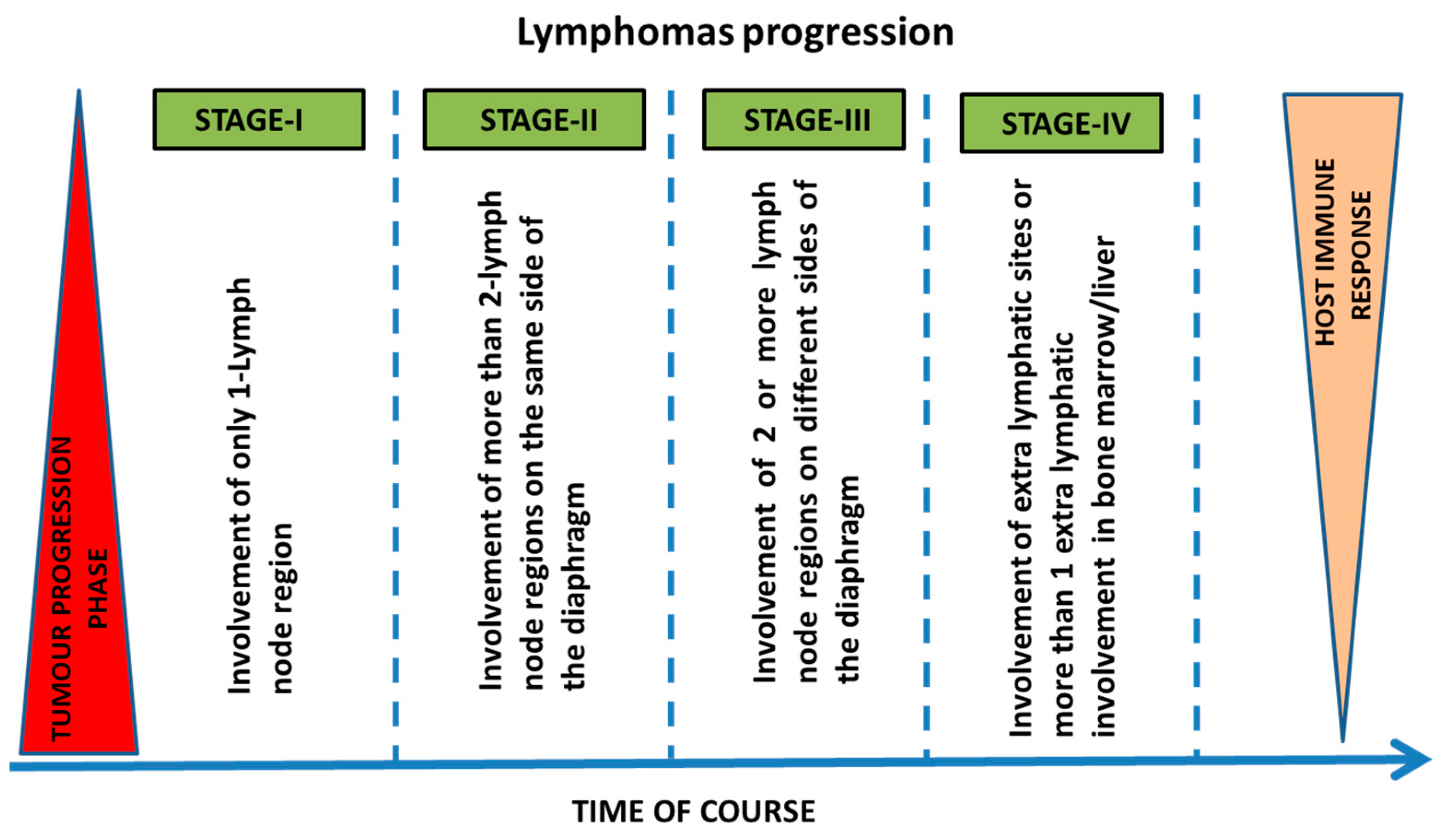
2.1. Stages and Progression of Lymphomas

Lymphomas are generally transformed over time into aggressive forms of lymphomas (Richter’s transformation) and their etiology was studied and diagnosed. The Ann Arbor staging system is used to describe lymphoma progression. It mainly describes Hodgkin lymphomas and, to some extent, non-Hodgkin lymphomas (Figure 1). The Arbor staging schema considers the number of lymph node sites, involvement of extranodal sites, disease above or below the diaphragm, and the presence or absence of systemic symptoms, such as (1) fever, (2) frequent weight loss, (3) sweating of individuals, (4) alopecia, and (5) anemia conditions [22]. Lymphomas were staged mainly based on the characterization of blood and the expression levels of lactate dehydrogenase (LDH) enzyme [23,24].
Many environmental and genetic factors have been associated with specific lymphomas, including herbicides, pesticides, and microbes (Helicobacter pylori, Chlamydia psittaci, Borrelia burgdorferi, and Campylobacter jejuni). It is reported that the human T-cell lymphotropic virus can lead to the development of adult T-cell lymphoma in humans. Moreover, evidence suggests that lymphomas are most prominent in those who are immune deficient. Long-term usage of some drugs, such as tumor necrosis factor-alpha (TNFα) inhibitors, has also been associated with an increased incidence of lymphomas in patients with inflammatory bowel disease and rheumatoid arthritis [25,26]. Lymphomas commonly exhibit some of the stereotypical immunophenotypic molecular lesions that can aid in accurate classification and better treatment patterns for lymphoma progression (Figure 2) [27]. Currently available treatments for lymphomas are mainly based on: (1) histological type, (2) clinical aggressiveness, (3) stage of the disease, (4) molecular markers, (5) single agent therapies, (6) chemotherapy, and (7) hematopoietic stem cell transplantation (HSCT).

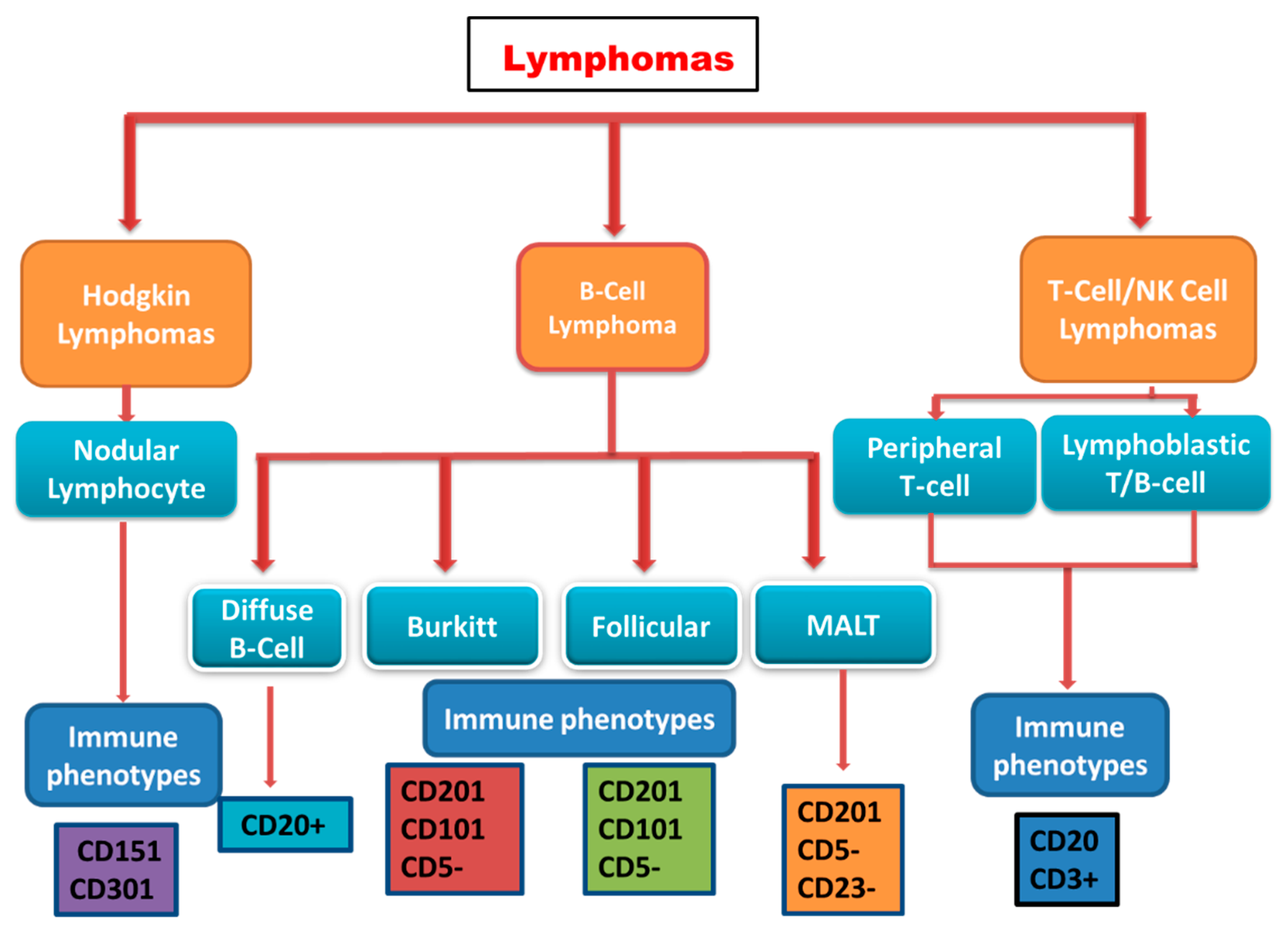
2.2. Classification of Lymphomas

2.2.1. Hodgkin Lymphoma

Hodgkin lymphomas are a large family of a unique type of lymphomas, in which the nature of the neoplastic cell was enigmatic for several years. Samuel Wilks first coined these lymphomas to recognize the earlier reports generated by Thomas Hodgkin [28]. The Hodgkin’s disease, now renamed Hodgkin lymphoma, famous for many years, reflected uncertainty regarding the cellular lineage of the Hodgkin lymphomas and questions regarding their reactive or neoplastic nature. Hodgkin lymphomas are a rare B-cell malignant neoplasm affecting approximately 9000 new patients annually. This disease represents approximately 11% of all lymphomas [29]. The American Cancer Society (ACS) estimates about 8830 new cases (4830 in males and 4000 in females) and about 960 deaths (570 males and 390 females) associated with Hodgkin lymphoma in the United States in 2021. According to recent studies, lymphomas are more predominant in males, mostly in western countries [30]. Progression and the incidence of lymphoma depend on several factors, including heritable characteristics, the abundance and presence of human leukocyte antigen (HLA), which can lead to genetic aberration, and infection with Epstein Barr virus and HIV [31,32,33,34]. Hodgkin lymphomas are mainly sub-divided into Classical Hodgkin lymphoma and Nodular Hodgkin lymphoma (Non-classical). Classical lymphomas accounted for 95% of Hodgkin lymphomas that were primarily characterized by the presence of both Hodgkin and Reed-Sternberg cells (large and abnormal lymphocytes that may contain more than one nucleus). The presence of lymphocyte-predominant cells, sometimes termed “popcorn cells”, a variant of Reed–Sternberg cells, characterizes Nodular Hodgkin lymphoma.

2.2.2. Non-Hodgkin Lymphoma

Non-Hodgkin Lymphoma (NHL) is a type of cancer that develops in the lymph nodes and lymphatic tissue in the skin, stomach, and intestines, sometimes blood and bone marrow. NHL-type lymphomas have a higher incidence rate and are clinically more aggressive with poor prognosis. NHL is further classified into more than 60 subtypes, either indolent (slow-growing) or aggressive (fast-growing). NHL subtypes have been identified by their genetic features; for example, lymphoma cells are characterized by the presence of proteins on the cell surface. Diffuse large B-cell lymphoma (DLBCL) can be classified as an aggressive NHL subtype. Indolent lymphomas account for about 40 percent of all NHL cases and tend to grow more slowly with fewer symptoms when first diagnosed. Follicular lymphoma (FL) is also one of the most common indolent subtypes of NHL. About 90 percent of NHL cases develop in the B lymphocytes (B cells) among different types of lymphocytes. In addition to DLBCL and FL, Mantle cell lymphoma (MCL), Burkitt lymphoma (BL), and Marginal zone lymphoma (MZL) represent other B-cell NHL subtypes that are less frequently diagnosed (https://www.lls.org/lymphoma accessed on 16 May 2021).

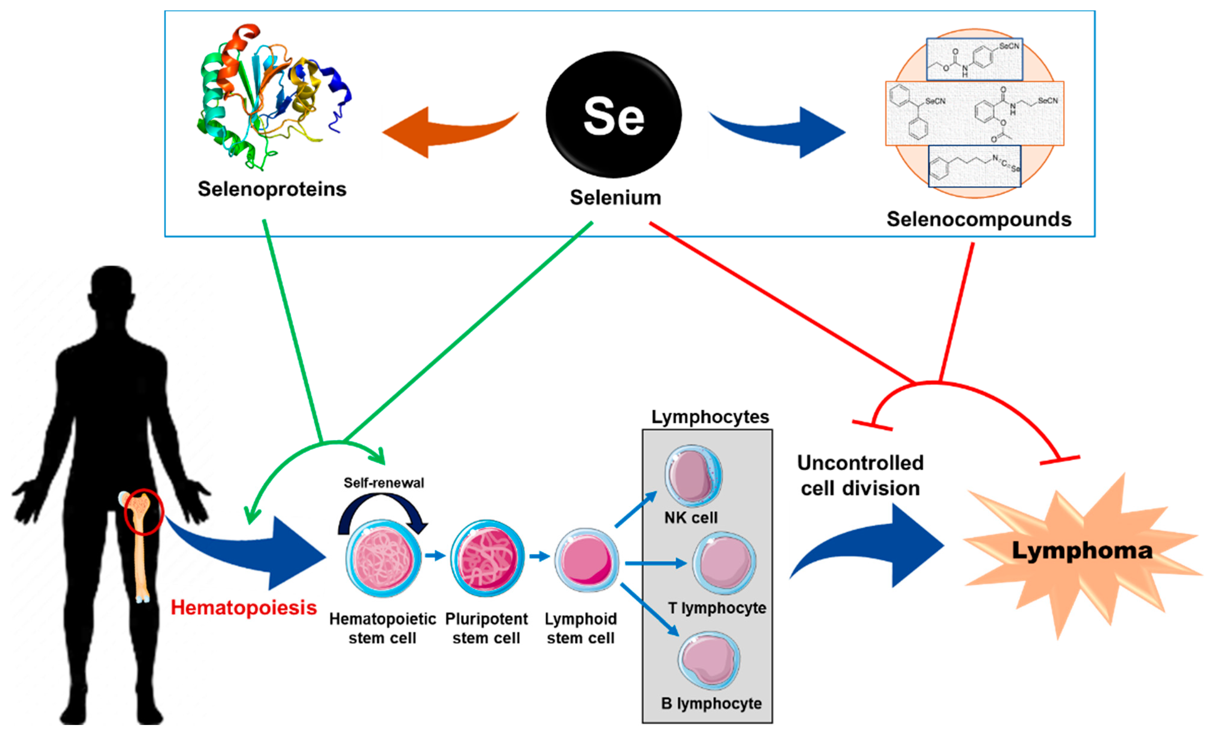
3. Biological Role of Se and SePs in Hematopoiesis and Lymphoma

As stated above, Se is an essential trace element in human metabolism that has beneficial roles in promoting human health after being incorporated into SePs and has been extensively reviewed in the past [5,6,7,8,9,10,11]. SePs play an important physiological role in attaining human health by regulating redox homeostasis, Se transport, immunity, and thyroid hormone metabolism. The deficiency of Se/SeC and dysregulation of SePs can lead to several severe disorders such as cancer, cardiovascular disease (Keshan disease), osteoarthritis, liver disease (hepatopathy), arthropathy (Kashin–Beck disease), and defective immunity against viral infections [9,35,36]. However, it is noteworthy that excessive consumption of Se causes selenosis, which is accompanied by several adverse symptoms such as diarrhea, fatigue, nausea, increased heart rate, necrosis in liver and kidney, and neurological damage. Chronic selenosis results in a defective immune and reproductive system in humans [37].

3.1. Role of Se and SePs in Hematopoiesis

Hematopoiesis is a complex process of blood cell formation, which has a high demand for nutrients due to the rapid turnover time and shorter life span of these cells in the circulation. Hematopoiesis occurs during embryonic development and throughout adulthood to produce and replenish the blood system. In adults, the hematopoietic stem cells (HSCs) proliferate by self-renewal and differentiate into different blood lineages such as erythrocytes, leucocytes (basophils, eosinophils, lymphocytes, monocytes, and neutrophils), and platelets [38]. ROS are byproducts of normal cellular metabolism and have a variety of effects on cellular functions, signaling, and homeostasis. Therefore, the precise regulation of free intra- and extracellular ROS by different antioxidants and specific regulatory pathways plays an important role in maintaining cellular integrity. SePs, including GPxs and TRxs, play a critical role in achieving redox homeostasis, thus avoiding oxidative stress by neutralizing free ROS. As reviewed by Kaweme NM et al., the dysregulation of redox systems plays a critical role in both acute-, and chronic homological malignancies [17]. The process of Hematopoiesis requires tight redox regulation in order to avoid oxidative stress and thus hematological disorders. In fact, Marcus Conrad M et al. deciphered the essential role of mitochondrial TRx2 in hematopoiesis and cardiac development by using ubiquitous Cre-mediated inactivation of TrxR2 in mouse models. The mice lacking mitochondrial TrxR2 exhibited embryonic lethality with a drastic reduction in hematopoietic colonies along with cardiomyocyte proliferation [39].
In addition to redox regulation in HSCs during hematopoiesis, the erythropoiesis (production of new erythrocytes) process is constantly prone to oxidative stress due to the presence of iron, heme, and unpaired globin chains in erythrocytes, which are detrimental to erythroid development and can lead to anemia. The beneficial role of Se and SePs in stabilizing the erythropoiesis process through GPxs has been reviewed in [40]. Earlier, it was reported that the dietary supplementation of Se protects erythrocytes from oxidative damage in animal models and loss of SePs leads to hemolysis of erythrocytes. The extensive study by Liao C et al. explored the critical role of dietary Se and SePs in stress erythropoiesis using murine models. It was found that the loss of SePs functionality either by Se deficiency or mutation of the Sec tRNA (tRNA[Sec]) gene (Trsp) affected stress erythropoiesis at two stages. In line, the authors correlated these findings with the loss of selenoprotein W (SelW), and SelW mutations in bone marrow cells and the murine erythroblast (G1E) cell line resulted in defective terminal differentiation [41]. The deficiency of Se or lack of SePs drastically affects the stress erythropoiesis intensifying the anemia in rodents and human patients. Therefore, the micronutrient Se and SePs play a critical role in the development and expansion of hematopoietic and erythroid cells, in addition to the erythroid niche during acute anemia recovery by neutralizing oxidative stress produced by excessive ROS generation.

3.2. Role of Se and SePs in Lymphoma

As cited above, Se is an important micronutrient that mediates a range of biological functions through SePs and, thus, affects the overall quality of human health. Accordingly, the deficiency of Se in serum is linked with aberrant immune functions, cancer, cardiac, and inflammatory diseases [42]. Recently, we reviewed the anticancer effects of Se and SeCs alone or in combination with standard therapy (chemo- or radiation) for different hematological malignancies including Leukemias [43]. The supplementation of Se and/or Se-containing food was shown to have chemopreventive effects in animals and humans with some contradictory reports [44]. In 2003, Last KW et al. analyzed Se levels in the sera frozen at presentation, from 100 lymphoma patients who received chemo-, radiation therapy, or both, using inductively coupled plasma mass spectrometry. Interestingly, their findings concluded that Se levels in the serum at presentation could act as a prognostic factor, predicting positively dose delivery, treatment response, and long-term survival in diffuse large B-cell lymphoma [45]. Based on the findings, the authors have hypothesized that the Se supplementation might provide a novel treatment strategy for aggressive non-Hodgkin’s lymphoma. In a similar line, Deffuant C et al. tested the predictive value of serum Se levels before and after therapeutic response in 200 melanoma (81 stage I, 63 stage II, and 56 stage III) and 51 epidermotropic cutaneous T-cell lymphoma (CTCL) (8 stage I, 24 stage II, 10 stage III, and 9 stage IV) patients using atomic absorption spectrophotometry. The study findings clearly demonstrated that serum Se levels have prognostic values in the follow-up of both melanoma and CTCL [46].
The study by Ozgen et al. reported that the Se level in hair was significantly lower in children with lymphoma or leukemia when compared to that of the healthy control group [47]. On a similar note, decreased serum Se levels were well correlated in adult patients with hematological malignancies such as acute myeloid leukemia (AML) [48] or advanced chronic lymphocytic leukemia (CLL) [49], while increased or normalized serum Se levels in AML patients were found following complete remission [48,50]. This evidence is further supported by testing the hypothesis in a large cohort of patients with hematological malignancies in a study by Stevens J et al. The authors included a total of 430 patients (163-AML; 156-Hodgkin Lymphoma; and 111-Follicular Lymphoma) to test serum Se levels. Interestingly, the treatment response was in accordance with serum levels of Se, and low levels of Se correlated with worse outcomes in hematological malignancies, but it was not independently predictive [18].
Se and SePs play important role in achieving cellular redox homeostasis. It was reported previously that cellular oxidative stress negatively impacts the chemotherapy of cancers. The authors tested four different chemotherapy drugs (Ara-C, cisplatin, doxorubicin, and VP-16) to induce apoptosis in human Burkitt lymphoma cells and found out that H2O2-induced oxidative stress negatively impacts their therapeutic response [51]. So, the use of antioxidants could enhance chemotherapy-induced apoptosis and phagocytosis in the treatment of lymphomas and other cancers. In growing evidence, a recent study by Wu W et al. analyzed the expression of gene-encoding SePs in different types of cancer (between/within). The findings clearly demonstrated that the expression of SePs genes correlated well with tumor mutagenicity, drug sensitivity, and drug resistance. Further, SePs could be considered as potential therapeutic targets for the treatment of different cancers [16]. Accordingly, the overexpression of one of the SePs, glutathione peroxidase 4 (GPX4; an enzyme that suppresses peroxidation of membrane phospholipids) was shown to be a poor prognostic predictor of DLBC lymphomas. Very recently, it was ruled out that the downstream regulator of GPX4, namely, SECISBP2 (Selenocysteine Insertion Sequence-Binding Protein 2), which regulates various SePs as a novel prognostic predictor, might be a novel therapeutic target for DLBC lymphomas treatment [52]. Altogether, these studies highlighted the role of biological levels of Se and SePs in prognosis, disease progression, and treatment response in hematological malignancies including lymphomas.

3.3. Effect of Se Supplementation in Patients Undergoing Hematopoietic Stem Cell Transplantation

Hematopoietic stem cell transplantation (HSCT) is one of many effective supportive cares for the treatment of several hematological disorders including lymphoma. Despite recent advances in the understanding of transplant immunology, HSCT has limitations due to severe complications such as graft-versus-host disease (GVHD), hepatic veno-occlusive disease, oral mucositis (OM), and infections [53]. These complications are triggered by pro-inflammatory cytokines, such as interleukins (IL-6/IL-1b) and tumor necrosis factor-alpha (TNF-α), released due to high-dose chemotherapy (HDC) prior to HSCT [54,55]. The ROS and oxidative stress (OS) produced by chemotherapy drugs are known to activate transcriptional factors including NF-kB, which, in turn, upregulate genes, which results in increased pro-inflammatory cytokines. Accordingly, the agents including Amifostine and TNF-α inhibitors that suppress oxidative stress (OS) or pro-inflammatory cytokine levels have been demonstrated to prevent or ameliorate complications related to HSCT [56,57]. Since Se has anti-inflammatory properties [58] and constitutes an important role in the human antioxidant system in the form of SePs (GPx/TRx), Daeian N et al. explored the effect of Se supplementation on pro-inflammatory cytokine levels in a randomized double-blind placebo-controlled clinical trial involving 74 patients undergoing HSCT. According to study findings, Se supplementation prevented severe OM in HSCT patients without exhibiting significant differences in the plasma levels of inflammatory cytokines. So, the authors concluded that earlier administration and/or using larger doses of Se might result in beneficial effects during HSCT [59].

4. Physico-Chemical and Anticancer Properties of Se

Selenium (Se) is a nonmetallic trace element that was considered a poison until the 19th century, and was later identified as an essential micronutrient and dietary supplement with anticancer activity. The biological importance of Se for the effective functionality of several SePs has led to the exploitation of various SeCs to mimic and modulate biological effects mediated by Se.

4.1. The Historical Perspectives of Se

In the year 1817, Jons Jacob Berzelius first isolated, identified, and analyzed the Se element [6]. After analyzing the properties of Se, interestingly, Marco Polo, in 1295, described the first record of Se toxicity and described a disease called “hoof rot” in cattle and horses in the Tien Shan and Nan Shan mountains of Turkestan, where the soil concentration of Se was high [60,61]. Some studies reported in the 20th century showed that high levels of Se in soil were strongly associated with the gradual development of skin lesions, neuropathy in horses, sheep, cattle, and some of the plants such as the genera Astragalus, Xylorrhiza, Oonopsis, and Stanleya [62,63,64]. Se toxicity is also associated with diseases called “blind staggers”, the symptoms of which mainly include weight loss, blindness, ataxia, anorexia, and respiratory distress [65]. In the year 1950, Se was replaced by vitamin E in the diet of some experimental animals, which did not show any adverse, abnormal effects [66]. Se is considered an essential trace element, and this was confirmed through a pathological clinical condition known as KESHAN disease, which is mainly implicated by the inadequate levels of Se presence in the diet. Some of the early reports in the 20th century reported that potassium selenate (around a concentration of 300 µg/day) and selenium dioxide (3 mg/day) are promising drugs for the treatment of hematological malignancies [67]. Accordingly, recent studies have also strongly recommended Se and SeCs for the prevention of different types of cancers [5,68,69].

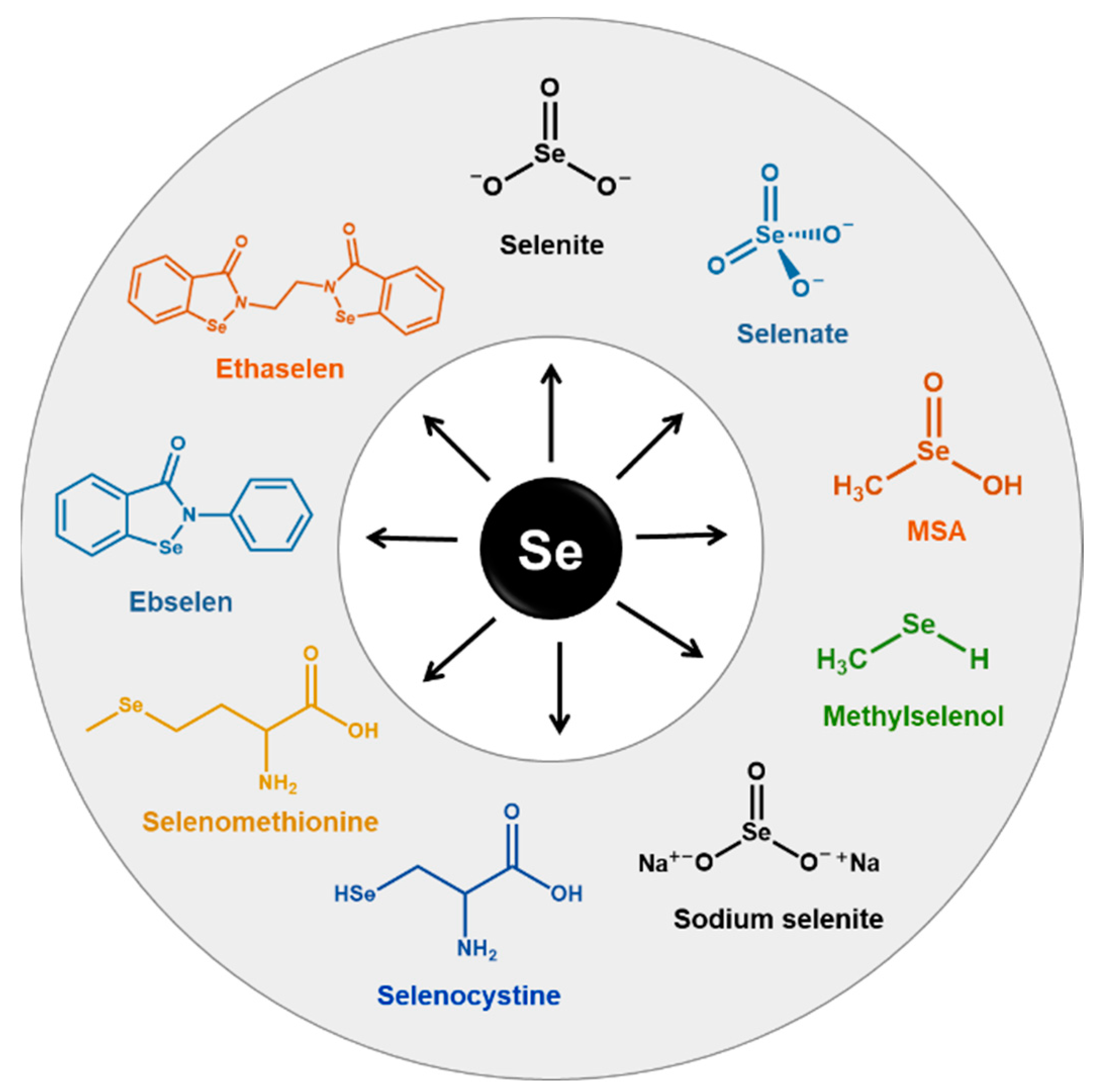
4.2. Physico-Chemical Properties of Se

Se exists in three allotropic forms, namely, a (1) Deep red crystal; (2) red amorphous powder; and (3) black vitreous form. Mostly, Se is insoluble in water while inorganic alkali selenites and selenates are soluble in water [70]. Se shares similar chemical properties with sulfur, and to a lesser extent with tellurium, all belonging to group 16 of the periodic table. Se and SeCs have a strong tendency to make complexes with heavy metals. SeCs such as selenate are the most stable oxidized form in alkaline and also oxidizing solutions among the other SeCs, and alkyl SeCs are among the least toxic compounds (dimethyl selenide and trimethyl selenide), and these are mainly released as products of detoxification of selenium in the body [71]. Se has the capability to alter sulfur into different forms of organic SeCs, which include mainly dimethylselenide and trimethylselenonium; mostly Se occurs as a form of selenides and selenocysteine at physiological pH [72].

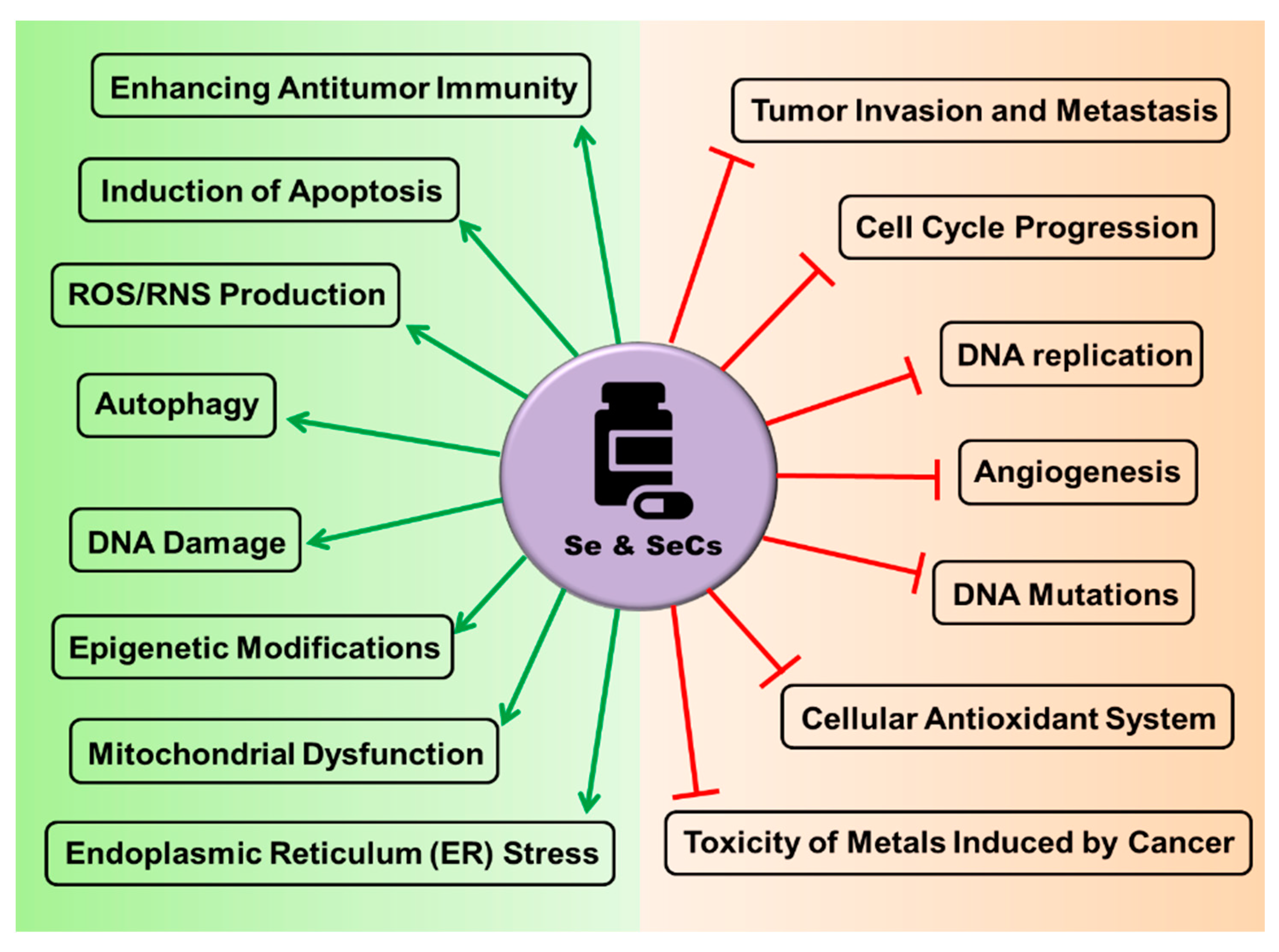
4.3. The Rationale behind the Use of Se and SeCs as Anticancer Agents

Normally, healthy cells can be differentiated from cancer cells by a number of molecular, physiological, and pathological functions. Most healthy cells can exhibit tightly regulated systems and low steady states of ROS/RNS production and reducing equivalents, while in cancer cells, mainly increased levels of ROS production and reduced equivalents such as NADPH and NADH through several uncontrolled glycolysis processes are observed. In general, cancer cells also exhibit irregular functions that are associated with the up- or down-regulation of protein synthesis, deregulated ROS generation, and enhanced antioxidant capacity to counteract ROS-induced cell death, etc. According to several recent studies, the induction of high levels of oxidative stress and downregulation of target genes involved in the antioxidant capacity within the tumor cells have shown to be a promising therapeutic strategy for the treatment of different cancers [73,74]. However, several recent plausible observations have been put forward that the therapeutic role of SeCs as potential anticancer agents is typically by inducing DNA strand breaks, cell cycle arrest, and apoptosis [69]. Some of the important and well-tested SeCs are illustrated in Figure 3.
Recent findings from pre-clinical studies and human clinical trials have strongly associated preventive and therapeutic roles of Se in cancer therapeutic development in addition to inhibition of tumor progression. It was shown recently that the supplementation of a Se-rich diet significantly lowered the incidence of cancer in mice [75]. The anticancer effects of Se and SeCs are very diverse, but findings from earlier studies hinted that Se exhibits anticancer activity by increasing ROS production, thiol group modifications, and chromatin binding and modifications [74,75,76,77,78,79]. So, the anticancer activity of Se was majorly attributed to organic Se, and associated mechanisms include antioxidant and pro-oxidant activities in different cancers including breast, lung, colon cancers, etc. In addition, the anticancer activity of Se was exhibited through several mechanisms, which mainly included protection against DNA damage by dimethylbenz anthracene-induced aberration, which was shown mainly in breast, liver, and colon cancers [80,81]. According to recent reports, the selenomethionine (Se-met) compound is the most abundant form, showing the greatest biological activity and also the highest chemotherapeutic form [82]. Although the majority of literature encourages the use of Se and SeCs as anticancer agents either alone or in combination with standard therapy, a few contradictory studies have reported that Se alone failed to exhibit anti-cancer activity in randomized clinical trials and observational studies [83]. In addition, supplementation with Se has shown an increased risk of cancer and detrimental effects, with a widespread outbreak of acute Se toxicity [84,85]. The use of Se and SeCs as anticancer agents is limited due to poor pharmacokinetics such as rapid elimination from the body, narrow therapeutic window, and lack of distribution selectively [86]. So, the development of new formulations to enhance pharmacokinetics and the careful interpretation of results considering the distinct biological properties of organic and inorganic SeCs is necessary to avoid Se-mediated toxicity.
Several organo-selenium species have evolved as superior anticancer agents compared to natural SeCs: for example, “Ebselen”, is a synthetic drug that exhibits anticancer activities against different cancer types including breast, colon, and liver cancers. Ethaselen is a modified version of the parent molecule Ebselen, which showed an improved solubility and showed promising results against small lung carcinoma, and is currently in a phase-1 clinical trial [77,78,79,80]. SeCs also prevent free ROS generation sites, prevent the excess hydrogen peroxide that damages DNA, and also act as a nutrient, and maintain several physiological processes such as homeostasis, cell proliferation mechanisms, angiogenesis inhibition, and induction of apoptosis caused by the carcinogens to normal cells [14]. Se exhibits chemopreventive actions through the activation of apoptosis, mainly including the activation of multiple apoptotic pathways such as the activation of p53, specific Bax upregulation, and Bcl2 downregulation [87]. Some of the microarrays and genomic studies have reported Se to show strong anticancer activities through the activation of apoptosis and specific DNA damage [88]. Some well-tested Se-derived compounds and their anticancer activity mechanisms are listed in Table 2 and summarized in Figure 4.

5. Implication of Se and SeCs for the Treatment of Lymphomas

Lymphomas are currently treated using chemotherapy, immunotherapy, radiation, along with surgery. The dysregulation of Se levels in serum and aberrant expression of gene-encoding SePs has been shown to play a critical role in the development, prognosis, progression, and treatment response in the management of lymphomas (as described in Section 3.2) [16,18,45,46,47,52]. These studies support the implication of Se supplementation, Se, and SeCs as a potential therapeutic intervention for the treatment of several types of lymphomas (Figure 5). Although the precise anti-tumor mechanisms exerted by Se are not yet fully understood, the cellular effects of Se largely depend on the chemical form and concentrations/dose used. Several studies have reported the use of Se and SeCs against lymphomas and their probable mechanism of action was discussed.

5.1. Effect of Se and SeCs on Lymphoma Cultures In Vitro

According to earlier studies, Se and SeCs initially showed cytotoxicity through the induction of several death apoptotic pathways in several cancer cells including most lymphomas [99,100]. Previous studies reported that organic SeCs such as SDG and MSA showed a dose-dependent decrease in cell viability and increased cytotoxicity (lymphoma cells are more sensitive to SDG than MSA) in different lymphomas such as mantle cell lymphoma, follicular lymphoma, chronic lymphocytic leukemia cell lines and primary lymphoma cultures [99]. Moreover, Jüliger S et al. reported that combining the minimally toxic concentrations (EC5–10) of MSA with that of other chemotherapeutic agents (e.g., doxorubicin, etoposide, and melphalan) increased their cytotoxicity by 2.5-fold in B-cell lymphoma cell lines. The results suggest the development of Se as a potential modulator of chemotherapy drug activity in lymphomas [101]. In addition, sodium selenite caused a significant decrease in cell proliferation and cell viability leading to cell death in murine B lymphoma (A20) cells [102]. Recently, Kumar S et al. achieved the carboxylic group-induced synthesis of selenium nanoparticles (SeNPs) using selenosulfate as a precursor. Following the characterization of SeNPs, the authors tested their cytotoxicity against Dalton’s lymphoma (DL) cells. The SeNPs exerted cytotoxicity in DL cells as evidenced by decreased cell viability, altered nuclear morphology, and increased apoptosis [103]. In continuation, Gautam PK et al. reported that SeNPs exerted cytotoxicity in DL cells by inducing apoptosis [104]. Very recently, SeNPs exhibited potent antitumor activity through the upregulation of miR-16 in prostate cancer cells. Serum Se levels are positively correlated with that of miR-16 expression, and with the overall and disease-free survival rates [105]. These studies demonstrate the cytotoxicity of Se and SeCs in lymphoma cells and cultures.

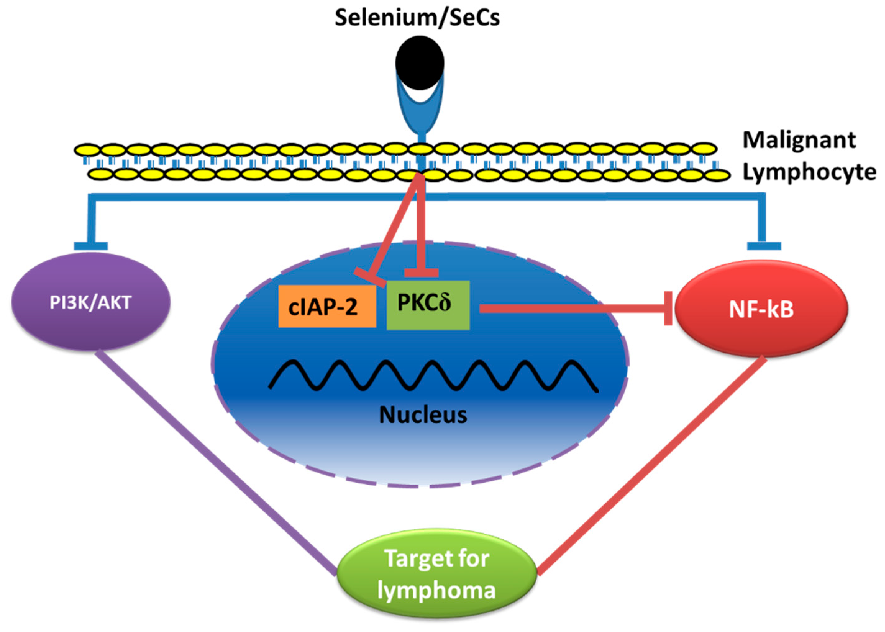
5.2. Effects of Se/SeCs on the PI3-Kinase/Akt, and MAP Kinase Pathways

The phosphoinositide 3-kinase (PI3K)/Akt pathway is a well-known key pro-survival pathway in all cells. The PI3K/Akt pathway is highly upregulated in different human cancer cells, and drugs targeting this pathway result in increased proliferation and reduced apoptosis [106,107,108]. Gonzalez-Moreno et al. reported that organic Se compound MSA exhibited dose-dependent cytotoxicity specifically in tumor cells by decreasing Akt phosphorylation. Moreover, the combination treatment of low doses of etoposide or docetaxel, with low doses of MSA, exhibited synergetic cytotoxicity and enhanced apoptosis in tumor cells [109]. Therefore, the Se/SeCs might exert chemopreventive effects through the targeting of the PI3k/Akt pathway, thus sensitizing lymphoma cells to anticancer drugs.

5.3. Effects of Se/SeCs through the Nuclear Factor Kappa B (NF-κB) Pathway

NF-κB is a well-known important signaling pathway, which is mainly involved in cancer development and cancer progression. NF-κB mainly controls the expression of different target genes, including TNF-α, IL-6, BCLXL, BCL-2, BCLXS, XIAP, and VEGF. Typically, NF-κB mediates several pathological functions such as tumor-cell proliferation, survival, angiogenesis, etc. [110]. Baldwin AS concluded that NF-κB is a family that consists of five different regulatory proteins: Rel-A (p65), Rel-B, c-Rel, NF-κB1 (p105/p50), and NF-κB2 (p100/52), and is kept in an inactive state in the cytoplasm by the IκB family of inhibitory proteins [111]. Various stimuli, such as cytokines, TNFα, oxidative stress, and infection with viruses, resulted in the translocation of NF-κB into the nucleus, which bound to specific regulatory genes and resulted in higher transcription levels [112]. It has been reported that NF-κB activity was unconditionally upregulated in many cancer types including lymphoma and NF-κB inhibitors sensitizes the cancer cells to chemotherapy [113,114]. Jüliger S et al. demonstrated that the inhibitory effects of Se on NF-κB in MSA-treated DLBC lymphoma cell lines significantly reduced cell viability, and enhanced cytotoxic activities of different chemotherapeutic agents (e.g., doxorubicin, etoposide). Moreover, it was evidenced that the inhibition of NF-κB activity in B-cell lymphoma cells by MSA results in apoptosis and chemo-sensitivity pathways associated with TNF-α [101]. In another study, sodium selenite has been shown to modulate several intracellular signaling molecules such as protein kinase-C (PKC), NF-κB, and inhibition apoptosis protein (IAP) in murine B lymphoma (A20) cells [102]. Therefore, Se and SeCs exert cytotoxicity effects in lymphoma cells by targeting PI3k/Akt, PKC, IAP, and NF-κB signaling pathways (Figure 6).

6. Conclusions

In conclusion, it was evident from previous investigations that low levels of Se in serum and the modulation of SePs activity or expression have a significant association with several lymphomas in patients. As such, Se is a well-studied micronutrient/dietary supplement for its chemopreventive effects in different cancers including lymphoma cell lines and patients. Over the last few years, several SeCs have been developed to mimic and alter the functions of seleno-enzymes to achieve different pharmacological interventions including anticancer activity. Different oncogenic signaling pathways such as NF-κB, PI3K/Akt, Notch1, and PKC were targeted by these SeCs and Se to impart chemopreventive effects. Though the toxicity associated with Se is limited, both the pharmacotherapy and efforts to minimize the side effects of SeCs through suitable chemical substitution are promising. Moreover, a combination of minimal doses of Se with that of other standard cancer therapies, such as chemotherapy, radiotherapy, and HSCT, exhibited a synergistic effect in inducing cytotoxicity specifically in tumor cells sparing normal healthy cells. In addition, emerging pan-cancer studies reveal the prognostic role of various SePs in cancer development and progression, with a scope for future therapeutics targeting seleno-enzymes for the treatment of cancers including hematological malignancies (Figure 5). The data reviewed in this article may not be exclusively extrapolated at this time to human situations because the difference between the desirable intake of Se and that of toxic levels is relatively minute. Despite the existence of abundant and promising literature supporting Se and SeCs as anticancer agents, few contradictory studies warn about the adverse effects associated with the deregulated use of Se supplementation. Therefore, further studies are warranted in large population (genetic or nutritional background) settings to strengthen and confirm the definitive role of Se, SePs, and SeCs in tumor prognosis, progression, safety, treatment response, and drug resistance.

Author Contributions

U.G. conceived the structure of this review, investigated key resources for the review, edited, and revised the manuscript. U.G. and S.D. wrote the first draft, prepared all figures, edited the manuscript, and contributed equally. All authors have read and agreed to the published version of the manuscript.

Funding

This research received no external funding.

Institutional Review Board Statement

Not applicable.

Informed Consent Statement

Not applicable.

Data Availability Statement

Not applicable.

Acknowledgments

The authors thank Arun Sharma (Penn State Cancer Institute) for his valuable comments to improve the manuscript. The authors are grateful to their respective universities/institutions for their support.

Conflicts of Interest

The authors declare no conflict of interest.

References

  1. Haque, A.; Brazeau, D.; Amin, A.R. Perspectives on natural compounds in chemoprevention and treatment of cancer: An update with new promising compounds. Eur. J. Cancer 2021, 149, 165–183. [Google Scholar] [CrossRef]
  2. Golla, U. Emergence of nutraceuticals as the alternative medications for pharmaceuticals. Int. J. Complement Alt. Med. 2018, 11, 155–158. [Google Scholar] [CrossRef] [Green Version]
  3. Amin, A.R.; Kucuk, O.; Khuri, F.R.; Shin, D.M. Perspectives for cancer prevention with natural compounds. J. Clin. Oncol 2009, 27, 2712–2725. [Google Scholar] [CrossRef] [Green Version]
  4. Harvie, M. Nutritional supplements and cancer: Potential benefits and proven harms. Am. Soc. Clin. Oncol. Educ. Book 2014, 34, e478–e486. [Google Scholar] [CrossRef] [Green Version]
  5. Evans, S.O.; Khairuddin, P.F.; Jameson, M.B. Optimising Selenium for Modulation of Cancer Treatments. Anticancer Res. 2017, 37, 6497–6509. [Google Scholar] [CrossRef] [Green Version]
  6. Foster, L.; Sumar, S. Selenium in health and disease: A review. Crit. Rev. Food Sci. Nutr. 1997, 37, 211–228. [Google Scholar] [CrossRef] [PubMed]
  7. El-Bayoumy, K.; Sinha, R. Molecular chemoprevention by selenium: A genomic approach. Mutat. Res. Fundam. Mol. Mech. Mutagenesis 2005, 591, 224–236. [Google Scholar] [CrossRef] [PubMed]
  8. Fairweather-Tait, S.J.; Bao, Y.; Broadley, M.R.; Collings, R.; Ford, D.; Hesketh, J.E.; Hurst, R. Selenium in human health and disease. Antioxid Redox Signal 2011, 14, 1337–1383. [Google Scholar] [CrossRef]
  9. Brown, K.M.; Arthur, J.R. Selenium, selenoproteins and human health: A review. Public Health Nutr. 2001, 4, 593–599. [Google Scholar] [CrossRef] [Green Version]
  10. Kieliszek, M.; Blazejak, S. Current Knowledge on the Importance of Selenium in Food for Living Organisms: A Review. Molecules 2016, 21, 609. [Google Scholar] [CrossRef] [Green Version]
  11. Kang, D.; Lee, J.; Wu, C.; Guo, X.; Lee, B.J.; Chun, J.S.; Kim, J.H. The role of selenium metabolism and selenoproteins in cartilage homeostasis and arthropathies. Exp. Mol. Med. 2020, 52, 1198–1208. [Google Scholar] [CrossRef] [PubMed]
  12. Chambers, I.; Frampton, J.; Goldfarb, P.; Affara, N.; McBain, W.; Harrison, P.R. The structure of the mouse glutathione peroxidase gene: The selenocysteine in the active site is encoded by the ‘termination’ codon, TGA. EMBO J. 1986, 5, 1221–1227. [Google Scholar] [CrossRef]
  13. Zhang, J.; Zhou, H.; Li, H.; Ying, Z.; Liu, X. Research progress on separation of selenoproteins/Se-enriched peptides and their physiological activities. Food Funct. 2021, 12, 1390–1401. [Google Scholar] [CrossRef]
  14. Ganther, H.E. Selenium metabolism, selenoproteins and mechanisms of cancer prevention: Complexities with thioredoxin reductase. Carcinogenesis 1999, 20, 1657–1666. [Google Scholar] [CrossRef] [PubMed] [Green Version]
  15. Bellinger, F.P.; Raman, A.V.; Reeves, M.A.; Berry, M.J. Regulation and function of selenoproteins in human disease. Biochem. J. 2009, 422, 11–22. [Google Scholar] [CrossRef] [Green Version]
  16. Wu, W.; Li, D.; Feng, X.; Zhao, F.; Li, C.; Zheng, S.; Lyu, J. A pan-cancer study of selenoprotein genes as promising targets for cancer therapy. BMC Med. Genomics 2021, 14, 78. [Google Scholar] [CrossRef]
  17. Kaweme, N.M.; Zhou, S.; Changwe, G.J.; Zhou, F. The significant role of redox system in myeloid leukemia: From pathogenesis to therapeutic applications. Biomark Res. 2020, 8, 63. [Google Scholar] [CrossRef]
  18. Stevens, J.; Waters, R.; Sieniawska, C.; Kassam, S.; Montoto, S.; Fitzgibbon, J.; Rohatiner, A.; Lister, A.; Joel, S. Serum selenium concentration at diagnosis and outcome in patients with haematological malignancies. Br. J. Haematol. 2011, 154, 448–456. [Google Scholar] [CrossRef]
  19. Sung, H.; Ferlay, J.; Siegel, R.L.; Laversanne, M.; Soerjomataram, I.; Jemal, A.; Bray, F. Global Cancer Statistics 2020: GLOBOCAN Estimates of Incidence and Mortality Worldwide for 36 Cancers in 185 Countries. CA Cancer J. Clin. 2021, 71, 209–249. [Google Scholar] [CrossRef]
  20. Shanbhag, S.; Ambinder, R.F. Hodgkin lymphoma: A review and update on recent progress. CA Cancer J. Clin. 2018, 68, 116–132. [Google Scholar] [CrossRef]
  21. Swerdlow, S.H.; Campo, E.; Harris, N.L.; Jaffe, E.S.; Pileri, S.A.; Stein, H.; Thiele, J. WHO Classification of Tumours of Haematopoietic and Lymphoid Tissues, 4th ed.; IARC Press: Lyon, France, 2008; Volume 2. [Google Scholar]
  22. Skarin, A.T.; Dorfman, D.M. Non-Hodgkin’s lymphomas: Current classification and management. CA Cancer J. Clin. 1997, 47, 351–372. [Google Scholar] [CrossRef] [Green Version]
  23. Endrizzi, L.; Fiorentino, M.V.; Salvagno, L.; Segati, R.; Pappagallo, G.L.; Fosser, V. Serum lactate dehydrogenase (LDH) as a prognostic index for non-Hodgkin’s lymphoma. Eur. J. Cancer Clin. Oncol. 1982, 18, 945–949. [Google Scholar] [CrossRef]
  24. Elstrom, R.; Guan, L.; Baker, G.; Nakhoda, K.; Vergilio, J.-A.; Zhuang, H.; Pitsilos, S.; Bagg, A.; Downs, L.; Mehrotra, A. Utility of FDG-PET scanning in lymphoma by WHO classification. Blood J. Am. Soc. Hematol. 2003, 101, 3875–3876. [Google Scholar] [CrossRef]
  25. Dahmus, J.; Rosario, M.; Clarke, K. Risk of Lymphoma Associated with Anti-TNF Therapy in Patients with Inflammatory Bowel Disease: Implications for Therapy. Clin. Exp. Gastroenterol. 2020, 13, 339–350. [Google Scholar] [CrossRef]
  26. Mercer, L.K.; Galloway, J.B.; Lunt, M.; Davies, R.; Low, A.L.; Dixon, W.G.; Watson, K.D.; Consortium, B.C.C.; Symmons, D.P.; Hyrich, K.L. Risk of lymphoma in patients exposed to antitumour necrosis factor therapy: Results from the British Society for Rheumatology Biologics Register for Rheumatoid Arthritis. Ann. Rheum. Dis. 2017, 76, 497–503. [Google Scholar] [CrossRef] [Green Version]
  27. Clinical Practice Guidelines in Oncology: B-Cell Lymphomas. 2018. Available online: www.nccn.org. (accessed on 12 March 2021).
  28. Wilks, S. Cases of enlargement of the lymphatic glands and spleen (or, Hodgkin’s disease) with remarks. Guy’s Hosp. Rep. 1856, 11, 56–67. [Google Scholar]
  29. Siegel, R.L.; Miller, K.D.; Jemal, A. Cancer statistics, 2015. CA A Cancer J. Clin. 2015, 65, 5–29. [Google Scholar] [CrossRef]
  30. Morton, L.M.; Wang, S.S.; Devesa, S.S.; Hartge, P.; Weisenburger, D.D.; Linet, M.S. Lymphoma incidence patterns by WHO subtype in the United States, 1992–2001. Blood 2006, 107, 265–276. [Google Scholar] [CrossRef]
  31. Harty, L.C.; Lin, A.Y.; Goldstein, A.M.; Jaffe, E.S.; Carrington, M.; Tucker, M.A.; Modi, W.S. HLA-DR, HLA-DQ, and TAP genes in familial Hodgkin disease. Blood J. Am. Soc. Hematol. 2002, 99, 690–693. [Google Scholar] [CrossRef] [Green Version]
  32. Hooper, W.C.; Holman, R.C.; Clarke, M.; Chorba, T.L. Trends in non-hodgkin lymphoma (NHL) and HIV-associated NHL deaths in the United States. Am. J. Hematol. 2001, 66, 159–166. [Google Scholar] [CrossRef]
  33. Tirelli, U.; Spina, M.; Gaidano, G.; Vaccher, E.; Franceschi, S.; Carbone, A. Epidemiological, biological and clinical features of HIV-related lymphomas in the era of highly active antiretroviral therapy. Aids 2000, 14, 1675–1688. [Google Scholar] [CrossRef] [Green Version]
  34. International Collaboration on HIV and Cancer. Highly active antiretroviral therapy and incidence of cancer in human immunodeficiency virus-infected adults. J. Natl. Cancer Inst. 2000, 92, 1823–1830. [Google Scholar] [CrossRef]
  35. Hatfield, D.L.; Yoo, M.H.; Carlson, B.A.; Gladyshev, V.N. Selenoproteins that function in cancer prevention and promotion. Biochim. Biophys. Acta 2009, 1790, 1541–1545. [Google Scholar] [CrossRef] [Green Version]
  36. Schweizer, U.; Fradejas-Villar, N. Why 21? The significance of selenoproteins for human health revealed by inborn errors of metabolism. FASEB J. 2016, 30, 3669–3681. [Google Scholar] [CrossRef] [Green Version]
  37. Nuttall, K.L. Evaluating selenium poisoning. Ann. Clin. Lab. Sci. 2006, 36, 409–420. [Google Scholar]
  38. Elsaid, R.; Soares-da-Silva, F.; Peixoto, M.; Amiri, D.; Mackowski, N.; Pereira, P.; Bandeira, A.; Cumano, A. Hematopoiesis: A Layered Organization Across Chordate Species. Front. Cell Dev. Biol. 2020, 8, 606642. [Google Scholar] [CrossRef]
  39. Conrad, M.; Jakupoglu, C.; Moreno, S.G.; Lippl, S.; Banjac, A.; Schneider, M.; Beck, H.; Hatzopoulos, A.K.; Just, U.; Sinowatz, F.; et al. Essential role for mitochondrial thioredoxin reductase in hematopoiesis, heart development, and heart function. Mol. Cell Biol. 2004, 24, 9414–9423. [Google Scholar] [CrossRef] [Green Version]
  40. Liao, C.; Carlson, B.A.; Paulson, R.F.; Prabhu, K.S. The intricate role of selenium and selenoproteins in erythropoiesis. Free Radic. Biol. Med. 2018, 127, 165–171. [Google Scholar] [CrossRef]
  41. Liao, C.; Hardison, R.C.; Kennett, M.J.; Carlson, B.A.; Paulson, R.F.; Prabhu, K.S. Selenoproteins regulate stress erythroid progenitors and spleen microenvironment during stress erythropoiesis. Blood 2018, 131, 2568–2580. [Google Scholar] [CrossRef]
  42. Brigelius-Flohé, R. Selenium in Human Health and Disease: An Overview. In Selenium; Michalke, B., Ed.; Molecular and Integrative Toxicology; Springer: Berlin/Heidelberg, Germany, 2018. [Google Scholar]
  43. Ehudin, M.A.; Golla, U.; Trivedi, D.; Potlakayala, S.D.; Rudrabhatla, S.V.; Desai, D.; Dovat, S.; Claxton, D.; Sharma, A. Therapeutic Benefits of Selenium in Hematological Malignancies. Int. J. Mol. Sci. 2022, 23, 7972. [Google Scholar] [CrossRef]
  44. Muecke, R.; Schomburg, L.; Buentzel, J.; Kisters, K.; Micke, O.; German Working Group Trace Elements and Electrolytes in Oncology-AKTE. Selenium or no selenium—That is the question in tumor patients: A new controversy. Integr. Cancer Ther. 2010, 9, 136–141. [Google Scholar] [CrossRef] [Green Version]
  45. Last, K.W.; Cornelius, V.; Delves, T.; Sieniawska, C.; Fitzgibbon, J.; Norton, A.; Amess, J.; Wilson, A.; Rohatiner, A.Z.; Lister, T.A. Presentation serum selenium predicts for overall survival, dose delivery, and first treatment response in aggressive non-Hodgkin’s lymphoma. J. Clin. Oncol. 2003, 21, 2335–2341. [Google Scholar] [CrossRef]
  46. Deffuant, C.; Celerier, P.; Boiteau, H.; Litoux, P.; Dreno, B. Serum selenium in melanoma and epidermotropic cutaneous T-cell lymphoma. Acta Derm. Venereol. 1994, 74, 90–92. [Google Scholar]
  47. Ozgen, I.T.; Dagdemir, A.; Elli, M.; Saraymen, R.; Pinarli, F.G.; Fisgin, T.; Albayrak, D.; Acar, S. Hair selenium status in children with leukemia and lymphoma. J Pediatr Hematol Oncol 2007, 29, 519–522. [Google Scholar] [CrossRef]
  48. Beguin, Y.; Weber, G.; Delbrouck, J.M.; Roelandts, I.; Robaye, G.; Bury, J.; Fillet, G. Serum trace elements during chemotherapy for acute myelogenous leukemia. Acta Pharmacol Toxicol 1986, 59 (Suppl. 7), 270–273. [Google Scholar] [CrossRef]
  49. Beguin, Y.; Brasseur, F.; Weber, G.; Bury, J.; Delbrouck, J.M.; Roelandts, I.; Robaye, G.; Fillet, G. Observations of serum trace elements in chronic lymphocytic leukemia. Cancer 1987, 60, 1842–1846. [Google Scholar] [CrossRef]
  50. Asfour, I.A.; El-kholy, N.M.; Ayoub, M.S.; Ahmed, M.B.; Bakarman, A.A. Selenium and glutathione peroxidase status in adult Egyptian patients with acute myeloid leukemia. Biol. Trace Elem. Res. 2009, 132, 85–92. [Google Scholar] [CrossRef]
  51. Shacter, E.; Williams, J.A.; Hinson, R.M.; Senturker, S.; Lee, Y.J. Oxidative stress interferes with cancer chemotherapy: Inhibition of lymphoma cell apoptosis and phagocytosis. Blood 2000, 96, 307–313. [Google Scholar] [CrossRef]
  52. Taguchi, T.; Kurata, M.; Onishi, I.; Kinowaki, Y.; Sato, Y.; Shiono, S.; Ishibashi, S.; Ikeda, M.; Yamamoto, M.; Kitagawa, M.; et al. SECISBP2 is a novel prognostic predictor that regulates selenoproteins in diffuse large B-cell lymphoma. Lab. Investig. 2021, 101, 218–227. [Google Scholar] [CrossRef]
  53. Tabbara, I.A.; Zimmerman, K.; Morgan, C.; Nahleh, Z. Allogeneic hematopoietic stem cell transplantation: Complications and results. Arch. Intern. Med. 2002, 162, 1558–1566. [Google Scholar] [CrossRef] [Green Version]
  54. Schots, R.; Kaufman, L.; Van Riet, I.; Ben Othman, T.; De Waele, M.; Van Camp, B.; Demanet, C. Proinflammatory cytokines and their role in the development of major transplant-related complications in the early phase after allogeneic bone marrow transplantation. Leukemia 2003, 17, 1150–1156. [Google Scholar] [CrossRef]
  55. Takatsuka, H.; Takemoto, Y.; Yamada, S.; Wada, H.; Tamura, S.; Fujimori, Y.; Okamoto, T.; Suehiro, A.; Kanamaru, A.; Kakishita, E. Complications after bone marrow transplantation are manifestations of systemic inflammatory response syndrome. Bone Marrow Transpl. 2000, 26, 419–426. [Google Scholar] [CrossRef] [Green Version]
  56. Spencer, A.; Horvath, N.; Gibson, J.; Prince, H.M.; Herrmann, R.; Bashford, J.; Joske, D.; Grigg, A.; McKendrick, J.; Prosser, I.; et al. Prospective randomised trial of amifostine cytoprotection in myeloma patients undergoing high-dose melphalan conditioned autologous stem cell transplantation. Bone Marrow Transpl. 2005, 35, 971–977. [Google Scholar] [CrossRef] [Green Version]
  57. Levine, J.E.; Paczesny, S.; Mineishi, S.; Braun, T.; Choi, S.W.; Hutchinson, R.J.; Jones, D.; Khaled, Y.; Kitko, C.L.; Bickley, D.; et al. Etanercept plus methylprednisolone as initial therapy for acute graft-versus-host disease. Blood 2008, 111, 2470–2475. [Google Scholar] [CrossRef]
  58. Duntas, L.H. Selenium and inflammation: Underlying anti-inflammatory mechanisms. Horm. Metab. Res. 2009, 41, 443–447. [Google Scholar] [CrossRef]
  59. Daeian, N.; Radfar, M.; Jahangard-Rafsanjani, Z.; Hadjibabaie, M.; Ghavamzadeh, A. Selenium supplementation in patients undergoing hematopoietic stem cell transplantation: Effects on pro-inflammatory cytokines levels. Daru 2014, 22, 51. [Google Scholar] [CrossRef] [Green Version]
  60. Dickerson, O.; Smith, T. Selenium, tellurium, and osmium. In Occupational Medicine, 3rd ed.; Mosby: St. Louis, MO, USA, 1994; Volume 9. [Google Scholar]
  61. Mihajlovic, M. Selenium toxicity in domestic animals. Glas. Srp. Akad. Nauka Med. 1992, 42, 131–144. [Google Scholar]
  62. Draize, J.; Beath, O. Observations on the pathology of blind staggers and alkali disease. J. Am. Vet. Med. Assoc. 1935, 86, 753–763. [Google Scholar]
  63. Frastke, K.; Painter, E. Selenium in proteins from toxic foodstuffs. 1. Remarks on the occurrence and nature of the selenium present in a number of foodstuffs or their derived products. Cereal Chem. 1936, 13, 67–70. [Google Scholar]
  64. Rosenfeld, I.; Beath, O.A. Pathology of Selenium Poisoning; University of Wyoming, Agricultural Experiment Station: Laramie, WY, USA, 1946. [Google Scholar]
  65. O’Toole, D.; Raisbeck, M.; Case, J.; Whitson, T. Selenium-induced “blind staggers” and related myths. A commentary on the extent of historical livestock losses attributed to selenosis on western US rangelands. Vet. Pathol. 1996, 33, 104–116. [Google Scholar] [CrossRef]
  66. Combs, G., Jr. Growing interest in selenium. West. J. Med. 1990, 153, 192. [Google Scholar] [PubMed]
  67. Walker, C.; Klein, F. Selenium—Its therapeutic value, especially in cancer, American Med. 628, as cited by Schrauzer, CH, 1979, Trace elements in carcinogenesis. Adv. Nutr. Res. 1915, 2, 219–244. [Google Scholar]
  68. Bartolini, D.; Sancineto, L.; Fabro de Bem, A.; Tew, K.D.; Santi, C.; Radi, R.; Toquato, P.; Galli, F. Selenocompounds in Cancer Therapy: An Overview. Adv. Cancer Res. 2017, 136, 259–302. [Google Scholar] [CrossRef] [PubMed]
  69. Gandin, V.; Khalkar, P.; Braude, J.; Fernandes, A.P. Organic selenium compounds as potential chemotherapeutic agents for improved cancer treatment. Free Radic. Biol. Med. 2018, 127, 80–97. [Google Scholar] [CrossRef] [PubMed]
  70. Tripathi, G.; Srivastava, D.K.; Mishra, V. Positive and Negative Impacts of Selenium on Human Health and Phytotoxicity. In Selenium Contamination in Water, 1st ed.; John Wiley & Sons Ltd.: Hoboken, NJ, USA, 2021; pp. 73–90. [Google Scholar] [CrossRef]
  71. Roychowdhury, M. A review of safety and health hazards of metalorganic compounds. Am. Ind. Hyg. Assoc. J. 1993, 54, 607–614. [Google Scholar] [CrossRef]
  72. Betz, A.L.; Firth, J.A.; Goldstein, G.W. Polarity of the blood-brain barrier: Distribution of enzymes between the luminal and antiluminal membranes of brain capillary endothelial cells. Brain Res. 1980, 192, 17–28. [Google Scholar] [CrossRef] [Green Version]
  73. Montero, A.J.; Jassem, J. Cellular redox pathways as a therapeutic target in the treatment of cancer. Drugs 2011, 71, 1385–1396. [Google Scholar] [CrossRef]
  74. Cairns, R.A.; Harris, I.S.; Mak, T.W. Regulation of cancer cell metabolism. Nat. Rev. Cancer 2011, 11, 85–95. [Google Scholar] [CrossRef] [Green Version]
  75. Abdulah, R.; Miyazaki, K.; Nakazawa, M.; Koyama, H. Chemical forms of selenium for cancer prevention. J. Trace Elem. Med. Biol. 2005, 19, 141–150. [Google Scholar] [CrossRef]
  76. Dwivedi, C.; Shah, C.P.; Singh, K.; Kumar, M.; Bajaj, P.N. An organic acid-induced synthesis and characterization of selenium nanoparticles. J. Nanotechnol. 2011, 2011, 651971. [Google Scholar] [CrossRef] [Green Version]
  77. Ingole, A.R.; Thakare, S.R.; Khati, N.; Wankhade, A.V.; Burghate, D. Green synthesis of selenium nanoparticles under ambient condition. Chalcogenide Lett. 2010, 7, 485–489. [Google Scholar]
  78. Nasrolahi Shirazi, A.; Tiwari, R.K.; Oh, D.; Sullivan, B.; Kumar, A.; Beni, Y.A.; Parang, K. Cyclic peptide–selenium nanoparticles as drug transporters. Mol. Pharm. 2014, 11, 3631–3641. [Google Scholar] [CrossRef]
  79. Weekley, C.M.; Harris, H.H. Which form is that? The importance of selenium speciation and metabolism in the prevention and treatment of disease. Chem. Soc. Rev. 2013, 42, 8870–8894. [Google Scholar] [CrossRef]
  80. El-Bayoumy, K. The protective role of selenium on genetic damage and on cancer. Mutat. Res. Fundam. Mol. Mech. Mutagenesis 2001, 475, 123–139. [Google Scholar] [CrossRef]
  81. Combs Jr, G.F.; Gray, W.P. Chemopreventive agents: Selenium. Pharmacol. Ther. 1998, 79, 179–192. [Google Scholar] [CrossRef]
  82. Sanmartín, C.; Plano, D.; Sharma, A.K.; Palop, J.A. Selenium compounds, apoptosis and other types of cell death: An overview for cancer therapy. Int. J. Mol. Sci. 2012, 13, 9649–9672. [Google Scholar] [CrossRef]
  83. Vinceti, M.; Filippini, T.; Cilloni, S.; Crespi, C.M. The Epidemiology of Selenium and Human Cancer. Adv. Cancer Res. 2017, 136, 1–48. [Google Scholar] [CrossRef]
  84. MacFarquhar, J.K.; Broussard, D.L.; Melstrom, P.; Hutchinson, R.; Wolkin, A.; Martin, C.; Burk, R.F.; Dunn, J.R.; Green, A.L.; Hammond, R.; et al. Acute selenium toxicity associated with a dietary supplement. Arch. Intern. Med. 2010, 170, 256–261. [Google Scholar] [CrossRef] [Green Version]
  85. Vinceti, M.; Filippini, T.; Wise, L.A.; Rothman, K.J. A systematic review and dose-response meta-analysis of exposure to environmental selenium and the risk of type 2 diabetes in nonexperimental studies. Environ. Res. 2021, 197, 111210. [Google Scholar] [CrossRef]
  86. Guan, B.; Yan, R.; Li, R.; Zhang, X. Selenium as a pleiotropic agent for medical discovery and drug delivery. Int J Nanomedicine 2018, 13, 7473–7490. [Google Scholar] [CrossRef] [Green Version]
  87. Zeng, H.; Combs, G.F., Jr. Selenium as an anticancer nutrient: Roles in cell proliferation and tumor cell invasion. J. Nutr. Biochem. 2008, 19, 1–7. [Google Scholar] [CrossRef] [PubMed]
  88. Dong, Y.; Ganther, H.E.; Stewart, C.; Ip, C. Identification of molecular targets associated with selenium-induced growth inhibition in human breast cells using cDNA microarrays. Cancer Res. 2002, 62, 708–714. [Google Scholar] [PubMed]
  89. Lee, Y.K.; Park, S.Y.; Kim, Y.M.; Kim, D.C.; Lee, W.S.; Surh, Y.J.; Park, O.J. Suppression of mTOR via Akt-dependent and -independent mechanisms in selenium-treated colon cancer cells: Involvement of AMPKalpha1. Carcinogenesis 2010, 31, 1092–1099. [Google Scholar] [CrossRef] [PubMed]
  90. Smith, M.L.; Lancia, J.K.; Mercer, T.I.; Ip, C. Selenium compounds regulate p53 by common and distinctive mechanisms. Anticancer Res. 2004, 24, 1401–1408. [Google Scholar] [PubMed]
  91. Han, B.; Wei, W.; Hua, F.; Cao, T.; Dong, H.; Yang, T.; Yang, Y.; Pan, H.; Xu, C. Requirement for ERK activity in sodium selenite-induced apoptosis of acute promyelocytic leukemia-derived NB4 cells. J. Biochem. Mol. Biol. 2007, 40, 196–204. [Google Scholar] [CrossRef] [Green Version]
  92. Jiang, C.; Wang, Z.; Ganther, H.; Lu, J. Distinct effects of methylseleninic acid versus selenite on apoptosis, cell cycle, and protein kinase pathways in DU145 human prostate cancer cells. Mol. Cancer Ther. 2002, 1, 1059–1066. [Google Scholar]
  93. Ren, Y.; Huang, F.; Liu, Y.; Yang, Y.; Jiang, Q.; Xu, C. Autophagy inhibition through PI3K/Akt increases apoptosis by sodium selenite in NB4 cells. BMB Rep. 2009, 42, 599–604. [Google Scholar] [CrossRef] [Green Version]
  94. Fernandes, A.P.; Gandin, V. Selenium compounds as therapeutic agents in cancer. Biochim. Biophys. Acta Gen. Subj. 2015, 1850, 1642–1660. [Google Scholar] [CrossRef]
  95. Zeng, H.; Briske-Anderson, M.; Wu, M.; Moyer, M.P. Methylselenol, a selenium metabolite, plays common and different roles in cancerous colon HCT116 cell and noncancerous NCM460 colon cell proliferation. Nutr. Cancer 2012, 64, 128–135. [Google Scholar] [CrossRef]
  96. Zeng, H.; Wu, M.; Botnen, J.H. Methylselenol, a selenium metabolite, induces cell cycle arrest in G1 phase and apoptosis via the extracellular-regulated kinase 1/2 pathway and other cancer signaling genes. J. Nutr. 2009, 139, 1613–1618. [Google Scholar] [CrossRef] [Green Version]
  97. Gopalakrishna, R.; Chen, Z.H.; Gundimeda, U. Selenocompounds induce a redox modulation of protein kinase C in the cell, compartmentally independent from cytosolic glutathione: Its role in inhibition of tumor promotion. Arch. Biochem. Biophys. 1997, 348, 37–48. [Google Scholar] [CrossRef] [PubMed]
  98. Ghose, A.; Fleming, J.; El-Bayoumy, K.; Harrison, P.R. Enhanced sensitivity of human oral carcinomas to induction of apoptosis by selenium compounds: Involvement of mitogen-activated protein kinase and Fas pathways. Cancer Res. 2001, 61, 7479–7487. [Google Scholar] [PubMed]
  99. Last, K.; Maharaj, L.; Perry, J.; Strauss, S.; Fitzgibbon, J.; Lister, T.A.; Joel, S. The activity of methylated and non-methylated selenium species in lymphoma cell lines and primary tumours. Ann. Oncol. 2006, 17, 773–779. [Google Scholar] [CrossRef] [PubMed]
  100. Lu, J.; Jiang, C. Antiangiogenic activity of selenium in cancer chemoprevention: Metabolite-specific effects. Nutr. Cancer 2001, 40, 64–73. [Google Scholar] [CrossRef]
  101. Juliger, S.; Goenaga-Infante, H.; Lister, T.A.; Fitzgibbon, J.; Joel, S.P. Chemosensitization of B-cell lymphomas by methylseleninic acid involves nuclear factor-kappaB inhibition and the rapid generation of other selenium species. Cancer Res. 2007, 67, 10984–10992. [Google Scholar] [CrossRef] [Green Version]
  102. Gopee, N.V.; Johnson, V.J.; Sharma, R.P. Sodium selenite-induced apoptosis in murine B-lymphoma cells is associated with inhibition of protein kinase C-delta, nuclear factor kappaB, and inhibitor of apoptosis protein. Toxicol. Sci. 2004, 78, 204–214. [Google Scholar] [CrossRef] [Green Version]
  103. Kumar, S.; Tomar, M.S.; Acharya, A. Carboxylic group-induced synthesis and characterization of selenium nanoparticles and its anti-tumor potential on Dalton’s lymphoma cells. Colloids Surf. B Biointerfaces 2015, 126, 546–552. [Google Scholar] [CrossRef]
  104. Gautam, P.K.; Kumar, S.; Tomar, M.S.; Singh, R.K.; Acharya, A.; Kumar, S.; Ram, B. Selenium nanoparticles induce suppressed function of tumor associated macrophages and inhibit Dalton’s lymphoma proliferation. Biochem. Biophys. Rep. 2017, 12, 172–184. [Google Scholar] [CrossRef]
  105. Liao, G.; Tang, J.; Wang, D.; Zuo, H.; Zhang, Q.; Liu, Y.; Xiong, H. Selenium nanoparticles (SeNPs) have potent antitumor activity against prostate cancer cells through the upregulation of miR-16. World J. Surg. Oncol. 2020, 18, 81. [Google Scholar] [CrossRef]
  106. Cantley, L.C. The phosphoinositide 3-kinase pathway. Science 2002, 296, 1655–1657. [Google Scholar] [CrossRef]
  107. Golla, U.; Sesham, K.; Dallavalasa, S.; Manda, N.K.; Unnam, S.; Sanapala, A.K.; Nalla, S.; Kondam, S.; Kumar, R. ABHD11-AS1: An Emerging Long Non-Coding RNA (lncRNA) with Clinical Significance in Human Malignancies. Noncoding RNA 2022, 8, 21. [Google Scholar] [CrossRef] [PubMed]
  108. He, Y.; Sun, M.M.; Zhang, G.G.; Yang, J.; Chen, K.S.; Xu, W.W.; Li, B. Targeting PI3K/Akt signal transduction for cancer therapy. Signal Transduct Target Ther. 2021, 6, 425. [Google Scholar] [CrossRef] [PubMed]
  109. Gonzalez-Moreno, O.; Segura, V.; Serrano, D.; Nguewa, P.; de las Rivas, J.; Calvo, A. Methylseleninic acid enhances the effect of etoposide to inhibit prostate cancer growth in vivo. Int. J. Cancer 2007, 121, 1197–1204. [Google Scholar] [CrossRef]
  110. Baud, V.; Karin, M. Is NF-kappaB a good target for cancer therapy? Hopes and pitfalls. Nat. Rev. Drug Discov. 2009, 8, 33–40. [Google Scholar] [CrossRef] [PubMed] [Green Version]
  111. Baldwin, A.S., Jr. The NF-kappa B and I kappa B proteins: New discoveries and insights. Annu. Rev. Immunol. 1996, 14, 649–683. [Google Scholar] [CrossRef] [PubMed] [Green Version]
  112. Karin, M. How NF-kappaB is activated: The role of the IkappaB kinase (IKK) complex. Oncogene 1999, 18, 6867–6874. [Google Scholar] [CrossRef] [Green Version]
  113. Bentires-Alj, M.; Barbu, V.; Fillet, M.; Chariot, A.; Relic, B.; Jacobs, N.; Gielen, J.; Merville, M.P.; Bours, V. NF-kappaB transcription factor induces drug resistance through MDR1 expression in cancer cells. Oncogene 2003, 22, 90–97. [Google Scholar] [CrossRef] [Green Version]
  114. Nakanishi, C.; Toi, M. Nuclear factor-kappaB inhibitors as sensitizers to anticancer drugs. Nat. Rev. Cancer 2005, 5, 297–309. [Google Scholar] [CrossRef]
Figure 1. Illustration of different stages in Ann Arbor classification of lymphomas.
Biomed 02 00025 g001
Figure 2. A summarized graphical representation of stereotypical immunophenotypic molecular lesions in different lymphomas.
Biomed 02 00025 g002
Figure 3. The spectrum of SeCs that are implicated as anticancer agents.
Biomed 02 00025 g003
Figure 4. An overview of the mechanisms through which Se and SeCs exhibit anticancer activity. Se-based compounds inhibit carcinogenesis by negatively regulating cell cycle progression, DNA replication, oncogenic DNA mutations, cellular antioxidant system, metal toxicity induced by cancer, tumor invasion, and metastasis processes. These SeCs exhibit anticancer effects through induction of antitumor immunity, oxidative stress, apoptosis, Mitochondrial/ER stress, autophagy, DNA damage, and chromatin modifications. The precise mechanism of action of SeCs is based on the type of cancer cells, dosage, and chemical nature of the molecule.
Biomed 02 00025 g004
Figure 5. Illustration of Se, SePs, and SeCs role in the prognosis, progression, and treatment of lymphomas. Selenoproteins (SePs) and optimal levels of Se in the body play important role in hematopoiesis process. The aberrant expression and activity of SePs negatively affect the development, expansion, and differentiation of hematopoietic cells, which results in hematological malignancies such as lymphomas. The use of Se and SeCs for the treatment of lymphomas was found to be effective alone or in conjunction with standard anticancer therapies.
Biomed 02 00025 g005
Figure 6. Targeting of PI3k/Akt and NF-κB signaling pathways by Se and SeCs to exert anticancer activity in malignant lymphoma cells. The lymphomas exhibit dysregulation of several oncogenic signaling pathways mediated through PI3k/Akt, Notch1, PKC, and NF- κB. Implication of Se and SeCs is shown to target such signaling pathways to inhibit lymphomas progression, thus impart anticancer effects.
Biomed 02 00025 g006
Table 1. The localization and tissue distribution of different SePs in humans.
S. No.SePLocalizationTissue DistributionClassification
1GPx1CytosolUbiquitousAntioxidant enzymes
2GPx2CytosolEpithelium of the gastrointestinal
3GPx3PlasmaPlasma
4GPx4Cytosol; mitochondria; nucleusUbiquitous
5GPx6CytosolOlfactory epithelium and embryonic tissues
6SelKERHeart, skeletal muscle, and endoplasmic reticulum
7SelRCytosolCytosol, nucleus
8SelWCytosolUbiquitous
9TrxR1CytosolCytosol, nucleusRedox signaling
10TrxR2CytosolMitochondria
11TrxR3MitochondriaTestis
12DIO1Plasma MembraneLiver, kidney, thyroid, and pituitary glandThyroid hormone metabolism
13DIO2ERThyroid, the central nervous system, the pituitary gland, and skeletal muscle
14DIO3Plasma membranePregnant uterus, placenta, embryonic liver, embryonic and neonatal brain, and neonatal skin
15SPS2CytosolLiver, kidney, and testisSec synthesis
16SelPPlasmaPlasmaTransport and storage of Se
17Sep15ERBrain, lung, testis, liver, thyroid, and kidneyProtein folding (potential)
18SelNER membraneTissues
19SelMEREndoplasmic reticulum
20SelSEREndoplasmic reticulum, plasma membrane
21SelHNucleus
22SelIMembrane
23SelOMitochondria
24SelTER and golgi
25SelVCytosol
Table 2. A brief Summary of the anticancer mechanisms mediated by Se and SeCs.
S. No.Se/SeCsAntitumor MechanismOutcome of Effect
1SelenateUp regulation of p-53
Down regulation of Bcl2
Suppression of mTOR
Activation of Apoptotic pathway [89]
2Se-metUp regulation of p-53 dependent and non-dependent proteins
Up regulation of PARP Cleavage
Activation of Apoptotic pathway [90]
3SeleniteUp regulation of Bax
Down regulation of Bcl2
G2/M Phase arrest
slight effects on p38
Activation of Apoptotic pathway and Cell death [91]
4Methylseleninic acid (MSA)Activation of caspase activity, cytotoxicity, Dephosphorylation of Akt and involvement of PI3k, ERK1/2, and p38DNA damage [92]
5Sodium seleniteInduction of autophagyCell death [93]
6EbselenActivation of caspase activityActivation of Apoptotic pathway [94]
7EthaselenActivation of caspase activityActivation of Apoptotic pathway [94]
8MethylselenolInhibition of the ERK1/2 pathway activation and c-myc expressionCell death [95,96]
9SelenocystineCatalyze the oxidation of active site Cys thiols in protein kinase C (PKC)Cell death [97]
10selenodiglutathione (SDG)JNK and p38 kinases; activate ERKs and AktActivation of Apoptotic pathway [98]
Publisher’s Note: MDPI stays neutral with regard to jurisdictional claims in published maps and institutional affiliations.

