Communication

Assessing Lymph Node Involvement in Muscle-Invasive Bladder Cancer: Proposal of a Predictive Model Using Clinical Variables

by
William A. Barragán Flores
*,
Carlos Carrillo George
,
José María Sandoval
,
Claudia Cívico Sánchez
,
Cristina Flores
,
Victoria Muñoz
and
Tomás Fernández Aparicio
Department of Urology, Hospital General Universitario J. M. Morales Meseguer, 30008 Murcia, Spain
*
Author to whom correspondence should be addressed.
BioMed 2024, 4(3), 213-219; https://doi.org/10.3390/biomed4030017
Submission received: 10 June 2024 / Revised: 7 July 2024 / Accepted: 8 July 2024 / Published: 10 July 2024

Abstract

:
Background: Lymph node involvement (N+) in bladder cancer indicates a poor prognosis. Current preoperative evaluations of N+ are often inaccurate. We aimed to develop a predictive model for N+ using basic clinical variables and assess the diagnostic accuracy of Computed Tomography (CT). Methods: A retrospective cohort study was conducted. We include 62 MIBC patients who underwent radical cystectomy (RC) from 2010 to 2019 in our center. We evaluated diagnostic concordance between CT and histopathology for extravesical extension (T3a≥) and N+. Univariate and multivariate logistic regressions were used to create a predictive model, with an ROC curve and nomogram developed. Results: We found 59% sensitivity and 69% specificity for CT for staging cT3≥ and a sensitivity of 22% and a specificity of 21% for N+. NLR > 2.60 (OR 6.03, p = 0.02) and lymphovascular invasion (LVInv) in the TURB sample (OR 9.26, p = 0.04) were correlated with N+. Both fundus lesions (OR 0.21, p = 0.04) and creatinine > 0.94 mg/dL (OR 0.17, p = 0.025) were associated with reduced risk. The ROC curve of the model showed 80.4% AUC. Conclusions: A predictive model with good diagnostic performance for N+ can be developed from basic clinical data. CT sensitivity and specificity for the detection of N+ patients are limited.

1. Introduction

Bladder cancer (BC) is among the most common cancers of the genitourinary system and is the fourth tumor with the highest incidence in men and the third when taking into account both sexes in Europe [1]. Approximately 75% of patients will be diagnosed with a non-muscle-invasive bladder cancer (NMIBC), of which around 70% will have a recurrence during the first year and 20–30% will progress to muscle-invasive bladder carcinoma (MIBC) [2]. It is the last major cause of morbidity and mortality worldwide and its incidence increases as the population ages [2].
The gold standard for the management of NMIBC is transurethral resection of the bladder (TURB), which is a therapeutic procedure and a diagnostic procedure from which the grade, histology, and tumor stage can be determined [3]. Depending on the staging of the disease, the most appropriate management for the patient can be determined, either with treatments with endovesical instillations or radical surgical treatment with or without neoadjuvant chemotherapy, depending on the characteristics of the tumor and comorbidities of the patient.
A recent meta-analysis found that both metastatic lymph node involvement and advanced T stage in BC are important factors of poor clinical prognosis in these patients [4]. Approximately, one-third of pN+ patients after cystectomy will not survive for more than 3 years [5]. Similarly, between 42% and 44% of patients with stage ≥ pT3 and between 65% and 67% of those with pN+ will present a recurrence in less than 5 years [6]. Currently, approximately 25% of lymph node metastases are not diagnosed, which is associated with a higher risk of recurrence and higher mortality after the initial treatment [7]. These findings demonstrate that the current methods by which the preoperative evaluation is performed are not completely accurate and can be improved.
The European guideline on muscle-invasive bladder cancer (MIBC) currently recommends the use of Computed Tomography (CT) for tumor staging [8]. However, given the potential improvement in staging, we conducted an analysis of the diagnostic accuracy of CT in our setting and also developed a predictive model of lymph node involvement from clinical variables.

2. Materials and Methods

A retrospective, analytic cohort study was conducted with 114 consecutive patients diagnosed with MIBC who underwent radical cystectomy (RC) and extended Pelvic Lymph Node Dissection (PLND) from 2010 to 2019 at our center. Patients with fewer than 12 nodes in the pathology specimen were excluded, in accordance with the American Urological Association (AUA) guidelines for adequate staging. Approval for this study was obtained from our hospital’s Bioethics Committee.
Demographic variables such as age, sex, and BMI were collected. Pre-cystectomy TURB variables (lesion appearance and size, number of lesions, and location) and histopathology report details (histological type, T stage, grade, presence of carcinoma in situ (Cis), and lymphovascular invasion (LVInv)) were also gathered. Additionally, CT staging and pre-surgical blood analysis variables (creatinine and neutrophil to lymphocyte ratio (NLR)) were included. A thoracoabdominal contrast-enhanced CT was performed at around 2–3 weeks after MIBC diagnosis and was evaluated by two radiologists who specialized in urological imaging. To categorize pathological nodes, size (>10 mm short axis) and morphological characteristics were used. We categorized continuous variables, such as creatinine, by taking its median as the cut-off point (above or below 0.94 mg/dL). For the NLR, an ROC curve was performed by taking 2.60 as the cut-off point, with the highest performance in Youden’s index analysis. The diagnostic concordance between CT and histopathology findings was evaluated in terms of extravesical extension (T3a or greater) and lymph node involvement (N+) by two-by-two tables to analyze the diagnostic performance of CT. Subsequently, univariate and multivariate logistic regressions were performed to obtain a predictive model of lymph node (LN) involvement in the cystectomy specimen. Hosmer and Lemeshow tests were performed for the model. Finally, an ROC curve and a nomogram were obtained. Statistical analysis was performed with IBM SSPS v22 (SPSS, Chicago, IL, USA) and Stata v14 (StataCorp LLC, College Station, TX, USA).

3. Results

After applying the exclusion criteria, 62 patients were included in this study. The mean age of the patients was 65 years (median 66). Of these, fifty-eight (93.5%) were male and four (6.5%) were female. The median creatinine level was 0.94 mg/dL. The mean NLR was 2.94. Only five (8.1%) of the patients received neoadjuvant chemotherapy. Open cystectomy was performed in 21 (35.6%) cases, and 36 (61%) cases were laparoscopic. The mean number of lymph nodes obtained in the cystectomy was 19.84. Of the 62 patients, 44 (71%) exhibited no pathological LN involvement following cystectomy, while 18 (29%) demonstrated LN metastases (Table 1).
A diagnostic concordance analysis between the CT and histopathology findings for cT3 or higher staging revealed a sensitivity of 59% and a specificity of 69%, with a PPV of 71% and an NPV of 56% (Table 2). The diagnostic concordance for N+ had a sensitivity of 22%, a specificity of 21%, a PPV of 31%, and an NPV of 70% (Table 3).
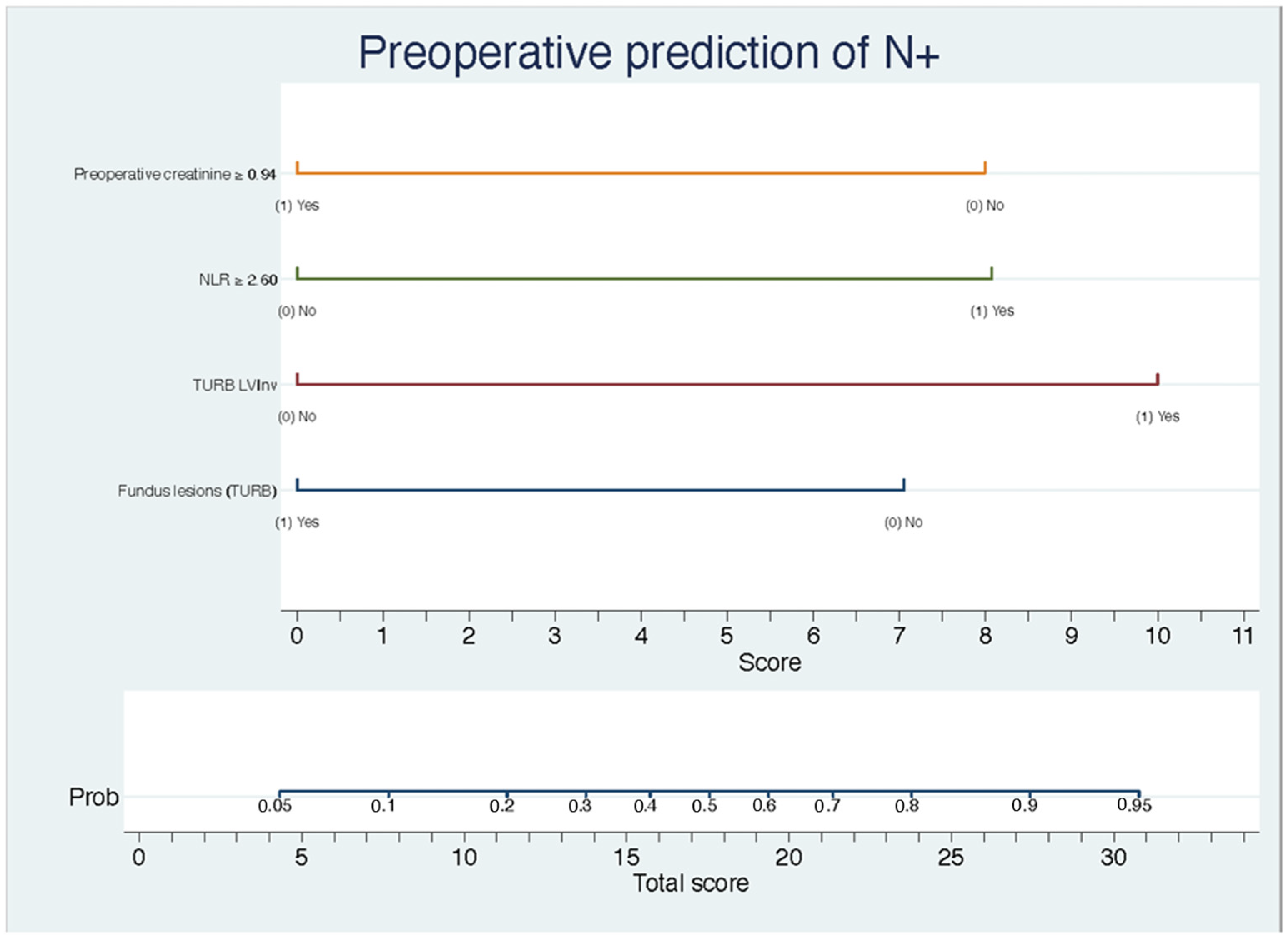
The univariate logistic regression demonstrated that the presence of LVInv exhibited a statistically significant relationship with LN+ (OR 5.25, 1.10–25 95% CI, p = 0.037). A multivariable logistic regression was performed, obtaining a predictive model of LN involvement (X2 = 16.84, p = 0.002) that exhibits a sensitivity and specificity of 41.2% and 94.7%, respectively (Table 4). The Hosmer and Lemeshow test was not significant (p = 0.20). The presence of an NLR greater than 2.60 (OR 6.03, 1.29–28.3 95% CI, p = 0.02) and the presence of LVInv in the TURB sample (OR 9.26, 1.11–77.3 95% CI, p = 0.04) were found to be significantly correlated with the presence LN+. Both fundus lesions (OR 0.21, 0.05–0.93 95% CI, p = 0.04) and creatinine values > 0.94 mg/dL (OR 0.17, 0.03–0.80 95% CI, p = 0.025) were found to be associated with a decreased likelihood of LN+ (Table 4). An ROC curve was generated for the model, resulting in a predictive capacity of 80.4% (area under the curve, AUC), with a 95% CI between 66.6% and 94.2%. This was statistically significant (p < 0.005) (Figure 1). A nomogram from this model was obtained (Figure 2).

4. Discussion

Several studies have shown that the presence of lymph node metastases is associated with poor prognosis in survival and recurrence in patients after radical cystectomy. In almost 30% of patients with pN+, overall survival is less than 3 years, and recurrences occur in less than 5 years in about 65% of cases [5,6]. On the other hand, it has been demonstrated that approximately a quarter of patients with MIBC without clinical lymph node involvement on pretreatment imaging subsequently have lymph node metastases in the cystectomy specimen [7,9,10]. This latter finding correlates with the results of our analysis of the diagnostic accuracy of CT in our center, which has a sensitivity of only 25% for identifying lymph node involvement. This shows the need for enhanced diagnostic tools to facilitate a precise preoperative identification of lymph node involvement, thereby enabling more accurate evaluation for the use of neoadjuvant and/or adjuvant treatments in patients presenting with adverse or unfavorable factors. Conversely, if we rule out lymph node metastases, we can offer our patients alternatives in terms of urinary diversion, such as neobladders.
Based on clinical, laboratory, and histopathology data that are usually obtained prior to cystectomy in patients with MIBC, the model demonstrated an acceptable predictive performance of lymph node metastatic involvement with an AUC of 80.4%. Our findings are consistent with other studies that reported nomograms to predict lymph node involvement from preoperative data. Karakiewicz et al. reported a predictive model of lymph node involvement with a predictive performance of 63% (AUC) based on tumor stage and tumor grade after TURB [11]. A similar study by Green et al. evaluated a predictive model based on clinical tumor stage, the presence of LVInv, and radiological findings, and reported an 83% AUC [6].
Our model includes LVInv in the pathology specimen of TURB, lesion location in the fundus (OR 0.21), the NLR (OR 6.03), and preoperative creatinine (OR 0.17). The inclusion of LVInv (OR 9.26) in our model is consistent with other studies in which LVInv has been found to be related to the presence of lymph node metastasis both independently and as part of predictive models. These findings are consistent with those reported by Green D. et al. [6]. This is supported by the hypothesis that the invasion of lymphatic vessels occurs prior to lymph node involvement [6]. Likewise, the NLR has been studied as a prognostic marker in different carcinomas, including bladder cancer. In this context, higher levels have been found to be associated with recurrence and progression in non-muscle-invasive bladder carcinoma [12]. Studies such as that by Aoyama et al. have demonstrated a correlation between the NLR and the presence of lymph node metastases in endometrial cancer [13]. This finding suggests that the NLR has significant potential as a prognostic marker for lymph node involvement in various carcinomas.
It should be noted that the limitations of this study include it being conducted at single institution, its retrospective nature, and its relatively small sample. The limited number of patients included in our study may affect the generalizability of our findings. The patient population from a single center might not adequately represent the broader population of patients with muscle-invasive bladder cancer (MIBC). Furthermore, the specific characteristics of the patients must be taken into account when trying to generalize these results. Thus, our predictive model may perform differently in other settings or with different patient demographics. Several studies have demonstrated that predictive nomograms often do not have the same diagnostic value in other populations due to variations in baseline characteristics or due to treatment modalities specific to each center [14]. Therefore, it is necessary to evaluate the predictive performance of our model in different populations from ours. Despite the limited number of patients, our model has a good predictive performance and is comparable to that of other studies with larger sample sizes. The inclusion of the NLR in our model is noteworthy, as it is a marker that is being widely studied for its predictive association with various types of malignancies. Our results are encouraging, particularly given that the NLR is a marker that is relatively easy to collect at a minimal cost. Further studies should address the usefulness of the NLR as a predictor of lymph node metastasis, either alone or as part of other predictive models.

5. Conclusions

Our data show that the diagnostic accuracy of a preoperative CT scan is low for the evaluation of lymph node metastases, which demonstrates the need to develop better tools for the evaluation of lymph node involvement prior to radical cystectomy. This should help with a better individualization of neoadjuvant and adjuvant treatments, as well as in the choice of the type of urinary diversion to perform. Our predictive model of lymph node involvement developed from variables that are routinely obtained in patients with MIBC before radical cystectomy shows good diagnostic performance; although, given our study limitations, the model should be evaluated in different populations. Finally, it is important to highlight the presence of the NLR in our model of, supporting its role as a marker of lymph node metastasis.

Author Contributions

Conceptualization, W.A.B.F. and C.C.G.; methodology, T.F.A.; formal analysis, W.A.B.F.; investigation, W.A.B.F. and J.M.S.; writing—original draft preparation, C.C.S. and C.F.; writing—review and editing, W.A.B.F. and V.M.; supervision, C.C.G. and T.F.A. All authors have read and agreed to the published version of the manuscript.

Funding

This research did not receive any specific grant from funding agencies in the public, commercial, or not-for-profit sectors.

Institutional Review Board Statement

The study was conducted in accordance with the Declaration of Helsinki, and approved by the Institutional Ethics Committee of Hospital Universitario General Morales Meseguer (CETI Code 48/19, date of approval: 29/10/2019).

Informed Consent Statement

Patient consent was waived due to the retrospective character of the study and did not involve any intervention.

Data Availability Statement

All results and statistical analysis of the results are available with W. Barragán (corresponding author for the present article).

Conflicts of Interest

The authors declare no conflicts of interest.

References

  1. Ferlay, J.; Colombet, M.; Soerjomataram, I.; Dyba, T.; Randi, G.; Bettio, M.; Gavin, A.; Visser, O.; Bray, F. Cancer incidence and mortality patterns in Europe: Estimates for 40 countries and 25 major cancers in 2018. Eur. J. Cancer 2018, 103, 356–387. [Google Scholar] [CrossRef] [PubMed]
  2. Yuk, H.D.; Jeong, C.W.; Kwak, C.; Kim, H.H.; Ku, J.H. Lymphovascular invasion have a similar prognostic value as lymph node involvement in patients undergoing radical cystectomy with urothelial carcinoma. Sci. Rep. 2018, 8, 1–6. [Google Scholar] [CrossRef]
  3. Yafi, F.A.; Aprikian, A.G.; Chin, J.L.; Fradet, Y.; Izawa, J.; Estey, E.; Fairey, A.; Rendon, R.; Cagiannos, I.; Lacombe, L.; et al. Impact of concomitant carcinoma in situ on upstaging and outcome following radical cystectomy for bladder cancer. World J. Urol. 2014, 32, 1295–1301. [Google Scholar] [CrossRef] [PubMed]
  4. Zhang, L.; Wu, B.; Zha, Z.; Qu, W.; Zhao, H.; Yuan, J. Clinicopathological factors in bladder cancer for cancer-specific survival outcomes following radical cystectomy: A systematic review and meta-analysis. BMC Cancer 2019, 19, 1–13. [Google Scholar] [CrossRef] [PubMed]
  5. Cha, E.K.; Sfakianos, J.P.; Sukhu, R.; Yee, A.M.; Sjoberg, D.D.; Bochner, B.H. Poor prognosis of bladder cancer patients with occult lymph node metastases treated with neoadjuvant chemotherapy. BJU Int. 2018, 122, 627–632. [Google Scholar] [CrossRef] [PubMed]
  6. Green, D.A.; Rink, M.; Hansen, J.; Cha, E.K.; Robinson, B.; Tian, Z.; Chun, F.K.; Tagawa, S.; Karakiewicz, P.I.; Fisch, M.; et al. Accurate preoperative prediction of non-organ-confined bladder urothelial carcinoma at cystectomy. BJU Int. 2012, 111, 404–411. [Google Scholar] [CrossRef] [PubMed]
  7. Seiler, R.; Lam, L.L.; Erho, N.; Takhar, M.; Mitra, A.P.; Buerki, C.; Davicioni, E.; Skinner, E.C.; Daneshmand, S.; Black, P.C. Prediction of Lymph Node Metastasis in Patients with Bladder Cancer Using Whole Transcriptome Gene Expression Signatures. J. Urol. 2016, 196, 1036–1041. [Google Scholar] [CrossRef] [PubMed]
  8. Witjes, J.A.; Bruins, M.; Cathomas, R.; Compérat, E.; Cowan, N.C.; Gakis, G.; Thalmann, G.N. EAU Guidelines on: Muscle-Invasive and Metastatic Bladder Cancer; European Association of Urology: Arnhem, The Netherlands, 2019. [Google Scholar]
  9. Chang, S.S.; Bochner, B.H.; Chou, R.; Dreicer, R.; Kamat, A.M.; Lerner, S.P.; Holzbeierlein, J.M. Tratamiento del Cáncer de Vejiga Músculo Invasivo y No Metastásico: Guía de AUA/ASCO/ASTRO/SUO; Spanish Version; American Urological Association Education and Research: Linthicum, MD, USA, 2019. [Google Scholar]
  10. Madersbacher, S.; Hochreiter, W.; Burkhard, F.; Thalmann, G.N.; Danuser, H.; Markwalder, R.; Studer, U.E. Radical cystectomy for bladder cancer today—A homogeneous series without neoadjuvant therapy. J. Clin. Oncol. 2003, 21, 690–696. [Google Scholar] [CrossRef] [PubMed]
  11. Karakiewicz, P.I.; Shariat, S.F.; Palapattu, G.S.; Perrotte, P.; Lotan, Y.; Rogers, C.G.; Amiel, G.E.; Vazina, A.; Gupta, A.; Bastian, P.J.; et al. Precystectomy Nomogram for Prediction of Advanced Bladder Cancer Stage. Eur. Urol. 2006, 50, 1254–1262. [Google Scholar] [CrossRef]
  12. Vartolomei, M.D.; Porav-Hodade, D.; Ferro, M.; Mathieu, R.; Abufaraj, M.; Foerster, B.; Kimura, S.; Shariat, S.F. Prognostic Role of Pretreatment Neutrophil-To-Lymphocyte Ratio (NLR) in Patients With Non-Muscle-Invasive Bladder Cancer (NMIBC): A Systematic Review and Meta-Analysis. Urol. Oncol. Semin. Orig. Investig. 2018, 36, 389–399. [Google Scholar] [CrossRef] [PubMed]
  13. Aoyama, T.; Takano, M.; Miyamoto, M.; Yoshikawa, T.; Kato, K.; Sakamoto, T.; Takasaki, K.; Matsuura, H.; Soyama, H.; Hirata, J.; et al. Pretreatment Neutrophil-to-Lymphocyte Ratio Was a Predictor of Lymph Node Metastasis in Endometrial Cancer Patients. Oncology 2019, 96, 259–267. [Google Scholar] [CrossRef] [PubMed]
  14. Kluth, L.A.; Black, P.C.; Bochner, B.H.; Catto, J.; Lerner, S.P.; Stenzl, A.; Sylvester, R.; Vickers, A.J.; Xylinas, E.; Shariat, S.F. Prognostic and Prediction Tools in Bladder Cancer: A Comprehensive Review of the Literature. Eur. Urol. 2015, 68, 238–253. [Google Scholar] [CrossRef] [PubMed]
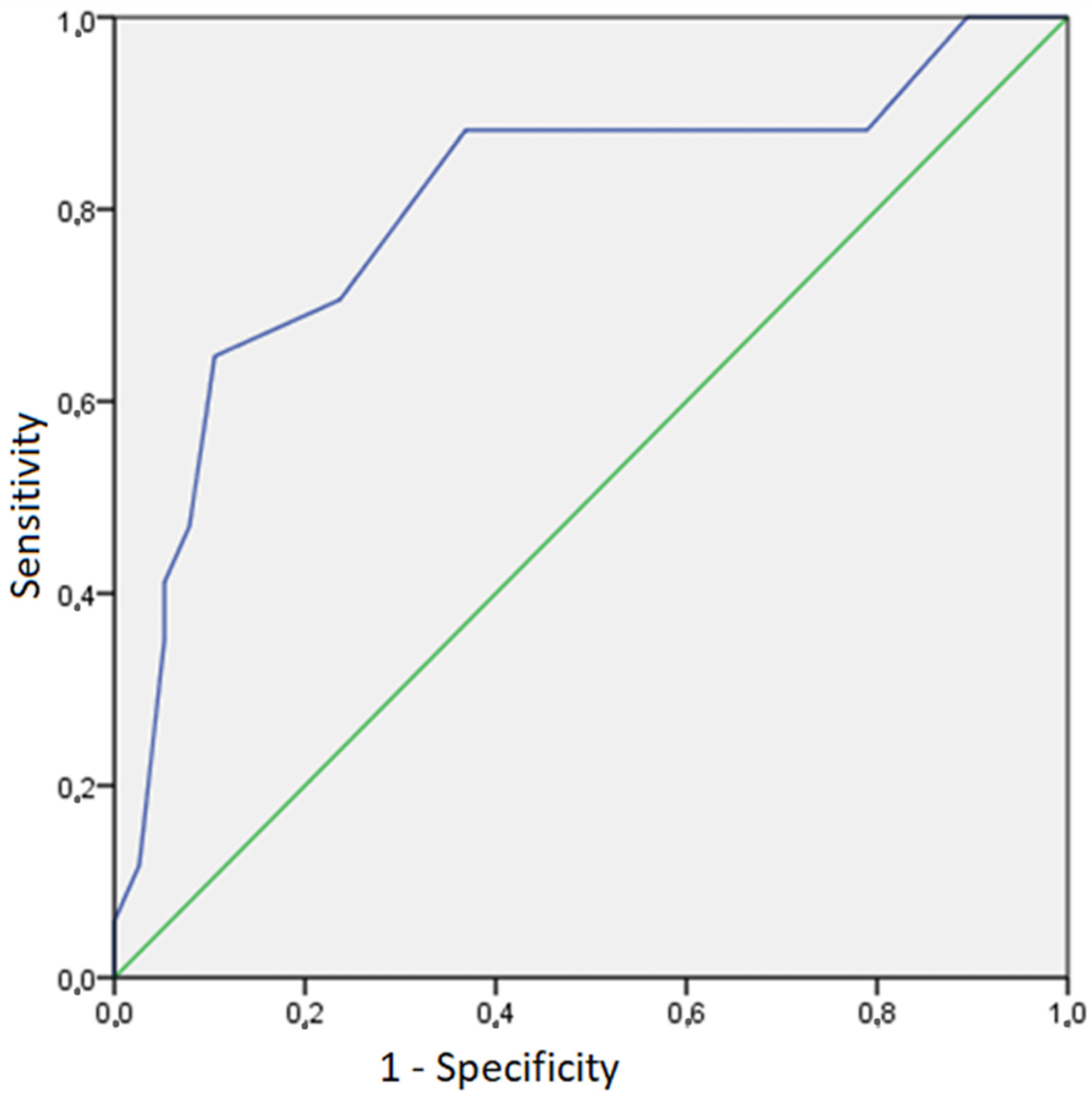
Figure 1. ROC curve of the prediction model of lymph node involvement. Green line, represents the performance of a random classifier with an AUC of 0.50. Blue line, represents the ROC curve of the model, with an AUC of 0.804.
Biomed 04 00017 g001
Figure 2. The developed preoperative Nomogram of N+. There are included the variables obtained by multivariable logistic regression for prediction of N+.
Biomed 04 00017 g002
Table 1. Patients demographic and clinicopathological characteristics.
VariablesN° (%) Patients
AgeMean (Median)65 (66)
Range33–83
GenderMale58 (93.50)
Female4 (6.50)
Presurgical creatinineMean (Median)1.04 (0.94)
Range0.46–2.31
TURB T stageTx1 (1.60)
Tis0 (0)
Ta0 (0)
T15 (8.20)
T255 (90.20)
CIS presence after TURB 17 (27.40)
LVInv presence after TURB 8 (12.90)
cT stageTx5 (8.50)
T0-T1-T227 (45.70)
T320 (33.90)
T47 (11.90)
cN stageN046 (79.30)
N16 (10.30)
N23 (5.20)
N33 (5.20)
Presurgical NLRMean (Median)2.94 (2.25)
Range0.59–13.50
Neoadjuvant Chemotherapy 5 (8.10)
Lymph nodes obtainedMean (Median)19.84 (19)
pT stageTis8 (12.90)
T05 (8.10)
T18 (12.90)
T27 (11.30)
T321 (33.90)
T413 (21)
pN stageN044 (71)
N16 (9.70)
N212 (19.40)
N30 (0)
CIS, Carcinoma in situ; LVIns, Linfovascular invasion; NLR, neutrophil-to-lymphocyte ratio.
Table 2. Diagnostic concordance between CT and histopathology findings in extravesical extension (T3a or greater).
pT3 ≥ stage
NoYesTotal
cT3 ≥ stageNo181432
Yes82028
Total263460
Table 3. Diagnostic concordance between CT and histopathology findings in lymph node involvement (N+).
pN+
NoYesTotal
cN+ No331447
Yes9413
Total421860
Table 4. Logistic regression of variables included in the prediction model of lymph node involvement *.
VariablesORI.C. 95%p-Value
Preoperative creatinine0.170.03–0.800.02
NLR6.031.29–28.300.02
LVInv (TURB)9.261.11–77.300.04
Fundus lesion (RTU)0.210.05–0.930.04
Model CalibrationX2 = 16.84p = 0.002
* Model calibration: Sensitivity 41.2%, Specificity 94.7%, PPV 77.7%, NPV 78.26%. LVInv, Linfovascular invasion; NLR, neutrophil-to-lymphocyte ratio.
Disclaimer/Publisher’s Note: The statements, opinions and data contained in all publications are solely those of the individual author(s) and contributor(s) and not of MDPI and/or the editor(s). MDPI and/or the editor(s) disclaim responsibility for any injury to people or property resulting from any ideas, methods, instructions or products referred to in the content.