Review

Targeting Cancer Stem Cells with Radioimmunotherapy: The Case of the Ovarian Cancer Stemness-Associated Biomarker L1CAM

1
Center for Radiopharmaceutical Sciences ETH-PSI, Paul Scherrer Institute, 5232 Villigen, Switzerland
2
Department of Chemistry and Applied Biosciences, ETH Zurich, 8093 Zurich, Switzerland
*
Author to whom correspondence should be addressed.
Int. J. Transl. Med. 2024, 4(3), 463-485; https://doi.org/10.3390/ijtm4030031
Submission received: 31 May 2024 / Revised: 8 July 2024 / Accepted: 15 July 2024 / Published: 18 July 2024

Abstract

:
Cancer stem cells (CSCs) are a dynamic population of tumor cells characterized by long-term self-renewal, high tumorigenicity, resistance to conventional therapies such as radio- and chemotherapy, and capacity to recapitulate the tumor heterogeneity. Similar to other tumor cells, CSCs need to carry critical mutations and epigenetic changes to acquire their aberrant phenotype. Confirmed in various hematologic and solid malignancies, the critical need to deepen our understanding of CSC biology, including identification of CSC biomarkers, and develop novel CSC-targeted therapies has been clearly recognized. Here, we review the L1 cell adhesion molecule (L1CAM) as a CSC-associated biomarker in ovarian cancer. Furthermore, we inform on the promising potential of anti-L1CAM radioimmunotherapy with 161Tb as a novel CSC-targeted therapeutic approach to overcome CSC radioresistance in comparison to 177Lu.

1. Introduction

Ovarian cancer (OC) is a severe disease which represents the most common and lethal gynecologic malignancy in developed countries [1,2,3]. High-grade serous ovarian carcinoma (HGSOC) is the most common OC histotype characterized by relapse in >70% of the cases and poor survival outcomes despite the successful first line of therapy [2]. The development and progression of OC, including HGSOC, points to the cancer stem cell (CSC) disease maintenance. More aggressive and therapy-resistant relapsed OC as well as accumulation of ascitic fluid with high tumor cell content could be explained by the presence of ovarian CSCs, which could acquire dynamic states, allowing them to survive conventional therapies in anchorage-independent cell growth conditions and invade and metastasize to distant sites [2,4,5,6]. This highlights the necessity to investigate CSCs and CSC mechanisms of therapy resistance, including radioresistance, and identify CSC biomarkers to inform the development of novel targeted therapies for clinical application.
The L1 cell adhesion molecule (L1CAM) has been confirmed as a CSC-associated biomarker in various malignancies, including OC [7,8,9,10]. A recent study validated in vitro and in vivo L1CAM+/CD133+ cells as an ovarian CSC population while also identifying the L1CAM as a biomarker of radioresistance in OC [11]. Previous work demonstrated the efficacy of anti-L1CAM radioimmunotherapy (RIT) with the monoclonal antibody chCE7 radiolabeled with the β−/conversion (CE)/Auger electron (AE)-emitter Terbium-161 (161Tb) in comparison to the β−-emitter Lutetium-177 (177Lu) [12]. Due to their high cytotoxicity, CE/AE have the potential to overcome the radioresistance of CSCs and, therefore, propose a promising novel alternative for CSC-targeted therapies [13,14].
In this paper, we review OC with a focus on HGSOC, cancer stem cell properties and current methods to study and characterize CSCs. Furthermore, we specifically discuss the role of L1CAM in health and disease including cancer stemness as well as anti-L1CAM RIT as a novel therapeutic modality against CSCs.

2. Ovarian Cancer—Epidemiology, Histotypes, and Current Standard of Treatment of Epithelial Ovarian Cancer

2.1. Epidemiology, Histological Subtypes, and Molecular Features of HGSOC

The latest worldwide cancer statistics by the Global Cancer Observatory (GLOBOCAN) reported 313,959 new cases and 207,252 death cases of OC for 2020 [3,15]. OC ranks eighth in terms of both incidence and mortality rate among women while it remains the deadliest gynecologic malignancy [2,16]. It is difficult to detect due to asymptomatic early stages (International Federation of Gynecology and Obstetrics (FIGO) stages I and II) and limited screening options. For most cases, it is diagnosed at later stages (FIGO stages III and IV) when the cancer has already metastasized [2]. Late-stage OC symptoms are non-specific and overlap with symptoms of more common diseases. These symptoms could encompass abdominal swelling, pelvic pain, weight loss, nausea, fatigue, and urinary frequency or urgency [17].
OC is a heterogeneous disease with distinct sites of origin, morphology, molecular alterations, treatment, and prognosis [2,18]. Generally, it is divided into non-epithelial and epithelial cancers. Non-epithelial OC represents approximately 10% of the cases and includes ovarian germ cell cancer, ovarian sex cord–stromal cancer, and small carcinoma of the ovary (Figure 1A). Epithelial OC (EOC) represents more than 90% of all ovarian malignancies (Figure 1A). According to the most up-to-date classification by the World Health Organization (WHO), EOC is divided into five histological subtypes (Figure 1B)—high-grade serous ovarian carcinoma (HGSOC), low-grade serous ovarian carcinoma (LGSOC), clear cell carcinoma (CCC), endometrioid carcinoma (EC), and mucinous carcinoma (MC) [4,18]. Each EOC histotype is characterized by distinct clinical and molecular characteristics represented by the cancer incidence, age at diagnosis, origin, histology, mutational profile, tumor microenvironment, major signaling pathways affected, treatment, and clinical outcome [2,4,16,19]. In parallel, FIGO has determined the surgical stages I-IV of EOC as follows: stage I (tumor confined to the ovaries), stage II (tumor involves one or both ovaries with pelvic extension or primary peritoneal cancer), stage III (tumor involves the ovaries, or primary peritoneal cancer, with spread to the peritoneum outside pelvis and/or metastasis to the retroperitoneal lymph nodes), and stage IV (distant metastasis) [19].
HGSOC is the most common and lethal EOC histotype with >75% (Figure 1B) of the cases diagnosed at advanced FIGO stages III and IV, while the rest of EOC cases are shared between LGSOC, CCC, EC, and MC. For HGSOC, the overall patient prognosis is poor with a high risk of aggressive and treatment-resistant relapse [2,16]. HGSOC is characterized by a high frequency of somatic TP53 mutation. As a result, the TP53 protein loses its tumor-suppressive function. In addition, 15–20% of the patients carry germline mutations in breast cancer gene 1 and 2 (BRCA1, BRCA2). The BRCA genes participate in the homologous recombination (HR) repair of double-strand DNA breaks. Further analysis points out that most of the cases with germline BRCA mutations also have somatic mutations in other HR genes such as Fanconi anemia genes, mismatch repair genes, DNA damage response genes, and RAD family genes collectively referred to as HR deficiency (HRD) genes [20]. Cells with non-functioning HR utilize other pathways of DNA repair such as non-homologous end-joining (NHEJ) which is more error-prone and could further increase the genomic instability. An analysis by The Cancer Genome Atlas Research Network (TCGA) reports that almost 50% of the HGSOC cases are HRD [21]. A recent report on the origin of HGSOC demonstrated that it can originate from both the fallopian tube epithelium (FTE) as well as the ovarian surface epithelium (OSE). The results point out the prognostic value of the tissue of origin providing evidence that FTE- and OSE-originated HGSOC possess specific transcriptional profiles, with OSE-originated cancer correlating with a higher invasiveness and immunomodulatory phenotype [22].

2.2. Current Standard of Treatment of Epithelial Ovarian Cancer and Therapies under Investigation

To improve EOC treatment and increase patient survival, it is crucial to consider the clinicopathological characteristics and molecular heterogeneity of EOC. Although early-stage OC is curable, most of the patients are diagnosed at advanced stages III-IV due to the display of non-specific or no symptoms and the lack of effective screening methods. The OC risk factors include inherited risk (germline mutations in the BRCA1/BRCA2 breast cancer susceptibility genes), infertility, endometriosis, obesity, age, and nulliparity [16]. In addition, the dissemination of OC to the peritoneal cavity and the abdomen, which results in accumulation of ascetic fluid and bowel obstruction, is facilitated by the lack of an anatomical barrier around the ovaries [18].
The current gold standard of care for EOC patients includes a debulking cytoreductive surgery followed by chemotherapy as a first line of treatment. This strategy has remained the same in clinics for the past three decades. The aim of the debulking surgery is to reduce the amount of cancer in the patient by full or partial removal of the cancer-affected organs. It is also key in the accurate determination of the diagnosis. However, the cytoreductive surgery is rarely curative, especially in patients with advanced disease. It is currently combined with adjuvant chemotherapy. The standard combination chemotherapy comprises taxane-based (paclitaxel, docetaxel, cabazitaxel) and platinum-based (cisplatin, carboplatin, oxaliplatin) drugs (Table 1).
Although the patients respond well to the first line of therapy and 60–80% enter remission, 70–80% relapse, developing more aggressive and therapy-resistant OC [23]. Recurrent OC is categorized into platinum-refractory, resistant, partially sensitive, or fully sensitive in accordance with the period between the last platinum treatment and the relapse. Second-line chemotherapy with various drugs such as carboplatin, gemcitabine, topotecan, vinorelbine, or other therapeutic approaches, including targeted therapies, has been tested [24]. Unfortunately, the efforts up to date in overcoming the therapy resistance in recurrent OC have led to minimal improvements in patient survival. Radiotherapy has shown a limited success in OC treatment. Its current application is for patients with oligometastatic and oligoprogressive disease as well as palliative care, although there are ongoing clinical trials studying modern radiotherapy techniques alone or in combination with other therapeutic approaches [25].
Two targeted therapeutic strategies are approved up to date by the U.S. Food and Drug Administration (FDA) and the European Medicines Agency (EMA): bevacizumab, a recombinant humanized monoclonal anti-vascular endothelial growth factor (VEGF) antibody, and poly (ADP-ribose) polymerase inhibitors (PARPi) (Table 1). The establishment of PARPi has revolutionized EOC patient treatment. They specifically inhibit the enzyme family of poly (ADP-ribose) polymerases (PARPs), including PARP1, PARP2, and PARP3 as its most well-studied members. PARP enzymes play a main role in the repair of single-strand DNA breaks (SSDBs) by recruiting components of the DNA repair machinery. PARPi compete with the nicotinamide NAD+ in the PARP catalytic site, rendering the enzymes inactive. As a result, the SSDBs persist while double-strand DNA breaks accumulate. Healthy cells can repair the double-strand DNA breaks via the error-free HR. However, HRD cancers such as HGSOC lack that pathway due to its BRCA1/BRCA2 deficiency. The BRCA mutational status together with the PARPi result in the synthetic lethality of the cancer cells. The inhibition of the SSDB repair, replication fork stalling, PARP trapping, and activation of the more error-prone NHEJ DNA repair have been proposed as PARPi mechanisms [26]. Other targeted therapies against various EOC targets have been under investigation, a selection of which is summarized in Table 1. To date, there are no approved immunotherapies against OC.
Table 1. Selected therapeutic agents and targets for treatment of epithelial ovarian cancer.
Therapeutic AgentTarget StatusRef.
Taxane-based drugsTubulin (inhibition of the microtubule disassembly)Approved[27]
Platinum-based drugsDNA cross-linking agentApproved
BevasizumabAnti-VEGFApproved[28,29,30]
PARP inhibitorsPARP enzymesApproved[16,30,31,32]
Mirvetuximab
soravtansine
Folate receptor alphaNCT020631876, Phase III[33]
Anti-PD-1/PD-L1, anti-CTLA-4PD-1/PD-L1, CTLA-4NCT02580058, Phase III
NCT02811497, Phase II
NCT02657889, Phase 1/2
[16]
CAR T cellsFolate receptor alpha, mesothelin, MUC16 (also known as CA125), CD70Multiple ongoing or completed clinical and preclinical studies[34]
The treatment of EOC and the observed high therapy resistant relapse with an increased mortality rate continue to pose a clinical challenge. There is an urgent need to develop novel therapies that are more successful. A key factor in achieving this goal is to deepen our knowledge and understanding of the biology of EOC heterogeneity followed by tailoring the therapies to the specific features of the disease. Such novel therapies could target cancer stem cells (CSCs), as one known source of tumor recurrence, aggressiveness, and therapy resistance, either by CSC-specific drugs or CSC-targeted radionuclide therapy such as radioimmunotherapy utilizing suitable particle radiation emitters [35].

3. Cancer Stem Cells—Definition, Concept, Study, and Identification, Ovarian Cancer Stem Cells

3.1. Definition and Concept of Cancer Stem Cells

CSCs are a population of tumor cells defined by their functional properties of long-term self-renewal, tumor propagation and recapitulation of the tumor heterogeneity, and radio- and chemotherapy resistance [5,6] (Figure 2). Similar to other tumor cells, CSCs need to carry critical mutations and epigenetic changes to acquire their aberrant phenotype.
The CSC concept started to emerge at the beginning of the 19th century by observing that a certain subset of tumor cells in teratomas, teratocarcinomas, and embryonic stem cells had the ability to produce several distinct differentiated cell populations as well as retained tumorigenicity after multiple passages [36]. In the late 90s, Dick et al. refocused the spotlight back to CSCs by isolating CD34+/CD38− from acute myeloid leukaemia and confirming their major CSC characteristics [37,38]. The first confirmation of CSCs in a human solid tumor was in the 2000s in breast cancer in which CD44+/CD24−/low/Lineage cells isolated from breast mouse xenografts or cancer patients [39]. Up to date CSCs have been verified in multiple hematologic and solid tumors together with their role in fundamental tumor processes, including tumor development and recurrence, invasion and metastasis, and therapy resistance (Table 2).
The development of the cancer stemness hypothesis advanced the view of the hierarchical organization of tumors similar to normal adult stem cells and healthy tissue. Such a hierarchy recognizes the tumor heterogeneity as well as the heterogenous functions within the various tumor cell populations. According to this hierarchical model, only the CSCs, which possess extensive self-renewal and multipotency, can give rise to transit-amplifying cells with lower proliferative capacity and could further differentiate into non-proliferative cancer cells [40]. Therefore, the tumor formation and growth are maintained by the CSCs. As with normal stem cells, a CSC niche is required to provide a favorable environment for the survival and maintenance of their functions. In comparison, the stochastic model, also known as the clonal evolution model, of tumor growth proposes the equipotency of all tumor cells to self-renew and generate differentiated tumor progeny. Importantly, the plasticity model combines the previous two models by introducing the plasticity between the transition of CSCs and non-CSCs as a result of spontaneous, intrinsic, and/or induced transformation between these populations, therefore pointing out to the dynamic rather than the stable and unidirectional CSC phenotype. Non-CSCs have been shown to de-differentiate into CSCs in vitro and in vivo, highlighting the plasticity and bidirectionality between these two populations [41]. There is a growing body of evidence in support of the plasticity model which creates a biological and clinical challenge for deeper understanding of the underlying mechanisms and to devise novel cancer therapies [40].
In addition to the plasticity, the dynamic phenotype of CSCs is also demonstrated by acquiring other reversible states which include epithelial-to-mesenchymal transition (EMT), dormancy (quiescence), and senescence (Figure 2). EMT is one of the key cellular programs in which cells enter various discrete states along the epithelial–mesenchymal spectrum, allowing the cells to migrate to distant sites [42]. While in health, EMT is important during embryogenesis, wound healing for tissue regeneration and repair, and recruitment of immune cells to sites of inflammation and infection, in cancer, the EMT program could contribute to malignant progression by leading to tumor initiation, motility, ability to disseminate and colonize distant sites, as well as therapy resistance, which are major CSC properties, as discussed above [43]. The close association of EMT and CSCs has already been documented in different cancers such as breast, lung, prostate, pancreatic, head and neck, and colon cancer [43].
CSC dormancy (quiescence) as well as senescence are two other states related to the dynamic CSC phenotype that have emerged in recent years (Figure 2). Dormancy refers to a reversible entry into the G0 resting phase of the cell cycle where the cell remains viable but not proliferating, triggered by various microenvironmental cues such as hypoxia, nutrient deprivation, oxidative stress, growth factors leading to maintaining stemness cells, and surviving unfavorable conditions [44]. Currently, at least three settings are considered in which dormancy-competent CSCs could undergo dormancy to survive and adapt: primary tumor dormancy, metastatic dormancy, and therapy-induced dormancy [45]. In breast cancer, the activity of Src family kinase and the mitogen-activated protein kinase (MAPK) pathway is required for the proliferative outgrowth and metastasis formation of dormant cancer cells [46]. A CSC population in glioblastoma entered into quiescence upon temozolomide treatment while ablation of these cells resulted in susceptibility to the combined chemotherapy with temozolomide and ganciclovir [47].
Cellular senescence acts as a failsafe program by entry into cell cycle arrest in the G1-S phase as a result of increased DNA damage and persistent oxidative and oncogenic stress [44]. In addition to its anti-tumorigenic effects to protect the organismal integrity by preventing the further propagation and eradication of (pre-) malignant cells, it could also have a pro-tumorigenic role supporting CSC generation. Uncleared senescent cancer cells could accumulate more mutations, allowing them to escape the cell cycle arrest and clearance while acquiring CSC properties, or could secrete various factors which could lead to the reprogramming and activation of CSC TFs [48]. Senescence and cancer stemness have been found to be co-regulated by overlapping signaling networks including p16Ink4a, p21 or p53, demonstrating a link between them [49]. Therapy-induced senescent cancer cells acquired phenotypic and functional stemness in hematologic cancers and after re-entry into the cell cycle, these cells showed a higher tumor-initiating capacity than their never-senescent counterparts [50]. Senescent human colon cancer cells induced by repeated cycle with doxorubicin acquired a CSC phenotype by first entering a dormant state and later triggering tumor regrowth [51]. Taken all together, this clearly indicates that it is crucial to consider CSC plasticity, dormancy, and senescence for the effective eradication of CSCs and bulk tumor population.
Table 2. Selected reported cancer stem cell populations in different malignancies and their associated properties.
CSC PopulationCancer TypeAssociated CSC PropertiesRef.
L1CAM+/CD133+OvarianIn vitro—high clonogenicity, spherogenicity, invasiveness, radioresistance. Upregulation of stem cell and EMT genes (Oct-4, CXCR4, ABCG2, TGF-1β, β-catenin, vimentin).
In vivo—high tumorigenicity, self-renewal.
[11]
CD44+/CD117+OvarianIn vitro—anchorage-independent and self-renewing sphere formation in CSC selective conditions, chemoresistance. Expression of stem cell genes (Oct-4, Nestin, Nanog, Notch-1, Bmi-1).
In vivo—tumorigenicity and serial propagation, histological recapitulation of the original tumor.
[52]
ALDH+/CD133+OvarianIn vitro—high and long-term spherogenicity in CSC selective conditions. Upregulation of stem cell genes (Sox2, Oct4, Nanog).
In vivo—generate heterogenous tumors.
[53]
CD44+/CD133+Pancreatic In vitro—high sphere formation in CSC selective conditions, proliferation, chemoresistance, recapitulation of the other tumor populations. Upregulation of inflammation and EMT genes (Sparc, Col1a1, Ccl2, Cxcl1, Cxcl2); mRNAs * in CSC-related pathways.
In vivo—high tumorigenicity.
[54]
CXCR4+/CD133+Colorectal

Pancreatic
In vitro—high migratory capacity. Upregulation of EMT genes (vimentin, N-cadherin, Snail).
In vivo—high tumorigenicity and metastasis formation blocked with the CXCR4 antagonist AMD3100.
In vivo—high tumorigenicity, invasiveness, and metastasis formation, inhibited by AMD3100.
[55]

[56]
CD44+/CD24+GastricIn vitro—enhanced sphere formation in CSC selective conditions. Upregulation of stemness genes (Shh, Ptch1).
In vivo—high tumorigenicity; regeneration of the tumor heterogeneity.
[57]
EpCAM+/CD166+/
CD44+
Non-small cell lungIn vitro—higher proliferation, clonogenicity, sphere formation, migration, chemoresistance. Upregulated stem cell genes (Rex1, Ssea4).[58]
CD44+/CD24−
/ALDH1+
Head and neckIn vitro—high spherogenicity in CSC selective conditions, increased invasion, radioresistance. Upregulation of the EMT and stem cell genes (Snail, Oct-4, Nanog, Sox2).
In vivo—high tumorigenicity. Knockdown of Snail reduced the CSC properties.
[59]
CD34+/CD38-AMLIn vivo—high tumorigenicity, self-renewal, differentiation into other tumor cells populations.[37,38] **
CD44+/CD24−/lowBreast cancerIn vivo—high tumorigenicity, regeneration of the tumor heterogeneity.[39] **
ABCG2, ATP-binding cassette super-family G member 2; ALDH, aldehyde dehydrogenase; AML, acute myeloid leukemia; CXCR4, CXC motif chemokine receptor 4; EMT, epithelial-to-mesenchymal transition; L1CAM, L1 cell adhesion molecule; TGF-1β, transforming growth factor beta 1; Oct-4, octamer binding transcription factor 4; Shh, sonic hedgehog; Ptch1, patched 1. * The mRNA expression differences were determined by genome-wide analysis of mRNA—long non-coding RNA (lncRNA) co-expression networks. ** These were the first reports of CSCs in a hematologic and solid malignancy.

3.2. Study and Identification of Cancer Stem Cells

The identification and characterization of CSCs rely on the biomarker combination of cell surface or intracellular markers (Table 2). The need for such marker combinations rather than a single marker is emphasized by the heterogeneity within the CSC populations as well as the fact that most of the currently reported CSC markers are also expressed on normal embryonic and adult stem cells [60]. This is clearly supported by multiple reports, a selection of which is summarized in Table 3, in which only the double positive populations demonstrated major CSC features when compared to any of the single positive or the double negative cells.
An array of key in vitro and in vivo methods has been established to study and identify CSCs (Table 3). These methods rely on marker-dependent or -independent CSC enrichment followed by confirming the key CSC features of the populations of interest [61]. To study CSCs, it is pivotal to combine in vitro and in vivo methods while considering the limitations of each method (Table 3). A potential workflow following the selection of clinically relevant CSC biomarkers could start with cell enrichment based on the expression of cell surface markers via fluorescence-activated cell sorting (FACS) or magnetic-activated cell sorting (MACS). An alternative starting point could be CSC enrichment from bulk cells by seeding in CSC selective conditions which include cell culture serum-free medium supplemented with growth factors. Sorted cells could also be cultured under CSC selective conditions in order to assess their cancer stemness properties. Subsequently, various key in vitro (anchorage-independent cell growth (AICG), clonogenic survival, radioresistance) and in vivo (limiting dilution, serial xenotransplantation, lineage tracing) assays are performed to verify the CSC phenotype of the cell populations of interest.
Following the application of in vitro or in vivo methods, with or without treatment, the cells can be further investigated on a molecular level via qPCR, RNA-Sequencing, and omics technologies such as genomics, transcriptomics, proteomics, phosphoproteomics, metabolomics, and interactomics. Such approaches are increasingly implemented in CSC research and could provide invaluable knowledge.
Table 3. Key selected methods for identification and functional study of cancer stem cells and their limitations.
MethodCSC PropertyMethod LimitationsRef.
In vitro
Anchorage -independent cell growth (spherogenicity in non-adherent conditions)Tumorigenicity
Anchorage-independent survival
Long-term self-renewal (sphere passaging)
  • Required factors for the growth of CSCs in an organism might not be provided in vitro.
  • Might not fully replicate the 3D structure of a tumor or its environment.
  • Spheres could be a result of aggregation and not of single clone proliferation.
  • Dormant cells might not form spheres.
  • Non-CSCs could also have spherogenic capacity.
  • Cells within a population might respond differently to mitogens.
  • The cell line might not form spheres or have low sphere forming capacity.
[62,63,64,65,66]
ClonogenicityClonogenic survival
(Unlimited proliferation capacity)
  • The CSC could be in a dormant state, and therefore be proliferatively inactive.
  • Plating efficiency-based analysis assumes a linear correlation between the number of seeded cells and formed colonies while some cell lines could demonstrate a non-linear correlation due to cellular cooperation.
[67,68]
Radio- and chemoresistanceTherapy resistance/sensitivity
  • The therapy resistance might not be restricted only to a single CSC population or to CSCs.
  • Often based on clonogenicity or spherogenicity.
In vivo
Limiting dilution Tumorigenicity
CSC frequency
  • The tumor propagating ability might not be restricted only to a single CSC population or to CSCs.
  • Procedures such as cell sorting and dissociation might change the cell metabolism, development, and role in the tumor hierarchy.
  • Selection of suitable immunodeficient mouse models—strains and injection sites.
  • Lack of immunocompetent environment as immunodeficient mice lack intact immune systems and natural immunosurveillance.
  • Lack of tumor-specific microenvironment and cytokines for CSC stimulation.
  • Low CSC frequency might reflect the limited cell survival in a xenoenvironment.
[5,6,40,69,70,71]
Serial transplantation Long-term self-renewal
Lineage tracing Cancer cell of origin
Tumorigenicity
Clonal expansion
Cellular heterogeneity
CSC frequency
Localization
  • The cell labeling efficiency is variable.
  • Can only be performed in mice while there are significant differences between human and mouse organs and cells.
  • In mouse models of human cancer, the oncogenic promoter is immediately switched on, resulting in all-at-once genetic defects which does not recapitulate real cancer development.

3.3. Cancer Stem Cell Determinants of Radioresistance

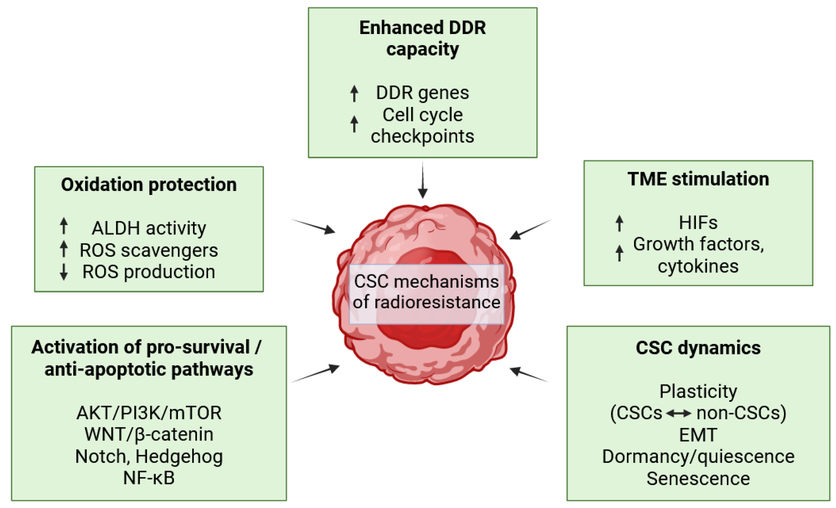
DNA damage and the induced DNA damage response (DDR) play a critical role in the biological effects of ionizing radiation (IR) and radioresistance of CSCs (Figure 3). IR could damage nucleotide bases, induce DNA singe-strand breaks or the most lethal DNA lesions, DSBs. To preserve their genomic integrity, the cells initiate the DDR to repair the radiation-induced damage or, if not repairable, to enter cell cycle arrest and programmed cell death. The DDR induces upregulation of DDR genes (for example BRCA genes, PARP, Rad51, or DNA dependent protein kinases (DNAPKs)) and cell cycle checkpoint kinases (such as ataxia telangiectasia mutated (ATM) checkpoint kinase 2 (Chk2) and ATM-Rad3-related (ATR)-checkpoint kinase (Chk1)) which delay cell cycle progression to allow DNA repair [72,73]. CSCs have been reported to have an increased DNA repair capacity following IR-induced damage in multiple solid malignancies by activation of the ATR-Chk1 and ATM-Chk2 signaling pathways [74,75,76,77].
The generation of reactive oxygen species (ROS) is another mechanism of radiation-induced damage (Figure 3). ROS are chemical species that are highly reactive with biomolecules such as proteins, lipids, and DNA. Physiologically, ROS homeostasis is tightly regulated as they play a key role in signaling transduction, metabolism, cell growth, differentiation, enzyme regulation, and mediation of inflammation [78]. Excessive ROS production leads to oxidative stress and could eventually result in cell death, which is beneficial for radiotherapy. Oxidation protection by increased ROS scavenging systems, such as superoxide dismutases, glutathione peroxidase, peroxiredoxins, glutaredoxin, thioredoxin, and catalase, as well as generally lower ROS production, have been described as CSC mechanisms of radioresistance [79,80,81]. Aldehyde dehydrogenase (ALDH), which participates in free radical scavenging in oxidative stress, has also been implicated in CSC radioresistance in various solid malignancies [53,59,82,83].
CSC radioresistance could also be attributed to the activation of pro-survival and anti-apoptotic pathways which lead to cell growth, proliferation, and protection from cell death (Figure 3). Aberrant upregulation of multiple signaling pathways, such as wingless and INT-1 (WNT)/β-catenin, AKT/phosphoinositide 3-kinase (PI3K)/mammalian target of rapamycin (mTOR), Notch, Hedgehog, nuclear factor κB (NF-κB), and Janus kinase (JAK)/signal transducer and activator of transcription (STAT), could lead to the development and maintenance of cancer stemness [84].
The TME also plays a critical role in providing extrinsic determinants of CSC radioresistance (Figure 3). The TME is a structural and functional niche rich in various components such as blood vessels, extracellular matrix (ECM), and diverse cell types such as fibroblasts, cancer associated fibroblasts, stromal cells, immune cells, and endothelial cells [85]. The plethora of secreted factors and the complexity of interactions between the TME components could modulate a pro-tumorigenic effect including CSC radioresistance. As a result of the TME responding to radiotherapy, multiple factors could be secreted which activate ROS-scavenging, pro-survival, and anti-apoptotic pathways in CSCs [85]. The diverse immune cell subsets present could also promote cancer stemness either by direct interaction with the CSCs or secretion of growth factors and cytokines [86]. Upregulation of the transcriptional factors hypoxia inducible factor (HIF) 1 and HIF2 activates pro-survival pathways resulting in protection and radioresistance of CSCs located in hypoxic niches [87].
The capacity of CSCs to acquire multiple reversible states due to their dynamic phenotype is essential for radioresistance which could be either primary or acquired following stimulation from microenvironmental cues [44] (Figure 3). Radiotherapy induces dedifferentiation of non-CSC tumor cells into CSCs in breast cancer and squamous cell carcinoma [88,89]. Conventional therapies have been shown to induce quiescence or senescence leading to the pro-tumorigenic role, development, maintenance, and radioresistance of CSCs [48,50,51,90]. A clear link between CSCs and the EMT has also been established in multiple cancer types where the activation of the EMT program in CSCs led to development of multiple CSC properties including radioresistance [43].
The nature of the IR used in targeted radionuclide therapy (TRT) (α, β, Auger electrons (AE), and conversion electrons (CE)) in contrast to external radiotherapy (X–rays, γ, protons) requires one to specifically study the radioresistance of cancer cells in a TRT setting since it could elicit different responses. However, the currently published studies focus mainly on CSC radioresistance in the context of radiotherapy. There is a lack of research investigating CSC mechanisms of radioresistance in TRT.

3.4. Ovarian Cancer Stem Cells—Origin and Biomarkers

The identification and characterization of ovarian CSCs is intricately connected to the challenges related to OC biology represented by heterogenous OC histotypes with diverse clinical evolutions and molecular features [2,16]. This emphasizes the need for careful validation of ovarian CSCs in clinically relevant settings. The occurrence and development of OC support the notion that it is driven and maintained by CSCs. A major hallmark of HGSOC is the accumulation of peritoneal ascitic fluid with a high tumor cell burden [4] where the tumor cells survive anoikis and proliferate, forming spheroids in an anchorage-independent manner (Table 3), which is recognized as a key CSC property [63]. Moreover, the high OC relapse rate of more aggressive and therapy-resistant disease [2] could be accounted for by the presence of chemo- and radiotherapy-resistant ovarian CSCs which could also acquire different states such as quiescence or senescence to survive therapeutic pressure.
The origin of ovarian CSCs remains a debated question. Recognizing the molecular and functional similarities between normal adult stem cells and ovarian CSCs, one trajectory of ovarian CSC generation is upon the malignant transformation of normal stem cells. The CSC plasticity model points to CSC generation via the dedifferentiation of tumor cells [40,41]. Evidence suggests that, similar to HGSOC originating not only from the OSE but also from non-ovary tissue such as the FTE, ovarian CSCs could also originate from the OSE and FTE niches [22,91]. It is important to recognize that these models are not mutually exclusive, and it is likely that they could occur simultaneously even within the same tumor, probably generating distinct ovarian CSCs.
A panel of ovarian CSC-associated biomarkers has been reported—selected surface markers include the L1 cell adhesion molecule (L1CAM), CD44, CD133, and CD117, while intracellular markers comprise ALDH and the upregulation of stemness genes such as Sox2, Nanog, and Oct4 (Table 2) [92]. Systematic studies through in vitro and in vivo methodology verified L1CAM+/CD133+ [11], CD44+/CD117+ [52], and ALDH+/CD133+ [53] cells as ovarian CSC populations (Table 2). A lack of unified ovarian CSC biomarker panels most probably reflects the high heterogeneity of OC as well as the presence of various subsets of ovarian CSCs.

3.5. Current Therapeutic Strategies against Ovarian Cancer Stem Cells

A variety of drugs against ovarian CSCs has been under investigation. Metformin, a first-line diabetic drug, demonstrated promising results targeting ovarian CSCs. Neoadjuvant application of metformin in a non-randomized phase II clinical trial (NCT01579812) with thirty eight patients in stage II/III/IV relapse OC, twenty nine of whom had HGSOC, resulted in a 2.4-fold decrease of ALDH+/CD133+ ovarian CSCs and increased cisplatin sensitivity ex vivo [93]. Low metformin doses selectively decreased CD44+/CD117+ ovarian CSCs in vitro and in vivo including in patient-derived mouse xenografts by inhibiting the CSC EMT and self-renewal while enhancing the efficacy of cisplatin chemotherapy [94].
Drugs against CSC signaling pathways including the Notch pathway (γ-secretase inhibitors, withaferin A (WFA), eugenol); WNT/β-catenin (calcitriol, theaflavin-3,3′-digallate (TF3), ginsenoside-Rb1, ipafricept (OMP-54F28)); the Hedgehog pathway (sonidegib, vismodegib); PI3K/phosphatase and the tensin homolog (PTEN)/AKT/mechanistic target of rapamycin (mTOR) (N-t-boc-Daidzein, genistein derivative DFOG, LY294002); NF-κB (PFK158, eriocalyxin B (EriB), 3PO); the Hippo pathway (Verteporfin (VP) YAP inhibitor); and JAK2/STAT3 (Jak2 inhibitors TG101209, CYT387) demonstrated inhibitory effects against ovarian CSCs in vitro and in vivo [95,96]. Only a few of these drugs have advanced to clinical trials. The γ-secretase inhibitor RO4929097 was tested in a phase II clinical trial (NCT01175343) in patients with platinum-resistant recurrent EOC but showed insufficient activity as a single agent [97]. Calcitriol, an active metabolite of vitamin D, depleted ALDH+ and CD44+/CD117+ ovarian CSCs by targeting the WNT/β-catenin pathway [98] and participated in a phase I dose escalation study in a combination with taxane-based chemotherapy (NCT01588522). A phase I clinical trial of ipafricept in combination with paclitaxel and carboplatin against recurrent platinum-sensitive EOC (NCT02092363) showed that the drug combination is tolerated but limited by bone toxicity at efficacy doses, which prevented further development in EOC [99]. The Hedgehog pathway inhibitor sonidegib demonstrated antitumoral activity combined with paclitaxel in a phase I clinical trial in combination with PTX (NCT01954355) [100]. Vismodegib, another Hedgehog inhibitor, was used in a phase II clinical trial as maintenance therapy for patients diagnosed with OC in a second or third complete remission (NCT00739661), but no significant effect was achieved [101]. PFK158, a 6-phosphofructo-2-kinase/fructose-2,3-biphosphatase 3 (PFKFB3) inhibitor, impaired the stemness of ALDH+/CD44+ ovarian CSCs by inhibiting inhibitors of apoptosis (IAP) proteins and NF-κB [102]. PFK158 was tested in a phase I clinical trial against solid malignancies (NCT02044861). None of the drugs have been approved for patient treatment to date.
All-trans retinoic acid (ATRA) inhibited the CSC properties of ALDH-high ovarian CSCs in vitro and in vivo [103]. Interestingly, DNA damage-binding protein 2 (DDB2) was shown in vitro and in vivo to suppress to the dedifferentiation of non-CSCs to CSCs by ALDH1A1 transcription repression [104]. The same study reported that the ALDH1A1 inhibitor NCT-501 reduced the ovarian CSC population possessing low DDB2 levels. Epigenetic targeting of ovarian CSCs including DNA methyltransferase (DNMT) 1 inhibitors such as guadecitabine [105,106], histone deacetylase (HDAC) inhibitors [107], or bromodomain and extraterminal (BET) inhibitors which suppress ALDH activity [108] alone or in combination with chemotherapy showed efficacy against ovarian CSCs.
Third generation anti-CD24 and anti-mesothelin dual-CAR-natural killer (NK) cells were developed and functionally validated in vitro against OC cell lines and patient-derived primary OC samples [109]. Since CD24 is a recognized ovarian CSC-associated marker with limited expression in healthy tissue [110] and mesothelin is overexpressed in OC where it regulates invasion and peritoneal dissemination [111], this dual-CAR-NK therapeutic modality could present an attractive approach targeting both the bulk tumor and the CSCs. Anti-mesothelin CAR-T cell immunotherapy has been recently validated in a cohort of three OC patients, providing a basis for clinical trials [112]. Although these CAR-cell-based therapies showed increased cytotoxicity and specificity against the marker-expressing cells, they are yet to be validated specifically against ovarian CSCs. A nanoscale-based drug delivery system with siRNA against CD44 mRNA, another CSC-associated biomarker [113], successfully knocked down CD44 and sensitized OC towards paclitaxel in vitro and in vivo including in an ascites-derived patient xenograft model [114].
Most reported studies focus on targeting cells selected based on one CSC-associated biomarker. Due to the heterogeneity of the CSC populations and the expression of many CSC-associated biomarkers on normal tissue [60], together with empirical data confirming that cell populations selected based on a combination of markers demonstrate stronger CSC properties on a functional and molecular level (Table 2 and Table 3), it is crucial to select CSCs and study therapies against them based on a combination of biomarkers. However, such studies are still scarce, while even fewer have reached validation in clinical trials. More efforts are required to develop novel CSC-targeted therapies for clinical application. RIT with suitable medically relevant radionuclides could fill in this gap by overcoming radioresistance and eradicating CSCs [35].

4. L1CAM as a Promising Target for Radioimmunotherapy against Ovarian Cancer Stem Cells

4.1. The Ovarian Cancer Stemness-Associated Biomarker L1CAM

4.1.1. L1CAM in Health and Disease

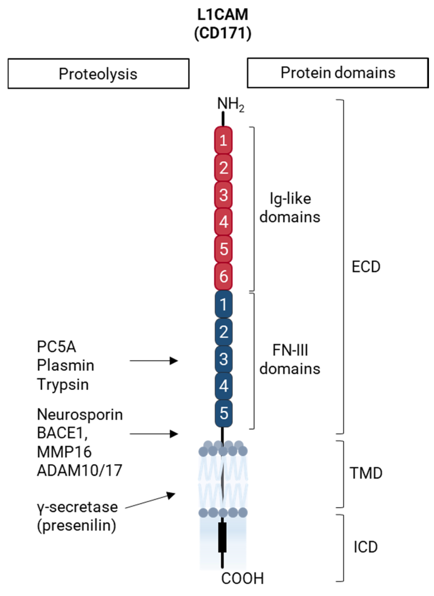
The L1CAM, also known as CD171, is the founding member of the L1 subfamily which consists of closely related cell adhesion molecules (CAMs) belonging to the immunoglobulin (Ig) superfamily of CAMs. In vertebrates, it includes close homolog of L1 (CHL1), neuronal cell adhesion molecule (NrCAM), neurofascin, and L1CAM [115]. L1CAM is a type I transmembrane glycoprotein with a molecular weight of 200–220 kDa. Its extracellular domains consist of N-terminal six Ig-like domains and five fibronectin (FN) III-like domains followed by a single transmembrane domain which connects to the highly conserved cytoplasmic domain (Figure 4). Multiple antibodies against various regions along the L1CAM polypeptide chain have been developed and applied for imaging and/or therapy [116,117,118,119,120].
The L1CAM was first described in the central nervous system [121,122], where it plays a pivotal role in its development and plasticity by participating in processes such as neuronal migration and differentiation, neurite outgrowth, fasciculation of axons and dendrites, axon guidance, myelination, synaptogenesis cell adhesion, and survival [123,124,125,126,127]. Mutations in the human L1CAM gene localized on the X chromosome Xq28 lead to the development of L1 syndrome—a complex X-linked neurological disorder characterized by hydrocephalus, intellectual disability, adducted thumbs, and leg spasticity [128]. The L1CAM also has a crucial role in the formation and maintenance of the epithelium of the gastrointestinal and urogenital tract by supporting cell-to-cell adhesion as well as cell migration [129,130,131]. Postembryonically, the L1CAM is expressed in the kidney tubules, the central nervous system and peripheral nerves while low L1CAM levels have also been reported on B lymphocytes, T lymphocytes, dendritic cells, and monocytes [132,133]. Furthermore, the L1CAM was found to be expressed and required for the intestinal epithelial regeneration following loss of epithelial integrity while it was not expressed in homeostatic intestinal epithelium [134]. Although not essential for vascular homeostasis, the L1CAM could also be upregulated in pericytes in the context of vascular stress and neovascularization [135].
The L1CAM lacks enzymatic activity for downstream signal transduction. To mediate signaling, it interacts with a wide interactome [7,136]. There are currently three distinguished modes of L1CAM-mediated signaling which are activated by the different L1CAM forms upon proteolytic cleavage (Figure 4) [136], all of which have been reported in cancer. L1CAM-assisted signaling leads to the activation of L1CAM interactions with the extracellular signal-regulated kinase (ERK)/MAPK signaling pathway via full length or soluble extracellular L1CAM interactions with other L1CAM molecules, integrins, and Receptor Tyrosine Kinases such as epithelial growth factor receptors (EGFRs) and fibroblast growth factor receptors (FGFRs) [137,138]. L1CAM forward signaling, cleavage-dependent, is based on the proteolytic cleavage-generating L1CAM soluble extracellular domain and intracellular domain [139,140,141]. The extracellular domain functions as an integrin ligand contributing to L1CAM-assisted signaling or reverse signaling [142,143]. The intracellular domain translocates into the nucleus where it regulates L1CAM-dependent genes including PI3K/AKT, ERK/MAPK, and interleukin 1β (IL-1β), an activator of the NF-κB pathway), which could promote cancer cell survival, migration, and apoptosis resistance [140,144]. L1CAM reverse signaling, cleavage-independent, involves a full-length membrane-bound L1CAM which interacts with integrins and cytoskeleton crosslinking proteins such as ezrin leading to activation of the NF-κB pathway either directly or by production of IL-1β [145,146,147,148]. As a result, this signaling mode contributes to cancer cell survival, proliferation, invasion and metastasis, and therapy resistance [136].
Diverse L1CAM membrane-bound and soluble intra- or extracellular forms have been identified, reflecting its multiple roles in health and disease, including cancer. These forms could be generated through proteolytic cleavage by a variety of proteases [139,140,141,149,150], summarized with their cleavage sites in Figure 4. L1CAM alternative splicing has also been reported for exon 2 and exon 27, which affects L1CAM interactions and endocytosis [151], as well as those of exon 25, which results in a soluble variant without a transmembrane domain [116,152].
L1CAM-aberrant expression in cancer, including that of its diverse forms, has been linked to key tumor processes such as cell survival, cell migration and invasion, tumor and metastasis formation, therapy resistance, cancer stemness, cell adhesion, and angiogenesis in multiple solid malignancies, including glioblastoma, colorectal cancer, pancreatic cancer, prostate cancer, gynecologic cancers, retinoblastoma, oral squamous cell carcinoma, endometrial cancer, and breast cancer [7,8,116,134,135,153,154,155,156,157,158].
These studies also point to the clinical relevance of the L1CAM as a diagnostic and prognostic marker. L1CAM-targeted therapeutic approaches, including monoclonal antibodies, radioimmunotherapy, and CAR T-cells alone or in combination with other treatments, have already been documented [7,10,159,160,161].

4.1.2. L1CAM as a Cancer Stem Cell-Associated Biomarker in Epithelial Ovarian Cancer

L1CAM overexpression and a correlation with poor outcome and decreased patient survival in EOC has already been reported [10,132,162]. Accumulated research from the past 30 years has confirmed the role of L1CAM in major tumor processes in such as cell migration, resistance to apoptosis, chemoresistance, and increased tumorigenicity, all representing important steps in OC development and progression [142,144,163].
Importantly, the role of the L1CAM as a CSC-associated biomarker in EOC has also been validated. The L1CAM was found to promote ovarian cancer stemness in patient-derived ovarian CSCs and OC cell lines in which it mediated tumor initiation, AICG growth, and chemoresistance via the FGFR/SRC/STAT3 signaling pathway [10]. Another recent study conducted on L1CAM+/CD133+ cells derived from OC cell lines verified the CSC phenotype of this population on a molecular (upregulation of stemness genes) and functional level in vivo (limiting dilution and serial transplantation) and in vitro (clonogenicity, spherogenicity in CSC-selective conditions, radioresistance) (Table 2 and Table 3) [11]. In addition, the same study identified the L1CAM as a marker of radioresistance, since CRISPR-Cas9-mediated L1CAM knockout led to increase in radiosensitivity, which was significantly restored after L1CAM rescue. These findings created a basis for investigation of the underlining molecular mechanisms of radioresistance which remain to be elucidated. Despite the confirmed importance of the L1CAM as a clinically relevant and targetable CSC-associated biomarker in EOC, to date L1CAM-targeted therapies have not been transferred into the clinics.

4.2. Anti-L1CAM Radioimmunotherapy against Ovarian Cancer Stem Cells

4.2.1. Clinical Radioimmunotherapy against Solid Tumors

Clinical RIT is a type of TRT which utilizes a monoclonal antibody (mAb) as a targeting vector to deliver coupled therapeutic radiation to cancer-relevant antigens. It allows the target-specific delivery of particle radiation—α, β, CE/AE—while minimizing toxicity and off-target effects, making it a promising therapeutic modality due to its advantages over conventional radiotherapy [14]. Optimally, the mAb should possess high specificity and binding affinity to a cell surface antigen abundantly expressed on the cancer cells with limited to no expression on healthy tissue [164]. The selection of an appropriate radionuclide is also essential since its physical and biological properties significantly determine the therapeutic efficacy. The radiolabeled mAb should be produced with high radiochemical purity, retain high immunoreactivity, and demonstrate biodistribution with high tumor uptake and low uptake into the radiosensitive organs such as the bone marrow, spleen, liver, and kidneys.
Several major factors are to be considered for the effective delivery of RIT to solid tumors. The mAb high molecular weight of 150 kDa restricts its penetration into the tumor and the mAb could also elicit unwanted immune responses [165]. The dense ECM and the tight cell-to-cell junctions could form a physical barrier influencing mAb penetration [166]. Low tumor vascularization, compressed or leaky blood vessels, and high interstitial pressure which creates gradients opposing macromolecular diffusion also limit the drug delivery [167]. Various strategies have been explored to address these obstacles, including the generation of smaller antibody fragments, pretargeting, combination treatments with modulators of the ECM, tight cell-to-cell junctions, immune responses, and blood vessels to increase delivery to the tumors [165,166,168,169].
Although many RIT approaches have been in clinical trials for non-solid and solid cancers in the last decade, only a few have been approved for clinical application up to date, only against hematologic malignancies [169]. The clinical trials of RIT in OC are scarce. A phase I clinical trial (NCT04461457, completed 2005–2012) investigated the vector MX35 F(ab’)2 radiolabeled with the α-emitter Astatine-211 targeting the cell surface glycoprotein NaPi2b in relapsed OC [170]. The study showed no radiation-induced toxicity while it also pointed to the requirement for optimization of the dosimetry calculations. Another phase I trial (NCT03507452, completed 2018–2022) studied the mAb BAY2287411 radiolabeled with the α-emitter Thorium-227 against mesothelin-expressing OC [169]. Results are yet to be reported. Other potential targets for RIT against EOC include MUC16 (also known as CA125), folate receptor alpha, HER2, and EGFR [171,172]. Only a few studies have investigated RIT for OC against the targets discussed above, while no studies to date have validated RIT against ovarian CSCs. A main reason is the limited research verifying ovarian CSC biomarkers and populations. The thorough work on the L1CAM, as reviewed above, has pointed out to its promising potential for CSC-targeted RIT.

4.2.2. Anti-L1CAM Radioimmunotherapy as a Novel Therapeutic Modality against Ovarian Cancer Stem Cells

The validation of the L1CAM in OC as a CSC-associated biomarker [7,9,10,142] and the identification of L1CAM+/CD133+ cells as ovarian CSCs together with confirmation of L1CAM as a marker of radioresistance [11] provided a solid base for the development of anti-L1CAM RIT.
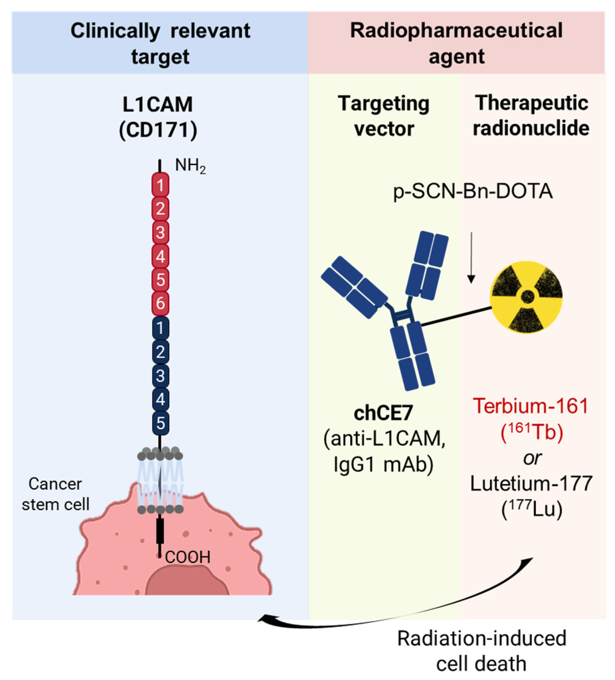
Previous work has already verified key characteristics of the components (Figure 5) and the therapeutic efficacy of anti-L1CAM RIT with the chimeric monoclonal antibody (mAb) chCE7 against bulk L1CAM-expressing tumors. The anti-L1CAM chCE7 is a full-length mAb (150 kDa) which can be produced in high yields [118]. The first and only todate clinical application of chCE7 was to test its imaging potential in neuroblastoma patients [173]. The study showed that chCE7 radiolabeled with the β/γ emitter Iodine-131 (131I) had a superior sensitivity in detecting primary tumors and metastasis [173]. Because there was not enough clinically approved chCE7 for therapeutic application, no further clinical studies were conducted. Hence, substantial effort has focused on chCE7 radiolabeled with the clinically relevant 177Lu and 161Tb. Both [177Lu]Lu-DOTA-chCE7 and [161Tb]Tb-DOTA-chCE7 were reproducibly obtained with high radiochemical purity and immunoreactivity, and showed high cell and tumor uptake with minimal accumulation in healthy tissue [12,161,174,175]. A combination of paclitaxel or protein kinase inhibitors with [177Lu]Lu-DOTA-chCE7 RIT improved the therapeutic efficacy against OC [174,175]. [161Tb]Tb-DOTA-chCE7 demonstrated stronger therapeutic potency in vitro and in vivo against bulk L1CAM-expressing ovarian tumors which is due to the substantial emission of highly cytotoxic CE/AE by 161Tb [12].
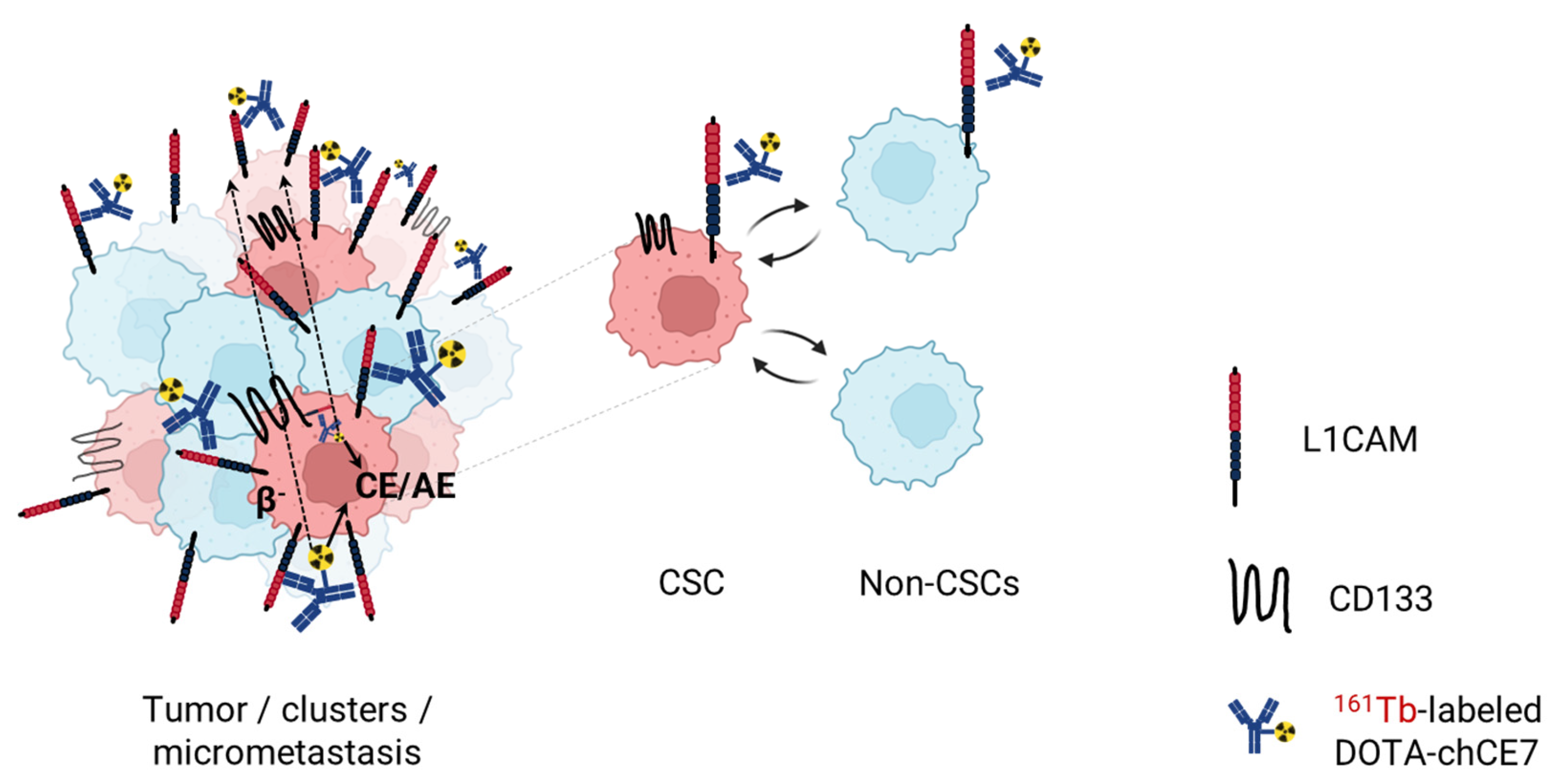
Because of 161Tb co-emission of long-ranged β radiation and short-ranged CE/AE, it has emerged as a highly attractive radionuclide for TRT. 161Tb-based radiopharmaceuticals are currently being validated in several clinical trials—REALITY (NCT04833517) and VIOLET (NCT05521412) against metastatic castration-resistant prostate cancer, and Beta plus study (NCT05359146) against neuroendocrine tumours. 161Tb-based RIT could leverage the high cytotoxicity of CE/AE to eradicate radioresistant CSCs while the β radiation could kill bulk tumor cells (Figure 6). This could prevent the dynamic transition between bulk tumor cells and CSCs and thus lead to complete tumor eradication. In our most recent work, we successfully validated the efficacy of 161Tb-based anti-L1CAM RIT against L1CAM+/CD133+ ovarian CSCs in a CSC xenograft model in nude mice (MS ID#: JNUMED/2024/267864, under revision). Our research provides an important link between targeting and eradicating the bulk tumor as well as the CSCs which to date continues to pose a clinical challenge. Furthermore, it highlights the promising potential of anti-L1CAM RIT with β/CE/AE-emitters such as 161Tb establishing a novel therapeutic modality against ovarian CSCs.

5. Conclusions

There is a critical demand for novel CSC-targeted therapies in clinics. Research to identify clinically relevant CSC biomarkers as well as deepen our understanding of CSC biology and therapy-resistant mechanisms is key to the development of successful CSC-targeted therapies. Radioimmunotherapy with suitable radionuclides such as 161Tb-emitting highly cytotoxic CE/AE could potentially overcome the radioresistance of L1CAM-expressing ovarian CSCs, providing a promising novel approach which could ultimately improve patient care.

Author Contributions

Conceptualization: T.Z.T. and J.G.; writing—first manuscript draft: T.Z.T.; writing—review and editing: T.Z.T., R.S., M.B. and J.G.; visualization: T.Z.T. All authors have read and agreed to the published version of the manuscript.

Funding

This work was funded by the Swiss Cancer Research (Krebsforschung Schweiz, KFS-4876-08-2019) to Jürgen Grünberg and Mahari-Stiftung, Switzerland to Tihomir Todorov.

Conflicts of Interest

The authors declare no conflicts of interest. The funders had no role in the design of the study; in the collection, analyses, or interpretation of data; in the writing of the manuscript; or in the decision to publish the results.

References

  1. Reid, F. World Ovarian Cancer Coalition Atlas 2023—Global Trends in Incidence, Mortality, and Survival; World Ovarian Cancer Coalition: Toronto, ON, Canada, 2023. [Google Scholar]
  2. Hollis, R.L. Molecular characteristics and clinical behaviour of epithelial ovarian cancers. Cancer Lett. 2023, 555, 216057. [Google Scholar] [CrossRef]
  3. Cabasag, C.J.; Fagan, P.J.; Ferlay, J.; Vignat, J.; Laversanne, M.; Liu, L.; van der Aa, M.A.; Bray, F.; Soerjomataram, I. Ovarian cancer today and tomorrow: A global assessment by world region and Human Development Index using GLOBOCAN 2020. Int. J. Cancer 2022, 151, 1535–1541. [Google Scholar] [CrossRef] [PubMed]
  4. Duska, L.R.; Kohn, E.C. The new classifications of ovarian, fallopian tube, and primary peritoneal cancer and their clinical implications. Ann. Oncol. 2017, 28, viii8–viii12. [Google Scholar] [CrossRef] [PubMed]
  5. Valent, P.; Bonnet, D.; De Maria, R.; Lapidot, T.; Copland, M.; Melo, J.V.; Chomienne, C.; Ishikawa, F.; Schuringa, J.J.; Stassi, G.; et al. Cancer stem cell definitions and terminology: The devil is in the details. Nat. Rev. Cancer 2012, 12, 767–775. [Google Scholar] [CrossRef] [PubMed]
  6. Loh, J.J.; Ma, S. Hallmarks of cancer stemness. Cell Stem Cell 2024, 31, 617–639. [Google Scholar] [CrossRef] [PubMed]
  7. Giordano, M.; Cavallaro, U. Different shades of L1CAM in the pathophysiology of cancer stem cells. J. Clin. Med. 2020, 9, 1502. [Google Scholar] [CrossRef] [PubMed]
  8. Altevogt, P.; Doberstein, K.; Fogel, M. L1CAM in human cancer. Int. J. Cancer 2016, 138, 1565–1576. [Google Scholar] [CrossRef] [PubMed]
  9. Doberstein, K.; Spivak, R.; Reavis, H.D.; Hooda, J.; Feng, Y.; Kroeger, P.T., Jr.; Stuckelberger, S.; Mills, G.B.; Devins, K.M.; Schwartz, L.E.; et al. L1CAM is required for early dissemination of fallopian tube carcinoma precursors to the ovary. Commun. Biol. 2022, 5, 1362. [Google Scholar] [CrossRef] [PubMed]
  10. Giordano, M.; Decio, A.; Battistini, C.; Baronio, M.; Bianchi, F.; Villa, A.; Bertalot, G.; Freddi, S.; Lupia, M.; Jodice, M.G.; et al. L1CAM promotes ovarian cancer stemness and tumor initiation via FGFR1/SRC/STAT3 signaling. J. Exp. Clin. Cancer Res. 2021, 40, 319. [Google Scholar] [CrossRef]
  11. Terraneo, N.; Jacob, F.; Peitzsch, C.; Dubrovska, A.; Krudewig, C.; Huang, Y.L.; Heinzelmann-Schwarz, V.; Schibli, R.; Behe, M.; Grünberg, J. L1 cell adhesion molecule confers radioresistance to ovarian cancer and defines a new cancer stem cell population. Cancers 2020, 12, 217. [Google Scholar] [CrossRef]
  12. Grünberg, J.; Lindenblatt, D.; Dorrer, H.; Cohrs, S.; Zhernosekov, K.; Koster, U.; Turler, A.; Fischer, E.; Schibli, R. Anti-L1CAM radioimmunotherapy is more effective with the radiolanthanide terbium-161 compared to lutetium-177 in an ovarian cancer model. Eur. J. Nucl. Med. Mol. Imaging 2014, 41, 1907–1915. [Google Scholar] [CrossRef]
  13. Ku, A.; Facca, V.J.; Cai, Z.; Reilly, R.M. Auger electrons for cancer therapy—A review. EJNMMI Radiopharm. Chem. 2019, 4, 27. [Google Scholar] [CrossRef] [PubMed]
  14. Pouget, J.P.; Navarro-Teulon, I.; Bardies, M.; Chouin, N.; Cartron, G.; Pelegrin, A.; Azria, D. Clinical radioimmunotherapy—The role of radiobiology. Nat. Rev. Clin. Oncol. 2011, 8, 720–734. [Google Scholar] [CrossRef] [PubMed]
  15. Sung, H.; Ferlay, J.; Siegel, R.L.; Laversanne, M.; Soerjomataram, I.; Jemal, A.; Bray, F. Global Cancer Statistics 2020: GLOBOCAN estimates of incidence and mortality worldwide for 36 cancers in 185 countries. CA Cancer J. Clin. 2021, 71, 209–249. [Google Scholar] [CrossRef] [PubMed]
  16. Lheureux, S.; Braunstein, M.; Oza, A.M. Epithelial ovarian cancer: Evolution of management in the era of precision medicine. CA Cancer J. Clin. 2019, 69, 280–304. [Google Scholar] [CrossRef] [PubMed]
  17. Ebell, M.H.; Culp, M.B.; Radke, T.J. A systematic review of symptoms for the diagnosis of ovarian cancer. Am. J. Prev. Med. 2016, 50, 384–394. [Google Scholar] [CrossRef] [PubMed]
  18. Ravindran, F.; Choudhary, B. Ovarian Cancer: Molecular Classification and Targeted Therapy; InTechOpen: London, UK, 2021. [Google Scholar] [CrossRef]
  19. Prat, J.; FIGO Committee on Gynecologic Oncology. FIGO’s staging classification for cancer of the ovary, fallopian tube, and peritoneum: Abridged republication. J. Gynecol. Oncol. 2015, 26, 87–89. [Google Scholar] [CrossRef] [PubMed]
  20. Frey, M.K.; Pothuri, B. Homologous recombination deficiency (HRD) testing in ovarian cancer clinical practice: A review of the literature. Gynecol. Oncol. Res. Pract. 2017, 4, 4. [Google Scholar] [CrossRef]
  21. Cancer Genome Atlas Research Network. Integrated genomic analyses of ovarian carcinoma. Nature 2011, 474, 609–615. [Google Scholar] [CrossRef] [PubMed]
  22. Lo Riso, P.; Villa, C.E.; Gasparoni, G.; Vingiani, A.; Luongo, R.; Manfredi, A.; Jungmann, A.; Bertolotti, A.; Borgo, F.; Garbi, A.; et al. A cell-of-origin epigenetic tracer reveals clinically distinct subtypes of high-grade serous ovarian cancer. Genome Med. 2020, 12, 94. [Google Scholar] [CrossRef]
  23. Torre, L.A.; Trabert, B.; DeSantis, C.E.; Miller, K.D.; Samimi, G.; Runowicz, C.D.; Gaudet, M.M.; Jemal, A.; Siegel, R.L. Ovarian cancer statistics, 2018. CA Cancer J. Clin. 2018, 68, 284–296. [Google Scholar] [CrossRef]
  24. Pignata, S.; Cecere, S.C.; Du Bois, A.; Harter, P.; Heitz, F. Treatment of recurrent ovarian cancer. Ann. Oncol. 2017, 28, viii51–viii56. [Google Scholar] [CrossRef]
  25. Flores-Balcazar, C.H.; Urias-Arce, D.M. Radiotherapy in women with epithelial ovarian cancer: Historical role, current advances, and indications. Chin. Clin. Oncol. 2020, 9, 49. [Google Scholar] [CrossRef] [PubMed]
  26. Rose, M.; Burgess, J.T.; O’Byrne, K.; Richard, D.J.; Bolderson, E. PARP Inhibitors: Clinical Relevance, Mechanisms of Action and Tumor Resistance. Front. Cell Dev. Biol. 2020, 8, 564601. [Google Scholar] [CrossRef] [PubMed]
  27. Sazonova, E.V.; Kopeina, G.S.; Imyanitov, E.N.; Zhivotovsky, B. Platinum drugs and taxanes: Can we overcome resistance? Cell Death Discov. 2021, 7, 155. [Google Scholar] [CrossRef] [PubMed]
  28. Tewari, K.S.; Burger, R.A.; Enserro, D.; Norquist, B.M.; Swisher, E.M.; Brady, M.F.; Bookman, M.A.; Fleming, G.F.; Huang, H.; Homesley, H.D.; et al. Final overall survival of a randomized trial of bevacizumab for primary treatment of ovarian cancer. J. Clin. Oncol. 2019, 37, 2317–2328. [Google Scholar] [CrossRef] [PubMed]
  29. Pfisterer, J.; Shannon, C.M.; Baumann, K.; Rau, J.; Harter, P.; Joly, F.; Sehouli, J.; Canzler, U.; Schmalfeldt, B.; Dean, A.P.; et al. Bevacizumab and platinum-based combinations for recurrent ovarian cancer: A randomised, open-label, phase 3 trial. Lancet Oncol. 2020, 21, 699–709. [Google Scholar] [CrossRef] [PubMed]
  30. Alvarez Secord, A.; O’Malley, D.M.; Sood, A.K.; Westin, S.N.; Liu, J.F. Rationale for combination PARP inhibitor and antiangiogenic treatment in advanced epithelial ovarian cancer: A review. Gynecol. Oncol. 2021, 162, 482–495. [Google Scholar] [CrossRef] [PubMed]
  31. Foo, T.; George, A.; Banerjee, S. PARP inhibitors in ovarian cancer: An overview of the practice-changing trials. Genes Chromosomes Cancer 2021, 60, 385–397. [Google Scholar] [CrossRef] [PubMed]
  32. Smith, M.; Pothuri, B. Appropriate selection of PARP inhibitors in ovarian cancer. Curr. Treat. Options Oncol. 2022, 23, 887–903. [Google Scholar] [CrossRef]
  33. Moore, K.N.; Vergote, I.; Oaknin, A.; Colombo, N.; Banerjee, S.; Oza, A.; Pautier, P.; Malek, K.; Birrer, M.J. FORWARD I: A Phase III study of mirvetuximab soravtansine versus chemotherapy in platinum-resistant ovarian cancer. Future Oncol. 2018, 14, 1669–1678. [Google Scholar] [CrossRef] [PubMed]
  34. Cutri-French, C.; Nasioudis, D.; George, E.; Tanyi, J.L. CAR-T cell therapy in ovarian cancer: Where are we now? Diagnostics 2024, 14, 819. [Google Scholar] [CrossRef] [PubMed]
  35. Terraneo, N.; Jacob, F.; Dubrovska, A.; Grunberg, J. Novel therapeutic strategies for ovarian cancer stem cells. Front. Oncol. 2020, 10, 319. [Google Scholar] [CrossRef]
  36. Capp, J.P. Cancer stem cells: From historical roots to a new perspective. J. Oncol. 2019, 2019, 5189232. [Google Scholar] [CrossRef]
  37. Lapidot, T.; Sirard, C.; Vormoor, J.; Murdoch, B.; Hoang, T.; Caceres-Cortes, J.; Mindent, M.; Paterson, B.; Caligiuri, M.A.; Dick, J.E. A cell initiating human acute myeloid leukaemia after transplantation into SCID mice. Nature 1994, 367, 645–648. [Google Scholar] [CrossRef]
  38. Bonnet, D.; Dick, J.E. Human acute myeloid leukemia is organized as a hierarchy that originates from a primitive hematopoietic cell. Nat. Med. 1997, 3, 730–737. [Google Scholar] [CrossRef] [PubMed]
  39. Al-Hajj, M.; Wicha, M.S.; Benito-Hernandez, A.; Morrison, S.J.; Clarke, M.F. Prospective identification of tumorigenic breast cancer cells. Proc. Natl. Acad. Sci. USA 2003, 100, 3983–3988. [Google Scholar] [CrossRef]
  40. Nassar, D.; Blanpain, C. Cancer stem cells: Basic concepts and therapeutic implications. Annu. Rev. Pathol. 2016, 11, 47–76. [Google Scholar] [CrossRef]
  41. Chaffer, C.L.; Brueckmann, I.; Scheel, C.; Kaestli, A.J.; Wiggins, P.A.; Rodrigues, L.O.; Brooks, M.; Reinhardt, F.; Su, Y.; Polyak, K.; et al. Normal and neoplastic nonstem cells can spontaneously convert to a stem-like state. Proc. Natl. Acad. Sci. USA 2011, 108, 7950–7955. [Google Scholar] [CrossRef]
  42. Dongre, A.; Weinberg, R.A. New insights into the mechanisms of epithelial-mesenchymal transition and implications for cancer. Nat. Rev. Mol. Cell Biol. 2019, 20, 69–84. [Google Scholar] [CrossRef]
  43. Shibue, T.; Weinberg, R.A. EMT, CSCs, and drug resistance: The mechanistic link and clinical implications. Nat. Rev. Clin. Oncol. 2017, 14, 611–629. [Google Scholar] [CrossRef]
  44. Paul, R.; Dorsey, J.F.; Fan, Y. Cell plasticity, senescence, and quiescence in cancer stem cells: Biological and therapeutic implications. Pharmacol. Ther. 2022, 231, 107985. [Google Scholar] [CrossRef]
  45. Crea, F.; Nur Saidy, N.R.; Collins, C.C.; Wang, Y. The epigenetic/noncoding origin of tumor dormancy. Trends Mol. Med. 2015, 21, 206–211. [Google Scholar] [CrossRef]
  46. El Touny, L.H.; Vieira, A.; Mendoza, A.; Khanna, C.; Hoenerhoff, M.J.; Green, J.E. Combined SFK/MEK inhibition prevents metastatic outgrowth of dormant tumor cells. J. Clin. Investig. 2014, 124, 156–168. [Google Scholar] [CrossRef] [PubMed]
  47. Chen, J.; Li, Y.; Yu, T.S.; McKay, R.M.; Burns, D.K.; Kernie, S.G.; Parada, L.F. A restricted cell population propagates glioblastoma growth after chemotherapy. Nature 2012, 488, 522–526. [Google Scholar] [CrossRef] [PubMed]
  48. Walcher, L.; Kistenmacher, A.K.; Suo, H.; Kitte, R.; Dluczek, S.; Strauss, A.; Blaudszun, A.R.; Yevsa, T.; Fricke, S.; Kossatz-Boehlert, U. Cancer stem cells—Origins and biomarkers: Perspectives for targeted personalized therapies. Front. Immunol. 2020, 11, 1280. [Google Scholar] [CrossRef] [PubMed]
  49. Zon, L.I. Intrinsic and extrinsic control of haematopoietic stem-cell self-renewal. Nature 2008, 453, 306–313. [Google Scholar] [CrossRef]
  50. Milanovic, M.; Fan, D.N.Y.; Belenki, D.; Dabritz, J.H.M.; Zhao, Z.; Yu, Y.; Dorr, J.R.; Dimitrova, L.; Lenze, D.; Monteiro Barbosa, I.A.; et al. Senescence-associated reprogramming promotes cancer stemness. Nature 2018, 553, 96–100. [Google Scholar] [CrossRef]
  51. Was, H.; Barszcz, K.; Czarnecka, J.; Kowalczyk, A.; Bernas, T.; Uzarowska, E.; Koza, P.; Kaminska, B.; Klejman, A.; Piwocka, K.; et al. Bafilomycin A1 triggers proliferative potential of senescent cancer cells in vitro and in NOD/SCID mice. Oncotarget 2017, 8, 9303–9322. [Google Scholar] [CrossRef]
  52. Zhang, S.; Balch, C.; Chan, M.W.; Lai, H.C.; Matei, D.; Schilder, J.M.; Yan, P.S.; Huang, T.H.; Nephew, K.P. Identification and characterization of ovarian cancer-initiating cells from primary human tumors. Cancer Res. 2008, 68, 4311–4320. [Google Scholar] [CrossRef]
  53. Kryczek, I.; Liu, S.; Roh, M.; Vatan, L.; Szeliga, W.; Wei, S.; Banerjee, M.; Mao, Y.; Kotarski, J.; Wicha, M.S.; et al. Expression of aldehyde dehydrogenase and CD133 defines ovarian cancer stem cells. Int. J. Cancer 2012, 130, 29–39. [Google Scholar] [CrossRef] [PubMed]
  54. Eptaminitaki, G.C.; Zaravinos, A.; Stellas, D.; Panagopoulou, M.; Karaliota, S.; Baltsavia, I.; Iliopoulos, I.; Chatzaki, E.; Iliopoulos, D.; Baritaki, S. Genome-wide analysis of lncRNA-mRNA co-expression networks in CD133+/CD44+ stem-like PDAC cells. Cancers 2023, 15, 1053. [Google Scholar] [CrossRef] [PubMed]
  55. Zhang, S.-s.; Han, Z.-p.; Jing, Y.-y.; Tao, S.-f.; Li, T.-j.; Wang, H.; Wang, Y.; Li, R.; Yang, Y.; Zhao, X.; et al. CD133+CXCR4+ colon cancer cells exhibit metastatic potential and predict poor prognosis of patients. BMC Med. 2012, 10, 85. [Google Scholar] [CrossRef] [PubMed]
  56. Hermann, P.C.; Huber, S.L.; Herrler, T.; Aicher, A.; Ellwart, J.W.; Guba, M.; Bruns, C.J.; Heeschen, C. Distinct populations of cancer stem cells determine tumor growth and metastatic activity in human pancreatic cancer. Cell Stem Cell 2007, 1, 313–323. [Google Scholar] [CrossRef] [PubMed]
  57. Zhang, C.; Li, C.; He, F.; Cai, Y.; Yang, H. Identification of CD44+CD24+ gastric cancer stem cells. J. Cancer Res. Clin. Oncol. 2011, 137, 1679–1686. [Google Scholar] [CrossRef] [PubMed]
  58. Satar, N.A.; Fakiruddin, K.S.; Lim, M.N.; Mok, P.L.; Zakaria, N.; Fakharuzi, N.A.; Rahman, A.Z.A.; Zakaria, Z.; Yahaya, B.H.; Baharuddin, P. Novel triple-positive markers identified in human non-small cell lung cancer cell line with chemotherapy-resistant and putative cancer stem cell characteristics. Oncol. Rep. 2018, 40, 669–681. [Google Scholar] [CrossRef] [PubMed]
  59. Chen, Y.C.; Chen, Y.W.; Hsu, H.S.; Tseng, L.M.; Huang, P.I.; Lu, K.H.; Chen, D.T.; Tai, L.K.; Yung, M.C.; Chang, S.C.; et al. Aldehyde dehydrogenase 1 is a putative marker for cancer stem cells in head and neck squamous cancer. Biochem. Biophys. Res. Commun. 2009, 385, 307–313. [Google Scholar] [CrossRef] [PubMed]
  60. Kim, W.T.; Ryu, C.J. Cancer stem cell surface markers on normal stem cells. BMB Rep. 2017, 50, 285–298. [Google Scholar] [CrossRef]
  61. Peitzsch, C.; Nathansen, J.; Schniewind, S.I.; Schwarz, F.; Dubrovska, A. Cancer stem cells in head and neck squamous cell carcinoma: Identification, characterization and clinical implications. Cancers 2019, 11, 616. [Google Scholar] [CrossRef]
  62. Pastrana, E.; Silva-Vargas, V.; Doetsch, F. Eyes wide open: A critical review of sphere-formation as an assay for stem cells. Cell Stem Cell 2011, 8, 486–498. [Google Scholar] [CrossRef]
  63. Ishiguro, T.; Ohata, H.; Sato, A.; Yamawaki, K.; Enomoto, T.; Okamoto, K. Tumor-derived spheroids: Relevance to cancer stem cells and clinical applications. Cancer Sci. 2017, 108, 283–289. [Google Scholar] [CrossRef]
  64. Borowicz, S.; Van Scoyk, M.; Avasarala, S.; Karuppusamy Rathinam, M.K.; Tauler, J.; Bikkavilli, R.K.; Winn, R.A. The soft agar colony formation assay. J. Vis. Exp. 2014, 92, e51998. [Google Scholar] [CrossRef]
  65. Bahmad, H.F.; Cheaito, K.; Chalhoub, R.M.; Hadadeh, O.; Monzer, A.; Ballout, F.; El-Hajj, A.; Mukherji, D.; Liu, Y.N.; Daoud, G.; et al. Sphere-formation assay: Three-dimensional in vitro culturing of prostate cancer stem/progenitor sphere-forming cells. Front. Oncol. 2018, 8, 347. [Google Scholar] [CrossRef]
  66. Lim, Y.C.; Oh, S.Y.; Cha, Y.Y.; Kim, S.H.; Jin, X.; Kim, H. Cancer stem cell traits in squamospheres derived from primary head and neck squamous cell carcinomas. Oral Oncol. 2011, 47, 83–91. [Google Scholar] [CrossRef] [PubMed]
  67. Franken, N.A.; Rodermond, H.M.; Stap, J.; Haveman, J.; van Bree, C. Clonogenic assay of cells in vitro. Nat. Protoc. 2006, 1, 2315–2319. [Google Scholar] [CrossRef]
  68. Brix, N.; Samaga, D.; Hennel, R.; Gehr, K.; Zitzelsberger, H.; Lauber, K. The clonogenic assay: Robustness of plating efficiency-based analysis is strongly compromised by cellular cooperation. Radiat. Oncol. 2020, 15, 248. [Google Scholar] [CrossRef]
  69. Rycaj, K.; Tang, D.G. Cell-of-origin of cancer versus cancer stem cells: Assays and interpretations. Cancer Res. 2015, 75, 4003–4011. [Google Scholar] [CrossRef]
  70. Visvader, J.E. Cells of origin in cancer. Nature 2011, 469, 314–322. [Google Scholar] [CrossRef] [PubMed]
  71. Clevers, H. The cancer stem cell: Premises, promises and challenges. Nat. Med. 2011, 17, 313–319. [Google Scholar] [CrossRef]
  72. Huang, R.X.; Zhou, P.K. DNA damage response signaling pathways and targets for radiotherapy sensitization in cancer. Signal Transduct. Target. Ther. 2020, 5, 60. [Google Scholar] [CrossRef]
  73. Gorodetska, I.; Kozeretska, I.; Dubrovska, A. BRCA genes: The role in genome stability, cancer stemness and therapy resistance. J. Cancer 2019, 10, 2109–2127. [Google Scholar] [CrossRef] [PubMed]
  74. Krause, M.; Dubrovska, A.; Linge, A.; Baumann, M. Cancer stem cells: Radioresistance, prediction of radiotherapy outcome and specific targets for combined treatments. Adv. Drug Deliv. Rev. 2017, 109, 63–73. [Google Scholar] [CrossRef] [PubMed]
  75. Zhang, P.; Wei, Y.; Wang, L.; Debeb, B.G.; Yuan, Y.; Zhang, J.; Yuan, J.; Wang, M.; Chen, D.; Sun, Y.; et al. ATM-mediated stabilization of ZEB1 promotes DNA damage response and radioresistance through CHK1. Nat. Cell. Biol. 2014, 16, 864–875. [Google Scholar] [CrossRef] [PubMed]
  76. Bao, S.; Wu, Q.; McLendon, R.E.; Hao, Y.; Shi, Q.; Hjelmeland, A.B.; Dewhirst, M.W.; Bigner, D.D.; Rich, J.N. Glioma stem cells promote radioresistance by preferential activation of the DNA damage response. Nature 2006, 444, 756–760. [Google Scholar] [CrossRef] [PubMed]
  77. Wang, W.J.; Wu, S.P.; Liu, J.B.; Shi, Y.S.; Huang, X.; Zhang, Q.B.; Yao, K.T. MYC regulation of CHK1 and CHK2 promotes radioresistance in a stem cell-like population of nasopharyngeal carcinoma cells. Cancer Res. 2013, 73, 1219–1231. [Google Scholar] [CrossRef] [PubMed]
  78. Trachootham, D.; Alexandre, J.; Huang, P. Targeting cancer cells by ROS-mediated mechanisms: A radical therapeutic approach? Nat. Rev. Drug Discov. 2009, 8, 579–591. [Google Scholar] [CrossRef] [PubMed]
  79. Lendeckel, U.; Wolke, C. Redox-regulation in cancer stem cells. Biomedicines 2022, 10, 2413. [Google Scholar] [CrossRef] [PubMed]
  80. Chang, C.W.; Chen, Y.S.; Chou, S.H.; Han, C.L.; Chen, Y.J.; Yang, C.C.; Huang, C.Y.; Lo, J.F. Distinct subpopulations of head and neck cancer cells with different levels of intracellular reactive oxygen species exhibit diverse stemness, proliferation, and chemosensitivity. Cancer Res. 2014, 74, 6291–6305. [Google Scholar] [CrossRef]
  81. Diehn, M.; Cho, R.W.; Lobo, N.A.; Kalisky, T.; Dorie, M.J.; Kulp, A.N.; Qian, D.; Lam, J.S.; Ailles, L.E.; Wong, M.; et al. Association of reactive oxygen species levels and radioresistance in cancer stem cells. Nature 2009, 458, 780–783. [Google Scholar] [CrossRef]
  82. Singh, S.; Brocker, C.; Koppaka, V.; Chen, Y.; Jackson, B.C.; Matsumoto, A.; Thompson, D.C.; Vasiliou, V. Aldehyde dehydrogenases in cellular responses to oxidative/electrophilic stress. Free Radic. Biol. Med. 2013, 56, 89–101. [Google Scholar] [CrossRef]
  83. Schwarz, F.M.; Schniewind, I.; Besso, M.J.; Lange, S.; Linge, A.; Patil, S.G.; Lock, S.; Klusa, D.; Dietrich, A.; Voss-Bohme, A.; et al. Plasticity within aldehyde dehydrogenase-positive cells determines prostate cancer radiosensitivity. Mol. Cancer Res. 2022, 20, 794–809. [Google Scholar] [CrossRef] [PubMed]
  84. Matsui, W.H. Cancer stem cell signaling pathways. Medicine 2016, 95, S8–S19. [Google Scholar] [CrossRef] [PubMed]
  85. Jarosz-Biej, M.; Smolarczyk, R.; Cichon, T.; Kulach, N. Tumor microenvironment as a “Game Changer” in cancer radiotherapy. Int. J. Mol. Sci. 2019, 20, 3212. [Google Scholar] [CrossRef] [PubMed]
  86. Muller, L.; Tunger, A.; Plesca, I.; Wehner, R.; Temme, A.; Westphal, D.; Meier, F.; Bachmann, M.; Schmitz, M. Bidirectional crosstalk between cancer stem cells and immune cell subsets. Front. Immunol. 2020, 11, 140. [Google Scholar] [CrossRef] [PubMed]
  87. Schoning, J.P.; Monteiro, M.; Gu, W. Drug resistance and cancer stem cells: The shared but distinct roles of hypoxia-inducible factors HIF1alpha and HIF2alpha. Clin. Exp. Pharmacol. Physiol. 2017, 44, 153–161. [Google Scholar] [CrossRef] [PubMed]
  88. Lagadec, C.; Vlashi, E.; Della Donna, L.; Dekmezian, C.; Pajonk, F. Radiation-induced reprogramming of breast cancer cells. Stem Cells 2012, 30, 833–844. [Google Scholar] [CrossRef] [PubMed]
  89. Gao, X.; Sishc, B.J.; Nelson, C.B.; Hahnfeldt, P.; Bailey, S.M.; Hlatky, L. Radiation-induced reprogramming of pre-senescent mammary epithelial cells enriches putative CD44+/CD24−/low stem cell phenotype. Front. Oncol. 2016, 6, 138. [Google Scholar] [CrossRef] [PubMed]
  90. Deutsch, E.; Chargari, C.; Galluzzi, L.; Kroemer, G. Optimising efficacy and reducing toxicity of anticancer radioimmunotherapy. Lancet Oncol. 2019, 20, e452–e463. [Google Scholar] [CrossRef] [PubMed]
  91. Lupia, M.; Cavallaro, U. Ovarian cancer stem cells: Still an elusive entity? Mol. Cancer 2017, 16, 64. [Google Scholar] [CrossRef]
  92. Varier, L.; Sundaram, S.M.; Gamit, N.; Warrier, S. An overview of ovarian cancer: The role of cancer stem cells in chemoresistance and a precision medicine approach targeting the Wnt pathway with the antagonist sFRP4. Cancers 2023, 15, 1275. [Google Scholar] [CrossRef]
  93. Brown, J.R.; Chan, D.K.; Shank, J.J.; Griffith, K.A.; Fan, H.; Szulawski, R.; Yang, K.; Reynolds, R.K.; Johnston, C.; McLean, K.; et al. Phase II clinical trial of metformin as a cancer stem cell-targeting agent in ovarian cancer. JCI Insight 2020, 5, e133247. [Google Scholar] [CrossRef]
  94. Zhang, R.; Zhang, P.; Wang, H.; Hou, D.; Li, W.; Xiao, G.; Li, C. Inhibitory effects of metformin at low concentration on epithelial-mesenchymal transition of CD44+CD117+ ovarian cancer stem cells. Stem Cell Res. Ther. 2015, 6, 262. [Google Scholar] [CrossRef]
  95. Ma, H.; Tian, T.; Cui, Z. Targeting ovarian cancer stem cells: A new way out. Stem Cell Res. Ther. 2023, 14, 28. [Google Scholar] [CrossRef] [PubMed]
  96. Munoz-Galvan, S.; Carnero, A. Targeting cancer stem cells to overcome therapy resistance in ovarian cancer. Cells 2020, 9, 1402. [Google Scholar] [CrossRef] [PubMed]
  97. Diaz-Padilla, I.; Wilson, M.K.; Clarke, B.A.; Hirte, H.W.; Welch, S.A.; Mackay, H.J.; Biagi, J.J.; Reedijk, M.; Weberpals, J.I.; Fleming, G.F.; et al. A phase II study of single-agent RO4929097, a gamma-secretase inhibitor of Notch signaling, in patients with recurrent platinum-resistant epithelial ovarian cancer: A study of the Princess Margaret, Chicago and California phase II consortia. Gynecol. Oncol. 2015, 137, 216–222. [Google Scholar] [CrossRef] [PubMed]
  98. Srivastava, A.K.; Rizvi, A.; Banerjee, A.; Naseem, I.; Cui, T.; Zheng, Y.; Wani, A.A.; Han, C.; Wang, Q.-E. Depleting ovarian cancer stem cells with calcitriol. Oncotarget 2018, 9, 14481–14491. [Google Scholar] [CrossRef] [PubMed]
  99. Moore, K.N.; Gunderson, C.C.; Sabbatini, P.; McMeekin, D.S.; Mantia-Smaldone, G.; Burger, R.A.; Morgan, M.A.; Kapoun, A.M.; Brachmann, R.K.; Stagg, R.; et al. A phase 1b dose escalation study of ipafricept (OMP54F28) in combination with paclitaxel and carboplatin in patients with recurrent platinum-sensitive ovarian cancer. Gynecol. Oncol. 2019, 154, 294–301. [Google Scholar] [CrossRef] [PubMed]
  100. Stathis, A.; Hess, D.; von Moos, R.; Homicsko, K.; Griguolo, G.; Joerger, M.; Mark, M.; Ackermann, C.J.; Allegrini, S.; Catapano, C.V.; et al. Phase I trial of the oral smoothened inhibitor sonidegib in combination with paclitaxel in patients with advanced solid tumors. Investig. New Drugs 2017, 35, 766–772. [Google Scholar] [CrossRef] [PubMed]
  101. Kaye, S.B.; Fehrenbacher, L.; Holloway, R.; Amit, A.; Karlan, B.; Slomovitz, B.; Sabbatini, P.; Fu, L.; Yauch, R.L.; Chang, I.; et al. A phase II, randomized, placebo-controlled study of vismodegib as maintenance therapy in patients with ovarian cancer in second or third complete remission. Clin. Cancer Res. 2012, 18, 6509–6518. [Google Scholar] [CrossRef] [PubMed]
  102. Jiang, Y.X.; Siu, M.K.Y.; Wang, J.J.; Leung, T.H.Y.; Chan, D.W.; Cheung, A.N.Y.; Ngan, H.Y.S.; Chan, K.K.L. PFKFB3 regulates chemoresistance, metastasis and stemness via IAP proteins and the NF-kappaB signaling pathway in ovarian cancer. Front. Oncol. 2022, 12, 748403. [Google Scholar] [CrossRef]
  103. Kim, D.; Choi, B.H.; Ryoo, I.G.; Kwak, M.K. High NRF2 level mediates cancer stem cell-like properties of aldehyde dehydrogenase (ALDH)-high ovarian cancer cells: Inhibitory role of all-trans retinoic acid in ALDH/NRF2 signaling. Cell Death Dis. 2018, 9, 896. [Google Scholar] [CrossRef] [PubMed]
  104. Cui, T.; Srivastava, A.K.; Han, C.; Wu, D.; Wani, N.; Liu, L.; Gao, Z.; Qu, M.; Zou, N.; Zhang, X.; et al. DDB2 represses ovarian cancer cell dedifferentiation by suppressing ALDH1A1. Cell Death Dis. 2018, 9, 561. [Google Scholar] [CrossRef]
  105. Matei, D.; Ghamande, S.; Roman, L.; Alvarez Secord, A.; Nemunaitis, J.; Markham, M.J.; Nephew, K.P.; Jueliger, S.; Oganesian, A.; Naim, S.; et al. A Phase I clinical trial of guadecitabine and carboplatin in platinum-resistant, recurrent ovarian cancer: Clinical, pharmacokinetic, and pharmacodynamic analyses. Clin. Cancer Res. 2018, 24, 2285–2293. [Google Scholar] [CrossRef] [PubMed]
  106. Wang, Y.; Cardenas, H.; Fang, F.; Condello, S.; Taverna, P.; Segar, M.; Liu, Y.; Nephew, K.P.; Matei, D. Epigenetic targeting of ovarian cancer stem cells. Cancer Res. 2014, 74, 4922–4936. [Google Scholar] [CrossRef]
  107. Suraweera, A.; O’Byrne, K.J.; Richard, D.J. Combination therapy with histone deacetylase inhibitors (HDACi) for the treatment of cancer: Achieving the full therapeutic potential of HDACi. Front. Oncol. 2018, 8, 92. [Google Scholar] [CrossRef] [PubMed]
  108. Yokoyama, Y.; Zhu, H.; Lee, J.H.; Kossenkov, A.V.; Wu, S.Y.; Wickramasinghe, J.M.; Yin, X.; Palozola, K.C.; Gardini, A.; Showe, L.C.; et al. BET inhibitors suppress ALDH activity by targeting ALDH1A1 super-enhancer in ovarian cancer. Cancer Res. 2016, 76, 6320–6330. [Google Scholar] [CrossRef]
  109. Klapdor, R.; Wang, S.; Morgan, M.; Dork, T.; Hacker, U.; Hillemanns, P.; Buning, H.; Schambach, A. Characterization of a novel third-generation anti-CD24-CAR against ovarian cancer. Int. J. Mol. Sci. 2019, 20, 660. [Google Scholar] [CrossRef]
  110. Muinao, T.; Deka Boruah, H.P.; Pal, M. Diagnostic and prognostic biomarkers in ovarian cancer and the potential roles of cancer stem cells—An updated review. Exp. Cell Res. 2018, 362, 1–10. [Google Scholar] [CrossRef]
  111. Coelho, R.; Ricardo, S.; Amaral, A.L.; Huang, Y.L.; Nunes, M.; Neves, J.P.; Mendes, N.; Lopez, M.N.; Bartosch, C.; Ferreira, V.; et al. Regulation of invasion and peritoneal dissemination of ovarian cancer by mesothelin manipulation. Oncogenesis 2020, 9, 61. [Google Scholar] [CrossRef]
  112. Chen, J.; Hu, J.; Gu, L.; Ji, F.; Zhang, F.; Zhang, M.; Li, J.; Chen, Z.; Jiang, L.; Zhang, Y.; et al. Anti-mesothelin CAR-T immunotherapy in patients with ovarian cancer. Cancer Immunol. Immunother. 2023, 72, 409–425. [Google Scholar] [CrossRef]
  113. Sacks, J.D.; Barbolina, M.V. Expression and function of CD44 in epithelial ovarian carcinoma. Biomolecules 2015, 5, 3051–3066. [Google Scholar] [CrossRef]
  114. Shah, V.; Taratula, O.; Garbuzenko, O.B.; Taratula, O.R.; Rodriguez-Rodriguez, L.; Minko, T. Targeted nanomedicine for suppression of CD44 and simultaneous cell death induction in ovarian cancer: An optimal delivery of siRNA and anticancer drug. Clin. Cancer Res. 2013, 19, 6193–6204. [Google Scholar] [CrossRef]
  115. Sytnyk, V.; Leshchyns’ka, I.; Schachner, M. Neural cell adhesion molecules of the immunoglobulin superfamily regulate synapse formation, maintenance, and function. Trends Neurosci. 2017, 40, 295–308. [Google Scholar] [CrossRef]
  116. Maten, M.V.; Reijnen, C.; Pijnenborg, J.M.A.; Zegers, M.M. L1 cell adhesion molecule in cancer, a systematic review on domain-specific functions. Int. J. Mol. Sci. 2019, 20, 4180. [Google Scholar] [CrossRef]
  117. Meli, M.L.; Carrel, F.O.; Waibel, R.; Amstutz, H.; Crompton, N.; Jaussi, R.; Moch, H.; Schubiger, P.A.; Novak-Hofer, I. Anti-neuroblastoma antibody chCE7 binds to an isoform of L1-CAM present in renal carcinoma cells. Int. J. Cancer 1999, 83, 401–408. [Google Scholar] [CrossRef]
  118. Grünberg, J.; Knogler, K.; Waibel, R.; Hofer-Novak, I. High-yield production of recombinant antibody fragments in HEK-293 cells using sodium butyrate. BioTechniques 2003, 35, 968–972. [Google Scholar] [CrossRef] [PubMed]
  119. Fogel, M.; Harari, A.; Muller-Holzner, E.; Zeimet, A.G.; Moldenhauer, G.; Altevogt, P. A standardized staining protocol for L1CAM on formalin-fixed, paraffin-embedded tissues using automated platforms. Int. J. Biol. Markers 2014, 29, e180–e183. [Google Scholar] [CrossRef] [PubMed]
  120. Weidle, U.H.; Eggle, D.; Klostermann, S. L1-CAM as a target for treatment of cancer with monoclonal antibodies. Anticancer Res. 2009, 29, 4919–4932. [Google Scholar]
  121. Rathjen, F.G.; Schachner, M. Immunocytological and biochemical characterization of a new neuronal cell surface component (L1 antigen) which is involved in cell adhesion. EMBO J. 1984, 3, 1–10. [Google Scholar] [CrossRef] [PubMed]
  122. Moos, M.; Tacke, R.; Scherer, H.; Teplowt, D.; Friih, K.; Schachner, M. Neural adhesion molecule L1 as a member of the immunoglobulin superfamily with binding domains similar to fibronectin. Nature 1988, 334, 701–703. [Google Scholar] [CrossRef]
  123. Patzke, C.; Acuna, C.; Giam, L.R.; Wernig, M.; Sudhof, T.C. Conditional deletion of L1CAM in human neurons impairs both axonal and dendritic arborization and action potential generation. J. Exp. Med. 2016, 213, 499–515. [Google Scholar] [CrossRef] [PubMed]
  124. Schmid, R.S.; Maness, P.F. L1 and NCAM adhesion molecules as signaling coreceptors in neuronal migration and process outgrowth. Curr. Opin. Neurobiol. 2008, 18, 245–250. [Google Scholar] [CrossRef]
  125. Dahme, M.; Bartsch, U.; Martini, R.; Anliker, B.; Schachner, M.; Mantei, N. Disruption of the mouse L1 gene leads to malformations of the nervous system. Nat. Genet. 1997, 17, 346–349. [Google Scholar] [CrossRef] [PubMed]
  126. Brümmendorf, T.; Kenwrickt, S.; Rathjen, F.G. Neural cell recognition molecule L1: From cell biology to human hereditary brain malformations. Development 1998, 8, 87–97. [Google Scholar] [CrossRef]
  127. Valente, P.; Lignani, G.; Medrihan, L.; Bosco, F.; Contestabile, A.; Lippiello, P.; Ferrea, E.; Schachner, M.; Benfenati, F.; Giovedi, S.; et al. Cell adhesion molecule L1 contributes to neuronal excitability regulating the function of voltage-gated Na+ channels. J. Cell Sci. 2016, 129, 1878–1891. [Google Scholar] [CrossRef]
  128. Isik, E.; Onay, H.; Atik, T.; Akgun, B.; Cogulu, O.; Ozkinay, F. Clinical and genetic features of L1 syndrome patients: Definition of two novel mutations. Clin. Neurol. Neurosurg. 2018, 172, 20–23. [Google Scholar] [CrossRef] [PubMed]
  129. Pechriggl, E.J.; Concin, N.; Blumer, M.J.; Bitsche, M.; Zwierzina, M.; Dudas, J.; Koziel, K.; Altevogt, P.; Zeimet, A.G.; Fritsch, H. L1CAM in the early enteric and urogenital system. J. Histochem. Cytochem. 2017, 65, 21–32. [Google Scholar] [CrossRef] [PubMed]
  130. Debiec, H.; Christensen, E.I.; Ronco, P.M. The cell adhesion molecule L1 is developmentally regulated in the renal epithelium and is involved in kidney branching morphogenesis. J. Cell Biol. 1998, 143, 2067–2079. [Google Scholar] [CrossRef]
  131. Flaviana, C.; Monica, P.; Terenzio, C.; Raffaele, M.; Valentina, A.; Giulia, C.; Peter, V.E.; Giorgio, N.; Massimo, C.; Ferdinando, C.; et al. L1CAM expression in human gastrointestinal tract development: From tongue to colon-rectum. J. Public Health Res. 2023, 12, 22799036231165624. [Google Scholar] [CrossRef]
  132. Huszar, M.; Moldenhauer, G.; Gschwend, V.; Ben-Arie, A.; Altevogt, P.; Fogel, M. Expression profile analysis in multiple human tumors identifies L1 (CD171) as a molecular marker for differential diagnosis and targeted therapy. Hum. Pathol. 2006, 37, 1000–1008. [Google Scholar] [CrossRef]
  133. Maddaluno, L.; Verbrugge, S.E.; Martinoli, C.; Matteoli, G.; Chiavelli, A.; Zeng, Y.; Williams, E.D.; Rescigno, M.; Cavallaro, U. The adhesion molecule L1 regulates transendothelial migration and trafficking of dendritic cells. J. Exp. Med. 2009, 206, 623–635. [Google Scholar] [CrossRef]
  134. Ganesh, K.; Basnet, H.; Kaygusuz, Y.; Laughney, A.M.; He, L.; Sharma, R.; O’Rourke, K.P.; Reuter, V.P.; Huang, Y.H.; Turkekul, M.; et al. L1CAM defines the regenerative origin of metastasis-initiating cells in colorectal cancer. Nat. Cancer 2020, 1, 28–45. [Google Scholar] [CrossRef]
  135. Er, E.E.; Valiente, M.; Ganesh, K.; Zou, Y.; Agrawal, S.; Hu, J.; Griscom, B.; Rosenblum, M.; Boire, A.; Brogi, E.; et al. Pericyte-like spreading by disseminated cancer cells activates YAP and MRTF for metastatic colonization. Nat. Cell Biol. 2018, 20, 966–978. [Google Scholar] [CrossRef]
  136. Kiefel, H.; Pfeifer, M.; Bondong, S.; Hazin, J.; Altevogt, P. Linking L1CAM-mediated signaling to NF-kappaB activation. Trends Mol. Med. 2011, 17, 178–187. [Google Scholar] [CrossRef]
  137. Islam, R.; Kristiansen, L.V.; Romani, S.; Garcia-Alonso, L.; Hortsch, M. Activation of EGF receptor kinase by L1-mediated homophilic cell interactions. Mol. Biol. Cell 2004, 15, 2003–2012. [Google Scholar] [CrossRef]
  138. Kulahin, N.; Li, S.; Hinsby, A.; Kiselyov, V.; Berezin, V.; Bock, E. Fibronectin type III (FN3) modules of the neuronal cell adhesion molecule L1 interact directly with the fibroblast growth factor (FGF) receptor. Mol. Cell Neurosci. 2008, 37, 528–536. [Google Scholar] [CrossRef]
  139. Gutwein, P.; Mechtersheimer, S.; Riedle, S.; Stoeck, A.; Gast, D.; Joumaa, S.; Zentgraf, H.; Fogel, M.; Altevogt, D.P. ADAM10-mediated cleavage of L1 adhesion molecule at the cell surface and in released membrane vesicles. FASEB J. 2003, 17, 292–294. [Google Scholar] [CrossRef]
  140. Riedle, S.; Kiefel, H.; Gast, D.; Bondong, S.; Wolterink, S.; Gutwein, P.; Altevogt, P. Nuclear translocation and signalling of L1-CAM in human carcinoma cells requires ADAM10 and presenilin/gamma-secretase activity. Biochem. J. 2009, 420, 391–402. [Google Scholar] [CrossRef]
  141. Linneberg, C.; Toft, C.L.F.; Kjaer-Sorensen, K.; Laursen, L.S. L1cam-mediated developmental processes of the nervous system are differentially regulated by proteolytic processing. Sci. Rep. 2019, 9, 3716. [Google Scholar] [CrossRef]
  142. Stoeck, A.; Gast, D.; Sanderson, M.P.; Issa, Y.; Gutwein, P.; Altevogt, P. L1-CAM in a membrane-bound or soluble form augments protection from apoptosis in ovarian carcinoma cells. Gynecol. Oncol. 2007, 104, 461–469. [Google Scholar] [CrossRef]
  143. Friedli, A.; Fischer, E.; Novak-Hofer, I.; Cohrs, S.; Ballmer-Hofer, K.; Schubiger, P.A.; Schibli, R.; Grunberg, J. The soluble form of the cancer-associated L1 cell adhesion molecule is a pro-angiogenic factor. Int. J. Biochem. Cell Biol. 2009, 41, 1572–1580. [Google Scholar] [CrossRef] [PubMed]
  144. Gast, D.; Riedle, S.; Issa, Y.; Pfeifer, M.; Beckhove, P.; Sanderson, M.P.; Arlt, M.; Moldenhauer, G.; Fogel, M.; Kruger, A.; et al. The cytoplasmic part of L1-CAM controls growth and gene expression in human tumors that is reversed by therapeutic antibodies. Oncogene 2008, 27, 1281–1289. [Google Scholar] [CrossRef] [PubMed]
  145. Gavert, N.; Ben-Shmuel, A.; Lemmon, V.; Brabletz, T.; Ben-Ze’ev, A. Nuclear factor-kappaB signaling and ezrin are essential for L1-mediated metastasis of colon cancer cells. J. Cell Sci. 2010, 123, 2135–2143. [Google Scholar] [CrossRef] [PubMed]
  146. Kiefel, H.; Bondong, S.; Erbe-Hoffmann, N.; Hazin, J.; Riedle, S.; Wolf, J.; Pfeifer, M.; Arlt, A.; Schafer, H.; Muerkoster, S.S.; et al. L1CAM-integrin interaction induces constitutive NF-kappaB activation in pancreatic adenocarcinoma cells by enhancing IL-1beta expression. Oncogene 2010, 29, 4766–4778. [Google Scholar] [CrossRef]
  147. Kiefel, H.; Bondong, S.; Pfeifer, M.; Schirmer, U.; Erbe-Hoffmann, N.; Schafer, H.; Sebens, S.; Altevogt, P. EMT-associated up-regulation of L1CAM provides insights into L1CAM-mediated integrin signalling and NF-kappaB activation. Carcinogenesis 2012, 33, 1919–1929. [Google Scholar] [CrossRef] [PubMed]
  148. Zuo, C.; Hong, Y.; Qiu, X.; Yang, D.; Liu, N.; Sheng, X.; Zhou, K.; Tang, B.; Xiong, S.; Ma, M.; et al. Celecoxib suppresses proliferation and metastasis of pancreatic cancer cells by down-regulating STAT3/NF-kB and L1CAM activities. Pancreatology 2018, 18, 328–333. [Google Scholar] [CrossRef] [PubMed]
  149. Tatti, O.; Gucciardo, E.; Pekkonen, P.; Holopainen, T.; Louhimo, R.; Repo, P.; Maliniemi, P.; Lohi, J.; Rantanen, V.; Hautaniemi, S.; et al. MMP16 mediates a proteolytic switch to promote cell-cell adhesion, collagen alignment, and lymphatic invasion in melanoma. Cancer Res. 2015, 75, 2083–2094. [Google Scholar] [CrossRef]
  150. Matsumoto-Miyai, K.; Ninomiya, A.; Yamasaki, H.; Tamura, H.; Nakamura, Y.; Shiosaka, S. NMDA-dependent proteolysis of presynaptic adhesion molecule L1 in the hippocampus by neuropsin. J. Neurosci. 2003, 23, 7727–7736. [Google Scholar] [CrossRef] [PubMed]
  151. Jacob, J.; Haspel, J.; Kane-Goldsmith, N.; Grumet, M. L1 mediated homophilic binding and neurite outgrowth are modulated by alternative splicing of exon 2. J. Neurobiol. 2002, 51, 177–189. [Google Scholar] [CrossRef]
  152. Angiolini, F.; Belloni, E.; Giordano, M.; Campioni, M.; Forneris, F.; Paronetto, M.P.; Lupia, M.; Brandas, C.; Pradella, D.; Di Matteo, A.; et al. A novel L1CAM isoform with angiogenic activity generated by NOVA2-mediated alternative splicing. eLife 2019, 8, e44305. [Google Scholar] [CrossRef]
  153. Altevogt, P.; Ben-Ze’ev, A.; Gavert, N.; Schumacher, U.; Schafer, H.; Sebens, S. Recent insights into the role of L1CAM in cancer initiation and progression. Int. J. Cancer 2020, 147, 3292–3296. [Google Scholar] [CrossRef] [PubMed]
  154. Angiolini, F.; Cavallaro, U. The pleiotropic role of L1CAM in tumor vasculature. Int. J. Mol. Sci. 2017, 18, 254. [Google Scholar] [CrossRef] [PubMed]
  155. Kim, J.H.; Lee, K.W.; Ahn, D.G.; Oh, K.Y.; Yoon, H.J. Clinical significance of L1CAM expression and its biological role in the progression of oral squamous cell carcinoma. Oncol. Rep. 2023, 49, 67. [Google Scholar] [CrossRef]
  156. Bosse, T.; Nout, R.A.; Stelloo, E.; Dreef, E.; Nijman, H.W.; Jurgenliemk-Schulz, I.M.; Jobsen, J.J.; Creutzberg, C.L.; Smit, V.T. L1 cell adhesion molecule is a strong predictor for distant recurrence and overall survival in early stage endometrial cancer: Pooled PORTEC trial results. Eur. J. Cancer 2014, 50, 2602–2610. [Google Scholar] [CrossRef]
  157. Li, Y.; Galileo, D.S. Soluble L1CAM promotes breast cancer cell adhesion and migration in vitro, but not invasion. Cancer Cell Int. 2010, 10, 34. [Google Scholar] [CrossRef]
  158. Sung, S.-Y.; Wu, I.-H.; Chuang, P.-H.; Petros, J.A.; Wu, H.-C.; Zeng, H.-J.; Huang, W.-C.; Chung, L.W.K.; Hsieh, C.-L. Targeting L1 cell adhesion molecule expression using liposomeencapsulated siRNA suppresses prostate cancer bone metastasis and growth. Oncotarget 2014, 5, 9911–9929. [Google Scholar] [CrossRef] [PubMed]
  159. Ganesh, K.; Massague, J. Targeting metastatic cancer. Nat. Med. 2021, 27, 34–44. [Google Scholar] [CrossRef]
  160. Wolterink, S.; Moldenhauer, G.; Fogel, M.; Kiefel, H.; Pfeifer, M.; Luttgau, S.; Gouveia, R.; Costa, J.; Endell, J.; Moebius, U.; et al. Therapeutic antibodies to human L1CAM: Functional characterization and application in a mouse model for ovarian carcinoma. Cancer Res. 2010, 70, 2504–2515. [Google Scholar] [CrossRef]
  161. Fischer, E.; Grünberg, J.; Cohrs, S.; Hohn, A.; Waldner-Knogler, K.; Jeger, S.; Zimmermann, K.; Novak-Hofer, I.; Schibli, R. L1-CAM-targeted antibody therapy and 177Lu-radioimmunotherapy of disseminated ovarian cancer. Int. J. Cancer 2012, 130, 2715–2721. [Google Scholar] [CrossRef]
  162. Zecchini, S.; Bianchi, M.; Colombo, N.; Fasani, R.; Goisis, G.; Casadio, C.; Viale, G.; Liu, J.; Herlyn, M.; Godwin, A.K.; et al. The differential role of L1 in ovarian carcinoma and normal ovarian surface epithelium. Cancer Res. 2008, 68, 1110–1118. [Google Scholar] [CrossRef]
  163. Gast, D.; Riedle, S.; Riedle, S.; Schabath, H.; Schlich, S.; Schneider, A.; Issa, Y.; Stoeck, A.; Fogel, M.; Joumaa, S.; et al. L1 augments cell migration and tumor growth but not beta3 integrin expression in ovarian carcinomas. Int. J. Cancer 2005, 115, 658–665. [Google Scholar] [CrossRef] [PubMed]
  164. Larson, S.M.; Carrasquillo, J.A.; Cheung, N.K.; Press, O.W. Radioimmunotherapy of human tumours. Nat. Rev. Cancer 2015, 15, 347–360. [Google Scholar] [CrossRef] [PubMed]
  165. Funeh, C.N.; Bridoux, J.; Ertveldt, T.; De Groof, T.W.M.; Chigoho, D.M.; Asiabi, P.; Covens, P.; D’Huyvetter, M.; Devoogdt, N. Optimizing the Safety and Efficacy of Bio-Radiopharmaceuticals for Cancer Therapy. Pharmaceutics 2023, 15, 1378. [Google Scholar] [CrossRef] [PubMed]
  166. Zaheer, J.; Kim, H.; Lee, Y.J.; Kim, J.S.; Lim, S.M. Combination radioimmunotherapy strategies for solid tumors. Int. J. Mol. Sci. 2019, 20, 5579. [Google Scholar] [CrossRef] [PubMed]
  167. Gautam, S.K.; Dalal, V.; Jain, M.; Batra, S.K. Delivery of radioimmunotherapy for solid tumors. In Systemic Drug Delivery Strategies; Academic Press: Cambridge, MA, USA, 2022; pp. 437–461. [Google Scholar]
  168. Cheal, S.M.; Chung, S.K.; Vaughn, B.A.; Cheung, N.V.; Larson, S.M. Pretargeting: A path forward for radioimmunotherapy. J. Nucl. Med. 2022, 63, 1302–1315. [Google Scholar] [CrossRef] [PubMed]
  169. Rondon, A.; Rouanet, J.; Degoul, F. Radioimmunotherapy in oncology: Overview of the last decade clinical trials. Cancers 2021, 13, 5570. [Google Scholar] [CrossRef] [PubMed]
  170. Hallqvist, A.; Bergmark, K.; Back, T.; Andersson, H.; Dahm-Kahler, P.; Johansson, M.; Lindegren, S.; Jensen, H.; Jacobsson, L.; Hultborn, R.; et al. Intraperitoneal alpha-emitting radioimmunotherapy with 211At in relapsed ovarian cancer: Long-term follow-up with individual absorbed dose estimations. J. Nucl. Med. 2019, 60, 1073–1079. [Google Scholar] [CrossRef] [PubMed]
  171. Nimmagadda, S.; Penet, M.F. Ovarian cancer targeted theranostics. Front. Oncol. 2019, 9, 1537. [Google Scholar] [CrossRef]
  172. Chung, S.K.; Vargas, D.B.; Chandler, C.S.; Katugampola, S.; Veach, D.R.; McDevitt, M.R.; Seo, S.H.; Vaughn, B.A.; Rinne, S.S.; Punzalan, B.; et al. Efficacy of HER2-targeted Intraperitoneal 225Ac alpha-pretargeted radioimmunotherapy for small-volume ovarian peritoneal carcinomatosis. J. Nucl. Med. 2023, 64, 1439–1445. [Google Scholar] [CrossRef]
  173. Hoefnagel, C.A.; Rutgers, M.; Buitenhuis, C.K.; Smets, L.A.; Kraker, J.; Meli, M.; Carrel, F.; Amstutz, H.; Schubiger, P.A.; Novak-Hofer, I. A comparison of targetting of neuroblastoma with mIBG and anti L1-CAM antibody mAb chCE7: Therapeutic efficacy in a neuroblastoma xenograft model and imaging of neuroblastoma patients. Eur. J. Nucl. Med. 2001, 28, 359–368. [Google Scholar] [CrossRef]
  174. Lindenblatt, D.; Terraneo, N.; Pellegrini, G.; Cohrs, S.; Spycher, P.R.; Vukovic, D.; Behe, M.; Schibli, R.; Grünberg, J. Combination of lutetium-177 labeled anti-L1CAM antibody chCE7 with the clinically relevant protein kinase inhibitor MK1775: A novel combination against human ovarian carcinoma. BMC Cancer 2018, 18, 922. [Google Scholar] [CrossRef] [PubMed]
  175. Lindenblatt, D.; Fischer, E.; Cohrs, S.; Schibli, R.; Grünberg, J. Paclitaxel improved anti-L1CAM lutetium-177 radioimmunotherapy in an ovarian cancer xenograft model. EJNMMI Res. 2014, 4, 54. [Google Scholar] [CrossRef] [PubMed]
Figure 1. Representation of proportion of cases/diagnoses of ovarian cancer (A) and epithelial ovarian cancer (B). EOC, Epithelial OC; HGSOC, high-grade serous ovarian carcinoma; LGSOC, low-grade serous ovarian carcinoma. Sources: GLOBOCAN [3,15], World Ovarian Cancer Coalition [1].
Ijtm 04 00031 g001
Figure 2. Hallmarks of cancer stem cells. Cancer stem cells share common characteristics with adult stem cells and cancer cells. EMT, epithelial-to-mesenchymal transition.
Ijtm 04 00031 g002
Figure 3. Cancer stem cell mechanisms of radioresistance. ALDH, aldehyde dehydrogenase; DDR, DNA damage response; EMT, epithelial-to-mesenchymal transition; HIF, hypoxia inducible factor; NF-κB, nuclear factor κB; TME, tumor microenvironment; mTOR, mammalian target of rapamycin; PI3K, phosphoinositide 3-kinase; ROS, reactive oxygen species; WNT, wingless and INT-1.
Ijtm 04 00031 g003
Figure 4. L1CAM protein domain structure and known mediators of proteolytic cleavage. The arrows indicate the reported enzyme cleavage sites. ADAM10/17, a disintegrin and metalloproteinase domain-containing protein 10 or 17; BACE1, β-secretase 1; ECD, extracellular domain; FN, fibronectin; ICD, intracellular domain; Ig, immunoglobulin; MMP16, matrix metalloproteinase 16; PC5A, proprotein convertase type 5; TMD, transmembrane domain.
Ijtm 04 00031 g004
Figure 5. Components of anti-L1CAM radioimmunotherapy with the monoclonal antibody chCE7 and the radiolanthanides 161Tb and 177Lu. IgG1, immunoglobulin subclass G1; mAb, monoclonal antibody; chelator p-SCN-Bn-DOTA, S-2 (4-Isothiocyanatobenzyl)- 1,4,7,10-tetraazacyclo-dodecane tetraacetic acid.
Ijtm 04 00031 g005
Figure 6. Anti-L1CAM 161Tb-based radioimmunotherapy against ovarian cancer and ovarian cancer stem cells. Following targeting L1CAM-expressing tumor cells with chCE7 radiolabeled with 161Tb, the long-ranged β radiation could kill the bulk tumor cells while the highly cytotoxic short-ranged conversion (CE)/Auger electrons (AE) could kill the L1CAM-expressing cancer stem cells (CSCs), thus preventing CSC dynamics and eradicating the whole tumor. DOTA, S-2-(4-Isothiocyanatobenzyl)-1,4,7,10-tetraazacyclododecane tetraacetic acid.
Ijtm 04 00031 g006
Disclaimer/Publisher’s Note: The statements, opinions and data contained in all publications are solely those of the individual author(s) and contributor(s) and not of MDPI and/or the editor(s). MDPI and/or the editor(s) disclaim responsibility for any injury to people or property resulting from any ideas, methods, instructions or products referred to in the content.