Abstract
Tick-borne fever in sheep is caused by Anaplasma phagocytophilum and this obligate intracellular bacterium multiplies mostly in neutrophil granulocytes of its host. In Europe, the pathogen is mainly transmitted by the tick species Ixodes ricinus. Data on the dissemination of A. phagocytophilum in the German sheep population are scarce. Hence, this pilot study aimed to investigate the presence of A. phagocytophilum in ovine serum samples retrospectively. The use of sera is beneficial because the specimens are available in large numbers and usually stored over a long period of time. Ten sheep flocks located in five federal states of Germany (Baden-Württemberg, Bavaria, Lower Saxony, North Rhine-Westphalia, Schleswig-Holstein) were included in the investigations, due to the high intra-flock seroprevalence (>63%) of antibodies against the Anaplasma species. In total, 357 serum samples from seropositive sheep were analysed by 16S rRNA real-time PCR. DNA of A. phagocytophilum was detected in two ewes from the same farm in the northern German federal state of Schleswig-Holstein and represented an overall detection rate of 0.6%. Serum samples can be used to determine A. phagocytophilum, but sensitivity might be reduced in comparison to whole blood samples. Moreover, the sampling time may influence the detection of A. phagocytophilum in sheep, due to the seasonal pattern of tick activity in Germany. Our results support the findings of previous studies and demonstrate that A. phagocytophilum is also present in sheep from northern Germany (Schleswig-Holstein).
1. Introduction
The obligatory intracellular bacterium Anaplasma phagocytophilum is a tick-borne pathogen and the Ixodes persulcatus complex is considered to be the main vector [1]. In Europe, A. phagocytophilum is transmitted by the widely distributed Ixodes ricinus tick species [1,2]. In North America, Ixodes pacificus and Ixodes spinipalpis are the primary vectors, while the species Ixodes persulcatus mainly transmits A. phagocytophilum in Asia [1,2]. An infection results in granulocytic anaplasmosis, due to the proliferation of A. phagocytophilum in the vacuoles of neutrophil granulocytes but rarely in lymphocytes in many domestic animals [3], such as dogs [4,5], cats [6,7], horses [8,9], cattle [10,11], goats [12] and sheep [13,14]. Moreover, A. phagocytophilum has zoonotic potential and causes granulocytic anaplasmosis in humans (HGA), occurring mostly in the USA and less frequently in Europe and Asia [15,16]. Wild ruminants might act as possible reservoir hosts [14,17,18,19].
Clinical signs of an A. phagocytophilum infection in sheep are also known as tick-borne fever (TBF) because infected sheep suffer from high fever, loss of appetite, emaciation and apathy [20,21,22]. The haematological key findings are neutropenia and thrombocytopenia [20,23]. Mild courses of TBF resulted in reduced growth rates in lambs [21]. The suppression of cellular innate immunity caused by A. phagocytophilum makes lambs more susceptible to other pathogens, such as Mannheimia haemolytica and Bibersteinia trehalosi [24] or staphylococcal bacteria [23], which lead to respiratory distress and polyarthritis, respectively, and can end fatally. The pathogen circulates in an undulatory manner in the blood of sheep and triggers recurrent bacteraemia [25,26]. While primary bacteraemia lasts about 15 days, secondary and subsequent cycles of bacteraemia are only a few days long [26]. Moreover, there is evidence of long-term persistence of A. phagocytophilum in the peripheral blood of sheep [26]. Detection of antibodies against A. phagocytophilum in sheep is possible between two- and twelve-weeks post infection with almost constantly high titres [25,27].
The vector I. ricinus is widespread throughout Germany and detection rates of A. phagocytophilum in I. ricinus range between 1.9% and 5.4% [28,29,30]. Although TBF is an emerging disease in the German small ruminant population, knowledge about the distribution of A. phagocytophilum in German sheep flocks is still limited. Detection of A. phagocytophilum DNA ranged from 4% (n = 255) in sheep from north-west Germany [14] to 25% (n = 67) in subclinically affected sheep from the southern part of the country [13]. Furthermore, antibodies against Anaplasma spp. were detected in almost all sheep flocks (n = 71) from five different German federal states, thus indicating a very high exposure to intracellular Anaplasma species [31].
Various methods have been described to diagnose infections with Anaplasma spp. in sheep. Light microscopic examination of blood smears, obtained from blood samples taken during the initial fever phase of the animals, and subsequently stained with May–Grünwald–Giemsa enables the detection of A. phagocytophilum as a cytoplasmic inclusion in the polymorphonuclear leukocytes, in particular the neutrophils and monocytes [1,32], as well as lymphocytes [3]. Furthermore, immunohistochemistry was performed on tissue samples to confirm the diagnosis [33]. The antibody response against Anaplasma spp. was measured with serological tests, such as the indirect immunofluorescence antibody test (IFAT) and cELISA [13,31,34,35]. A clear identification of the Anaplasma species by cELISA is not possible. Detection of A. phagocytophilum DNA is usually carried out in EDTA-anticoagulated blood and tissue samples by polymerase chain reaction (PCR) to verify gene-specific sequences. Successfully applied sequences include the 16S rRNA locus, the groESL operon, and the genes coding for the major surface proteins (msp2 and msp4) [36]. Although sera ideally contain no blood cells, specific genome sequences of several Anaplasma spp. have already been detected in serum samples from cattle, deer, sheep and humans [37,38,39,40].
The aim of the present pilot study was to analyse ovine serum samples for the presence of A. phagocytophilum DNA to gain information about the dissemination of the emerging pathogen in the German sheep population. Based on a former study [31], sera from ten German small ruminant flocks (A–J) with a high Anaplasma spp. intra-flock seroprevalence (IFP) of above 63% were used for this approach.
2. Materials and Methods
2.1. Animals, Samples and Serological Examination
From autumn 2017 to early summer 2018, blood samples from 71 German small ruminant flocks were collected for a Q fever study [41]. The flocks were located in the following five German federal states: Baden-Württemberg, Bavaria, Lower Saxony, North Rhine-Westphalia and Schleswig-Holstein. The number of samples required from each flock to estimate the positivity rate was calculated on the assumption of 3% expected prevalence, 95% confidence interval, 80% power and 5% precision [41]. A maximum of 44 animals per flock had to be sampled. Sera were analysed for IgG antibodies against the msp5 protein of Anaplasma spp. using a competitive ELISA (Anaplasma Antibody Test Kit, cELISA v2, VMRD, Inc., Pullman, WA, USA), in accordance with the manufacturer’s instructions [31]. Results with an inhibition of ≥30% were specified as positive. Out of the 71 sheep flocks, two flocks with the highest Anaplasma spp. intra-flock seroprevalence (IFP) within each of the five federal states were included in further molecular investigations. The location and IFP of Anaplasma spp. of these ten flocks are shown in Figure 1. Blood samples from eight flocks (A–H) were collected at the pasture or shortly after housing for the winter season between early November 2017 and mid-January 2018. Two flocks, I and J, were visited at the pasture in the spring and early summer of 2018, respectively.
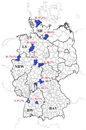
Figure 1.
Ten sheep flocks (A–J) were sampled in the following five German federal states: Baden-Württemberg: BW, Bavaria: BAV, Lower Saxony: LS, North Rhine-Westphalia: NRW, Schleswig-Holstein: SH. Shaded blue regions represent districts with participating farms. Anaplasma spp. intra-flock seroprevalences determined by cELISA [31] are indicated.
2.2. Molecular Analysis
Only Anaplasma spp. seropositive samples were examined to increase the probability of finding the pathogen’s DNA. In total, 357 ovine sera from ten small ruminant flocks were analysed. The number of tested samples per farm in cELISA and real-time PCR are shown in Table 1.

Table 1.
Overview of number of sampled and tested sheep from ten flocks (A–J). The Anaplasma spp. antibody response was determined by a commercial cELISA. The seropositive specimens were further analysed with a real-time PCR to detect Anaplasma phagocytophilum DNA.
DNA extraction was performed on stored serum samples using the DNeasy Blood and Tissue Mini Kit (Qiagen GmbH, Hilden, Germany), in accordance with the manufacturer’s instructions. Isolated genomic-DNA was stored at −20 °C until further use. Subsequently, a 16S rRNA real-time PCR was processed according to the primer–probe combinations by Sirigireddy and Ganta [42], but with modified reporter and quencher molecules of the probe (Table 2).
Table 2.
Reagents for the 16S rRNA real-time PCR based on the primer–probe combinations by Sirigireddy and Ganta [42], but with modified reporter and quencher molecules of the probe.
Amplification reactions were carried out as two-step singleplex real-time PCR reactions, combining 15 μL reaction mix with 10 μL DNA template. Thermocycling was conducted in an Agilent Stratagene Mx3005P thermocycler (Agilent Technologies, Inc., Santa Clara, CA, USA) and based on a protocol by Tappe and Strube [43], which was slightly modified as follows: initial denaturation at 95 °C for 15 min, followed by 45 cycles of 95 °C for 15 s and 58 °C for 60 s. Cycle quantification (Cq) values ≤ 45 were considered positive. All real-time PCR reactions were performed in duplicate, including positive and negative controls. The positive A. phagocytophilum control originated from a diseased lamb (GenBank no. MZ348289.1). Negative controls were available from sheep kept indoors at the Clinic for Swine and Small Ruminants, University of Veterinary Medicine Hannover, Foundation, Hannover, Germany. PCR specificity was successfully tested with A. ovis-positive PCR samples (GenBank nos. MZ348248, MZ348249) available from a previous study [13]. An internal control to assess the presence of PCR inhibitors was not applied.
3. Results
In total, 407 sheep were included in this pilot study and 357 showed an antibody response against Anaplasma spp. These 357 ovine serum samples were further analysed by 16S rRNA real-time PCR. DNA sequences of A. phagocytophilum were identified in two ewes (Table 3). Both sheep came from flock A in the northern German federal state of Schleswig-Holstein. The animals were sampled in early November 2017 at the pasture. All other samples tested A. phagocytophilum-PCR negative, although the intra-flock seroprevalence reached levels of up to 97.7%.
Table 3.
Information about age, cELISA result and the Cq-values (duplicates) of each A. phagocytophilum DNA positive ewe from flock A located in the northern German federal state of Schleswig-Holstein.
4. Discussion
Despite the worldwide occurrence of A. phagocytophilum in sheep [19,44,45,46], knowledge on the distribution of the pathogen in the German sheep population is still limited [13,14]. Hence, the aim of this study was to determine the distribution of A. phagocytophilum DNA in German sheep flocks with a very high Anaplasma spp. seroprevalence [31].
In this retrospective study, the detection rate of A. phagocytophilum was 0.6%, which is remarkably lower than that reported by Scharf and colleagues (4%) [14] and Bauer and colleagues (25%) [13] for the German sheep population. This discrepancy is possibly influenced by several factors.
Firstly, sensitivity of PCR amplification was reduced in serum samples compared to whole blood samples in previous studies [39,47,48]. Serum usually contains lower bacterial loads than whole blood due to the intracellular nature of Anaplasma [36]. Nevertheless, DNA sequences of A. phagocytophilum, A. marginale, A. bovis and A. centrale were successfully detected in serum samples from cattle, deer and sheep [37,38,40], probably due to the less inhibiting factors for the PCR reaction in serum, such as haem from the blood and the presence of host DNA [38].
Secondly, the DNA detection of A. phagocytophilum by PCR depended on the primers applied, and the variety of different primers and protocols hamper a direct comparison between the conducted studies [49,50,51]. We used the 16S rRNA gene fragment in contrast to an earlier study from Germany, which used msp2 [13]. The most common gene fragments in the A. phagocytophilum genome are msp2 and msp4, whereas there are only single copies of the 16S rRNA fragment [52,53]. This might explain the higher detection rate of the msp2 fragment compared to a 16S rRNA fragment. However, an earlier study identified no substantial differences among these targeting gene fragments [54]. More studies are needed to evaluate the sensitivity and specificity of different targeting genes and primers to detect A. phagocytophilum DNA from different sample matrices and to make the findings more comparable.
Thirdly, timing of sampling is crucial to detect A. phagocytophilum DNA in blood specimens. The pathogen circulates in an undulatory manner in the host, but primary bacteraemia only lasts up to two weeks [25,26]. Secondary and subsequent bacteraemia cycles are quite short-lived and rarely last longer than three days [26]. In contrast, antibodies against A. phagocytophilum can be detected from about week 2 post infection and are measured over a long period of up to 12 weeks with approximately similar titres [25,27]. In the present study, the IFP of antibodies against Anaplasma spp. was very high (>88%) in most of the sheep flocks (7/10), indicating that many animals had contact with the pathogen, but A. phagocytophilum DNA was hardly detected. The blood samples were collected from flock A-H at the pasture or shortly after housing between early November 2017 and mid-January 2018, which are seasons with low or even no tick activity, and pathogen transmission was probably reduced during this time of year [2,55,56]. Consequently, an acute infection might have taken place earlier. Thus, primary bacteraemia had already been completed and animals had recovered from the acute A. phagocytophilum infection. Furthermore, only samples from seropositive sheep were tested, so primary infections of seronegative animals were not detected. This reduced the likelihood of pathogen detection. Our assumption is supported by the absence of obvious clinical signs of TBF in sheep during sampling. The lack of clinical signs also resulted in the low detection rate (<7%) of A. phagocytophilum DNA in sheep from previous studies [14,38], whereas pathogen DNA was identified in a remarkable number (25%) of subclinical and clinical affected sheep [13,38]. Finally, only a small prevalence can be expected in sheep flocks with no acute cases of TBF [57,58].
The high IFP among the ten sheep flocks was determined by an Anaplasma spp. cELISA, which does not discriminate between the different Anaplasma species. Recently, DNA of Anaplasma ovis was identified for the first time in Germany in one sheep flock from southern Germany (Bavaria) [13] and this pathogen might influence the serological outcomes. However, A. ovis seems to occur only focally and A. phagocytophilum is the major Anaplasma species in the German small ruminant population [12,13,14]. Therefore, we suppose that most detected antibodies in the cELISA were induced by A. phagocytophilum.
5. Conclusions
DNA of A. phagocytophilum was detected in serum samples from two sheep from northern Germany (Schleswig-Holstein). Together with the findings from previous studies [13,14], we conclude that the pathogen occurs at least focally in the German sheep population, or at least in the north and south of Germany. The DNA detection of A. phagocytophilum might be lower in serum samples compared to EDTA-anticoagulated blood specimens. Nevertheless, sera are usually stored over a long period of time and are available in large numbers. Therefore, they can be used for pilot studies to determine the existence of the pathogen retrospectively [37]. Moreover, sheep should be sampled during the tick season to increase the likelihood of pathogen detection in blood specimens. Further research is needed to investigate the epidemiology and clinical impact of A. phagocytophilum as an emerging pathogen in the German sheep population. Such studies also support the One Health approach, since the pathogen is a zoonotic agent and HGA might be underreported in Europe [15].
Author Contributions
Conceptualisation, M.G. and B.U.B.; methodology, W.R. and B.U.B.; software, W.R.; validation, M.G. and B.U.B.; formal analysis, W.R.; investigation, W.R. and B.U.B.; resources, M.G.; data curation, W.R.; writing—original draft preparation, W.R.; writing—review and editing, M.G. and B.U.B.; visualisation, W.R.; supervision, M.G. and B.U.B.; project administration, M.G. and B.U.B. All authors have read and agreed to the published version of the manuscript.
Funding
This research received no external funding. This open access publication was funded by the Deutsche Forschungsgemeinschaft (DFG, German Research Foundation) 91094227 “Open Access Publication Funding” and the University of Veterinary Medicine Hannover Foundation.
Institutional Review Board Statement
Serum samples from sheep were available from a previously conducted study [41], which was approved by the federal state governments of Baden-Württemberg (AZ 35-9185.82/0351, AZ 35-9185.82/A-1/18), Bavaria (RUF-55.2.2-2532-2-651-5), Lower Saxony (Az. 33.8-42502-05-17A211), North Rhine-Westphalia (81-02.05.40.18.015) and Schleswig-Holstein (V 242-5191/2018). The study was conducted in accordance with German Animal Welfare Legislation and the EU Directive 2010/63/EU for animal experiments. All animals were handled in accordance with high ethical standards and national legislation.
Informed Consent Statement
Not applicable.
Data Availability Statement
The data are available upon request from the corresponding author. Parts of this manuscript are included in a doctoral thesis (Studies on the prevalence of Anaplasma spp. in German sheep flocks, Wiebke Rubel, 2022), which has been deposited in the institutional repository of the University of Veterinary Medicine Hannover Foundation (https://elib.tiho-hannover.de/receive/tiho_mods_00007168?q=rubel; accessed on 7 August 2022).
Acknowledgments
The authors would like to thank Ursula Küttler, Antje Polifka and Annika Wolf for their excellent technical support. Furthermore, we would like to thank Frances Sherwood-Brock for proofreading the manuscript.
Conflicts of Interest
The authors declare no conflict of interest.
References
- Stuen, S.; Granquist, E.G.; Silaghi, C. Anaplasma phagocytophilum—A widespread multi-host pathogen with highly adaptive strategies. Front. Cell. Infect. Microbiol. 2013, 3, 31. [Google Scholar] [CrossRef] [PubMed]
- Rymaszewska, A.; Grenda, S. Bacteria of the genus Anaplasma—Characteristics of Anaplasma and their vectors: A review. Vet. Med. 2008, 53, 573–584. [Google Scholar] [CrossRef]
- Henniger, T.; Henniger, P.; Grossmann, T.; Distl, O.; Ganter, M.; von Loewenich, F.D. Congenital infection with Anaplasma phagocytophilum in a calf in Northern Germany. Acta Vet. Scand. Suppl. 2013, 55, 38. [Google Scholar] [CrossRef] [PubMed]
- Jensen, J.; Simon, D.; Escobar, H.M.; Soller, J.T.; Bullerdiek, J.; Beelitz, P.; Pfister, K.; Nolte, I. Anaplasma phagocytophilum in dogs in Germany. Zoonoses Public Health 2007, 54, 94–101. [Google Scholar] [CrossRef] [PubMed]
- Kohn, B.; Silaghi, C.; Galke, D.; Arndt, G.; Pfister, K. Infections with Anaplasma phagocytophilum in dogs in Germany. Res. Vet. Sci. 2011, 91, 71–76. [Google Scholar] [CrossRef] [PubMed]
- Hamel, D.; Bondarenko, A.; Silaghi, C.; Nolte, I.; Pfister, K. Seroprevalence and bacteraemia of Anaplasma phagocytophilum in cats from Bavaria and Lower Saxony (Germany). Berl. Munch. Tierarztl. Wochenschr. 2012, 125, 163–167. [Google Scholar] [CrossRef] [PubMed]
- Schäfer, I.; Kohn, B.; Müller, E. Anaplasma phagocytophilum in domestic cats from Germany, Austria and Switzerland and clinical/laboratory findings in 18 PCR-positive cats (2008–2020). J. Feline Med. Surg. 2021, 24, 290–297. [Google Scholar] [CrossRef] [PubMed]
- von Loewenich, F.D.; Stumpf, G.; Baumgarten, B.U.; Röllinghoff, M.; Dumler, J.S.; Bogdan, C. A case of equine granulocytic ehrlichiosis provides molecular evidence for the presence of pathogenic Anaplasma phagocytophilum (HGE agent) in Germany. Eur. J. Clin. Microbiol. Infect. Dis. 2003, 22, 303–305. [Google Scholar] [CrossRef]
- Silaghi, C.; Liebisch, G.; Pfister, K. Genetic variants of Anaplasma phagocytophilum from 14 equine granulocytic anaplasmosis cases. Parasites Vectors 2011, 4, 161. [Google Scholar] [CrossRef]
- Silaghi, C.; Nieder, M.; Sauter-Louis, C.; Knubben-Schweizer, G.; Pfister, K.; Pfeffer, M. Epidemiology, genetic variants and clinical course of natural infections with Anaplasma phagocytophilum in a dairy cattle herd. Parasites Vectors 2018, 11, 20. [Google Scholar] [CrossRef]
- Tegtmeyer, P.; Ganter, M.; von Loewenich, F.D. Simultaneous infection of cattle with different Anaplasma phagocytophilum variants. Ticks Tick-Borne Dis. 2019, 10, 1051–1056. [Google Scholar] [CrossRef]
- Langenwalder, D.B.; Schmidt, S.; Gilli, U.; Pantchev, N.; Ganter, M.; Silaghi, C.; Aardema, M.L.; von Loewenich, F.D. Genetic characterization of Anaplasma phagocytophilum strains from goats (Capra aegagrus hircus) and water buffalo (Bubalus bubalis) by 16S rRNA gene, ankA gene and multilocus sequence typing. Ticks Tick-borne Dis. 2019, 10, 101267. [Google Scholar] [CrossRef] [PubMed]
- Bauer, B.U.; Raileanu, C.; Tauchmann, O.; Fischer, S.; Ambros, C.; Silaghi, C.; Ganter, M. Anaplasma phagocytophilum and Anaplasma ovis-Emerging pathogens in the German sheep population. Pathogens 2021, 10, 1298. [Google Scholar] [CrossRef] [PubMed]
- Scharf, W.; Schauer, S.; Freyburger, F.; Petrovec, M.; Schaarschmidt-Kiener, D.; Liebisch, G.; Runge, M.; Ganter, M.; Kehl, A.; Dumler, J.S.; et al. Distinct host species correlate with Anaplasma phagocytophilum ankA gene clusters. J. Clin. Microbiol. 2011, 49, 790–796. [Google Scholar] [CrossRef] [PubMed]
- Matei, I.A.; Estrada-Pena, A.; Cutler, S.J.; Vayssier-Taussat, M.; Varela-Castro, L.; Potkonjak, A.; Zeller, H.; Mihalca, A.D. A review on the eco-epidemiology and clinical management of human granulocytic anaplasmosis and its agent in Europe. Parasites Vectors 2019, 12, 599. [Google Scholar] [CrossRef]
- Zhang, L.; Liu, Y.; Ni, D.; Li, Q.; Yu, Y.; Yu, X.-j.; Wan, K.; Li, D.; Liang, G.; Jiang, X.; et al. Nosocomial transmission of human granulocytic anaplasmosis in China. JAMA 2008, 300, 2263–2270. [Google Scholar] [CrossRef] [PubMed]
- Dugat, T.; Lagrée, A.-C.; Maillard, R.; Boulouis, H.-J.; Haddad, N. Opening the black box of Anaplasma phagocytophilum diversity: Current situation and future perspectives. Front. Cell. Infect. Microbiol. 2015, 5, 61. [Google Scholar] [CrossRef]
- Kauffmann, M.; Rehbein, S.; Hamel, D.; Lutz, W.; Heddergott, M.; Pfister, K.; Silaghi, C. Anaplasma phagocytophilum and Babesia spp. in roe deer (Capreolus capreolus), fallow deer (Dama dama) and mouflon (Ovis musimon) in Germany. Mol. Cell. Probes 2017, 31, 46–54. [Google Scholar] [CrossRef] [PubMed]
- Stuen, S. Haemoparasites in small ruminants in European countries: Challenges and clinical relevance. Small Rumin. Res. 2016, 142, 22–27. [Google Scholar] [CrossRef]
- Gokce, H.I.; Woldehiwet, Z. Differential haematological effects of tick-borne fever in sheep and goats. J. Vet. Med. 1999, 46, 105–115. [Google Scholar] [CrossRef] [PubMed]
- Grøva, L.; Olesen, I.; Steinshamn, H.; Stuen, S. Prevalence of Anaplasma phagocytophilum infection and effect on lamb growth. Acta Vet. Scand. Suppl. 2011, 53, 30. [Google Scholar] [CrossRef] [PubMed]
- Stuen, S.; Bergström, K.; Palmer, E. Reduced weight gain due to subclinical Anaplasma phagocytophilum (formerly Ehrlichia phagocytophila) infection. Exp. Appl. Acarol. 2002, 28, 209–215. [Google Scholar] [CrossRef] [PubMed]
- Sargison, N.; Edwards, G. Tick infestations in sheep in the UK. In Pract. 2009, 31, 58–65. [Google Scholar] [CrossRef]
- Daniel, R.G.; Carson, A.; Evans, C.; Cookson, R.; Wessels, M. Pathological observations of tick-borne fever and intercurrent bacterial infections in lambs. Vet. Rec. Case Rep. 2016, 4, e000367. [Google Scholar] [CrossRef]
- Stuen, S.; Bergström, K.; Petrovec, M.; Van de Pol, I.; Schouls Leo, M. Differences in clinical manifestations and haematological and serological responses after experimental infection with genetic variants of Anaplasma phagocytophilum in sheep. Clin. Vaccine Immunol. 2003, 10, 692–695. [Google Scholar] [CrossRef] [PubMed]
- Thomas, R.J.; Birtles, R.J.; Radford, A.D.; Woldehiwet, Z. Recurrent bacteraemia in sheep infected persistently with Anaplasma phagocytophilum. J. Comp. Pathol. 2012, 147, 360–367. [Google Scholar] [CrossRef] [PubMed]
- Dreher, U.M.; de la Fuente, J.; Hofmann-Lehmann, R.; Meli, M.L.; Pusterla, N.; Kocan, K.M.; Woldehiwet, Z.; Braun, U.; Regula, G.; Staerk, K.D.C.; et al. Serologic cross-reactivity between Anaplasma marginale and Anaplasma phagocytophilum. Clin. Vaccine Immunol. 2005, 12, 1177–1183. [Google Scholar] [CrossRef]
- Hartelt, K.; Oehme, R.; Frank, H.; Brockmann, S.O.; Hassler, D.; Kimmig, P. Pathogens and symbionts in ticks: Prevalence of Anaplasma phagocytophilum (Ehrlichia spp.), Wolbachia spp., Rickettsia spp., and Babesia spp. in Southern Germany. Int. J. Med. Microbiol. Suppl. 2004, 293, 86–92. [Google Scholar] [CrossRef]
- Hildebrandt, A.; Krämer, A.; Sachse, S.; Straube, E. Detection of Rickettsia spp. and Anaplasma phagocytophilum in Ixodes ricinus ticks in a region of Middle Germany (Thuringia). Ticks Tick-borne Dis. 2010, 1, 52–56. [Google Scholar] [CrossRef]
- May, K.; Strube, C. Prevalence of Rickettsiales (Anaplasma phagocytophilum and Rickettsia spp.) in hard ticks (Ixodes ricinus) in the city of Hamburg, Germany. Parasitol. Res. 2014, 113, 2169–2175. [Google Scholar] [CrossRef] [PubMed]
- Rubel, W.; Schoneberg, C.; Wolf, A.; Ganter, M.; Bauer, B.U. Seroprevalence and risk factors of Anaplasma spp. in German small ruminant flocks. Animals 2021, 11, 2793. [Google Scholar] [CrossRef]
- Foggie, A. Studies on the infectious agent of tick-borne fever in sheep. J. Pathol. 1951, 63, 1–15. [Google Scholar] [CrossRef] [PubMed]
- Lepidi, H.; Bunnell, J.E.; Martin, M.E.; Madigan, J.E.; Stuen, S.; Dumler, J.S. Comparative pathology and immunohistology associated with clinical illness after Ehrlichia phagocytophila-group infections. Am. J. Trop. Med. Hyg. 2000, 62, 29–37. [Google Scholar] [CrossRef] [PubMed]
- Shabana, I.I.; Alhadlag, N.M.; Zaraket, H. Diagnostic tools of caprine and ovine anaplasmosis: A direct comparative study. BMC Vet. Res. 2018, 14, 165. [Google Scholar] [CrossRef]
- Torina, A.; Galindo, R.C.; Vicente, J.; Di Marco, V.; Russo, M.; Aronica, V.; Fiasconaro, M.; Scimeca, S.; Alongi, A.; Caracappa, S.; et al. Characterization of Anaplasma phagocytophilum and A. ovis infection in a naturally infected sheep flock with poor health condition. Trop. Anim. Health Prod. 2010, 42, 1327–1331. [Google Scholar] [CrossRef] [PubMed]
- Silaghi, C.; Santos, A.S.; Gomes, J.; Christova, I.; Matei, I.A.; Walder, G.; Domingos, A.; Bell- Sakyi, L.; Sprong, H.; von Loewenich, F.D.; et al. Guidelines for the Direct Detection of Anaplasma spp. in Diagnosis and Epidemiological Studies. Vector-Borne Zoonotic Dis. 2017, 17, 12–22. [Google Scholar] [CrossRef]
- Kawahara, M.; Rikihisa, Y.; Lin, Q.; Isogai, E.; Tahara, K.; Itagaki, A.; Hiramitsu, Y.; Tajima, T. Novel genetic variants of Anaplasma phagocytophilum, Anaplasma bovis, Anaplasma centrale, and a novel Ehrlichia spp. in wild deer and ticks on two major islands in Japan. Appl. Environ. Microbiol. 2006, 72, 1102–1109. [Google Scholar] [CrossRef]
- Kiilerich, A.M.; Christensen, H.; Thamsborg, S.M. Anaplasma phagocytophilum in Danish sheep: Confirmation by DNA sequencing. Acta Vet. Scand. Suppl. 2009, 51, 55. [Google Scholar] [CrossRef] [PubMed]
- Massung, R.F.; Slater, K.; Owens, J.H.; Nicholson, W.L.; Mather, T.N.; Solberg, V.B.; Olson, J.G. Nested PCR assay for detection of granulocytic Ehrlichiae. J. Clin. Microbiol. 1998, 36, 1090–1095. [Google Scholar] [CrossRef] [PubMed]
- Parvizi, O.; El-Adawy, H.; Melzer, F.; Roesler, U.; Neubauer, H.; Mertens-Scholz, K. Seroprevalence and molecular detection of bovine anaplasmosis in Egypt. Pathogens 2020, 9, 64. [Google Scholar] [CrossRef] [PubMed]
- Wolf, A.; Prufer, T.L.; Schoneberg, C.; Campe, A.; Runge, M.; Ganter, M.; Bauer, B.U. Prevalence of Coxiella burnetii in German sheep flocks and evaluation of a novel approach to detect an infection via preputial swabs at herd-level. Epidemiol. Infect. 2020, 148, e75. [Google Scholar] [CrossRef] [PubMed]
- Sirigireddy, K.R.; Ganta, R.R. Multiplex detection of Ehrlichia and Anaplasma species pathogens in peripheral blood by real-time reverse transcriptase-polymerase chain reaction. J. Mol. Diagn. 2005, 7, 308–316. [Google Scholar] [CrossRef]
- Tappe, J.; Strube, C. Anaplasma phagocytophilum and Rickettsia spp. infections in hard ticks (Ixodes ricinus) in the city of Hanover (Germany). Ticks Tick-Borne Dis. 2013, 4, 432–438. [Google Scholar] [CrossRef] [PubMed]
- Gorman, J.K.; Hoar, B.R.; Nieto, N.C.; Foley, J.E. Evaluation of Anaplasma phagocytophilum infection in experimentally inoculated sheep and determination of Anaplasma spp. seroprevalence in 8 free-ranging sheep flocks in California and Oregon. Am. J. Vet. Res. 2012, 73, 1029–1034. [Google Scholar] [CrossRef] [PubMed]
- Tumwebaze, M.A.; Byamukama, B.; Tayebwa, D.S.; Byaruhanga, J.; Angwe, M.K.; Galon, E.M.; Liu, M.; Lee, S.-H.; Ringo, A.E.; Adjou Moumouni, P.F.; et al. First molecular detection of Babesia ovis, Theileria spp., Anaplasma spp., and Ehrlichia ruminantium in goats from Western Uganda. Pathogens 2020, 9, 895. [Google Scholar] [CrossRef] [PubMed]
- Yang, J.; Liu, Z.; Niu, Q.; Liu, J.; Han, R.; Guan, G.; Li, Y.; Liu, G.; Luo, J.; Yin, H. Anaplasma phagocytophilum in sheep and goats in Central and Southeastern China. Parasites Vectors 2016, 9, 593. [Google Scholar] [CrossRef][Green Version]
- Dumler, J.S.; Bakken, J.S. Human granulocytic ehrlichiosis in Wisconsin and Minnesota: A frequent infection with the potential for persistence. J. Infect. Dis. 1996, 173, 1027–1030. [Google Scholar] [CrossRef] [PubMed][Green Version]
- Pancholi, P.; Kolbert, C.P.; Mitchell, P.D.; Reed, K.D.; Dumler, J.S.; Bakken, J.S.; Telford, S.R., III; Persing, D.H. Ixodes dammini as a potential vector of human granulocytic ehrlichiosis. J. Infect. Dis. 1995, 172, 1007–1012. [Google Scholar] [CrossRef]
- Chmielewska-Badora, J.; Zwolinski, J.; Cisak, E.; Wojcik-Fatla, A.; Buczek, A.; Dutkiewicz, J. Prevalence of Anaplasma phagocytophilum in Ixodes ricinus ticks determined by polymerase chain reaction with two pairs of primers detecting 16S rRNA and ankA genes. Ann. Agric. Environ. Med. 2007, 14, 281–285. [Google Scholar] [PubMed]
- Fingerle, V.; Munderloh, U.G.; Liegl, G.; Wilske, B. Coexistence of Ehrlichiae of the Phagocytophila group with Borrelia burgdorferi in Ixodes ricinus from Southern Germany. Med. Microbiol. Immunol. 1999, 188, 145–149. [Google Scholar] [CrossRef] [PubMed]
- Kang, J.G.; Ko, S.; Kim, Y.-J.; Yang, H.-J.; Lee, H.; Shin, N.-S.; Choi, K.-S.; Chae, J.-S. New genetic variants of Anaplasma phagocytophilum and Anaplasma bovis from Korean water deer (Hydropotes inermis argyropus). Vector Borne Zoonotic Dis. 2011, 11, 929–938. [Google Scholar] [CrossRef] [PubMed]
- Dunning Hotopp, J.C.; Lin, M.; Madupu, R.; Crabtree, J.; Angiuoli, S.V.; Eisen, J.A.; Seshadri, R.; Ren, Q.; Wu, M.; Utterback, T.R.; et al. Comparative genomics of emerging human ehrlichiosis agents. PLoS Genet. 2006, 2, e21. [Google Scholar] [CrossRef]
- Rymaszewska, A. PCR for detection of tick-borne Anaplasma phagocytophilum pathogens: A review. Vet. Med. 2011, 56, 529–536. [Google Scholar] [CrossRef]
- Massung, R.F.; Slater, K.G. Comparison of PCR assays for detection of the agent of human granulocytic ehrlichiosis, Anaplasma phagocytophilum. J. Clin. Microbiol. 2003, 41, 717–722. [Google Scholar] [CrossRef]
- Gethmann, J.; Hoffmann, B.; Kasbohm, E.; Süss, J.; Habedank, B.; Conraths, F.J.; Beer, M.; Klaus, C. Research paper on abiotic factors and their influence on Ixodes ricinus activity—Observations over a two-year period at several tick collection sites in Germany. Parasitol. Res. 2020, 119, 1455–1466. [Google Scholar] [CrossRef] [PubMed]
- Schulz, M.; Mahling, M.; Pfister, K. Abundance and seasonal activity of questing Ixodes ricinus ticks in their natural habitats in Southern Germany in 2011. J. Vector Ecol. 2014, 39, 56–65. [Google Scholar] [CrossRef] [PubMed]
- Hamzah, K.J.; Hasso, S.A. Molecular prevalence of Anaplasma phagocytophilum in sheep from Iraq. Open Vet. J. 2019, 9, 238–245. [Google Scholar] [CrossRef]
- Zhang, Y.; Lv, Y.; Zhang, F.; Zhang, W.; Wang, J.; Cui, Y.; Wang, R.; Jian, F.; Zhang, L.; Ning, C. Molecular and phylogenetic analysis of Anaplasma spp. in sheep and goats from six provinces of China. J. Vasc. Surg. Cases 2016, 17, 523–529. [Google Scholar] [CrossRef]

Publisher’s Note: MDPI stays neutral with regard to jurisdictional claims in published maps and institutional affiliations. |
© 2022 by the authors. Licensee MDPI, Basel, Switzerland. This article is an open access article distributed under the terms and conditions of the Creative Commons Attribution (CC BY) license (https://creativecommons.org/licenses/by/4.0/).