Review

Ocular Drug Delivery into the Eyes Using Drug-Releasing Soft Contact Lens

by
Toshihiko Tashima
Tashima Laboratories of Arts and Sciences, 1239-5 Toriyama-cho, Kohoku-ku, Yokohama 222-0035, Japan
Future Pharmacol. 2024, 4(2), 336-351; https://doi.org/10.3390/futurepharmacol4020019
Submission received: 1 April 2024 / Revised: 22 April 2024 / Accepted: 26 April 2024 / Published: 29 April 2024

Abstract

:
The impact of visual impairment, such as blindness, on quality of life is immeasurable. However, effective ocular drug delivery into the eyes has not yet been established, primarily due to the impermeability imposed by the blood–retinal barrier (BRB) based on the tight junctions and efflux transporters at the endothelium or the epithelium in oral or intravenous administration, as well as the dilution with tear fluid and excretion through the nasolacrimal duct in eye drop administration. Furthermore, intravitreous injections induce pain and fear in patients. Unmet medical needs persist in ocular diseases such as age-related macular degeneration and diabetic retinopathy. Therefore, innovative non-invasive administration methods should be developed. Drug-releasing soft contact lenses (DR-SCLs) affixed to the eye’s surface can continuously and locally deliver their loaded drugs to the eyes. The use of DR-SCLs is expected to greatly enhance the bioavailability and patient adherence to the drug regimen. It is known that several solute carrier (SLC) transporters are expressed in various parts of the eyes, including the cornea, the ciliary body, and the bulbar conjunctiva. Carrier-mediated transport through SLC transporters may occur in addition to passive diffusion. Moreover, nanoparticles can be loaded into DR-SCLs, offering various intelligent approaches based on modifications to induce receptor-mediated endocytosis/transcytosis or to control the loaded drug release within this delivery system. In this perspective review, I discuss the implementation and potential of DR-SCL-mediated ocular drug delivery, particularly focusing on low-molecular-weight compounds because of their fine distribution in living body, ease of handling, and ease of manufacturing.

1. Introduction

The eye is one of the body’s most vital organs, responsible for transmitting visual information to the brain as a sensory organ via the optic nerve. However, significant medical needs persist in ocular diseases such as age-related macular degeneration (AMD), diabetic retinopathy (DR), cataracts, glaucoma, dry eye disease, neurotrophic keratitis, and neuropathic corneal pain, largely due to barriers such as the blood–retinal barrier (BRB) at the endothelium composed of retinal capillary endothelial cells (the inner BRB) or the epithelium composed of retinal pigment epithelial cells (the outer BRB) [1], the ocular blood–aqueous barrier (BAB) in the iris and the ciliary body [2], and anatomical structures like the nasolacrimal duct, as indicated by structuralism [3,4]. The BRB and the BAB are basically based on tight junctions and efflux transporters. Drug membrane impermeability poses a significant challenge in drug discovery and development. While eye drop administration is relatively simple compared to local injection and is not hindered by the BRB or the BAB, only 1–7% of administered drugs actually penetrate into the eyes due to dilution with tear fluid, excretion through the nasolacrimal duct, and poor membrane permeability due to transportation via efflux transporters and low contact frequency to influx carriers [5]. Off-target side effects may arise when drugs enter the systemic circulation via the choroid, the bulbar conjunctiva, or the nasal mucosa. Furthermore, applying eye ointment, a dosage form intended for long-term effectiveness, can be somewhat challenging. Oral medication or intravenous injection is often hindered by the impermeability imposed by the BRB and can lead to off-target side effects due to incorrect distribution. Therefore, innovative drug delivery systems should be developed to enhance patient quality of life. Drug-releasing soft contact lenses (DR-SCLs) offer solutions to these challenges, addressing issues such as low bioavailability, off-target side effects, and drug adherence [Table 1]. Additionally, the potential for carrier-mediated eye drug transport across the BRB using cation transporters is an attractive prospect [6,7]. In this perspective review, I explore the possibilities and implementations of ocular drug delivery to the eyes using DR-SCLs loaded with potent drugs, particularly focusing on low-molecular-weight compounds because of their fine distribution in living body via carrier-mediated transport and passive diffusion, ease of handling to load into DR-SCLs, ease of manufacturing, and stability at room temperature.

2. Discussion

2.1. Potential Drug Pathways from DR-SCLs Based on Anatomical Eye Features

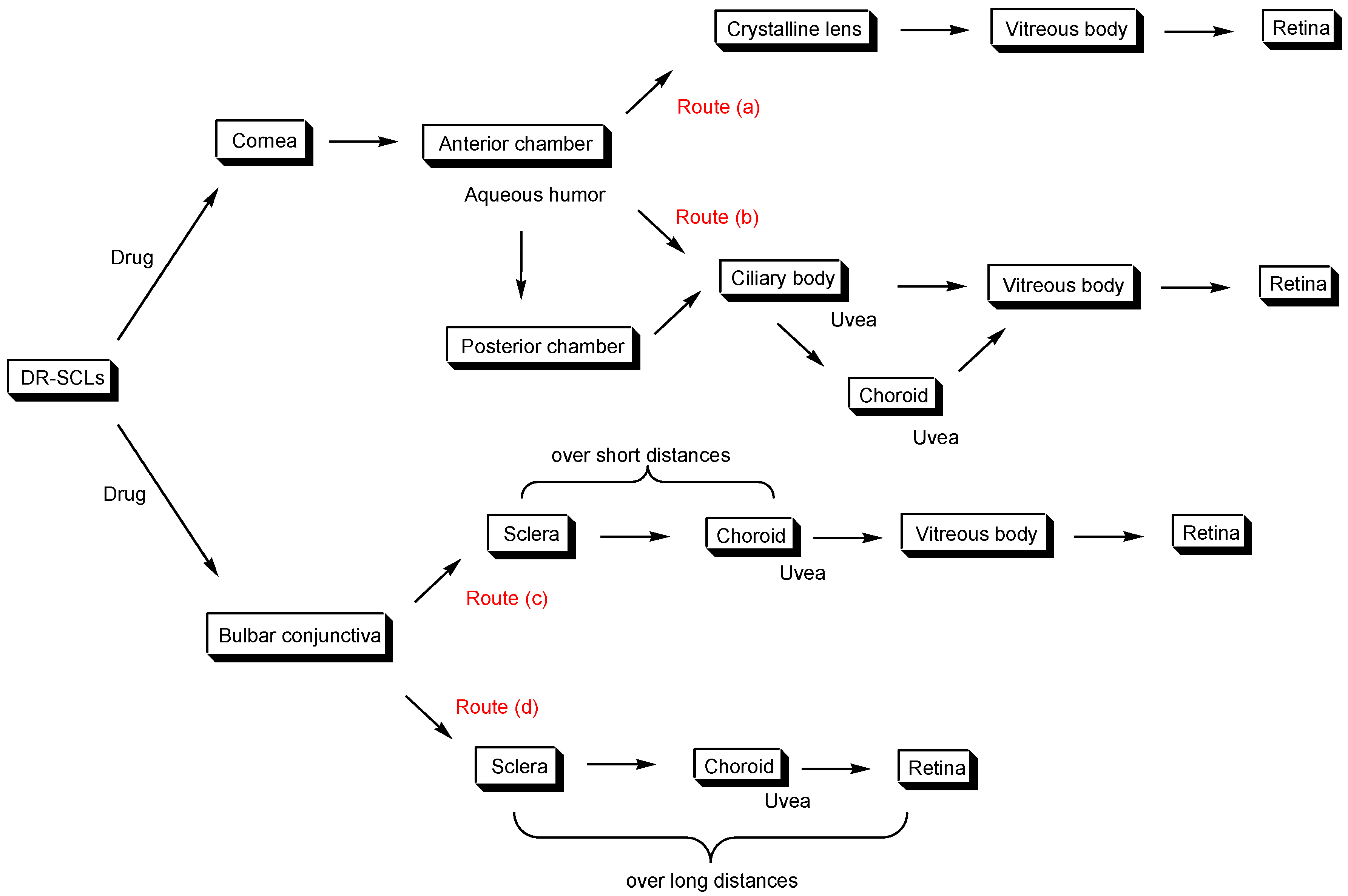
The eye is a complex organ, both anatomically and functionally [Figure 1]. Nevertheless, devising a drug delivery strategy for it poses significant challenges. The primary contact surface between a DR-SCL and the eye consists mainly of the cornea and the bulbar conjunctiva. Therefore, it is crucial to understand the anatomical features of the cornea and bulbar conjunctiva in order to develop DR-SCLs effectively. Several potential routes for drugs released from DR-SCLs exist. (a) Drugs traverse the cornea and eventually accumulate in the aqueous humor, which is the fluid found between the cornea and the crystalline lens in the anterior chamber of the eye, and between the iris and the crystalline lens in the posterior chamber of the eye. Subsequently, drugs within the chamber pass through either the crystalline lens or the ciliary zonule into the vitreous body, and then reach the retina [Figure 2].
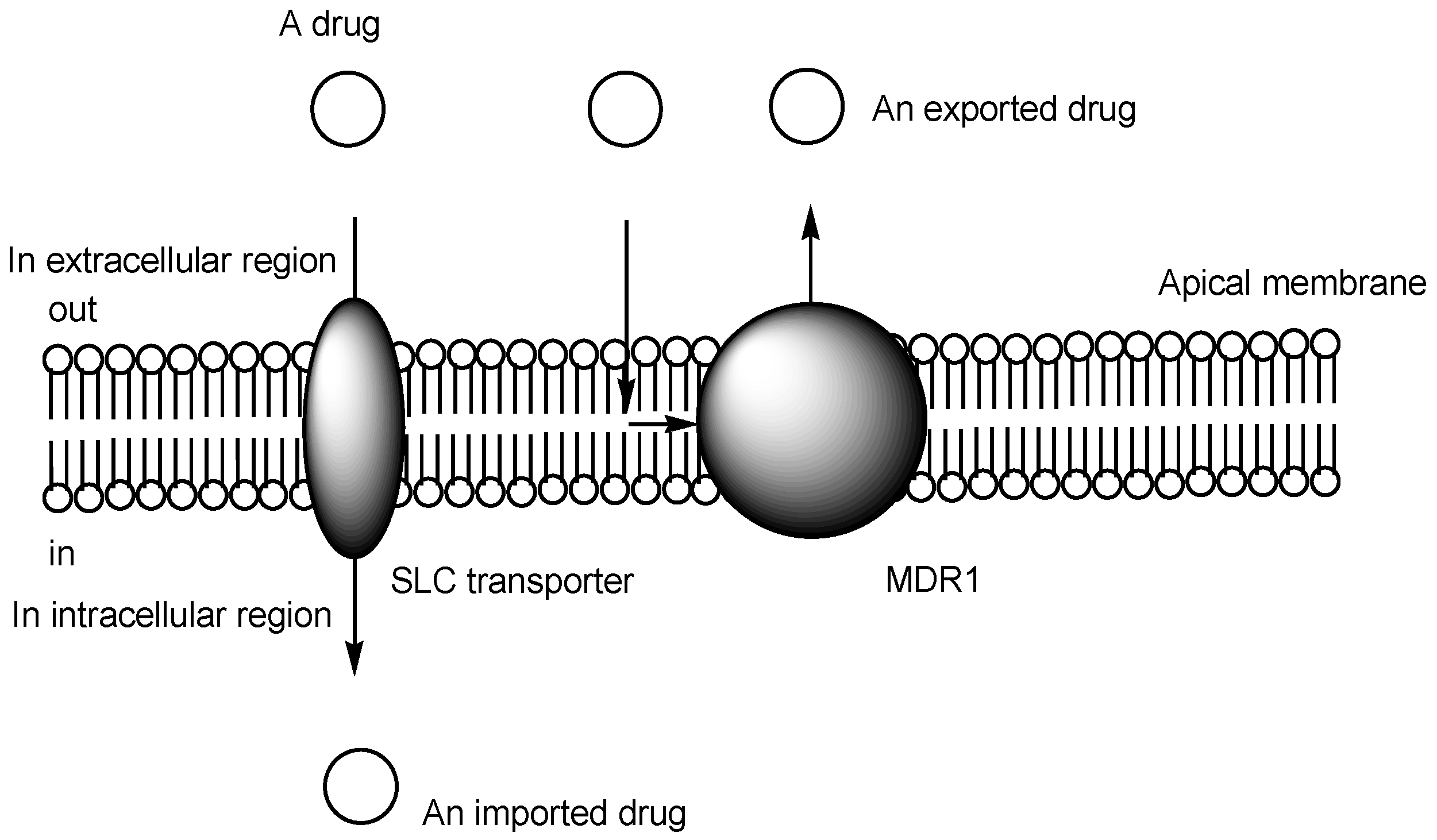
The cornea is composed of the corneal epithelium, the parenchyma of the cornea, and the corneal endothelium. Substances traverse the cornea through passive diffusion or active transport mechanisms in the transcellular pathway, as well as passive diffusion in the paracellular pathway. Certain amino acids cross the cornea via carrier-mediated transport utilizing L-amino acid transporter 1 (LAT1), ATB0,+, and alanine serine cysteine transporter 1 (ASCT1) [8]. ATB0,+ belongs to the amino acid transporter branch of the SLC6 family [Figure 3]. In the corneal epithelium, solute carrier (SLC) transporters such as LAT1, ATB0,+, peptide transporter 1 (PEPT1), monocarboxylate transporters (MCTs), and concentrative nucleoside transporter 3 (CNT3) are primarily expressed, while efflux transporters such as multiple drug resistance 1 (MDR1, P-glycoprotein) are also present [9]. MDR1 in the corneal epithelium captures hydrophobic low-molecular-weight substances that are passing through the plasma membrane [10], subsequently exporting them into tear fluid [Figure 3]. Therefore, the passive diffusion of hydrophobic low-molecular-weight substances across the corneal epithelium is hindered by MDR1. Carrier-mediated transport using SLC transporters or receptor-mediated endocytosis/transcytosis could serve as a promising strategy for facilitating the transepithelial transport of substances across the cornea. A study reported the internalization of Aspergillus flavus spores into corneal epithelial cells via actin-mediated endocytosis [11]. Moreover, bioadhesive glycosylated nanoformulations applied topically were observed to traverse corneal epithelial cells and reach the parenchyma of the cornea via transcytosis in in vivo assays using rats [12]. Nanoparticles encapsulating indomethacin have shown transcorneal penetration via three energy-dependent endocytosis pathways: clathrin-dependent endocytosis, caveolae-dependent endocytosis, and micropinocytosis. Among these pathways, caveolae-dependent endocytosis appears to be the most predominant in the penetration process [13].
Concerning the crystalline lens, lens epithelial cells and lens fiber cells, originating from lens epithelial cells, are enclosed within the lens capsule, which contains collagen IV. The main proteins in the crystalline lens are α-crystallin, β-crystallin, and γ-crystallin, comprising approximately 1/2 to 1/3 of the crystalline lens by weight. It is understood that intermediate-sized macromolecules, such as proteins, penetrate the crystalline lens through the lens capsule. Neutral compounds like dextrans penetrate the lens capsule more rapidly than negatively charged compounds such as recombinant epidermal growth factor and single-stranded DNAs [14]. Collagen IV carries a positive charge due to lysine and arginine [15]. Hydrophobic substances, including neutral compounds, cross the lipid bilayer via passive diffusion, whereas hydrophilic substances, including charged compounds, cannot cross it via passive diffusion. Thus, substances can traverse the crystalline lens into the vitreous body through the layers of the lens capsule, lens epithelial cells, and lens fiber cells, probably via passive diffusion due to experimental size dependence leading to a molecular sieve effect. The route via the cornea and the crystalline lens into the vitreous body is unlikely to be suitable for high-molecular-weight substances such as nanoparticles or monoclonal antibodies. Nanoparticles positioned between the DR-SCL and the cornea might cause discomfort to the patients. However, several cases of DR-SCLs loaded with nanoparticles have been reported. Nanoparticles can be loaded into DR-SCLs, offering generally various intelligent approaches based on modifications to induce receptor-mediated endocytosis/transcytosis or to control the loaded drug release [16,17]. This strategy employing nanoparticle-loaded DR-SCL is applicable to corneal diseases such as keratitis. If the encapsulated drugs are released from the nanoparticles before they reach the crystalline lens, the impermeability issue based on size would not occur in the crystalline lens.
(b) As a non-crystalline lens pathway, some substances might transition from the ciliary body or the choroid to the vitreous body and subsequently to the retina [Figure 2]. In fact, the pathway from the anterior chamber of the eye to the ciliary body might be more prevalent than that from the anterior chamber of the eye to the crystalline lens, as the crystalline lens is likely not suitable as a route for medicine due to its internal anatomy. The ciliary body plays a crucial role in producing aqueous humor to nourish the cornea and the crystalline lens, maintain eye pressure, and adjust the shape of the crystalline lens during focusing. It houses a variety of drug transporters, including SLC transporters such as organic anion transporters, organic anion-transporting polypeptides, bile acid transporters (such as apical sodium-dependent bile salt transporter (ASBT) and sodium taurocholate cotransporter), organic cation transporters (such as novel organic cation transporter (OCTN) and multidrug and toxin extrusion transporter (MATE)), and peptide transporters, as well as efflux transporters like MDR1 [Figure 3]. MDR1 in the capillary endothelium is associated with the BAB, whereas the tight junctions of the capillary endothelium of the iris and the ciliary body epithelium form the BAB [18]. The bilayered ciliary epithelium predominantly transports solutes and, secondarily, water from the underlying stroma to the aqueous humor [19]. Therefore, transport across the ciliary body from the anterior chamber of the eye may predominantly occur through routes involving passive diffusion. Hydrophobic substances in the anterior chamber of the eye would diffuse across the ciliary body membrane, as opposed to the crystalline lens, which is predominantly filled with water and exhibits thermodynamically stable behavior. Additionally, the choroid [20] is a thin layer situated between the sclera and the retina, rich in blood vessels that supply oxygen and nutrients to the retina. The choroid comprises four distinct layers: the innermost Bruch’s membrane, the choriocapillaris layer, Sattler’s layer, and the outermost Haller’s layer.
On the other hand, (c) drugs might successively cross the bulbar conjunctiva, the sclera, and the uvea, particularly the choroid, into the vitreous body [Figure 2]. The bulbar conjunctiva possesses relatively leaky epithelium, lacking tight junctions, thereby enabling the entry of not only low-molecular-weight substances but also larger hydrophilic drugs such as siRNAs and peptides [21]. Several transporters such as PEPT1, amino acid transporters, and MDR1 are recognized in the bulbar conjunctiva [Figure 3]. In fact, amino acids, D-glucose, monocarboxylates, nucleosides, and dipeptides are facilitated by several ion-coupled SLC transporters in the bulbar conjunctiva [22]. The uvea comprises the iris, the ciliary body, and the choroid. The uvea is rich in blood vessels, and some absorbed drugs might enter the systemic circulation.
Alternatively, (d) a hydrophobic pathway through the sclera via the bulbar conjunctiva, bypassing the vitreous body, is possible [Figure 2]. This strategy is likely restricted to non-high-molecular-weight amphipathic substances to traverse long distances along the sclera.
The main components of the vitreous body are water (>90%) and solid fibrillar components such as glycosaminoglycans (including anionic hyaluronic acid) and collagen, forming a viscoelastic hydrogel structure through which not only light but also substances move to reach the retina [23]. Overall, the trajectories of drugs administered from the outside are impeded by multilayered barriers consisting of hydrophobic membranes, hydrophilic cytosols, hydrophilic aqueous humor, and the hydrophilic vitreous body. Therefore, designed drugs should possess both moderate hydrophobicity and moderate hydrophilicity, in addition to being of a suitable size to pass through gel regions such as the crystalline lens and the vitreous body. As an extension of knowledge, their physical properties, such as the log partition coefficient (logP) and molecular weight (MW), could be defined according to Lipinski’s rule of five [24,25]. This rule evaluates drug-likeness concerning orally administered drugs that cross several barriers, such as (i) solubility in the small intestine and the systemic circulation and (ii) the permeability of the small intestinal epithelium and the portal vein. It empirically suggests calculated logP (less than 5) and MW (less than 500) as suitable physical properties. Compounds with a logP value less than 5 are not so hydrophobic that they can be dissolved in water. Compounds with MW less than 5 are not so large that they can go through certain substances such as phospholipids in the membrane without undergoing large steric hindrance.

2.2. Drug-Releasing Soft Contact Lenses (DR-SCLs)

DR-SCL therapy maintains medication concentrations at the ocular surface by inhibiting drainage through tear fluid and excretion via the nasolacrimal duct [26,27]. Surprisingly, the development history of DR-SCLs is relatively old. Ocusert® [28,29], a contact lens-like intraocular indwelling preparation, was launched in 1969 for the treatment of glaucoma and can sustain local intraocular pilocarpine concentration within the effective range for 1 week. Pilocarpine is released via a polymer membrane covering the drug reservoir. However, the development of DR-SCLs with drugs has been less extensive than expected, with exceptions for infections [30,31], inflammation in rabbits [32], and glaucoma as mentioned below. Therefore, research on DR-SCLs with drugs should be pursued more actively.
The simplest method for preparing drug-loaded CLs is the traditional immersion method. There is a wide range of CL materials available to meet users’ needs. Generally, silicone hydrogel CLs consist of silica-based materials such as polydimethylsiloxane (PDMS), tris(trimethylsiloxysilyl)propylvinylcarbamate (TPVC), and tris(trimethylsiloxy)methacryloxypropylsilane (TRIS), as well as hydrophilic materials such as 2-hydroxyethylmethacrylate (HEMA), N,N-dimethylacrylamide (DMA), and 1-vinyl-2-pyrrolidinone (NVP) [33]. HEMA is one of the most important hydrogel materials for CLs. Combination prescriptions can be freely adjusted depending on the type of drugs and applicable diseases. Menicon, a Japanese CL manufacturer, has developed DR-SCLs composed of 2-hydroxyethyl methacrylate (with a molar ratio of more than 0.5), methyl acrylate, and ethyl acrylate as monomers. These lenses are designed to accommodate anti-allergy drugs such as ketotifen, chlorpheniramine, olopatadine, and levocabastine. Ketotifen released from the DR-SCLs was evaluated using rabbit eyes. The importance of hydrophobicity in DR-SCLs for appropriately controlling drug release was suggested [34].

2.3. Ocular Diseases and DR-SCLs

2.3.1. Age-Related Macular Degeneration (AMD)

AMD [35] is a medical condition that leads to vision loss in older individuals due to macular degeneration in the retina. Wet AMD is characterized by bleeding associated with the development of new blood vessels, while dry AMD involves gradual atrophy caused by nutritional deficiencies and the accumulation of waste products due to retinal cell degeneration.
Abnormal vascular proliferation is induced by vascular endothelial growth factor (VEGF). Neovascular vessels do not exist in the normal retina and are so fragile that their components leak out and accumulate, or they are prone to bleeding. Thus, anti-VEGF therapy is a promising approach for wet AMD. Currently, anti-VEGF drugs such as bevacizumab (anti-VEGF monoclonal antibody), aflibercept (soluble decoy VEGF receptor), ranibizumab (anti-VEGF monoclonal antibody Fab fragment), brolucizumab (humanized anti-VEGF monoclonal single-chain variable fragment), and faricimab (bispecific monoclonal antibody targeting VEGF and Ang-2) are clinically used for the treatment of wet AMD [36,37]. However, they are administered via vitreous injection because monoclonal antibodies characterized as high-molecular-weight compounds cannot cross the membrane via passive diffusion due to large molecular size and hydrophilicity, which may not be patient-friendly. There is a need for more patient-friendly administration methods. It is well known that low-molecular-weight compounds can cross the membrane via passive diffusion or carrier-mediated transport using SLC transporters. At present, several low-molecular-weight VEGF inhibitors are clinically approved for cancer therapy, although they appear to be hydrophilic [Figure 4] [38]. DR-SCLs loaded with such low-molecular-weight VEGF inhibitors could be an alternative strategy for wet AMD. It is speculated that VEGF inhibitors released from DR-SCLs might pass through the vitreous body to the retina via (a) the crystalline lens and/or (b) the ciliary body [Figure 2].
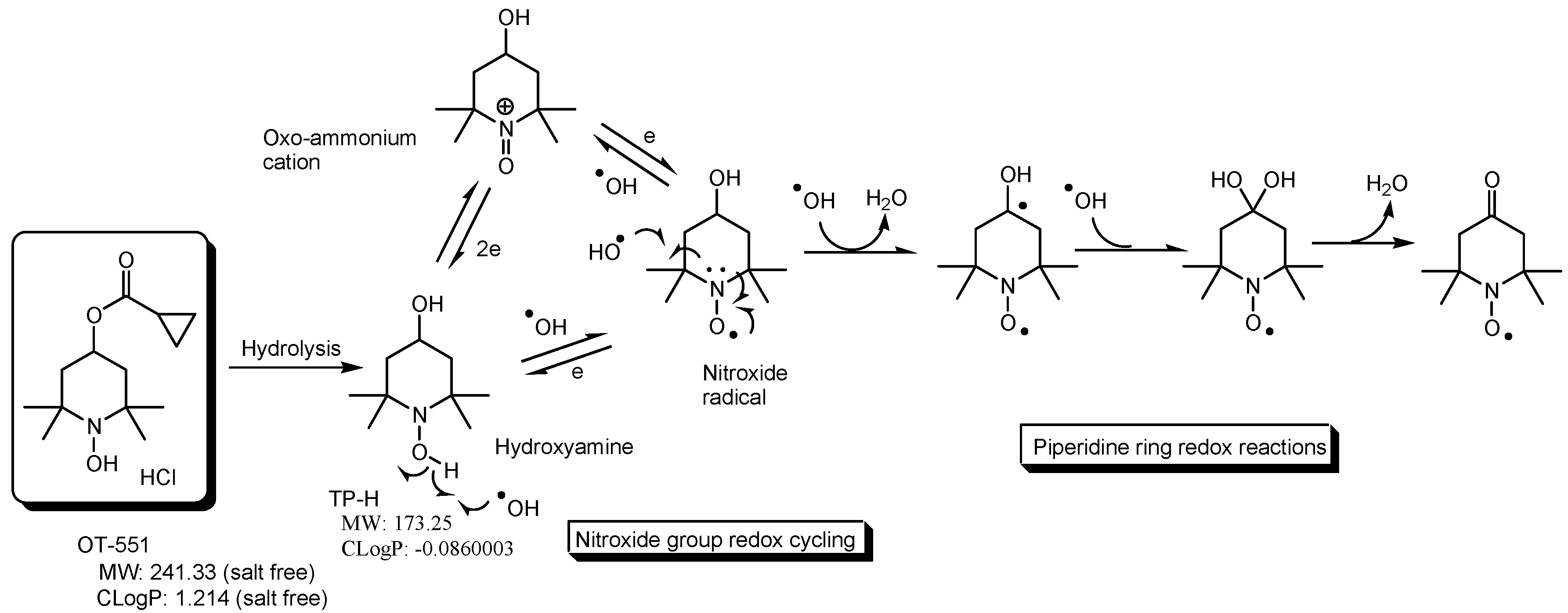
Several drugs for dry AMD are undergoing clinical trials [39]. Dry AMD is a complex multifactorial disease, and its pathogenesis is not completely understood. Oxidative stress, inflammation, and other factors are suggested to exacerbate its pathology. It is well known that lipoproteins called drusen accumulate behind the retina in the early stages of dry AMD. Complement components 3 (C3) and 5 (C5) in drusen are activated, leading to the progression of dry AMD through the induction of inflammation. Avacincaptad pegol, a pegylated RNA aptamer against C5, was approved for the treatment of geographic atrophy secondary to AMD by the U.S. Food and Drug Administration (FDA) in 2023. However, avacincaptad pegol requires administration via vitreous injection. Nonetheless, the investigation into DR-SCLs with RNAs is not likely to be active. Antioxidant agents such as supplements containing ZnO, CuO, vitamin C, vitamin E, and β-carotene (AREDS, NCT00000145), as well as supplements containing ZnO, CuO, vitamin C, vitamin E, lutein, and zeaxanthin (AREDS2, NCT00345176), completed phase 3 clinical trials with promising results. Another antioxidant agent, OT-551 [Figure 5] [40] (NCT00306488), completed a phase 2 clinical trial. Typically, OT-551 is formulated as a daily topical eye drop for patients with dry AMD. Therefore, DR-SCLs with supplements used in the AREDS2 clinical trial or OT-551 can enhance bioavailability in a non-invasive manner.

2.3.2. Diabetic Retinopathy (DR)

DR [41] is one of the most common complications of diabetes, resulting in vision loss and blindness. The aldehyde group of glucose is supposed to react with certain proteins through glycation and the Maillard reaction. As a result, glucotoxicity due to protein glycation in the retina occurs, thereby inducing microangiopathy in cases of diabetes where glucose metabolism is insufficient due to insulin deficiency VEGF released to restore microangiopathy ironically enhances the formation of brittle neovascular vessels that deteriorate DR due to bleeding. Currently, anti-VEGF therapy is effective in the late stages of DR. Clinically available anti-VEGF agents for the treatment of DR include bevacizumab (anti-VEGF monoclonal antibody), ranibizumab (anti-VEGF monoclonal antibody Fab fragment), and aflibercept (soluble decoy VEGF receptor) [42]. However, all anti-VEGF agents are regularly administered via intravitreal injections. Therefore, DR-SCLs with low-molecular-weight VEGF inhibitors [Figure 4] could be an alternative strategy for DR, as mentioned previously regarding AMD.

2.3.3. Cataract

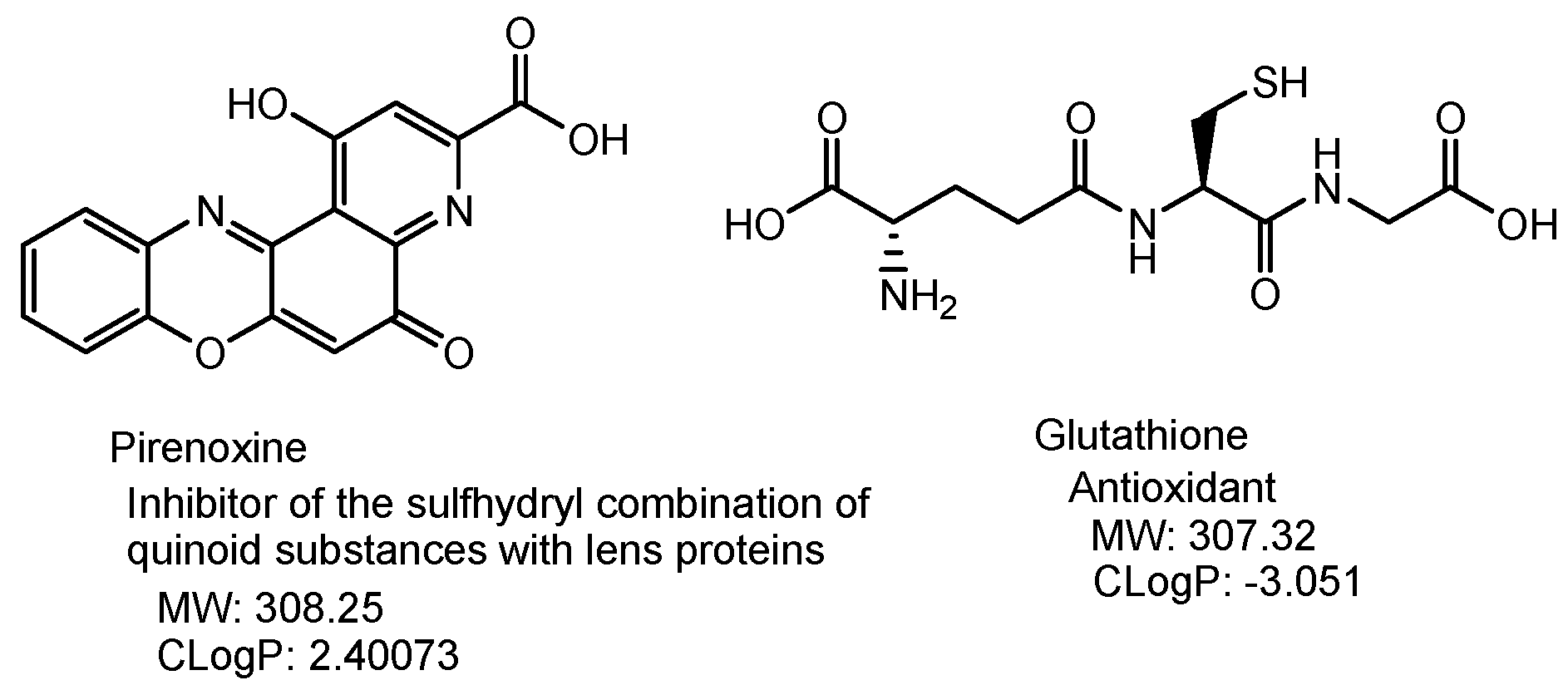
A cataract [43] is a dense clouding of the crystalline lens due to the aggregation of crystalline proteins, making it difficult to see clearly. Cataract treatment includes drug therapy and surgery. As symptoms progress, the cloudy parts are removed through surgical procedures. Phacoemulsification, a common cataract surgery method using ultrasound to remove the cloudy parts, was first developed by Dr. Charles Kelman in 1967 [44]. However, in the early stages of symptoms, eye drop treatments are administered to slow down progression. Pirenoxine (an inhibitor of the sulfhydryl combination of quinoid substances with lens proteins) or glutathione (an antioxidant) [Figure 6] are clinically used for early cataracts [45]. Cataracts are progressive chronic diseases. Therefore, DR-SCLs with pirenoxine or glutathione can be useful formulations to improve the bioavailability and drug adherence of patients.

2.3.4. Glaucoma

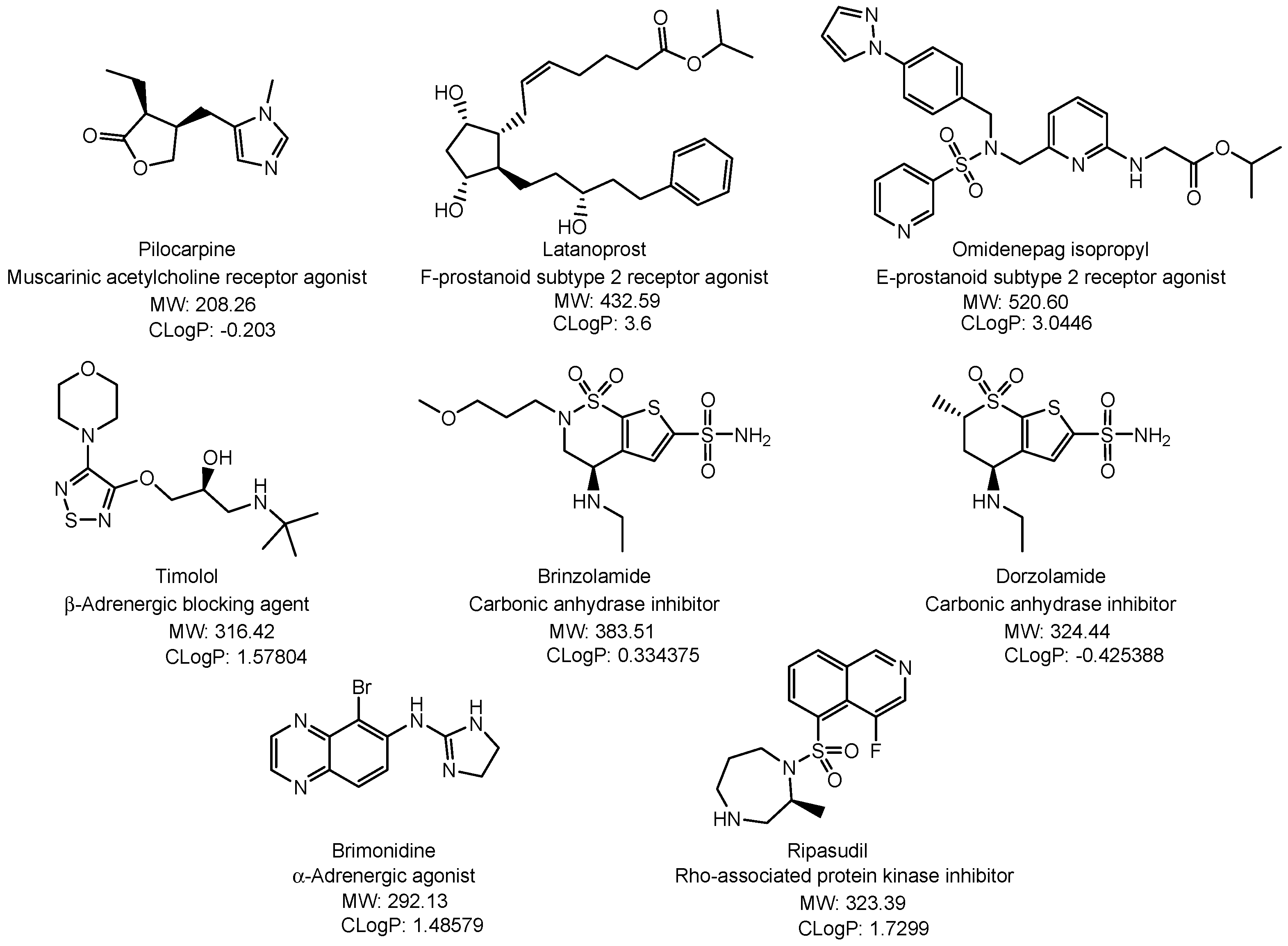
Glaucoma [46] is a progressive disease that leads to the death of retinal ganglion cells due to elevated intraocular pressure, resulting in a narrowed visual field and eventually blindness. Irregularities in aqueous fluid flow contribute to increased intraocular pressure. The prevalence of glaucoma increases with age. The most common treatment for glaucoma is prescription eye drops that lower intraocular pressure, in addition to laser treatment and surgery. The methods to lower the intraocular pressure are the enhancement of aqueous humor and the aqueous humor production inhibition (β-adrenergic blocking agents, carbonic anhydrase inhibitors, α-adrenergic agonists). Aqueous humor produced in the ciliary body is excreted through the main outflow channel from trabecular meshwork to blood vessels (trabecular meshwork outflow pathway) (muscarinic acetylcholine receptor agonists, Rho-associated protein kinase inhibitors) and the secondary outflow channel from root of the iris to the ciliary body muscle tissue (uveoscleral outflow pathway) (F-prostanoid subtype 2 receptor agonists, E-prostanoid subtype 2 receptor agonists) [47,48,49]. Drugs used to treat glaucoma include pilocarpine (muscarinic acetylcholine receptor agonist), latanoprost (F-prostanoid subtype 2 receptor agonist), omidenepag isopropyl (E-prostanoid subtype 2 receptor agonist), timolol (β-adrenergic blocking agent), brinzolamide, dorzolamide (carbonic anhydrase inhibitors), brimonidine (α-adrenergic agonist), and ripasudil (Rho-associated protein kinase inhibitor) [Figure 7] [47,48,49]. These drugs lower intraocular pressure via each mechanism shown in parentheses.
DR-SCLs with such low-molecular-weight drugs might enhance bioavailability. In fact, Ocusert® with pilocarpine was developed for the treatment of glaucoma [28,29], indicating the feasibility of this technology. Pilocarpine elicits shrinking pupils and intraocular pressure-lowering effects based on direct action to the pupillary sphincter and ciliary muscle by binding the muscarinic acetylcholine receptor of the parasympathetic nervous system via route (b) [Figure 2] from DR-SCLs. Ciliary muscle contractions make trabecular meshwork widen to enhance aqueous humor outflow, resulting in intraocular pressure reduction. Furthermore, latanoprost and timolol were simultaneously released from methoxypolyethylenglycol-polylactic acid (mPEG-PLA) micelles-laden CLs for glaucoma treatment, demonstrating a sustained reduction in intraocular pressure for over 168 h. In an in vivo pharmacokinetic study on rabbit eyes, the mean residence time and bioavailability of timolol and latanoprost delivered by CLs were improved 79.6-fold and 122.2-fold, respectively, compared to eye drops [50]. Similarly, brinzolamide and timolol were simultaneously released from methoxypolyethylenglycol-polycaprolactone (mPEG-PCL) micelles-laden CLs. The mean bioavailability for brinzolamide and timolol delivered by CLs was improved 1.41-fold and 2.71-fold, respectively, compared to eye drops in an in vivo pharmacokinetic study [51]. Additionally, timolol and dorzolamide released simultaneously from vitamin E-loaded CLs decreased intraocular pressure by inhibiting the production of aqueous humor in a Beagle model of glaucoma [52].
Prostanoids such as latanoprost and omidenepag isopropyl are typically designed based on prodrug strategy. Isopropyl esters are introduced to increase hydrophobicity to permeate the plasma membrane of the corneal epithelium and the corneal endothelium via passive diffusion. In aqueous humor, prostanoid prodrugs are de-esterified to form the parent drugs with free carboxylates. The parent drugs are too hydrophilic to permeate the plasma membrane via passive diffusion and, therefore, are located in the anterior chamber of the eye. Eventually, they bind receptors on the surface of cells in the ciliary muscle through the uveoscleral outflow pathway, leading to the increase in aqueous humor outflow due to ciliary muscle relaxation [53]. Thus, prostanoid prodrug-loaded DR-SCLs would enhance their bioavailability through route (b) [Figure 2] by avoiding the dilution with tear fluid and the excretion through the nasolacrimal duct and via the escape from MDR1 capture due to the repetition of passive diffusion and MDR1 transportation.
β-Adrenergic-blocking agents such as timolol inhibit aqueous humor secretion by binding β-adrenergic receptors as a blocker on the surface of ciliary epithelial cells [54]. Furthermore, carbonic anhydrase inhibitors such as brinzolamide and dorzolamide inhibit carbonic anhydrase in the ciliary epithelial cells of the ciliary body. HCO3 plays an important role in aqueous humor production, involving fluid transport of Na+ into the posterior chamber of the eye. Carbonic anhydrase inhibitors exhibit their activity in cells and, thus, have to cross the plasma membrane of ciliary epithelial cells, although brinzolamide and dorzolamide are likely to be hydrophilic. An extraction recovery of 30 μL of dorzolamide (dorzolamide hydrochloride ophthalmic solution, 2%) 1 h after a single topical ocular administration in rabbit ocular tissues showed that dorzolamide was distributed in the cornea (10.31 μg/g), conjunctiva (10.10 μg/g), aqueous humor (1.49 μg/g), sclera (anterior) (9.12 μg/g), sclera (posterior) (0.460 μg/g), retina (anterior) (1.02 μg/g), retina (posterior) (0.023 μg/g), vitreous (anterior) (0.07 μg/g), vitreous (posterior) (0.0096 μg/g), and optic nerve (0.657 μg/g). On the other hand, an extraction recovery of 30 μL of brinzolamide (brinzolamide ophthalmic suspension, 1%) 1 h after a single topical ocular administration in rabbit ocular tissues showed that that brinzolamide was distributed in the cornea (7.99 μg/g), conjunctiva (6.77 μg/g), aqueous humor (0.530 μg/g), sclera (anterior) (1.57 μg/g), sclera (posterior) (0.140 μg/g), retina (anterior) (0.821 μg/g), retina (posterior) (0.015 μg/g), vitreous (anterior) (0.034 μg/g), vitreous (posterior) (undetected drug level), and optic nerve (0.269 μg/g) [55]. Thus, these findings suggested the accuracy of the distribution of drugs released from DR-SCLs [Figure 2]. Moreover, α-adrenergic agonists such as brimonidine bind α-adrenergic receptors on the surface of ciliary epithelial cells and consequently reduce aqueous humor secretion. Intriguingly, a combination of brimonidine and β-blockers such as timolol reduced the intraocular pressure by 15.4% greater than the contralateral timolol-treated eye in a randomized, double-masked, placebo-controlled study of 20 human subjects [56].
Rho-associated protein kinase inhibitors such as ripasudil increase aqueous humor outflow through the trabecular meshwork outflow pathway [57]. Rho-associated protein kinases are inhibited in cells. Thus, inhibitors have to enter the cells across the plasma membrane. Rho-associated protein kinases are activated by the binding of the GTP-bound Rho phosphorylate myosin light chain (MLC) and Lin-1/Isl-1/Mec-3 kinase (LIMK), leading to tissue contraction and stiffness.
Low-molecular-weight drugs used in the treatment of glaucoma might be suitable for DR-SCLs, because the target sites are adjacent to the anterior chamber of the eye that drugs released from DR-SCLs can reach just across the cornea through route (b) [Figure 2].

2.3.5. Dry Eye Disease

Dry eye disease [53,54,58,59] is a condition where tears are unevenly distributed due to either aqueous tear deficiency or an imbalance in tear quality. Typical symptoms include a dry, gritty, burning, or bright sensation in the eyes, redness, eye fatigue, eye pain, or teary eyes.
SCLs physically prevent moisture evaporation to prevent corneal desiccation, although they are recognized as one of the causes of dry eye disease. SCLs used for treating dry eye disease do not necessarily need to be drug-loaded CLs. Silicone hydrogel lenses have very high oxygen permeability. Currently, products such as HYPER-CL (EyeYon Medical, Ness Tziona, Israel) for physically protecting the cornea, Acuvue Theravision with Ketotifen (ATK) (Johnson & Johnson Vision Care, Inc., Jacksonville, FL, USA) containing a histamine H1 receptor antagonist, Acuvue Oasys Lenses made from silicone hydrogel (Johnson & Johnson Vision Care, Inc.) for physically protecting the cornea, and others are commercially available for DR-SCLs [55,60].
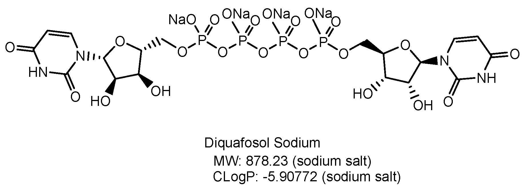
There are three targets for drug therapy for dry eye: the lipid layer, the aqueous/mucus layer, and the corneal epithelium [56,61]. Mild symptoms can be improved with eye drops. Diquafosol sodium [Figure 8] is a dinucleotide, purinoreceptor P2Y2 receptor agonist that promotes tear and mucin secretion via elevated intracellular Ca2+ concentrations [57,62]. This eye drug can be used while wearing normal SCLs because preservatives are also technologically difficult to adsorb to normal SCLs. When DR-SCLs with diquafosol sodium are used, they would extend the efficacy time of the drug longer than diquafosol sodium eye drops alone, with or without normal SCLs.

2.3.6. Neurotrophic Keratitis

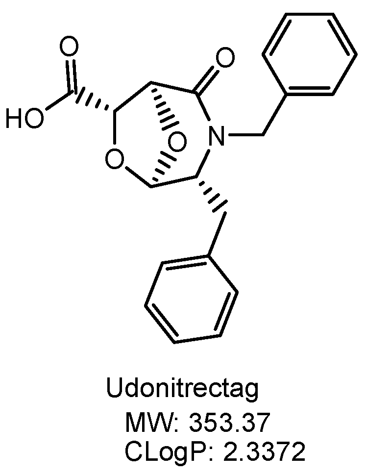
Neurotrophic keratitis [58,63] is a degenerative corneal disease characterized by damages such as corneal epithelial defects, corneal stromal melting, or corneal perforation due to disorders of the trigeminal nerve caused by the herpes virus and other factors. Consequently, significant vision loss occurs. Nerve growth factors via eye drops are currently being developed. Cenegermin [59,64] is a recombinant human nerve growth factor that is the first FDA-approved drug for the treatment of neurotrophic keratitis. Cenegermin (MW 13 kDa) is composed of 118 amino acids with three disulfide bonds [60,65]. Thus, DR-SCLs could be loaded with cenegermin. On the other hand, udonitrectag (MW 353.37 Da) [Figure 9] is a low-molecular-weight synthetic peptido-mimetic of human nerve growth factor [61,66]. DR-SCLs could be loaded with udonitrectag due to its smaller size compared to cenegermin.

2.3.7. Neuropathic Corneal Pain

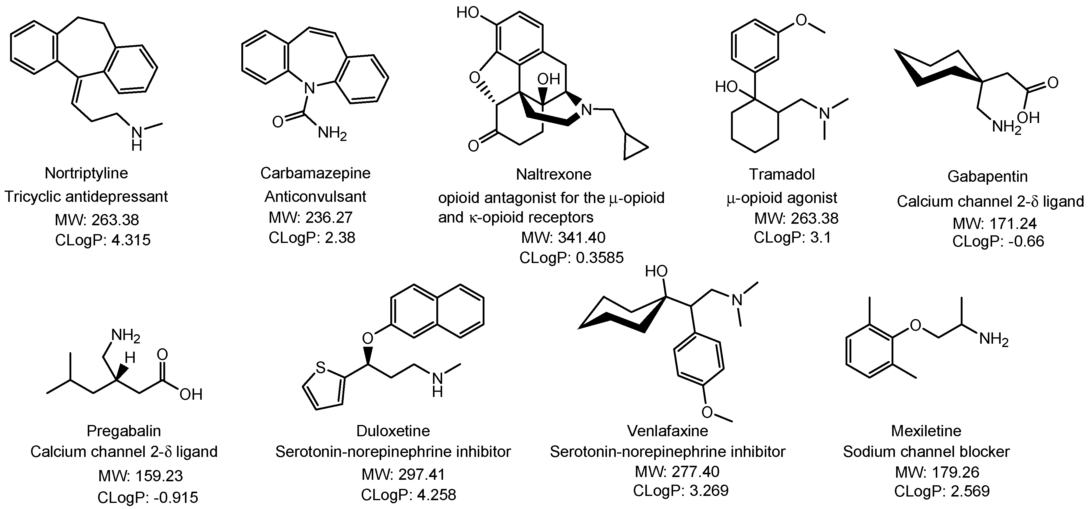
Pain is generally categorized into nociceptive pain, neuropathic pain, and psychogenic pain. Neuropathic corneal pain [62,67] is a condition characterized by pain without a noxious stimulus due to abnormal nerve function. Corneal nerve damage resulting from inflammatory diseases, neurological diseases, or surgical interventions often causes neuropathic corneal pain. The nerve terminals are located within the corneal epithelium [63,68]. While tricyclic antidepressants and carbamazepine (anticonvulsant) are first-line agents for neuropathic corneal pain, low-dose naltrexone (opioid antagonist for the μ-opioid and κ-opioid receptors) and tramadol (weak μ-opioid agonist) are second-line agents. Furthermore, gabapentin, pregabalin (calcium channel α 2-δ ligands), duloxetine, venlafaxine (serotonin-norepinephrine inhibitors), and mexiletine (sodium channel blocker) are third-line agents [Figure 10] [64,69]. Some of these low-molecular-weight drugs can be administered using DR-SCLs.
Nortriptyline [Figure 10], a tricyclic antidepressant, demonstrated efficacy in relieving neuropathic corneal pain symptoms in thirty patients when administered orally [65,70]. Thus, if nortriptyline can effectively target the ophthalmic nerve (CNV1) branching from the trigeminal nerve, DR-SCLs containing nortriptyline might offer better efficacy than oral administration, as they could potentially ensure more accurate distribution.

3. Conclusions

Many patients worldwide, particularly in developing countries, suffer from ocular diseases because unmet medical needs persist due to barriers such as the BRB and the BAB hindering intravenous and oral administration, or due to the low bioavailability of eye drops caused by dilution with tear fluid, excretion through the nasolacrimal duct, and poor membrane permeability. Therefore, innovative approaches to eye drug delivery are necessary. Although SCLs are typically used for correcting refractive errors, they can also serve as a vehicle for clinical treatment due to their continuous contact with the eyes. DR-SCLs can gradually release medications into the eyes either through the cornea or the bulbar conjunctiva, minimizing loss due to dilution with tear fluid and excretion through the nasolacrimal duct. Eye drugs released from DR-SCLs can be delivered into the eye in a non-invasive manner without intravitreous injection and without being impeded by the BRB. Thus, the use of DR-SCLs for treating eye diseases shows promise in addressing issues such as low bioavailability, off-target side effects, and medication adherence [Table 1]. However, research on DR-SCLs has not been pursued actively. The problem is whether there will be people who will try, although DR-SCLs were developed in the 1960s. I hope that this perspective review inspires readers to initiate such research efforts soon. Moreover, there are concerns that DR-SCLs also can cause infections similar to normal SCLs. Off-target side effects may arise when drugs enter the systemic circulation via well-vascularized areas such as the choroid, the bulbar conjunctiva, or the nasal mucosa.
The plausible pathways of drugs released from DR-SCLs to the retina pass through various parts of the eye, including the cornea, the vitreous body, the sclera, and the choroid. The uvea comprises the iris, the ciliary body, and the choroid. Briefly, route (a) follows the crystalline lens pathway. Route (b) traverses the non-crystalline lens pathway. Route (c) involves the vitreous body pathway, crossing the bulbar conjunctiva, sclera, and choroid. Route (d) follows the non-vitreous body pathway through the bulbar conjunctiva and layers of sclera and choroid [Figure 2]. Low-molecular-weight substances rather than high-molecular-weight substances might be well distributed through these routes due to size limitations. Nonetheless, the features of drugs would depend on the target sites such as the retina for wet AMD and the crystalline lens for cataracts. Therefore, the fine design of drug-loaded DR-SCLs is important for conducting appropriate drug delivery to the target sites.
Non-low-molecular-weight substances such as anti-VEGF monoclonal antibodies are extensively utilized in the treatment of AMD and DR, particularly through intravitreous injections that are burdensome on the patients. Therefore, DR-SCLs containing low-molecular-weight substances such as VEGF inhibitors, which have been approved for other medical conditions including cancers, can offer an alternative non-invasive approach. The indications for drugs will be expanded to eye diseases. Furthermore, the efficacy of treatments for ocular diseases, for which low-molecular-weight eye drop drugs are employed, can potentially be enhanced by utilizing DR-SCLs in conjunction with them. At present, eye drops are prescribed for ocular diseases such as cataracts, glaucoma, dry eye disease, neurotrophic keratitis, and neuropathic corneal pain.
Multidisciplinary approaches are particularly needed in eye drug discovery and development. Pharmaceutical scientists and medicinal chemists should possess knowledge of the anatomical structure and features of the eyes, as well as the physical properties of SCLs and drugs, and the biological features of cells comprising functional proteins, the mucin layer, membranes, the cytosols, aqueous humor, and other components. These aspects are regulated by the biological structuralism advocated by Dr. Lévi-Strauss [3,4]. The eyes are anatomically unique organs, and preliminary verification based on structuralism could be highly effective in their treatment. Consequently, substances created through properly conducted drug design can perform as intended. Ultimately, innovative eye drugs produced through such processes will reduce the incidence of blindness and offer bright prospects for disease recovery. Furthermore, smart contact lenses with built-in artificial intelligence would control drug release to the eye spatiotemporally in the future when technology advances. The materials composed of polymers used in DR-SCLs can exhibit the same function by implanting subcutaneously or by coming into contact with mucous membranes.

Funding

This research received no external funding.

Institutional Review Board Statement

Not applicable.

Informed Consent Statement

Not applicable.

Data Availability Statement

The data are available in a publicly accessible repository. The data presented in this study are openly available in the References below. ClinicalTrials.gov Identifier can be found at https://clinicaltrials.gov/ (accessed on 1 April 2024).

Acknowledgments

This review is just my opinion based on or inferred from available published articles and public knowledge. Thus, intellectual property rights are not infringed upon.

Conflicts of Interest

The author declares no conflicts of interest.

References

  1. O’Leary, F.; Campbell, M. The blood–retina barrier in health and disease. FEBS J. 2023, 290, 878–891. [Google Scholar] [CrossRef] [PubMed]
  2. Coca-Prados, M. The Blood-Aqueous Barrier in Health and Disease. J. Glaucoma 2014, 23 (Suppl. 1), S36–S38. [Google Scholar] [CrossRef] [PubMed]
  3. Laughlin, C.D.; D’Aquili, E.G. Biogenetic Structuralism; Columbia University Press: New York, NY, USA, 1974. [Google Scholar]
  4. Leavy, S.A. Biogenetic Structuralism. Yale J. Biol. Med. 1976, 49, 420–421. [Google Scholar]
  5. Ghate, D.; Edelhauser, H.F. Ocular drug delivery. Expert Opin. Drug Deliv. 2006, 3, 275–287. [Google Scholar] [CrossRef] [PubMed]
  6. Tashima, T. Carrier-Mediated Delivery of Low-Molecular-Weight N-Containing Drugs across the Blood–Brain Barrier or the Blood–Retinal Barrier Using the Proton-Coupled Organic Cation Antiporter. Future Pharmacol. 2023, 3, 742–762. [Google Scholar] [CrossRef]
  7. Tashima, T. Substance Delivery across the Blood-Brain Barrier or the Blood-Retinal Barrier using Organic Cation Transporter Novel Type 2 (OCTN2). Future Pharmacol. 2024. under review. [Google Scholar]
  8. Majumdar, S.; Hingorani, T.; Srirangam, R. Evaluation of Active and Passive Transport Processes in Corneas Extracted from Preserved Rabbit Eyes. J. Pharm. Sci. 2010, 99, 1921–1930. [Google Scholar] [CrossRef]
  9. Chemuturi, N.; Yanez, J.A. The Role of Xenobiotic Transporters in Ophthalmic Drug Delivery. J. Pharm. Pharm. Sci. 2013, 16, 683–707. [Google Scholar] [CrossRef] [PubMed]
  10. Kimura, Y.; Morita, S.; Matsuo, M.; Ueda, K. Mechanism of multidrug recognition by MDR1/ABCB1. Cancer Sci. 2007, 98, 1303–1310. [Google Scholar]
  11. Arunachalam, D.; Namperumalsamy, V.P.; Prajna, L.; Kuppamuthu, D. Human Corneal Epithelial Cells Internalize Aspergillus flavus Spores by Actin-Mediated Endocytosis. Infect Immun. 2021, 89, e00794-20. [Google Scholar] [CrossRef]
  12. Bioadhesive glycosylated nanoformulations for extended trans-corneal drug delivery to suppress corneal neovascularization. J. Mater. Chem. B 2021, 9, 4190–4200. [CrossRef] [PubMed]
  13. Nagai, N.; Ogata, F.; Otake, H.; Nakazawa, Y.; Kawasaki, N. Energy-dependent endocytosis is responsible for drug transcorneal penetration following the instillation of ophthalmic formulations containing indomethacin nanoparticles. Int. J. Nanomed. 2019, 14, 1213–1227. [Google Scholar] [CrossRef] [PubMed]
  14. Danysh, B.P.; Duncan, M.K. The Lens Capsule. Exp. Eye Res. 2009, 88, 151–164. [Google Scholar] [CrossRef] [PubMed]
  15. Koliakos, G.G.; Kouzi-Koliakos, K.; Furcht, L.T.; Reger, L.A.; Tsilibary, E.C. The Binding of Heparin to Type IV Collagen: Domain Specificity with Identification of Peptide Sequences from the al(1V) and a2(IV) Which Preferentially Bind Heparin. J. Biol. Chem. 1989, 264, 2313–2323. [Google Scholar] [CrossRef] [PubMed]
  16. Zhao, L.; Song, J.; Du, Y.; Ren, C.; Guo, B.; Bi, H. Therapeutic applications of contact lens-based drug delivery systems in ophthalmic diseases. Drug Deliv. 2023, 30, 2219419. [Google Scholar] [CrossRef] [PubMed]
  17. Toffoletto, N.; Saramago, B.; Serro, A.P.; Chauhan, A. A Physiology-Based Mathematical Model to Understand Drug Delivery from Contact Lenses to the Back of the Eye. Pharm. Res. 2023, 40, 1939–1951. [Google Scholar] [CrossRef] [PubMed]
  18. Lee, J.; Pelis, R.M. Drug Transport by the Blood-Aqueous Humor Barrier of the Eye. Drug Metab. Dispos. 2016, 44, 1675–1681. [Google Scholar] [CrossRef]
  19. Civan, M.M. Transport by the Ciliary Epithelium of the Eye. Physiology 1997, 12, 158–162. [Google Scholar] [CrossRef]
  20. Nickla, D.L.; Wallman, J. The multifunctional choroid. Prog. Retin. Eye Res. 2010, 29, 144–168. [Google Scholar] [CrossRef]
  21. Yang, H.; Zhao, M.; Xing, D.; Zhang, J.; Fang, T.; Zhang, F.; Nie, Z.; Liu, Y.; Yang, L.; Li, J.; et al. Contact lens as an emerging platform for ophthalmic drug delivery: A systematic review. Asian J. Pharm. Sci. 2023, 18, 100847. [Google Scholar] [CrossRef]
  22. Hosoya, K.; Lee, V.H.; Kim, K.J. Roles of the conjunctiva in ocular drug delivery: A review of conjunctival transport mechanisms and their regulation. Eur. J. Pharm. Biopharm. 2005, 60, 227–240. [Google Scholar] [CrossRef] [PubMed]
  23. Mishra, D.; Gade, S.; Glover, K.; Sheshala, R.; Singh, T.R.R. Vitreous Humor: Composition, Characteristics and Implication on Intravitreal Drug Delivery. Curr. Eye Res. 2023, 48, 208–218. [Google Scholar] [CrossRef]
  24. Lipinski, C.A.; Lombardo, F.; Dominy, B.W.; Feeney, P.J. Experimental and computational approaches to estimate solubility and permeability in drug discovery and development settings. Adv. Drug Deliv. Rev. 2001, 46, 3–26. [Google Scholar] [CrossRef] [PubMed]
  25. Lipinski, C.A. Lead- and drug-like compounds: The rule-of-five revolution. Drug Discov. Today Technol. 2004, 1, 337–341. [Google Scholar] [CrossRef]
  26. Zhang, X.; Cao, X.; Qi, P. Therapeutic contact lenses for ophthalmic drug delivery: Major challenges. J. Biomater. Sci. Polym Ed. 2020, 31, 549–560. [Google Scholar] [CrossRef] [PubMed]
  27. Holgado, M.A.; Anguiano-Domínguez, A.; Martín-Banderas, L. Contact lenses as drug-delivery systems: A promising therapeutic tool. Arch. Soc. Esp. Oftalmol (Engl Ed). 2020, 95, 24–33. [Google Scholar] [CrossRef] [PubMed]
  28. Armaly, M.F.; Rao, K.R. The effect of pilocarpine Ocusert with different release rates on ocular pressure. Investig. Ophthalmol. 1973, 1, 491–496. [Google Scholar]
  29. Sakuma, S. Controlled release-driven clinical benefits and next orientation. Drug Deliv. System 2016, 31, 183. [Google Scholar] [CrossRef]
  30. Kobayakawa, S.; Matsunaga, T. The Preventive Effect by the Drug Released Soft Contact Lens against Bacterial Endophthalmitis. Investig. Ophthalmol. Vis. Sci. 2013, 54, 2923. [Google Scholar]
  31. Kobayakawa, S.; Matsunaga, T.; Kakisu, K.; Yamazaki, Y.; Sato, T.; Tochikubo, T. Development of a Drug released Soft Contact Lens that Releases Antibiotics in a Sustained Manner. Investig. Ophthalmol. Vis. Sci. 2012, 53, 6102. [Google Scholar]
  32. Dixon, P.; Ghosh, T.; Mondal, K.; Konar, A.; Chauhan, A.; Hazra, S. Controlled delivery of pirfenidone through vitamin E-loaded contact lens ameliorates corneal inflammation. Drug Deliv. Transl. Res. 2018, 8, 1114–1126. [Google Scholar] [CrossRef] [PubMed]
  33. Tran, N.-P.-D.; Yang, M.-C. Synthesis and Characterization of Silicone Contact Lenses Based on TRIS-DMA-NVP-HEMA Hydrogels. Polymers 2019, 11, 944. [Google Scholar] [CrossRef] [PubMed]
  34. Mizoguchi, S.; Ogawa, S.; Fukuda, Y.; Niida, S.; Shimakawa, S. Slow-Drug-Release Ophthalmic Lens and Manufacturing Method Therefor. WO2014038004A1, 13 March 2014. [Google Scholar]
  35. Thomas, C.J.; Mirza, R.G.; Gill, M.K. Age-Related Macular Degeneration. Med. Clin. N. Am. 2021, 105, 473–491. [Google Scholar] [CrossRef] [PubMed]
  36. Moret, E.; Ambresin, A.; Gianniou, C.; Bijon, J.; Besse-Hayat, C.; Bogiatzi, S.; Hohl, D.; Spertini, F.; Mantel, I. Non-immediate drug hypersensitivity reactions secondary to intravitreal anti-vascular endothelial growth factors. Graefes. Arch. Clin. Exp. Ophthalmol. 2022, 260, 1005–1014. [Google Scholar] [CrossRef] [PubMed]
  37. Fogli, S.; Del Re, M.; Rofi, E.; Posarelli, C.; Figus, M.; Danesi, R. Clinical pharmacology of intravitreal anti-VEGF drugs. Eye 2018, 32, 1010–1020. [Google Scholar] [CrossRef] [PubMed]
  38. Liu, Y.; Li, Y.; Wang, Y.; Lin, C.; Zhang, D.; Chen, J.; Ouyang, L.; Wu, F.; Zhang, J.; Chen, L. Recent progress on vascular endothelial growth factor receptor inhibitors with dual targeting capabilities for tumor therapy. J. Hematol.Oncol. 2022, 15, 89. [Google Scholar] [CrossRef]
  39. Cabral de Guimaraes, T.A.; Daich Varela, M.; Georgiou, M.; Michaelides, M. Treatments for dry age-related macular degeneration:therapeutic avenues, clinical trials and future directions. Br. J. Ophthalmol. 2022, 106, 297–304. [Google Scholar] [CrossRef] [PubMed]
  40. Zarling, J.A.; Brunt, V.E.; Vallerga, A.K.; Li, W.; Tao, A.; Zarling, D.A.; Minson, C.T. Nitroxide pharmaceutical development for age-related degeneration and disease. Front. Genet. 2015, 6, 325. [Google Scholar] [CrossRef] [PubMed]
  41. Antonetti, D.A.; Silva, P.S.; Stitt, A.W. Current Understanding of the Molecular and Cellular Pathology of Diabetic Retinopathy. Nat. Rev. Endocrinol. 2021, 17, 195–206. [Google Scholar] [CrossRef]
  42. Bahr, T.A.; Bakri, S.J. Update on the Management of Diabetic Retinopathy: Anti-VEGF Agents for the Prevention of Complications and Progression of Nonproliferative and Proliferative Retinopathy. Life 2023, 13, 1098. [Google Scholar] [CrossRef]
  43. Asbell, P.A.; Dualan, I.; Mindel, J.; Brocks, D.; Ahmad, M.; Epstein, S. Age-related cataract. Lancet 2005, 365, 599–609. [Google Scholar] [CrossRef] [PubMed]
  44. Tahiri, R.; Albou-Ganem, C.; Gatinel, D.; Sandali, O. Editorial: Innovations in cataract surgery. Front. Med. 2024, 11, 1367577. [Google Scholar] [CrossRef] [PubMed]
  45. Xu, J.; Fu, Q.; Chen, X.; Yao, K. Advances in pharmacotherapy of cataracts. Ann. Transl. Med. 2020, 8, 1552. [Google Scholar] [CrossRef] [PubMed]
  46. Jayaram, H.; Kolko, M.; Friedman, D.S.; Gazzard, G. Glaucoma: Now and beyond. Lancet 2023, 402, 1788–1801. [Google Scholar] [CrossRef] [PubMed]
  47. Jayanetti, V.; Sandhu, S.; Lusthaus, J.A. The Latest Drugs in Development That Reduce Intraocular Pressure in Ocular Hypertension and Glaucoma. J. Exp. Pharmacol. 2020, 12, 539–548. [Google Scholar] [CrossRef]
  48. Shalaby, W.S.; Shankar, V.; Razeghinejad, R.; Katz, L.J. Current and new pharmacotherapeutic approaches for glaucoma. Expert. Opin. Pharmacother. 2020, 21, 2027–2040. [Google Scholar] [CrossRef] [PubMed]
  49. Matsuo, M.; Matsuoka, Y.; Tanito, M. Efficacy and Patient Tolerability of Omidenepag Isopropyl in the Treatment of Glaucoma and Ocular Hypertension. Clin. Ophthalmol. 2022, 16, 1261–1279. [Google Scholar] [CrossRef] [PubMed]
  50. Xu, J.; Ge, Y.; Bu, R.; Zhang, A.; Feng, S.; Wang, J.; Gou, J.; Yin, T.; He, H.; Zhang, Y.; et al. Co-delivery of latanoprost and timolol from micelles-laden contact lenses for the treatment of glaucoma. J. Control. Release 2019, 305, 18–28. [Google Scholar] [CrossRef]
  51. Yang, H.; Zhang, F.; Fan, Y.; Zhang, J.; Fang, T.; Xing, D.; Zhen, Y.; Nie, Z.; Liu, Y.; Wang, D.; et al. Co-delivery of Brinzolamide and Timolol from Micelles-laden Contact Lenses: In vitro and In Vivo Evaluation. Pharm. Res. 2024, 41, 531–546. [Google Scholar] [CrossRef]
  52. Hsu, K.H.; Carbia, B.E.; Plummer, C.; Chauhan, A. Dual drug delivery from vitamin E loaded contact lenses for glaucoma therapy. Eur. J. Pharm. Biopharm. 2015, 94, 312–321. [Google Scholar] [CrossRef]
  53. Winkler, N.S.; Fautsch, M.P. Effects of Prostaglandin Analogues on Aqueous Humor Outflow Pathways. J. Ocul Pharmacol. Ther. 2014, 30, 102–109. [Google Scholar] [CrossRef] [PubMed]
  54. McLaughlin, C.W.; Peart, D.; Purves, R.D.; Carré, D.A.; Peterson-Yantorno, K.; Mitchell, C.H.; Macknight, A.D.; Civan, M.M. Timolol may inhibit aqueous humor secretion by cAMP-independent action on ciliary epithelial cells. Am. J. Physiol. Cell Physiol. 2001, 281, C865–C875. [Google Scholar] [CrossRef] [PubMed]
  55. Kadam, R.S.; Jadhav, G.; Ogidigben, M.; Kompella, U.B. Ocular Pharmacokinetics of Dorzolamide and Brinzolamide After Single and Multiple Topical Dosing: Implications for Effects on Ocular Blood Flow. Drug Metab. Dispos. 2011, 39, 1529–1537. [Google Scholar] [CrossRef] [PubMed]
  56. Larsson, L.I. Aqueous Humor Flow in Normal Human Eyes Treated with Brimonidine and Timolol, Alone and in Combination. Arch. Ophthalmol. 2001, 119, 492–495. [Google Scholar] [CrossRef]
  57. Kusuhara, S.; Nakamura, M. Ripasudil Hydrochloride Hydrate in the Treatment of Glaucoma: Safety, Efficacy, and Patient Selection. Clin. Ophthalmol. 2020, 14, 1229–1236. [Google Scholar] [CrossRef] [PubMed]
  58. Tsubota, K.; Pflugfelder, S.C.; Liu, Z.; Baudouin, C.; Kim, H.M.; Messmer, E.M.; Kruse, F.; Liang, L.; Carreno-Galeano, J.T.; Rolando, M.; et al. Defining Dry Eye from a Clinical Perspective. Int. J. Mol. Sci. 2020, 21, 9271. [Google Scholar] [CrossRef] [PubMed]
  59. Dunn, J.D.; Karpecki, P.M.; Meske, M.E.; Reissman, D. Evolving knowledge of the unmet needs in dry eye disease. Am. J. Manag. Care 2021, 27 (Suppl. 2), S23–S32. [Google Scholar] [CrossRef]
  60. Chaudhary, S.; Ghimire, D.; Basu, S.; Agrawal, V.; Jacobs, D.S.; Shanbhag, S.S. Contact lenses in dry eye disease and associated ocular surface disorders. Indian J. Ophthalmol. 2023, 71, 1142–1153. [Google Scholar] [CrossRef] [PubMed]
  61. Nagai, N.; Otake, H. Novel drug delivery systems for the management of dry eye. Adv. Drug Deliv Rev. 2022, 191, 114582. [Google Scholar] [CrossRef]
  62. Nakamura, M.; Imanaka, T.; Sakamoto, A. Diquafosol Ophthalmic Solution for Dry Eye Treatment. Adv. Ther. 2012, 29, 579–589. [Google Scholar] [CrossRef]
  63. Saad, S.; Abdelmassih, Y.; Saad, R.; Guindolet, D.; Khoury, S.E.; Doan, S.; Cochereau, I.; Gabison, E.E. Neurotrophic keratitis: Frequency, etiologies, clinical management and outcomes. Ocul. Surf. 2020, 18, 231–236. [Google Scholar] [CrossRef] [PubMed]
  64. Deeks, E.D.; Lamb, Y.N. Cenegermin: A Review in Neurotrophic Keratitis. Drugs 2020, 80, 489–494. [Google Scholar] [CrossRef] [PubMed]
  65. Sheha, H.; Tighe, S.; Hashem, O.; Hayashida, Y. Update On Cenegermin Eye Drops In The Treatment Of Neurotrophic Keratitis. Clin. Ophthalmol. 2019, 13, 1973–1980. [Google Scholar] [CrossRef] [PubMed]
  66. Koay, S.Y.; Larkin, D.F.P. New Pharmacological Approaches for the Treatment of Neurotrophic Keratitis. Front. Pharmacol. 2022, 13, 796854. [Google Scholar] [CrossRef] [PubMed]
  67. Goyal, S.; Hamrah, P. Understanding Neuropathic Corneal Pain-Gaps and Current Therapeutic Approaches. Semin. Ophthalmol. 2016, 31, 59–70. [Google Scholar] [CrossRef] [PubMed]
  68. Guerrero-Moreno, A.; Baudouin, C.; Melik Parsadaniantz, S.; Réaux-Le Goazigo, A. Morphological and Functional Changes of Corneal Nerves and Their Contribution to Peripheral and Central Sensory Abnormalities. Front. Cell Neurosci. 2020, 14, 610342. [Google Scholar] [CrossRef]
  69. Dieckmann, G.; Goyal, S.; Hamrah, P. Neuropathic Corneal Pain: Approaches for Management. Ophthalmology 2017, 124, S34–S47. [Google Scholar] [CrossRef]
  70. Ozmen, M.C.; Dieckmann, G.; Cox, S.M.; Rashad, R.; Paracha, R.; Sanayei, N.; Morkin, M.I.; Hamrah, P. Efficacy and tolerability of nortriptyline in the management of neuropathic corneal pain. Ocul. Surf. 2020, 18, 814–820. [Google Scholar] [CrossRef]
Figure 1. The anatomy of the eye.
Futurepharmacol 04 00019 g001
Figure 2. The pathways of drugs released from drug-releasing soft contact lenses (DR-SCLs) to the retina involve various parts of the eye, including the cornea, the vitreous body, the sclera, and the choroid. The uvea comprises the iris, the ciliary body, and the choroid. Briefly, route (a) follows the crystalline lens pathway. Route (b) traverses the non-crystalline lens pathway. Route (c) involves the vitreous body pathway, crossing the bulbar conjunctiva, sclera, and choroid. Route (d) follows the non-vitreous body pathway through the bulbar conjunctiva and layers of sclera and choroid.
Futurepharmacol 04 00019 g002
Figure 3. The transcellular pathways of drugs via carrier-mediated transport using solute carrier (SLC) transporters or efflux by efflux transporters such as multiple drug resistance 1 (MDR1, P-glycoprotein) in the corneal epithelium, the ciliary body, or the bulbar conjunctiva.
Futurepharmacol 04 00019 g003
Figure 4. The structures of clinically available low-molecular-weight VEGF inhibitors for cancer therapy. MW stands for molecular weight. ClogP values are calculated using software (ChemDraw Ultra version 7.0.1. provided by CambridgeSoft Corporation, Cambridge, MA, USA).
Futurepharmacol 04 00019 g004
Figure 5. The structures of OT-551 and its de-esterified metabolite (tempol hydroxylamine, TP-H), along with the pathways of nitroxide group redox cycling and piperidine ring redox reactions. Reactive oxygen species such as hydroxyl radical (OH) are trapped by OT-551 metabolites. MW stands for molecular weight. ClogP value is calculated using software (ChemDraw Ultra version 7.0.1. provided by CambridgeSoft Corporation).
Futurepharmacol 04 00019 g005
Figure 6. The structures of pirenoxine and glutathione, used in eye drop treatments for early cataracts. MW stands for molecular weight. ClogP values are calculated using software (ChemDraw Ultra version 7.0.1. provided by CambridgeSoft Corporation).
Futurepharmacol 04 00019 g006
Figure 7. The structures of representative low-molecular-weight drugs used in the treatment of glaucoma, which employ various mechanisms to reduce intraocular pressure (IOP). MW stands for molecular weight. ClogP values are calculated using software (ChemDraw Ultra version 7.0.1. provided by CambridgeSoft Corporation).
Futurepharmacol 04 00019 g007
Figure 8. The structure of diquafosol sodium. MW stands for molecular weight. ClogP value is calculated using software (ChemDraw Ultra version 7.0.1. provided by CambridgeSoft Corporation).
Futurepharmacol 04 00019 g008
Figure 9. The structure of udonitrectag for the treatment of neurotrophic keratitis. MW stands for molecular weight. ClogP value is calculated using software (ChemDraw Ultra version 7.0.1. provided by CambridgeSoft Corporation).
Futurepharmacol 04 00019 g009
Figure 10. The structure of nortriptyline for the treatment of neuropathic corneal pain. MW stands for molecular weight. ClogP values are calculated using software (ChemDraw Ultra version 7.0.1. provided by CambridgeSoft Corporation).
Futurepharmacol 04 00019 g010
Table 1. The relationship of ocular drugs between administrations/formulations and pharmaceutical items. ◯ means positive correlation, while × means no positive correlation. △ means slightly positive correlation.
Eye DropEye OintmentLocal InjectionOral AdministrationIntravenous AdministrationDrug-Releasing Soft Contact Lenses (DR-SCLs)
Long-term effectiveness×××
Patient friendliness××
Patient adherence to the drug regimen×××
Low-molecular-weight drugs
High-molecular-weight drugs×××
Nanoparticles
Dilution with tear fluid××××
Excretion through the nasolacrimal duct××××
The blood–retinal barrier (BRB)××××
The ocular blood–aqueous barrier (BAB)××××
Disclaimer/Publisher’s Note: The statements, opinions and data contained in all publications are solely those of the individual author(s) and contributor(s) and not of MDPI and/or the editor(s). MDPI and/or the editor(s) disclaim responsibility for any injury to people or property resulting from any ideas, methods, instructions or products referred to in the content.