
Usefulness of Natural Phenolic Compounds in the Fight against Esophageal Cancer: A Systematic Review


Abstract
:1. Introduction
2. Methodology
2.1. Data Sources, Search Strategy, and Eligibility Criteria
2.2. Selection Process and Data Collection
2.3. Synthesis Methods
3. Results
3.1. Search Outcomes and Studies Characteristics
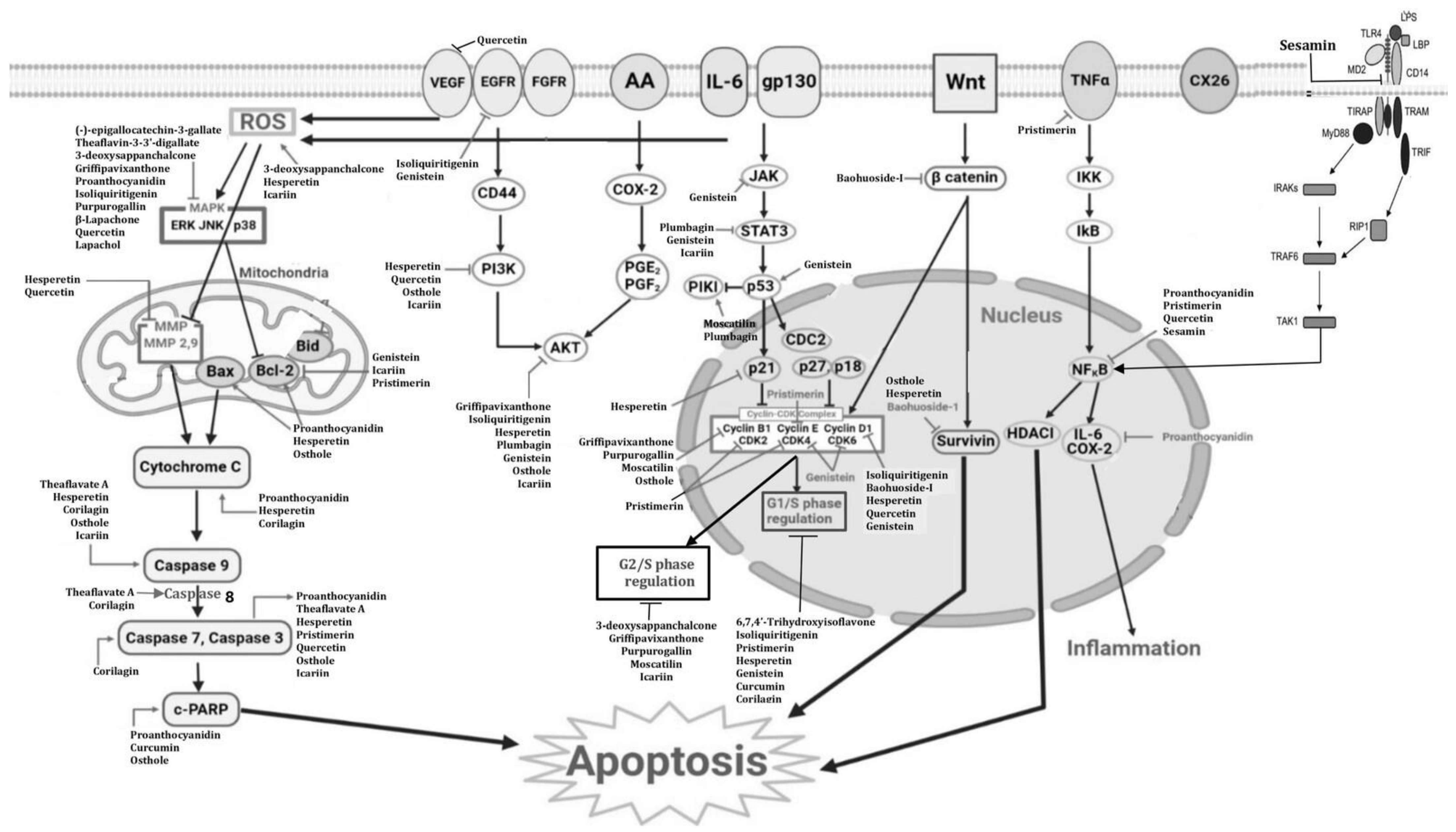
3.2. Description Phenolic Compounds with Anticancer Activities against Esophageal Cancer
3.2.1. Moscatilin
3.2.2. Isoliquiritigenin
3.2.3. 3-Deoxysappanchalcone (3-DSC)
3.2.4. Osthole
3.2.5. Quercetin
3.2.6. Icariin
3.2.7. Purpurogallin
3.2.8. (6,7,4′-THIF) or 6,7,4′-Trihydroxyisoflavone
3.2.9. Genistein
3.2.10. Hesperetin
3.2.11. Baohuoside-I
3.2.12. Curcumin
3.2.13. 2,6-Bis-Benzylidenocyclohexanone (BBCH)
3.2.14. Proanthocyanidin
3.2.15. Gallic Acid
3.2.16. Sesamin
3.2.17. (-)-Epigallocatechin-3-Gallate
3.2.18. Theaflavin-3-3′-Digallate
3.2.19. Theaflavate A
3.2.20. Lapachol
3.2.21. β-Lapachone
3.2.22. Pristimerin
3.2.23. Plumbagin
3.2.24. Corilagin
3.2.25. Griffipavixanthone
4. Discussion
5. Limitations and Futures Perspectives
6. Conclusions
Supplementary Materials
Author Contributions
Funding
Institutional Review Board Statement
Informed Consent Statement
Data Availability Statement
Conflicts of Interest
Correction Statement
References
- Ndebia, E.J.; Kamsu, G.T. Natural Alkaloids as Potential Treatments for Esophageal Squamous-Cell Cancer: A Comprehensive Review. Gastroenterol. Endosc. 2024, 2, 131–136. [Google Scholar] [CrossRef]
- Ntemafack, A.; Ayoub, M.; Hassan, Q.P.; Gandhi, S.G. A systematic review of pharmacological potential of phytochemicals from Rumex abyssinicus Jacq. S. Afr. J. Bot. 2023, 154, 11–25. [Google Scholar] [CrossRef]
- Ayad, R.; Akkal, S. Chapter 12—Phytochemistry and biological activities of algerian Centaurea and related genera. Stud. Nat. Prod. Chem. 2019, 63, 357–414. [Google Scholar]
- Randhir, R.; Lin, Y.T.; Shetty, K. Stimulation of phenolics, antioxidant and antimicrobial activities in dark germinated mung bean sprouts in response to peptide and phytochemical elicitors. Process Biochem. 2004, 39, 637–646. [Google Scholar] [CrossRef]
- Velderrain-Rodríguez, G.R.; Palafox-Carlos, H.; Wall-Medrano, A.; AyalaZavala, J.F.; Chen, C.-Y.O.; Robles-Sanchez, M.; Astiazaran-García, H.; Alvarez-Parrilla, E.; González-Aguilar, G.A. Phenolic compounds: Their journey after intake. Food Funct. 2014, 5, 189–197. [Google Scholar] [CrossRef]
- Xu, C.C.; Wang, B.; Pu, Y.Q.; Tao, J.S.; Zhang, T. Advances in extraction and analysis of phenolic compounds from plant materials. Chin. J. Nat. Med. 2017, 15, 721–731. [Google Scholar] [CrossRef]
- Alu’datt, M.H.; Rababah, T.; Alhamad, M.N.; Al-Mahasneh, M.A.; Almajwal, A.; Gammoh, S. A review of phenolic compounds in oil-bearing plants: Distribution, identification and occurrence of phenolic compounds. Food Chem. 2017, 218, 99–106. [Google Scholar] [CrossRef]
- Husain, N.; Gupta, S.A. critical study on chemistry and distribution of phenolic compounds in plants, and their role in human health. IOSR J. Environ. Sci. Toxicol. Food Technol. 2015, 1, 57–60. [Google Scholar]
- Šamec, D.; Karalija, E.; Šola, I.; Vujčić Bok, V.; Salopek-Sondi, B. The Role of Polyphenols in Abiotic Stress Response: The Influence of Molecular Structure. Plants 2021, 10, 118. [Google Scholar] [CrossRef]
- Leonhardt, S.D.; Chui, S.X.; Kuba, K. The role of non-volatile chemicals of floral rewards in plant-pollinator interactions. Basic Appl. Ecol. 2024, 75, 31–43. [Google Scholar] [CrossRef]
- Rahman, M.M.; Rahaman, M.S.; Islam, M.R.; Rahman, F.; Mithi, F.M.; Alqahtani, T. Role of Phenolic Compounds in Human Disease: Current Knowledge and Future Prospects. Molecules 2021, 27, 233. [Google Scholar] [CrossRef] [PubMed]
- Lutz, M.; Fuentes, E.; Ávila, F.; Alarcón, M.; Palomo, I. Roles of Phenolic Compounds in the Reduction of Risk Factors of Cardiovascular Diseases. Molecules 2019, 24, 366. [Google Scholar] [CrossRef] [PubMed]
- Caruso, G.; Godos, J.; Privitera, A.; Lanza, G.; Castellano, S.; Chillemi, A.; Bruni, O.; Ferri, R.; Caraci, F.; Grosso, G. Phenolic Acids and Prevention of Cognitive Decline: Polyphenols with a Neuroprotective Role in Cognitive Disorders and Alzheimer’s Disease. Nutrients 2022, 14, 819. [Google Scholar] [CrossRef] [PubMed]
- Bray, F.; Ferlay, J.; Soerjomataram, I.; Siegel, R.L.; Torre, L.A.; Jemal, A. Global cancer statistics 2018: GLOBOCAN estimates of incidence and mortality worldwide for 36 cancers in 185 countries. CA A Cancer J. Clin. 2018, 68, 394–424. [Google Scholar] [CrossRef]
- Sung, H.; Ferlay, J.; Siegel, R.L.; Laversanne, M.; Soerjomataram, I.; Jemal, A.; Bray, F. Global Cancer Statistics 2020: GLOBOCAN estimates of incidence and mortality worldwide for 36 cancers in 185 countries. CA Cancer J. Clin. 2021, 71, 209–249. [Google Scholar] [CrossRef]
- Li, J.; Xu, J.; Zheng, Y.; Gao, Y.; He, S.; Li, H.; Zou, K.; Li, N.; Tian, J.; Chen, W.; et al. Esophageal cancer: Epidemiology, risk factors and screening. Chin. J. Cancer Res. 2021, 33, 535–547. [Google Scholar] [CrossRef]
- Page, M.J.; McKenzie, J.E.; Bossuyt, P.M.; Boutron, I.; Hoffmann, T.C.; Mulrow, C.D.; Shamseer, L.; Tetzlaff, J.M.; Akl, E.A.; Brennan, S.E.; et al. The PRISMA 2020 statement: An updated guideline for reporting systematic reviews. BMJ 2021, 372, n71. [Google Scholar] [CrossRef]
- Ndebia, E.J.; Kamsu, G.T. A Comprehensive Meta-Analysis of Dietary and Culinary Practices on Esophageal Cancer Incidence in the East African Corridor. SVU-Int. J. Med. Sci. 2024, 7, 207–222. [Google Scholar] [CrossRef]
- Chen, C.A.; Chen, C.C.; Shen, C.C.; Chang, H.H.; Chen, Y.J. Moscatilin induces apoptosis and mitotic catastrophe in human esophageal cancer cells. J. Med. Food 2013, 16, 869–877. [Google Scholar] [CrossRef]
- Kresty, L.A.; Weh, K.M.; Zeyzus-Johns, B.; Perez, L.N.; Howell, A.B. Cranberry proanthocyanidins inhibit esophageal adenocarcinoma in vitro and in vivo through pleiotropic cell death induction and PI3K/AKT/mTOR inactivation. Oncotarget 2015, 6, 33438–33455. [Google Scholar] [CrossRef]
- Chen, W.K.; Chen, C.A.; Chi, C.W.; Li, L.H.; Lin, C.P.; Shieh, H.R.; Hsu, M.L.; Ko, C.C.; Hwang, J.J.; Chen, Y.J. Moscatilin Inhibits Growth of Human Esophageal Cancer Xenograft and Sensitizes Cancer Cells to Radiotherapy. J. Clin Med. 2019, 8, 187. [Google Scholar] [CrossRef] [PubMed]
- Ye, L.; Zhang, J.; Zhang, Y.; Gu, B.; Zhu, H.; Mao, X. Isoliquiritigenin Suppressed Esophageal Squamous Carcinoma Growth by Blocking EGFR Activation and Inducing Cell Cycle Arrest. Biomed. Res. Int. 2020, 2020, 9259852. [Google Scholar] [CrossRef] [PubMed]
- Kwak, A.W.; Lee, M.J.; Lee, M.H.; Yoon, G.; Cho, S.S.; Chae, J.I.; Shim, J.H. The 3-deoxysappanchalcone induces ROS-mediated apoptosis and cell cycle arrest via JNK/p38 MAPKs signaling pathway in human esophageal cancer cells. Phytomedicine 2021, 86, 153564. [Google Scholar] [CrossRef] [PubMed]
- Zhu, X.; Li, Z.; Li, T.; Long, F.; Lv, Y.; Liu, L.; Liu, X.; Zhan, Q. Osthole inhibits the PI3K/AKT signaling pathway via activation of PTEN and induces cell cycle arrest and apoptosis in esophageal squamous cell carcinoma. Biomed. Pharmacother. 2018, 102, 502–509. [Google Scholar] [CrossRef]
- Liu, Y.; Li, C.L.; Xu, Q.Q.; Cheng, D.; Liu, K.D.; Sun, Z.Q. Quercetin inhibits invasion and angiogenesis of esophageal cancer cells. Pathol. Res. Pract. 2021, 222, 153455. [Google Scholar] [CrossRef]
- Gu, Z.F.; Zhang, Z.T.; Wang, J.Y.; Xu, B.B. Icariin exerts inhibitory effects on the growth and metastasis of KYSE70 human esophageal carcinoma cells via PI3K/AKT and STAT3 pathways. Environ. Toxicol. Pharmacol. 2017, 54, 7–13. [Google Scholar] [CrossRef]
- Fan, C.; Yang, Y.; Liu, Y.; Jiang, S.; Di, S.; Hu, W.; Ma, Z.; Li, T.; Zhu, Y.; Xin, Z.; et al. Icariin displays anticancer activity against human esophageal cancer cells via regulating endoplasmic reticulum stress-mediated apoptotic signaling. Sci. Rep. 2016, 6, 21145. [Google Scholar] [CrossRef]
- Xie, X.; Zu, X.; Liu, F.; Wang, T.; Wang, X.; Chen, H.; Liu, K.; Wang, P.; Liu, F.; Zheng, Y.; et al. Purpurogallin is a novel mitogen-activated protein kinase kinase 1/2 inhibitor that suppresses esophageal squamous cell carcinoma growth in vitro and in vivo. Mol. Carcinog. 2019, 58, 1248–1259. [Google Scholar] [CrossRef]
- Sang, S.; Lambert, J.D.; Tian, S.; Hong, J.; Hou, Z.; Ryu, J.H.; Stark, R.E.; Rosen, R.T.; Huang, M.T.; Yang, C.S.; et al. Enzymatic synthesis of tea theaflavin derivatives and their anti-inflammatory and cytotoxic activities. Bioorg. Med. Chem. 2004, 12, 459–467. [Google Scholar] [CrossRef]
- Lim, T.-G.; Lee, S.-Y.; Duan, Z.; Lee, M.-H.; Chen, H.; Liu, F.; Liu, K.; Jung, S.K.; Kim, D.J.; Bode, A.M.; et al. The Prolyl Isomerase Pin1 Is a Novel Target of 6, 7, 40-Trihydroxyisoflavone for Suppressing Esophageal Cancer Growth. Cancer Prev. Res. 2017, 10, 308–318. [Google Scholar] [CrossRef]
- Gao, J.; Xia, R.; Chen, J.; Gao, J.; Luo, X.; Ke, C.; Ren, C.; Li, J.; Mi, Y. Inhibition of esophageal-carcinoma cell proliferation by genistein via suppression of JAK1/2-STAT3 and AKT/MDM2/p53 signaling pathways. Aging 2020, 12, 6240–6259. [Google Scholar] [CrossRef] [PubMed]
- Wu, D.; Zhang, J.; Wang, J.; Li, J.; Liao, F.; Dong, W. Hesperetin induces apoptosis of esophageal cancer cells via mitochondrial pathway mediated by the increased intracellular reactive oxygen species. Tumour Biol. 2016, 37, 3451–3459. [Google Scholar] [CrossRef] [PubMed]
- Wu, D.; Li, J.; Hu, X.; Ma, J.; Dong, W. Hesperetin inhibits Eca-109 cell proliferation and invasion by suppressing the PI3K/AKT signaling pathway and synergistically enhances the anti-tumor effect of 5-fluorouracil on esophageal cancer in vitro and in vivo. RSC Adv. 2018, 8, 24434–24443. [Google Scholar] [CrossRef] [PubMed]
- Wang, L.; Lu, A.; Liu, X.; Sang, M.; Shan, B.; Meng, F.; Cao, Q.; Ji, X. The flavonoid Baohuoside-I inhibits cell growth and downregulates survivin and cyclin D1 expression in esophageal carcinoma via β-catenin-dependent signaling. Oncol. Rep. 2011, 26, 1149–1156. [Google Scholar]
- Mizumoto, A.; Ohashi, S.; Kamada, M.; Saito, T.; Nakai, Y.; Baba, K.; Hirohashi, K.; Mitani, Y.; Kikuchi, O.; Matsubara, J.; et al. Combination treatment with highly bioavailable curcumin and NQO1 inhibitor exhibits potent antitumor effects on esophageal squamous cell carcinoma. J. Gastroenterol. 2019, 54, 687–698. [Google Scholar] [CrossRef]
- Alibeiki, F.; Jafari, N.; Karimi, M.; Peeri-Dogaheh, H. Potent anti-cancer effects of less polar Curcumin analogues on gastric adenocarcinoma and esophageal squamous cell carcinoma cells. Sci. Rep. 2017, 7, 2559. [Google Scholar] [CrossRef]
- Almanaa, T.N.; Geusz, M.E.; Jamasbi, R.J. Effects of curcumin on stem-like cells in human esophageal squamous carcinoma cell lines. BMC Complement. Altern. Med. 2012, 12, 195. [Google Scholar]
- Guo, F.; Hu, Y.; Niu, Q.; Li, Y.; Ding, Y.; Ma, R.; Wang, X.; Li, S.; Xie, J. Grape Seed Proanthocyanidin Extract Inhibits Human Esophageal Squamous Cancerous Cell Line ECA109 via the NF-κB Signaling Pathway. Mediators Inflamm. 2018, 2018, 3403972. [Google Scholar] [CrossRef]
- Faried, A.; Kurnia, D.; Faried, L.S.; Usman, N.; Miyazaki, T.; Kato, H.; Kuwano, H. Anticancer effects of gallic acid isolated from Indonesian herbal medicine, Phaleria macrocarpa (Scheff.) Boerl, on human cancer cell lines. Int. J. Oncol. 2007, 30, 605–613. [Google Scholar] [CrossRef]
- Wen, L.; Mao, W.; Xu, L.; Cai, B.; Gu, L. Sesamin exerts anti-tumor activity in esophageal squamous cell carcinoma via inhibition of TRIM44 and NF-κB signaling. Chem. Biol. Drug Des. 2022, 99, 118–125. [Google Scholar] [CrossRef]
- Gao, Y.; Li, W.; Jia, L.; Li, B.; Chen, Y.C.; Tu, Y. Enhancement of (-)-epigallocatechin-3-gallate and theaflavin-3-3′-digallate induced apoptosis by ascorbic acid in human lung adenocarcinoma SPC-A-1 cells and esophageal carcinoma Eca-109 cells via MAPK pathways. Biochem. Biophys. Res. Commun. 2013, 438, 370–374. [Google Scholar] [CrossRef] [PubMed]
- Sunassee, S.N.; Veale, C.G.; Shunmoogam, G.N.; Osoniyi, O.; Hendricks, D.T.; Caira, M.R.; de la Mare, J.A.; Edkins, A.L.; Pinto, A.V.; da Silva Júnior, E.N.; et al. Cytotoxicity of lapachol, β-lapachone and related synthetic 1, 4-naphthoquinones against oesophageal cancer cells. Eur. J. Med. Chem. 2013, 62, 98–110. [Google Scholar] [CrossRef] [PubMed]
- Zu, X.; Xie, X.; Zhang, Y.; Liu, K.; Bode, A.M.; Dong, Z.; Kim, D.J. Lapachol is a novel ribosomal protein S6 kinase 2 inhibitor that suppresses growth and induces intrinsic apoptosis in esophageal squamous cell carcinoma cells. Phytother. Res. 2019, 33, 2337–2346. [Google Scholar] [CrossRef] [PubMed]
- Huang, P.; Sun, L.Y.; Zhang, Y.Q. A Hopeful Natural Product, Pristimerin, Induces Apoptosis, Cell Cycle Arrest, and Autophagy in Esophageal Cancer Cells. Anal. Cell Pathol. 2019, 2019, 6127169. [Google Scholar] [CrossRef]
- Tu, Y.; Tan, F.; Zhou, J.; Pan, J. Pristimerin targeting NF-κB pathway inhibits proliferation, migration, and invasion in esophageal squamous cell carcinoma cells. Cell Biochem. Funct. 2018, 36, 228–240. [Google Scholar] [CrossRef]
- Cao, Y.Y.; Yu, J.; Liu, T.T.; Yang, K.X.; Yang, L.Y.; Chen, Q.; Shi, F.; Hao, J.J.; Cai, Y.; Wang, M.R.; et al. Plumbagin inhibits the proliferation and survival of esophageal cancer cells by blocking STAT3-PLK1-AKT signaling. Cell Death Dis. 2018, 9, 17. [Google Scholar] [CrossRef]
- Wu, C.; Huang, H.; Choi, H.-Y.; Ma, Y.; Zhou, T.; Peng, Y.; Pang, K.; Shu, G.; Yang, X. Anti-esophageal Cancer Effect of Corilagin Extracted from Phmllanthi Fructus via the Mitochondrial and Endoplasmic Reticulum Stress Pathways. J. Ethnopharmacol. 2021, 269, 113700. [Google Scholar] [CrossRef]
- Ding, Z.; Lao, Y.; Zhang, H.; Fu, W.; Zhu, L.; Tan, H.; Xu, H. Griffipavixanthone, a dimeric xanthone extracted from edible plants, inhibits tumor metastasis and proliferation via downregulation of the RAF pathway in esophageal cancer. Oncotarget 2016, 7, 1826–1837. [Google Scholar] [CrossRef]
- Cardile, V.; Avola, R.; Graziano, A.C.E.; Russo, A. Moscatilin, a bibenzyl derivative from the orchid Dendrobium loddigesii, induces apoptosis in melanoma cells. Chem. Biol. Interact. 2020, 323, 109075. [Google Scholar] [CrossRef]
- Aljeldah, M.M. Evaluation of the anticancer and antibacterial activities of moscatilin. Heliyon 2024, 10, e31131. [Google Scholar] [CrossRef]
- Zhang, Z.; Yung, K.K.; Ko, J.K. Therapeutic Intervention in Cancer by Isoliquiritigenin from Licorice: A Natural Antioxidant and Redox Regulator. Antioxidants 2022, 11, 1349. [Google Scholar] [CrossRef] [PubMed]
- Wang, N.; Wang, Z.; Peng, C.; You, J.; Shen, J.; Han, S.; Chen, J. Dietary compound isoliquiritigenin targets GRP78 to chemosensitize breast cancer stem cells via beta-catenin/ABCG2 signaling. Carcinogenesis 2014, 35, 2544–2554. [Google Scholar] [CrossRef] [PubMed]
- Wang, Z.; Wang, N.; Liu, P.; Chen, Q.; Situ, H.; Xie, T.; Zhang, J.; Peng, C.; Lin, Y.; Chen, J. MicroRNA-25 regulates chemoresistance associated autophagy in breast cancer cells, a process modulated by the natural autophagy inducer isoliquiritigenin. Oncotarget 2014, 5, 7013–7026. [Google Scholar] [CrossRef] [PubMed]
- Qiao, F.; Zhao, Y.; Mai, Y.; Guo, J.; Dong, L.; Zhang, W.; Yang, J. Isoliquiritigenin Nanosuspension Enhances Cytostatic Effects in A549 Lung Cancer Cells. Planta Med. 2020, 86, 538–547. [Google Scholar] [CrossRef] [PubMed]
- Na, A.-Y.; Yang, E.-J.; Jeon, J.M.; Ki, S.H.; Song, K.-S.; Lee, S. Protective Effect of Isoliquiritigenin against Ethanol-Induced Hepatic Steatosis by Regulating the SIRT1-AMPK Pathway. Toxicol. Res. 2018, 34, 23–29. [Google Scholar] [CrossRef]
- Wu, C.-H.; Chen, H.-Y.; Wang, C.-W.; Shieh, T.-M.; Huang, T.-C.; Lin, L.-C.; Wang, K.L.; Hsia, S.M. Isoliquiritigenin induces apoptosis and autophagy and inhibits endometrial cancer growth in mice. Oncotarget 2016, 7, 73432–73447. [Google Scholar] [CrossRef]
- Huang, F.; Wang, J.; Xu, Y.; Zhang, Y.; Xu, N.; Yin, L. Discovery of novel isoliquiritigenin analogue ISL-17 as a potential anti-gastric cancer agent. Biosci. Rep. 2020, 40, 20201199. [Google Scholar]
- Kwon, H.M.; Choi, Y.J.; Choi, J.S.; Kang, S.W.; Bae, J.Y.; Kang, I.J.; Jun, J.G.; Lee, S.S.; Lim, S.S.; Kang, Y.H. Blockade of cytokine induced endothelial cell adhesion molecule expression by licorice isoliquiritigenin through NF-kappaB signal disruption. Exp. Biol. Med. 2007, 232, 235–245. [Google Scholar]
- Hu, F.-W.; Yu, C.-C.; Hsieh, P.-L.; Liao, Y.-W.; Lu, M.-Y.; Chu, P.-M. Targeting oral cancer stemness and chemoresistance by isoliquiritigenin-mediated GRP78 regulation. Oncotarget 2017, 8, 93912–93923. [Google Scholar] [CrossRef]
- Zhang, X.; Yeung, E.D.; Wang, J.; Panzhinskiy, E.E.; Tong, C.; Li, W.; Li, J. Isoliquiritigenin, a natural anti-oxidant, selectively inhibits the proliferation of prostate cancer cells. Clin. Exp. Pharmacol. Physiol. 2010, 37, 841–847. [Google Scholar]
- Selvaraj, B.; Kim, D.W.; Huh, G.; Lee, H.; Kang, K.; Lee, J.W. Synthesis and biological evaluation of isoliquiritigenin derivatives as a neuroprotective agent against glutamate mediated neurotoxicity in HT22 cells. Bioorg. Med. Chem. Lett. 2020, 30, 127058. [Google Scholar] [CrossRef] [PubMed]
- Vij, T.; Anil, P.P.; Shams, R.; Dash, K.K.; Kalsi, R.; Pandey, V.K.; Harsányi, E.; Kovács, B.; Shaikh, A.M. A Comprehensive Review on Bioactive Compounds Found in Caesalpinia sappan. Molecules 2023, 28, 6247. [Google Scholar] [CrossRef] [PubMed]
- Kim, Y.E.; Choi, H.C.; Lee, I.C.; Yuk, D.Y.; Lee, H.; Choi, B.Y. 3-Deoxysappanchalcone Promotes Proliferation of Human Hair Follicle Dermal Papilla Cells and Hair Growth in C57BL/6 Mice by Modulating WNT/β-Catenin and STAT Signaling. Biomol. Ther. 2016, 24, 572–580. [Google Scholar] [CrossRef] [PubMed]
- Fu, X.; Zhao, R.; Yoon, G.; Shim, J.-H.; Choi, B.Y.; Yin, F.; Xu, B.; Laster, K.V.; Liu, K.; Dong, Z.; et al. 3-Deoxysappanchalcone Inhibits Skin Cancer Proliferation by Regulating T-Lymphokine-Activated Killer Cell-Originated Protein Kinase in vitro and in vivo. Front. Cell Dev. Biol. 2021, 9, 638174. [Google Scholar] [CrossRef]
- Sun, M.; Sun, M.; Zhang, J. Osthole: An overview of its sources, biological activities, and modification development. Med. Chem. Res. 2021, 30, 1767–1794. [Google Scholar] [CrossRef]
- Zhang, Z.R.; Leung, W.N.; Cheung, H.Y.; Chan, C.W. Osthole: A review on its bioactivities, pharmacological properties, and potential as alternative medicine. Evid. Based Complement. Altern. Med. 2015, 2015, 919616. [Google Scholar] [CrossRef]
- Singh, G.; Bhatti, R.; Mannan, R.; Singh, D.; Kesavan, A.; Singh, P. Osthole ameliorates neurogenic and inflammatory hyperalgesia by modulation of iNOS, COX-2, and inflammatory cytokines in mice. Inflammopharmacology 2019, 27, 949–960. [Google Scholar] [CrossRef]
- Shokoohinia, Y.; Bazargan, S.; Miraghaee, S.; Javadirad, E.; Farahani, F.; Hosseinzadeh, L. Safety Assessment of Osthole Isolated from Prangos ferulacea: Acute and Subchronic Toxicities and Modulation of Cytochrome P450. Jundishapur J. Nat. Pharm. Prod. 2017, 12, e63764. [Google Scholar] [CrossRef]
- Shen, Z.; Chen, J.; Lu, H. Osthole induced apoptosis in human normal liver cells by regulating cell proliferation and endoplasmic reticulum stress. Environ. Toxicol. 2019, 34, 768–776. [Google Scholar] [CrossRef]
- Callahan, B.N.; Kammala, A.K.; Syed, M.; Yang, C.; Occhiuto, C.J.; Nellutla, R.; Chumanevich, A.P.; Oskeritzian, C.A.; Das, R.; Subramanian, H. Osthole, a natural plant derivative inhibits mrgprx2 induced mast cell responses. Front. Immunol. 2020, 11, 703. [Google Scholar] [CrossRef]
- Kan, W.C.; Hwang, J.Y.; Chuang, L.Y.; Guh, J.Y.; Ye, Y.L.; Yang, Y.L.; Huang, J.S. Effect of osthole on advanced glycation end products-induced renal tubular hypertrophy and role of klotho in its mechanism of action. Phytomedicine 2019, 53, 205–212. [Google Scholar] [CrossRef] [PubMed]
- Anand-David, A.V.; Arulmoli, R.; Parasuraman, S. Overviews of Biological Importance of Quercetin: A Bioactive Flavonoid. Pharmacogn. Rev. 2016, 10, 84–89. [Google Scholar] [PubMed]
- Augoff, K.; Hryniewicz, J.A.; Tabola, R.; Stach, K. MMP9: A Tough Target for Targeted Therapy for Cancer. Cancers 2022, 14, 1847. [Google Scholar] [CrossRef] [PubMed]
- Cunningham, P.; Patton, E.; VanderVeen, B.N.; Unger, C.; Aladhami, A.; Enos, R.T.; Madero, S.; Chatzistamou, I.; Fan, D.; Murphy, E.A.; et al. Sub-chronic oral toxicity screening of quercetin in mice. BMC Complement. Med. Ther. 2022, 22, 279. [Google Scholar]
- Chen, R.; Lin, J.; Hong, J.; Han, D.; Zhang, A.D.; Lan, R.; Fu, L.; Wu, Z.; Lin, J.; Zhang, W.; et al. Potential toxicity of quercetin: The repression of mitochondrial copy number via decreased POLG expression and excessive TFAM expression in irradiated murine bone marrow. Toxicol. Rep. 2014, 1, 450–458. [Google Scholar] [CrossRef]
- Dibal, N.I.; Hyedima, G.S.; Watson, J.T. Acute Toxicity of Quercetin from Onion Skin in Mice. Pharm. Biomed. Res. 2020, 6, 269–276. [Google Scholar] [CrossRef]
- da Silva, S.V.S.; Barboza, O.M.; Souza, J.T.; Soares, É.N.; dos Santos, C.C.; Pacheco, L.V.; Santos, I.P.; Magalhães, T.B.d.S.; Soares, M.B.P.; Guimarães, E.T.; et al. Structural design, synthesis and antioxidant, antileishmania, anti-inflammatory and anticancer activities of a novel quercetin acetylated derivative. Molecules 2021, 26, 6923. [Google Scholar] [CrossRef]
- Shen, R.; Deng, W.; Li, C.; Zeng, G. A natural flavonoid glucoside icariin inhibits Th1 and Th17 cell differentiation and ameliorates experimental autoimmune encephalomyelitis. Int. Immunopharmacol. 2015, 24, 224–231. [Google Scholar] [CrossRef]
- Zhou, Y.D.; Hou, J.G.; Yang, G.; Jiang, S.; Chen, C.; Wang, Z.; Liu, Y.Y.; Ren, S.; Li, W. Icariin ameliorates cisplatin-induced cytotoxicity in human embryonic kidney 293 cells by suppressing ROS-mediated PI3K/Akt pathway. Biomed. Pharmacother. 2019, 109, 2309–2317. [Google Scholar] [CrossRef]
- Zhu, F.; Ren, Z. Icariin inhibits the malignant progression of lung cancer by affecting the PI3K/Akt pathway through the miR-205-5p/PTEN axis. Oncol. Rep. 2022, 47, 115. [Google Scholar] [CrossRef]
- Song, L.; Chen, X.; Mi, L.; Liu, C.; Zhu, S.; Yang, T.; Luo, X.; Zhang, Q.; Lu, H.; Liang, X. Icariin-induced inhibition of SIRT6/NF-κB triggers redox mediated apoptosis and enhances anti-tumor immunity in triple-negative breast cancer. Cancer Sci. 2020, 111, 4242–4256. [Google Scholar] [CrossRef] [PubMed]
- Liao, Y.C.; Kim, T.; Silva, J.L.; Chen, B.Y. A New Approach for Quantifying Purpurogallin in Brewed Beverages Using LC-MS in Combination with Solid Phase Extraction. Foods 2022, 11, 1429. [Google Scholar] [CrossRef] [PubMed]
- Wu, T.W.; Zeng, L.H.; Wu, J.; Carey, D. Purpurogallin--a natural and effective hepatoprotector in vitro and in vivo. Biochem. Cell Biol. 1991, 69, 747–750. [Google Scholar] [CrossRef] [PubMed]
- Prasad, K.; Mantha, S.V.; Kalra, J.; Kapoor, R.; Kamalarajan, B.R.C. Purpurogallin in the prevention of hypercholesterolemic atherosclerosis. Int. J. Angiol. 1997, 6, 157–166. [Google Scholar] [CrossRef]
- Zhen, A.X.; Piao, M.J.; Hyun, Y.J.; Kang, K.A.; Ryu, Y.S.; Cho, S.J.; Kang, H.K.; Koh, Y.S.; Ahn, M.J.; Kim, T.H.; et al. Purpurogallin protects keratinocytes from damage and apoptosis induced by Ultraviolet B radiation and particulate matter 2.5. Biomol. Ther. 2019, 27, 395–403. [Google Scholar] [CrossRef]
- NCBI (National Center for Biotechnology Information). PubChem Compound Summary for CID 5284649, 6, 7, 4′-Trihydroxyisoflavone. Available online: https://pubchem.ncbi.nlm.nih.gov/compound/6_7_4_-Trihydroxyisoflavone (accessed on 27 June 2024).
- Lee, H.-S.; Jeong, G.-S. Protective Effects of 6, 7, 4′ -Trihydroxyflavanone on Hypoxia-Induced Neurotoxicity by Enhancement of HO-1 through Nrf2 Signaling Pathway. Antioxidants 2021, 10, 341. [Google Scholar] [CrossRef]
- Sohel, M.; Biswas, P.; Al Amin, M.; Hossain, M.A.; Sultana, H.; Dey, D.; Aktar, S.; Setu, A.; Khan, M.S.; Paul, P.; et al. Genistein, a potential phytochemical against breast cancer treatment-insight into the molecular mechanisms. Processes 2022, 10, 415. [Google Scholar] [CrossRef]
- Yu, L.; Rios, E.; Castro, L.; Liu, J.; Yan, Y.; Dixon, D. Genistein: Dual Role in Women’s Health. Nutrients 2021, 13, 3048. [Google Scholar] [CrossRef]
- Singh, P.; Sharma, S.; Rath, S.K. Genistein induces deleterious effects during its acute exposure in Swiss mice. Biomed. Res. Int. 2014, 2014, 619617. [Google Scholar] [CrossRef]
- Okazaki, K.; Okazaki, S.; Nakamura, H.; Kitamura, Y.; Hatayama, K.; Wakabayashi, S.; Tsuda, T.; Katsumata, T.; Nishikawa, A.; Hirose, M. A repeated 28-day oral dose toxicity study of genistein in rats, based on the ‘Enhanced OECD Test Guideline 407′ for screening endocrine-disrupting chemicals. Arch. Toxicol. 2002, 76, 553–559. [Google Scholar] [CrossRef]
- McClain, R.M.; Wolz, E.; Davidovich, A.; Bausch, J. Genetic toxicity studies with genistein. Food Chem. Toxicol. 2006, 44, 42–55. [Google Scholar] [CrossRef] [PubMed]
- McClain, R.M.; Wolz, E.; Davidovich, A.; Pfannkuch, F.; Edwards, J.A.; Bausch, J. Acute, subchronic and chronic safety studies with genistein in rats. Food Chem. Toxicol. 2006, 44, 56–80. [Google Scholar] [CrossRef] [PubMed]
- NTP (National Toxicology Program). Toxicology and carcinogenesis studies of genistein (Cas No. 446-72-0) in Sprague-Dawley rats (feed study). Natl. Toxicol. Program Tech. Rep. Ser. 2008, 545, 1–240. [Google Scholar]
- Yang, Z.; Kulkarni, K.; Zhu, W.; Hu, M. Bioavailability and pharmacokinetics of genistein: Mechanistic studies on its ADME. Anticancer Agents Med. Chem. 2012, 12, 1264–1280. [Google Scholar] [CrossRef]
- Alipour, M.; Sharifi, S.; Samiei, M.; Shahi, S.; Aghazadeh, M.; Dizaj, S.M. Synthesis, characterization, and evaluation of Hesperetin nanocrystals for regenerative dentistry. Sci. Rep. 2023, 13, 2076. [Google Scholar] [CrossRef]
- Yang, H.; Wang, Y.; Xu, S.; Ren, J.; Tang, L.; Gong, J.; Lin, Y.; Fang, H.; Su, D. Hesperetin, a promising treatment option for diabetes and related complications: A literature review. J. Agric. Food Chem. 2022, 70, 8582–8592. [Google Scholar] [CrossRef]
- He, P.; Ma, J.; Liu, Y.; Deng, H.; Dong, W. Hesperetin Promotes Cisplatin−Induced Apoptosis of Gastric Cancer In Vitro and In Vivo by Upregulating PTEN Expression. Front. Pharmacol. 2020, 11, 1326. [Google Scholar] [CrossRef]
- Trivedi, P.P.; Kushwaha, S.; Tripathi, D.N.; Jena, G.B. Cardioprotective Effects of Hesperetin against Doxorubicin-Induced Oxidative Stress and DNA Damage in Rat. Cardiovasc. Toxicol. 2011, 11, 215–225. [Google Scholar] [CrossRef]
- Trivedi, P.P.; Tripathi, D.N.; Jena, G. Hesperetin protects testicular toxicity of doxorubicin in rat: Role of NFκB, p38 and caspase-3. Food Chem. Toxicol. 2011, 49, 838–847. [Google Scholar] [CrossRef]
- An, M.-F.; Shen, C.; Zhang, S.-S.; Wang, M.-Y.; Sun, Z.-R.; Fan, M.-S.; Zhang, L.J.; Zhao, Y.L.; Sheng, J.; Wang, X.J. Anti-hyperuricemia effect of hesperetin is mediated by inhibiting the activity of xanthine oxidase and promoting excretion of uric acid. Front. Pharmacol. 2023, 14, 1128699. [Google Scholar] [CrossRef]
- Geng, Y.; Wu, Z.; Buist-Homan, M.; Blokzijl, H.; Moshage, H. Hesperetin protects against palmitate-induced cellular toxicity via induction of GRP78 in hepatocytes. Toxicol. Appl. Pharmacol. 2020, 404, 115183. [Google Scholar] [CrossRef] [PubMed]
- Famurewa, A.C.; Renu, K.; Eladl, M.A.; Chakraborty, R.; Myakala, H.; El-Sherbiny, M.; Elsherbini, D.M.A.; Vellingiri, B.; Madhyastha, H.; Ramesh-Wanjari, U.; et al. Hesperidin and hesperetin against heavy metal toxicity: Insight on the molecular mechanism of mitigation. Biomed. Pharmacother. 2022, 149, 112914. [Google Scholar] [CrossRef] [PubMed]
- Ma, H.; He, X.; Yang, Y.; Li, M.; Hao, D.; Jia, Z. The genus Epimedium: An ethnopharmacological and phytochemical review. J. Ethnopharmacol. 2011, 134, 519–541. [Google Scholar] [CrossRef] [PubMed]
- Jin, X.; Zhang, Z.-h.; Sun, E.; Qian, Q.; Tan, X.-b.; Jia, X.-b. Preparation of a nanoscale baohuoside I-phospholipid complex and determination of its absorption: In vivo and in vitro evaluations. Int. J. Nanomed. 2012, 7, 4907–4916. [Google Scholar]
- Li, S.Y.; Ping, G.; Geng, L.; Seow, W.K.; Thong, Y.H. Immunopharmacology and toxicology of the plant flavonoid baohuoside-1 in mice. Int. J. Immunopharmacol. 1994, 16, 227–231. [Google Scholar] [CrossRef]
- Ni, F.; Tang, H.; Wang, C.; Zhang, H.; Zheng, C.; Zhang, N.; Chen, B.; Sun, L. Baohuoside I Inhibits the Proliferation of Pancreatic Cancer Cells via mTOR/S6K1-Caspases/Bcl2/Bax Apoptotic Signaling. Cancer Manag. Res. 2019, 11, 10609–10621. [Google Scholar] [CrossRef]
- Lestari, M.L.A.D.; Indrayanto, G. Curcumin. In Profiles of Drug Substances, Excipients, and Related Methodology; Elsevier Inc.: Amsterdam, The Netherlands, 2014; Volume 39, pp. 113–204. [Google Scholar]
- Kamsu, G.T.; Fodouop, S.P.; Tagne, R.S.; Kodjio, N.; Fakam, A.L.; Gatsing, D. Evaluation of the acute and sub-chronic toxicity of the ethanolic extract of Curcuma longa (Zingiberaceae) in wistar albino rats. Mod. Chem. Appl. 2019, 7, 267. [Google Scholar]
- Tiwari, R.; Siddiqui, M.H.; Mahmood, T.; Farooqui, A.; Bagga, P.; Ahsan, F.; Shamim, A. An exploratory analysis on the toxicity & safety profile of Polyherbal combination of curcumin, quercetin and rutin. Clin. Phytosci. 2020, 6, 82. [Google Scholar]
- Vitaglione, P.; Lumaga, R.B.; Ferracane, R.; Radetsky, I.; Mennella, I.; Schettino, R.; Koder, S.; Shimoni, E.; Fogliano, V. Curcumin bioavailability from enriched bread: The effect of microencapsulated ingredients. J. Agric. Food Chem. 2012, 60, 3357–3366. [Google Scholar] [CrossRef]
- Mannino, G.; Chinigò, G.; Serio, G.; Genova, T.; Gentile, C.; Munaron, L.; Bertea, C.M. Proanthocyanidins and where to find them: A meta-analytic approach to investigate their chemistry, biosynthesis, distribution, and effect on human health. Antioxidants 2021, 10, 1229. [Google Scholar] [CrossRef]
- Rauf, A.; Imran, M.; Abu-Izneid, T.; Iahtisham, U.-H.; Patel, S.; Pan, X.; Naz, S.; Sanches-Silva, A.; Saeed, F.; Rasul-Suleria, H.A. Proanthocyanidins: A comprehensive review. Biomed. Pharmacother. 2019, 116, 108999. [Google Scholar] [CrossRef] [PubMed]
- Yamakoshi, J.; Saito, M.; Kataoka, S.; Kikuchi, M. Safety evaluation of proanthocyanidin-rich extract from grape seeds. Food Chem. Toxicol. 2002, 40, 599–607. [Google Scholar] [CrossRef] [PubMed]
- Sano, A. Safety assessment of 4-week oral intake of proanthocyanidin-rich grape seed extract in healthy subjects. Food Chem. Toxicol. 2017, 108, 519–523. [Google Scholar] [CrossRef] [PubMed]
- Yang, K.; Zhang, L.; Liao, P.; Xiao, Z.; Zhang, F.; Sindaye, D.; Xin, Z.; Tan, C.; Deng, J.; Yin, Y.; et al. Impact of Gallic Acid on Gut Health: Focus on the Gut Microbiome, Immune Response, and Mechanisms of Action. Front. Immunol. 2020, 11, 580208. [Google Scholar] [CrossRef]
- Niho, N.; Shibutani, M.; Tamura, T.; Toyoda, K.; Uneyama, C.; Takahashi, N.; Hirose, M. Subchronic toxicity study of gallic acid by oral administration in F344 rats. Food Chem. Toxicol. 2001, 39, 1063–1070. [Google Scholar] [CrossRef]
- Booth, A.; Amen, R.J.; Scott, M.; Greenway, F.L. Oral dose-ranging developmental toxicity study of an herbal supplement (NT) and gallic acid in rats. Adv. Ther. 2010, 27, 250–255. [Google Scholar] [CrossRef]
- Dalibalta, S.; Majdalawieh, A.F.; Manjikian, H. Health benefits of sesamin on cardiovascular disease and its associated risk factors. Saudi Pharm. J. 2020, 28, 1276–1289. [Google Scholar] [CrossRef]
- Hori, H.; Takayanagi, T.; Kamada, Y.; Shimoyoshi, S.; Ono, Y.; Kitagawa, Y.; Shibata, H.; Nagao, M.; Fujii, W.; Sakakibara, Y. Genotoxicity evaluation of sesamin and episesamin. Mutat. Res. 2011, 719, 21–28. [Google Scholar] [CrossRef]
- Liu, C.-M.; Zheng, G.-H.; Ming, Q.-L.; Chao, C.; Sun, J.-M. Sesamin protects mouse liver against nickel-induced oxidative dna damage and apoptosis by the PI3K-Akt Pathway. J. Agric. Food Chem. 2013, 61, 1146–1154. [Google Scholar] [CrossRef]
- Cheng, F.C.; Jinn, T.R.; Hou, R.C.; Tzen, J.T. Neuroprotective effects of sesamin and sesamolin on gerbil brain in cerebral ischemia. Int. J. Biomed. Sci. 2006, 2, 284–288. [Google Scholar]
- Guo, H.; Liu, Y.; Wang, L.; Zhang, G.; Su, S.; Zhang, R.; Zhang, J.; Li, A.; Shang, C.; Bi, B.; et al. Alleviation of doxorubicin-induced hepatorenal toxicities with sesamin via the suppression of oxidative stress. Hum. Exp. Toxicol. 2016, 35, 1183–1193. [Google Scholar] [CrossRef] [PubMed]
- Srisongkram, T.; Weerapreeyakul, N. Route of intracellular uptake and cytotoxicity of sesamol, sesamin, and sesamolin in human melanoma SK-MEL-2 cells. Biomed. Pharmacother. 2022, 146, 112528. [Google Scholar] [CrossRef] [PubMed]
- Salas, F.; Rojas, J.; Morales, A.; Ramos-Nino, M.E.; Colmenares, N.G. In vitro Cytotoxic Activity of Sesamin Isolated from Vismia baccifera var. dealbata Triana & Planch (Guttiferae) Collected from Venezuela. Nat. Prod. Commun. 2008, 3, 1705–1708. [Google Scholar]
- Nagle, D.G.; Ferreira, D.; Zhou, Y.D. Epigallocatechin-3-gallate (EGCG): Chemical and biomedical perspectives. Phytochemistry 2006, 67, 1849–1855. [Google Scholar] [CrossRef]
- Bettuzzi, S.; Brausi, M.; Rizzi, F.; Castagnetti, G.; Peracchia, G.; Corti, A. Chemoprevention of human prostate cancer by oral administration of green tea catechins in volunteers with high-grade prostate intraepithelial neoplasia: A preliminary report from a one-year proof-of-principle study. Cancer Res. 2006, 66, 1234–1240. [Google Scholar] [CrossRef]
- Urusova, D.V.; Shim, J.H.; Kim, D.J.; Jung, S.K.; Zykova, T.A.; Carper, A.; Bode, A.M.; Dong, Z. Epigallocatechin-gallate suppresses tumorigenesis by directly targeting Pin1. Cancer Prev. Res. 2011, 4, 1366–1377. [Google Scholar] [CrossRef]
- Liu, Z.; de Bruijn, W.J.C.; Bruins, M.E.; Vincken, J.-P. Microbial metabolism of Theaflavin-3, 3′-digallate and its gut microbiota composition modulatory effects. J. Agric. Food Chem. 2021, 69, 232–245. [Google Scholar] [CrossRef]
- Way, T.-D.; Lee, H.-H.; Kao, M.-C.; Lin, J.-K. Black tea polyphenol Theaflavins inhibit aromatase activity and attenuate tamoxifen resistance in HER2/Neu-Transfected human breast cancer cells through tyrosine kinase suppression. Eur. J. Cancer 2004, 40, 2165–2174. [Google Scholar] [CrossRef]
- Li, Y.; Shi, J.; Sun, X.; Li, Y.; Duan, Y.; Yao, H. Theaflavic acid from black tea protects PC12 cells against ROS-mediated mitochondrial apoptosis induced by OGD/R via activating Nrf2/ARE signaling pathway. J. Nat. Med. 2020, 74, 238–246. [Google Scholar] [CrossRef]
- Lahiry, L.; Saha, B.; Chakraborty, J.; Adhikary, A.; Mohanty, S.; Hossain, D.M.S.; Banerjee, S.; Das, K.; Sa, G.; Das, T. Theaflavins Target Fas/Caspase-8 and Akt/PBad Pathways to Induce Apoptosis in P53-Mutated Human Breast Cancer Cells. Carcinogenesis 2010, 31, 259–268. [Google Scholar] [CrossRef]
- Epifano, F.; Genovese, S.; Fiorito, S.; Mathieu, V.; Kiss, R. Lapachol and its congeners as anticancer agents: A review. Phytochem. Rev. 2014, 13, 37–49. [Google Scholar] [CrossRef]
- Morrison, R.K.; Brown, D.E.; Oleson, J.J.; Cooney, D.A. Oral toxicology studies with lapachol. Toxicol. Appl. Pharmacol. 1970, 17, 1–11. [Google Scholar] [CrossRef] [PubMed]
- Guerra, M.O.; Mazoni, A.S.; Brandão, M.A.; Peters, V.M. Toxicology of Lapachol in rats: Embryolethality. Braz. J. Biol. 2001, 61, 171–174. [Google Scholar] [CrossRef] [PubMed]
- Sá, R.-D.-C.-d.-S.E.; Guerra, M.O. Reproductive toxicity of lapachol in adult male Wistar rats submitted to short-term treatment. Phytother. Res. 2007, 21, 658–662. [Google Scholar]
- Gomes, C.L.; de Albuquerque, W.S.V.; Gomes-de-Melo, C.; Ferreira-da-Silva, R.M.; Vicente, N.R.H.; Rolim, L.A.; Rolim, N.P.J. Beta-lapachone: Natural occurrence, physicochemical properties, biological activities, toxicity and synthesis. Phytochemistry 2021, 186, 112713. [Google Scholar] [CrossRef]
- Kim, K.H.; Park, S.H.; Adhikary, P.; Cho, J.H.; Kang, N.G.; Jeong, S.H. Stability of β-Lapachone upon Exposure to Various Stress Conditions: Resultant Efficacy and Cytotoxicity. Chem. Pharm. Bull. 2016, 64, 381–389. [Google Scholar]
- Oliveira, M.E.F.A.G.; Silva, É.C.G.M.; Câmara, C.A.; de Souza, I.A.; Amorim, R.V.S. Evaluation of acute toxicity of β-lapachone associated with chitosan as a cytoprotective agent. J. Bras. Patol. Med. Lab. 2018, 54, 279–287. [Google Scholar]
- de Almeida, E.R.; Lucena, F.R.S.; Silva, C.V.N.S.; da Silva, C.-J.W.; Cavalcanti, J.B.; Couto, G.B.L.; da Silva, L.L.; da Mota, D.L.; da Silveira, A.B.; de Sousa-Filho, S.D.; et al. Toxicological assessment of beta-lapachone on organs from pregnant and non-pregnant rats. Phyther. Res. 2009, 23, 1276–1280. [Google Scholar]
- Li, J.J.; Yan, Y.Y.; Sun, H.M.; Liu, Y.; Su, C.Y.; Chen, H.B.; Zhang, J.Y. Anti-Cancer Effects of Pristimerin and the Mechanisms: A Critical Review. Front. Pharmacol. 2019, 12, 746. [Google Scholar] [CrossRef]
- Gomes, J.P.M.; Cardoso, C.R.P.; Varanda, E.A.; Molina, J.-M.; Fernandez, M.F.; Olea, N.; Arlos, I.Z.; Vilegas, W. Antitumoral, mutagenic and (anti)estrogenic activities of tingenone and pristimerin. Rev. Bras. Farmacogn. 2011, 21, 963–971. [Google Scholar]
- Aziz, M.H.; Dreckschmidt, N.E.; Verma, A.K. Plumbagin, a medicinal plant-derived naphthoquinone, is a novel inhibitor of the growth and invasion of hormone-refractory prostate cancer. Cancer Res. 2008, 68, 9024–9032. [Google Scholar] [CrossRef] [PubMed]
- Padhye, S.; Dandawate, P.; Yusufi, M.; Ahmad, A.; Sarkar, F.H. Perspectives on medicinal properties of plumbagin and its analogs. Med. Res. Rev. 2012, 32, 1131–1158. [Google Scholar] [CrossRef] [PubMed]
- Sumsakul, W.; Plengsuriyakarn, T.; Na-Bangchang, K. Pharmacokinetics, toxicity, and cytochrome P450 modulatory activity of plumbagin. BMC Pharmacol. Toxicol. 2016, 17, 50. [Google Scholar] [CrossRef] [PubMed]
- Farr, S.B.; Natvig, D.O.; Kogoma, T. Toxicity and mutagenicity of plumbagin and the induction of a possible new DNA repair pathway in Escherichia coli. J. Bacteriol. 1985, 164, 1309–1316. [Google Scholar] [CrossRef] [PubMed]
- Zhang, L.; Jia, B.; Velu, P.; Wu, H. Corilagin induces apoptosis and inhibits HMBG1/PI3K/AKT signaling pathways in a rat model of gastric carcinogenesis induced by methylnitronitrosoguanidine. Environ. Toxicol. 2022, 37, 1222–1230. [Google Scholar] [CrossRef]
- Li, X.; Deng, Y.; Zheng, Z.; Huang, W.; Chen, L.; Tong, Q.; Ming, Y. Corilagin, a promising medicinal herbal agent. Biomed. Pharmacother. 2018, 99, 43–50. [Google Scholar] [CrossRef]
- Liu, Z.; Wang, D.; Chen, Y.; Ren, L.; Xu, J.; Li, K.; Wang, Q.; Zhang, W. Studies on anti-tumor activity, mutagenicity action by corilagin. China J. Cancer Prevent. Treat. 2003, 10, 469–472. [Google Scholar]
- Yang, C.M.; Cheng, H.Y.; Lin, T.C.; Chiang, L.C.; Lin, C.C. Hippomanin A from acetone extract of Phyllanthus urinaria inhibited HSV-2 but not HSV-1 infection in vitro. Phytother. Res. 2007, 21, 1182–1186. [Google Scholar] [CrossRef]
- Dong, X.R.; Luo, M.; Fan, L.; Zhang, T.; Liu, L.; Dong, J.H.; Wu, G. Corilagin inhibits the double strand break-triggered NF-κB pathway in irradiated microglial cells. Int. J. Mol. Med. 2010, 25, 531–536. [Google Scholar]
- Ming, Y.L.; Zheng, Z.Z.; Chen, L.H.; Zheng, G.H.; Liu, S.S.; Yu, Y.; Tong, Q.X. Corilagin inhibits hepatocellular carcinoma cell proliferation by inducing G2/M phase arrest. Cell. Biol. Int. 2013, 37, 1046–1054. [Google Scholar] [CrossRef]
- Reddy, B.U.; Mullick, R.; Kumar, A.; Sharma, G.; Bag, P.; Roy, C.L.; Sudha, G.; Tandon, H.; Dave, P.; Shukla, A.; et al. A natural small molecule inhibitor corilagin blocks HCV replication and modulates oxidative stress to reduce liver damage. Antivir. Res. 2018, 150, 47–59. [Google Scholar] [CrossRef] [PubMed]
- Xu, Y.; Cao, S.; Wu, X.; Lai, Y.H.; Tan, B.K.; Pereira, J.T.; Goh, S.H.; Venkatraman, G.; Harrison, L.J.; Sim, K.-Y. Griffipavixanthone, a novel cytotoxic bixanthone from Garcinia griffithii and G. pavifolia. Tetrahedron Lett. 1998, 39, 9103–9106. [Google Scholar] [CrossRef]
- Feng, S.; Jiang, Y.; Li, J.; Qiu, S.; Chen, T. A new bixanthone derivative from the bark of Garcinia oblongifolia. Nat. Prod. Res. 2013, 28, 81–85. [Google Scholar] [CrossRef] [PubMed]
- Tshisekedi, P.T.; Kapepula, P.M.; Muganza, D.M.; Ngombe, N.K.; Kalenda, D.T.; Tchinda, A.T.; Jansen, O.; Frédérich, M. Antiplasmodial Activity of Griffipavixanthone and Morelloflavone the Main Compounds from Garcinia Chromocarpa Engl. (Clusiaceae). Int. J. Pharmacogn. Chin. Med. 2022, 6, 000225. [Google Scholar] [CrossRef]
- An, J.; An, S.; Choi, M.; Jung, J.H.; Kim, B. Natural Products for Esophageal Cancer Therapy: From Traditional Medicine to Modern Drug Discovery. Int. J. Mol. Sci. 2022, 23, 13558. [Google Scholar] [CrossRef]
- Kuete, V.; Efferth, T. African flora has the potential to fight multidrug resistance of cancer. Biomed. Res. Int. 2015, 2015, 914813. [Google Scholar] [CrossRef]
- 1Herman, T.F.; Santos, C. First-Pass Effect. In StatPearls; StatPearls Publishing: Treasure Island, FL, USA, 2023. [Google Scholar]
- Susa, S.T.; Hussain, A.; Preuss, C.V. Drug Metabolism. In StatPearls [Internet]; StatPearls Publishing: Treasure Island, FL, USA, 2024. [Google Scholar]
- Koeberle, D.; Betticher, D.C.; von Moos, R.; Dietrich, D.; Brauchli, P.; Baertschi, D.; Matter, K.; Winterhalder, R.; Borner, M.; Anchisi, S.; et al. Bevacizumab continuation versus no continuation after first-line chemotherapy plus bevacizumab in patients with metastatic colorectal cancer: A randomized phase III non-inferiority trial (SAKK 41/06). Ann. Oncol. 2015, 26, 709–714. [Google Scholar] [CrossRef]
- Maleki-Dana, P.; Sadoughi, F.; Asemi, Z.; Yousefi, B. The role of polyphenols in overcoming cancer drug resistance: A comprehensive review. Cell. Mol. Biol. Lett. 2022, 27, 1. [Google Scholar] [CrossRef]
- Bonta; Ramesh, K. Anti-Cancer Agents. Med. Chem. 2020, 20, 29–48. [Google Scholar]
- He, S.; Xu, J.; Liu, X.; Zhen, Y. Advances and challenges in the treatment of esophageal cancer. Acta Pharm. Sin. B 2021, 11, 3379–3392. [Google Scholar] [CrossRef]
- Kamsu, G.T.; Chuisseu, D.P.D.; Fodouop, C.S.P.; Feudjio, H.B.L.; Famen, L.-C.N.; Kodjio, N.; Sokoudjou, J.B.; Gatsing, D. Toxicological profile of the aqueous extract of Tectona grandis L.F. (Verbenaceae) leaves: A medicinal plant used in the treatment of typhoid fever in traditional Cameroonian medicine. J. Toxicol. 2021, 2021, 6646771. [Google Scholar] [CrossRef]


| Class | Number | Compounds | Structure | Plants of Origin | Cancer Lines | References (Country) |
|---|---|---|---|---|---|---|
| Chalcone | 1 | Moscatilin |  | Stem of Dendrobium loddigesii | ESCC cells (CE81T/VGH) EAC cells (BE3) | (Taiwan) [19] |
| ESCC cells (CE81T/VGH) | (Taiwan) [21] | |||||
| 2 | Isoliquiritigenin | 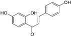 | Licorice root or Glycyrrhiza glabra | ESCC cells (KYSE140, KYSE520, TE-1) | (China) [22] | |
| 3 | 3-deoxysappanchalcone (3-DSC) |  | Caesalpinia sappan L. | ESCC cells (KYSE 70, KYSE 30, KYSE 410, KYSE 510, KYSE 450) | (Republic of Korea) [23] | |
| Coumarin | 4 | Osthole | 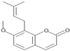 | Fruit of Fructus cnidii | ESCC cells (KYSE30, KYSE150, KYSE180, KYSE410, KYSE450) | (China) [24] |
| Polyphenolic Flavonoid | 5 | Quercetin | 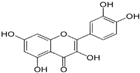 | Foods (grapes, onions, berries, broccoli, cherries, and citrus fruits) | ESCC Eca-109 cells | (China) [25] |
| 6 | Icariin | 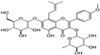 | Epimedium spp. | ESCC KYSE70 cell | (China) [26] | |
| ESCC cells (Eca109, TE-1) | (China) [27] | |||||
| 7 | Purpurogallin | 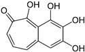 | Nutgalls and oak bark of Quercus spp | ESCC cells (KYSE70, KYSE30, KYSE410, KYSE450, KYSE510) | (China) [28] | |
| ESCC KYSE510 cells | (USA) [29] | |||||
| 8 | (6,7,4′-THIF) or 6,7,4′-Trihydroxyisoflavone |  | Glycine max L. Merr. (Soybean) | ESCC cells (KYSE 30, KYSE 450, KYSE 510) | (Republic of Korea) [30] | |
| 9 | Genistein | 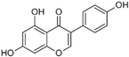 | Glycine max L. Merr. (Soybean) | ESCC cells (EC9706, Eca-109, CaES-17, Het-1A) | (China) [31] | |
| 10 | Hesperetin |  | Lemons and oranges | ESCC Eca-109 cells | (China) [32,33] | |
| 11 | Baohuoside-I | 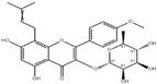 | Cortex periplocae | ESCC Eca-109 cells | (China) [34] | |
| 12 | Curcumin | 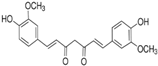 | Curcuma longa | ESCC cells (T.Tn, TE-1, TE-6, TE-5, TE-8, TE-11, TE-10, TE-11R, HCE-4) | (Japan) [35] | |
| ESCC cells (KYSE30) | (Iran) [36] | |||||
| ESCC cells (TE-1, TE-8, KY-5, KY-10, YES-1, YES-2) | (USA) [37] | |||||
| 13 | 2,6-Bis Benzylideno cyclohexanone | 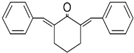 | Curcumin analogues | ESCC cells (KYSE30) | (Iran) [36] | |
| 14 | Proanthocyanidins | 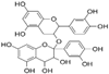 | Vitis vinifera L. (Grape seeds) | ESCC Eca-109 cells | (China) [38] | |
| Cranberry | EAC cells (OE19, JHAD1, OE33) | (USA) [20] | ||||
| 15 | Gallic acid |  | Phaleria macrocarpa (Scheff.) | ESCC TE-2 cells | (Japan) [39] | |
| 16 | Sesamin | 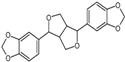 | Sesamum indicum | ESCC cells (KYSE150, EC9706, Eca-109, TE-2) | (China) [40] | |
| 17 | (-)-epigallocatechin-3-gallate |  | Green tea of Camellia sinensis L. | ESCC cells (Eca-109, KYSE 510) | (China) [41] (USA) [29] | |
| 18 | Theaflavin-3-3′-digallate | 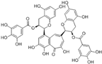 | Black tea of Camellia sinensis L. | ESCC cells (Eca-109, KYSE 510) | (China) [41] (USA) [29] | |
| 19 | Theaflavate A |  | ESCC KYSE 510 cells | (USA) [29] | ||
| Quinones | 20 | Lapachol | 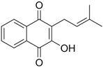 | Tabebuia avellanedae | ESCC WHCO1 cells | (South Africa) [42] |
| ESCC cells (KYSE30, KYSE450, KYSE510) | (China) [43] | |||||
| 21 | β-lapachone |  | Tabebuia avellanedae | ESCC WHCO1 cells | (South Africa) [42] | |
| 22 | Pristimerin | 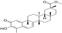 | Celastraceous and Hippocratic | ESCC Eca-109 cells | (China) [44] | |
| ESCC cells (EC9706, Eca-109, KYSE3) | (China) [45] | |||||
| 23 | Plumbagin |  | Plumbago zeylanica L. | ESCC cells (KYSE150, KYSE450) | (China) [46] | |
| Tannin | 24 | Corilagin | 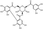 | Phyllanthus emblica L. | ESCC cells (ECA109, KYSE150) | (China) [47] |
| Xanthone | 25 | Griffipavixanthone |  | Garcinia esculenta | ESCC cells (TE-1, KYSE150) | (China) [48] |
| Compounds | In Vitro Cancer Activities | Animal Model | Cell Lines for In Vivo Assays | In Vivo Activities | Ref. |
|---|---|---|---|---|---|
| Moscatilin | CE81T/VGH (IC50 = 7.0 µM) and BE3 (IC50 = 6.7 µM) | / | / | / | [19] |
| Male nude mice | CE81T/VGH | A dose of 50 mg/kg reduces tumor mass by nearly 50% over 49 days. | [21] | ||
| Isoliquiritigenin | A concentration of 20 μM reduces proliferation of KYSE140, KYSE520, and TE-1 cells by 80%. | Femele Balb/c athymic nude mice | KYSE140 | A dose of 10 mg/kg reduces tumor mass by approximately 84% within 24 days. | [22] |
| 3-deoxysappanchalcone (3-DSC) | KYSE 30 (IC50 = 19.8 µM); KYSE 70 (IC50 = 20 µM); KYSE 410 (IC50 = 12.2 µM); KYSE 450 (IC50 = 24.7 µM); KYSE 510 (IC50 = 24.8 µM) | / | / | / | [23] |
| Osthole | KYSE150 (IC50 = 102.51 μM); KYSE410 (IC50 = 114.02 μM) | / | / | / | [24] |
| Quercetin | A concentration of 10 μM reduces proliferation of Eca-109 cells by approximately 92%. | / | / | / | [25] |
| Icariin | KYSE70 (IC50 = 40 μM) | Female immunodeficient mice | KYSE70 | A dose of 40 μg/g reduces tumor mass by approximately 71% over 4 weeks. | [26] |
| Eca109 (IC50 = 38.59 μM); TE-1 (IC50 = 42.21 μM) | Male athymic nude mice | Eca109 | A dose of 120 mg/kg reduces tumor mass by approximately 32% over 4 weeks. | [27] | |
| Purpurogallin | A concentration of 40 μM reduces proliferation by approximately 95% (KYSE30), 51%(KYSE70), 36% (KYSE410), 48% (KYSE450), and 59% (KYSE510) | Female mice | EG30 and LEG34 human ESCC | A dose of 100 mg/kg reduces tumor mass by approximately 64% in 21 days and 50% in 31 days, for tumors induced by EG30 and LEG34 cells, respectively. | [28] |
| KYSE510 (IC50 ≈ 7 µM) | / | / | / | [29] | |
| (6,7,4′-THIF) or 6,7,4′-Trihydroxyisoflavone | A concentration of 20 μmol/L, reduces proliferation by approximately 35% (KYSE 30); 42% (KYSE 450), and 82% (KYSE 510) | / | / | / | [30] |
| Genistein | Eca-109 (IC50 = 5 μM); EC9706 (IC50 = 15 μM); CaES-17 (IC50 = 12 μM); Het-1A (IC50 = 125 μM) | Nude mice | Eca-109 | A dose of 10 mg/kg reduces tumor mass by approximately 63% over 42 days. | [31] |
| Hesperetin | Eca-109 (IC50 = 200 μM) | Female Balb/c nude mice | Eca-109 | A dose of 90 mg/kg reduces tumor mass by approximately 74% over 30 days. | [33] |
| Baohuoside-I | Eca-109 (IC50 = 24.8 µg/mL) | Female Balb/c nude mice | Eca-109 | A dose of 25 mg/kg reduces tumor mass by approximately 83% over 21 days. | [34] |
| Curcumin | TE-1 (IC50 = 19.23 μM), TE-5 (IC50 = 19.45 μM), TE-6 (IC50 = 7.03 μM), TE-8 (IC50 = 8.88 μM), TE-10 (IC50 = 12.91 μM), TE-11 (IC50 = 8.98 μM), TE-11R (IC50 = 34.98 μM), T.Tn (IC50 = 19.66 μM), and HCE-4 (IC50 = 8.94 μM) | C57BL/6 male mice | TE-11R | A dose of 5000 ppm reduces tumor mass by approximately 43% over 49 days with intraperitoneal administration. | [35] |
| KYSE30 (IC50 = 5.42 µg/mL) | / | / | / | [36] | |
| A concentration of 60 μM reduces proliferation across all cell lines (TE-1, TE-8, KY-5, KY-10, YES-1, and YES-2), with the percentage of remaining cells ranging from 10.9% to 36.3%. | / | / | / | [37] | |
| Proanthocyanidins | Eca-109 (IC50 = 37.158 µg/mL) | / | / | / | [38] |
| IC50 between 50–100 µg/mL for OE19, JHAD1, and OE33 cells | Male NU/NU athymic mice | OE19 | A dose of 250 µg/mouse reduces tumor mass by approximately 67% over 19 days. | [20] | |
| Gallic acid | TE-2 (CPI50 = 0.3 mg/mL) | / | / | / | [39] |
| Sesamin | A concentration of 40 μM reduces proliferation by approximately 60% in KYSE150, EC9706, Eca-109, and TE-2 cells. | Female nude mice | Eca-109 | A dose of 150 mg/kg reduces tumor mass by approximately 48% over 21 days with oral administration. | [40] |
| (-)-epigallocatechin-3-gallate | KYSE 510 (IC50 = 18 μM) | / | / | / | [29] |
| Theaflavin-3-3′-digallate | KYSE 510 (IC50 = 18 μM) | / | / | / | [29,41] |
| Theaflavate A | KYSE 510 (IC50 = 18 μM) | / | / | / | [29] |
| Lapachol | WHCO1(IC50 = 24.1 µM) | / | / | / | [42] |
| KYSE30, KYSE450, KYSE510 (IC50 ≈ 2 µM) | / | / | / | [43] | |
| β-lapachone | WHCO1 (IC50 = 1.6 mM) | / | / | / | [42] |
| Pristimerin | A concentration of 1.5 μmol/L reduces cell proliferation in Eca-109 by 50%. | male BALB/c nude mice | Eca-109 | A dose of 1.5 μmol/L reduces tumor mass by approximately 70% over 21 days with intraperitoneal administration. | [44] |
| EC9706 (IC50 = 1.98 µM), Eca109 (IC50 = 1.76 µM), KYSE30 (IC50 = 1.13 µM) | / | / | / | [45] | |
| Plumbagin | KYSE150 (IC50 = 6.4 μM); KYSE450 (IC50 = 8.0 μM) | Female BALB/c nude mice | KYSE150 | A dose of 2 mg/kg reduces tumor mass by approximately 63% over 21 days with intraperitoneal administration. | [46] |
| Corilagin | Eca-109 (IC50 = 28.58 μM), and KYSE150 (IC50 = 35.05 μM) | Athymic nude mice | Eca109 | A dose of 20 mg/kg reduces tumor mass by approximately 75% over 21 days with oral administration. | [47] |
| Griffipavixanthone | A concentration of 10 μM reduces cell proliferation by 48% in TE-1 cells and 42% in KYSE150 cells. | / | / | / | [48] |
Disclaimer/Publisher’s Note: The statements, opinions and data contained in all publications are solely those of the individual author(s) and contributor(s) and not of MDPI and/or the editor(s). MDPI and/or the editor(s) disclaim responsibility for any injury to people or property resulting from any ideas, methods, instructions or products referred to in the content. |
© 2024 by the authors. Licensee MDPI, Basel, Switzerland. This article is an open access article distributed under the terms and conditions of the Creative Commons Attribution (CC BY) license (https://creativecommons.org/licenses/by/4.0/).
Share and Cite
Kamsu, G.T.; Ndebia, E.J. Usefulness of Natural Phenolic Compounds in the Fight against Esophageal Cancer: A Systematic Review. Future Pharmacol. 2024, 4, 626-650. https://doi.org/10.3390/futurepharmacol4030034
Kamsu GT, Ndebia EJ. Usefulness of Natural Phenolic Compounds in the Fight against Esophageal Cancer: A Systematic Review. Future Pharmacology. 2024; 4(3):626-650. https://doi.org/10.3390/futurepharmacol4030034
Chicago/Turabian StyleKamsu, Gabriel Tchuente, and Eugene Jamot Ndebia. 2024. "Usefulness of Natural Phenolic Compounds in the Fight against Esophageal Cancer: A Systematic Review" Future Pharmacology 4, no. 3: 626-650. https://doi.org/10.3390/futurepharmacol4030034
APA StyleKamsu, G. T., & Ndebia, E. J. (2024). Usefulness of Natural Phenolic Compounds in the Fight against Esophageal Cancer: A Systematic Review. Future Pharmacology, 4(3), 626-650. https://doi.org/10.3390/futurepharmacol4030034